cellular adaptations dr. peter anderson, uab pathology
TRANSCRIPT

Cellular Adaptations
Dr. Peter Anderson, UAB Pathology

Cellular AdaptationsEnvironmental Factors
• Increased or decreased stimulation• Increased or decreased work• Decreased blood flow • Abnormal materials

Response to Stress
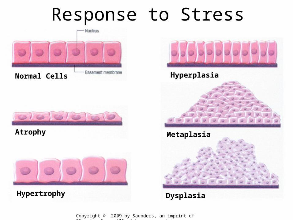
Normal Cells
Atrophy
Hypertrophy
Hyperplasia
Metaplasia
Dysplasia
Response to Stress
Copyright © 2009 by Saunders, an imprint of Elsevier Inc. All rights reserved

Atrophy• Shrinkage in the size of the cell by loss of
structural components

Atrophy
• Shrinkage in the size of the cell by loss of structural components– Decreased work load– Loss of innervation– Diminished blood supply– Inadequate nutrition – Loss of endocrine stimulation

Disuse Atrophy - Skeletal Muscle

Disuse Atrophy - Skeletal Muscle

Senile Atrophy

Hypertrophy• Increased size of cells & the organ

Hypertrophy
• Increased size of cells & the organ• Physiologic
– Hormonal stimulation e.g., uterus during pregnancy
• Pathologic– Increased functional demand e.g., Left
Ventricular Hypertrophy (LVH) - hypertension or valve stenosis

Hypertrophy

Hypertrophy

Hypertrophy
Normal Hypertrophy

Postpartum UterusHYPERTROPHY
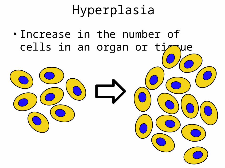
Hyperplasia
• Increase in the number of cells in an organ or tissue
Hyperplasia
• Increase in the number of cells in an organ or tissue– Physiologic hyperplasia
• hormonal induced – breast in pregnancy– Pathologic hyperplasia
• viral induced – papillomaviruses• excessive hormonal stimulation - prostate
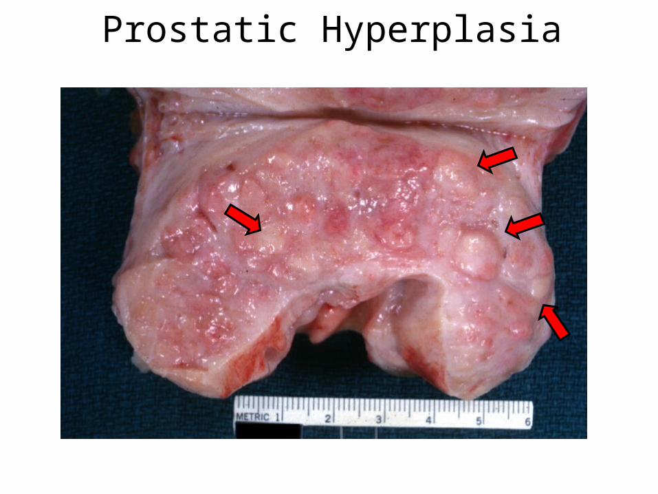
Prostatic Hyperplasia
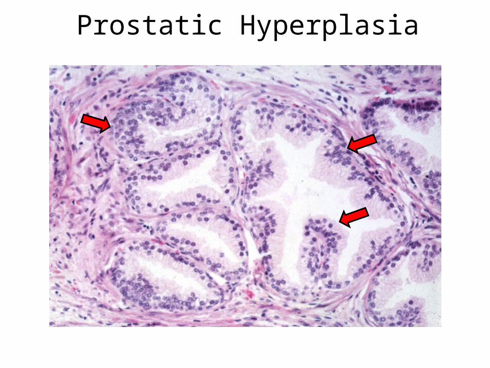
Prostatic Hyperplasia

Metaplasia• Reversible change in which one differentiated cell
type is replaced by another cell type.
Ciliated Columnar Epithelium
Squamous epithelium

Metaplasia
Ciliated Columnar Epithelium
Squamous epithelium
Stem Cells

Metaplasia
Stem CellsBasement Membrane
Ciliated Columnar Epithelium
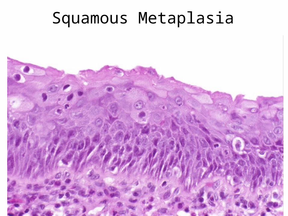
Squamous Metaplasia
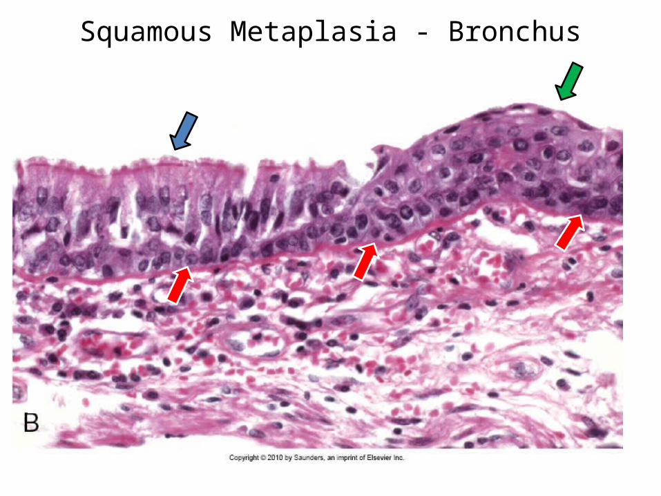
Squamous MetaplasiaSquamous Metaplasia - Bronchus
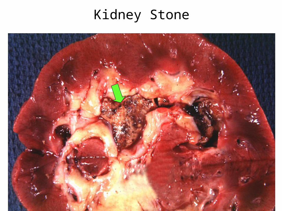
Kidney Stone
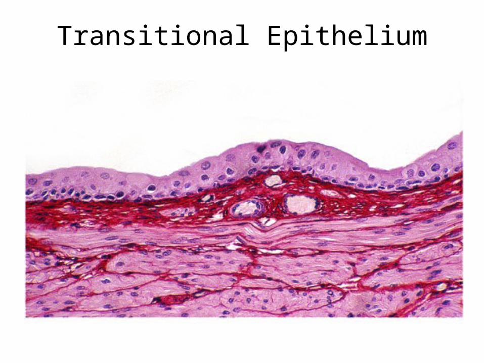
Transitional Epithelium

Squamous Metaplasia
Squamous Metaplasia

Metaplasia Summary
• Reversible change in which one differentiated cell type (epithelial or mesenchymal) is replaced by another cell type

Cellular Adaptations
AtrophyHypertrophyHyperplasiaMetaplasia

CellularAccumulations

Intracellular Accumulations
• Lipids• Proteins• Glycogen• Carbohydrates
• Carbon• Silica• Asbestos• Bacteria
Normal CellularConstituents
Abnormal or Exogenous

Fatty Change
• Lipid in macrophages– foam cells - atherosclerosis
• Lipid in parenchyma cells– alcoholic fatty liver
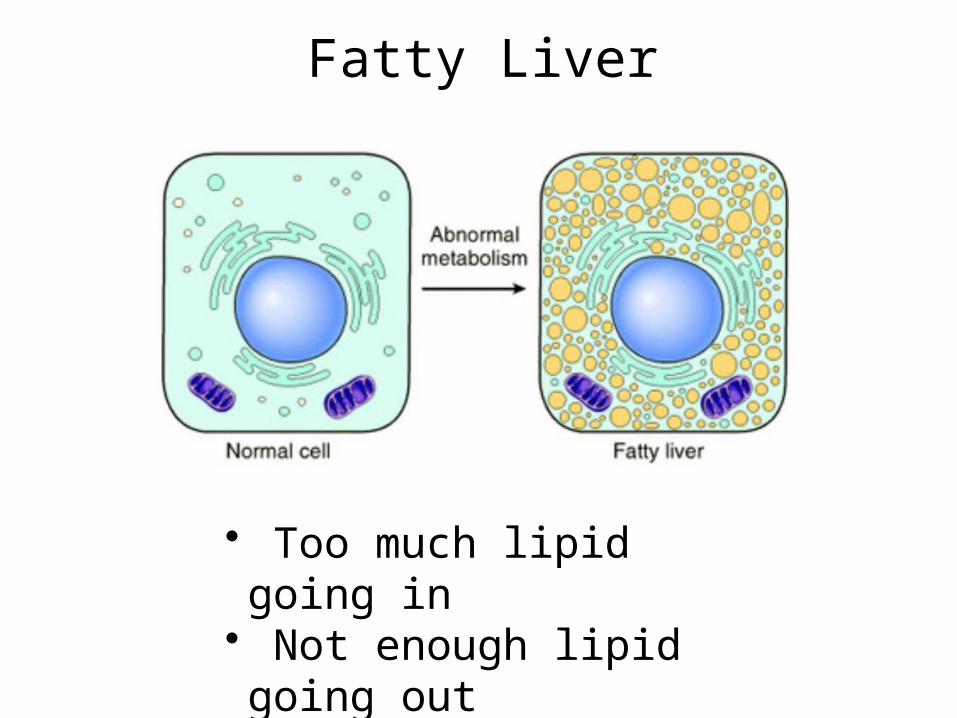
Fatty Liver
• Too much lipid going in• Not enough lipid going out

Early Fatty Change - Liver

Fatty Change - Liver

Fatty Change - Liver – Oil Red O Stain
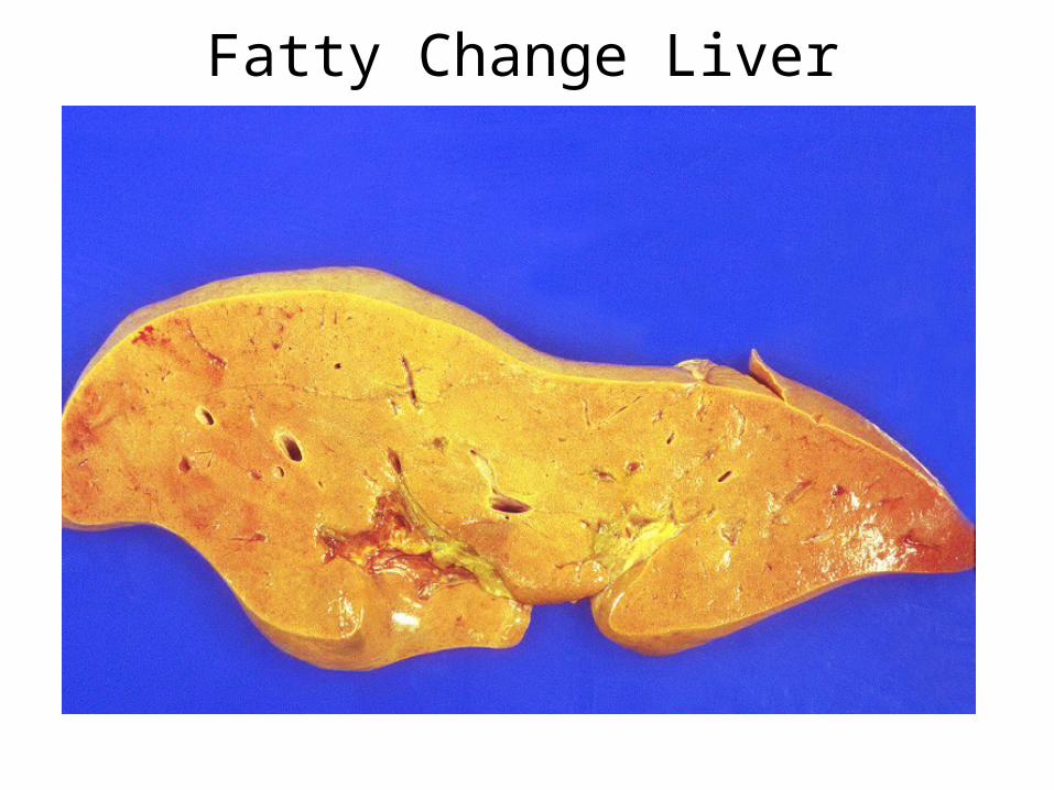
Fatty Change Liver

Normal Liver

Fatty Change Liver
Intracellular Proteins
• Kidney Proximal Tubules– hyaline droplets
• Plasma Cells– Russell bodies
• Alcoholic Hyaline
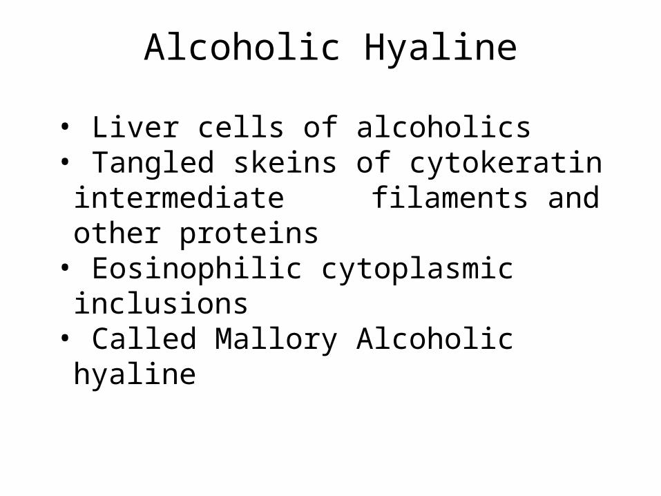
• Liver cells of alcoholics• Tangled skeins of cytokeratin intermediate
filaments and other proteins• Eosinophilic cytoplasmic inclusions• Called Mallory Alcoholic hyaline
Intracellular ProteinsAlcoholic Hyaline

Alcoholic Hyalin

Pigments
• Exogenous pigments– Carbon (anthracosis)– Tattooing– Natural substances
• b carotiene– Poisons
• lead (pica)

Anthracosis

Pigments
• Endogenous Pigments– Lipofuscin– Melanin – Hemosiderin

Melanin - Malignant Melanoma

Iron Overload
• Hemosiderosis– Iron overload in phagocytic cells– No tissue damage
• Hemochromatosis– Iron overload in parenchymal cells– Tissue damage

Iron - Hemochromatosis

Iron - Hemochromatosis
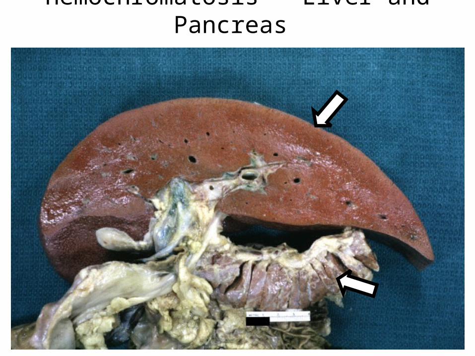
Hemochromatosis - Liver and Pancreas

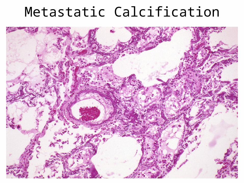
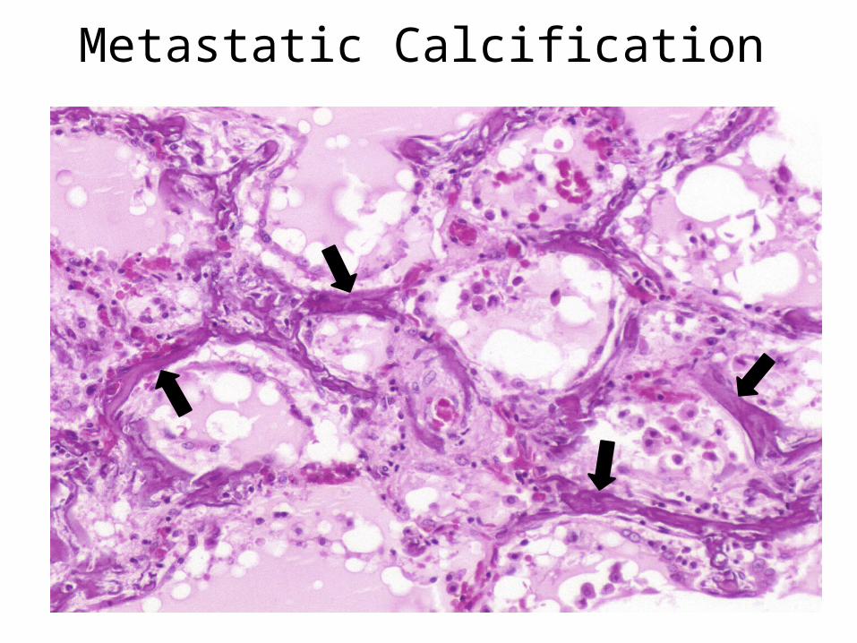
Metastatic Calcification
• Deposition of calcium in normal tissues due to hypercalcemia
• Interstitial tissues of blood vessels, kidneys, lungs, and gastric mucosa
Metastatic Calcification
Metastatic Calcification

Dystrophic Calcification• Deposition of calcium salts in necrotic
tissues• Intracellular, extracellular, or both• Heterotopic bone may form with time

Dystrophic Calcification

Dystrophic Calcification

The End
Cellular Adaptations&
Cellular Accumulations