celltiter-glo 2.0 assay - promega.co.jp 2 0 assay protocol.pdf · glo® 2.0 assay relies on the...
TRANSCRIPT
T E C H N I C A L M A N U A L
.TM403Printed 11/13
CellTiter-Glo® 2.0 AssayInstruc ons for use of Products G9241, G9242 AND G9243
1. Description ..........................................................................................................1
2. Product Components and Storage Conditions ............................................5
3. Performing the CellTiter-Glo® 2.0 Assay......................................................6A. Reagent Preparation.............................................................................................6B. Protocol for the Cell Viability Assay .................................................................7C. Protocol for Generating an ATP Standard Curve (optional).........................8D. Sequential Multiplexing of CellTox™ Green Cytotoxicity Assay
and CellTiter-Glo® 2.0..........................................................................................8
4. Appendix .............................................................................................................9A. Overview of the CellTiter-Glo® 2.0 Assay ........................................................9B. Additional Considerations................................................................................11C. References ............................................................................................................13D. Related Products.................................................................................................14
1. Description
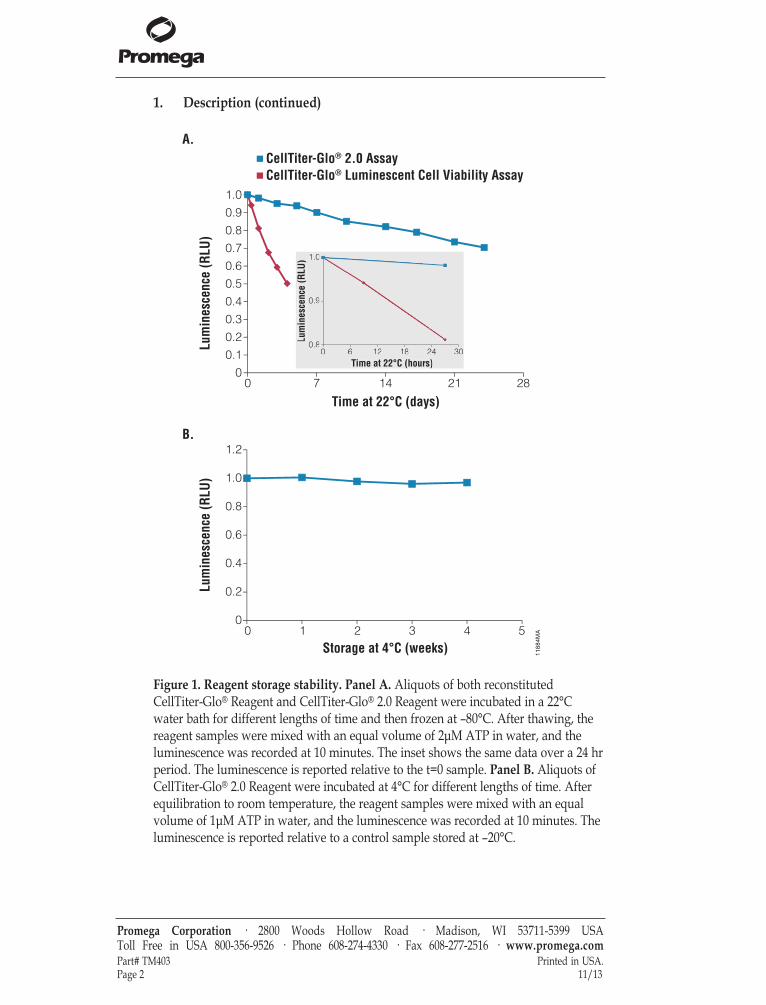
The CellTiter-Glo® 2.0 Assay(a–e) provides a homogeneous method to determinethe number of viable cells in culture by quantitating the amount of ATPpresent, which indicates the presence of metabolically active cells. This ready-to-use reagent is based on the original CellTiter-Glo® Luminescent Cell ViabilityAssay chemistry and eliminates the need to combine buffer with lyophilizedsubstrate when preparing reagent. Although similar to the CellTiter-Glo® OneSolution Assay, which needs to be stored frozen, the CellTiter-Glo® 2.0 Assay ismuch more stable and will maintain > 90% activity upon storage at 4°C for 4weeks or > 85% activity upon storage at room temperature for 7 days (Figure 1).The CellTiter-Glo® 2.0 Assay is designed for use with multiwell-plate formats,making it ideal for automated high-throughput screening (HTS) and cellproliferation and cytotoxicity assays. The homogeneous assay procedure(Figure 2) involves addition of a single reagent (CellTiter-Glo® 2.0 Reagent)directly to cells cultured in serum-supplemented medium. Cell washing,removal of medium and multiple pipetting steps are not required.
Promega Corporation · 2800 Woods Hollow Road · Madison, WI 53711-5399 USA Toll Free in USA 800-356-9526 · Phone 608-274-4330 · Fax 608-277-2516 · www.promega.comPrinted in USA. Part# TM40311/13 Page 1
CellTiter-Glo® 2.0 AssayAll technical literature is available on the Internet at: www.promega.com/protocols/ Please visit the web site to verify that you are using the most current version of this
Technical Manual. Please contact Promega Technical Services if you have questions on useof this system. E-mail: [email protected]
1. Description (continued)
Promega Corporation · 2800 Woods Hollow Road · Madison, WI 53711-5399 USA Toll Free in USA 800-356-9526 · Phone 608-274-4330 · Fax 608-277-2516 · www.promega.comPart# TM403 Printed in USA.Page 2 11/13
1188
4MA
0 7 14 21 28
Lum
ines
cenc
e (R
LU)
Lum
ines
cenc
e (R
LU)
Time at 22°C (days)
0 1 2 3 4 5
Storage at 4°C (weeks)
CellTiter-Glo® Luminescent Cell Viability AssayCellTiter-Glo® 2.0 Assay
0 6 12 18 24 30Time at 22°C (hours)
0.8
0.9
1.0
Lum
ines
cenc
e (R
LU)
0 6 12 18 24 30Time at 22°C (hours)
0.8
0.9
1.0
Lum
ines
cenc
e (R
LU)
0
0.2
0.4
0.6
0.8
1.0
1.2
A.
B.
0
0.1
0.2
0.3
0.4
0.5
0.6
0.7
0.8
0.9
1.0
Figure 1. Reagent storage stability. Panel A. Aliquots of both reconstitutedCellTiter-Glo® Reagent and CellTiter-Glo® 2.0 Reagent were incubated in a 22°Cwater bath for different lengths of time and then frozen at –80°C. After thawing, thereagent samples were mixed with an equal volume of 2μM ATP in water, and theluminescence was recorded at 10 minutes. The inset shows the same data over a 24 hrperiod. The luminescence is reported relative to the t=0 sample. Panel B. Aliquots ofCellTiter-Glo® 2.0 Reagent were incubated at 4°C for different lengths of time. Afterequilibration to room temperature, the reagent samples were mixed with an equalvolume of 1μM ATP in water, and the luminescence was recorded at 10 minutes. Theluminescence is reported relative to a control sample stored at –20°C.
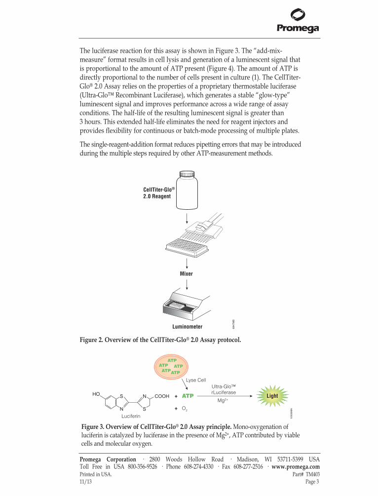
The luciferase reaction for this assay is shown in Figure 3. The “add-mix-measure” format results in cell lysis and generation of a luminescent signal thatis proportional to the amount of ATP present (Figure 4). The amount of ATP isdirectly proportional to the number of cells present in culture (1). The CellTiter-Glo® 2.0 Assay relies on the properties of a proprietary thermostable luciferase(Ultra-Glo™ Recombinant Luciferase), which generates a stable “glow-type”luminescent signal and improves performance across a wide range of assayconditions. The half-life of the resulting luminescent signal is greater than3 hours. This extended half-life eliminates the need for reagent injectors andprovides flexibility for continuous or batch-mode processing of multiple plates.
The single-reagent-addition format reduces pipetting errors that may be introducedduring the multiple steps required by other ATP-measurement methods.
Promega Corporation · 2800 Woods Hollow Road · Madison, WI 53711-5399 USA Toll Free in USA 800-356-9526 · Phone 608-274-4330 · Fax 608-277-2516 · www.promega.comPrinted in USA. Part# TM40311/13 Page 3
6847
MB
CellTiter-Glo®
2.0 Reagent
Mixer
Luminometer
Figure 2. Overview of the CellTiter-Glo® 2.0 Assay protocol.
1033
5MA
ATP ATP
ATP ATP
ATP
Lyse Cell
ATP +
+
Ultra-Glo™rLuciferase
N
S
S
N COOH Glo HO
ATPATP
ATPATP
ATP
ATP Light
Luciferin
O2
Mg2+
Figure 3. Overview of CellTiter-Glo® 2.0 Assay principle. Mono-oxygenation ofluciferin is catalyzed by luciferase in the presence of Mg2+, ATP contributed by viablecells and molecular oxygen.
1. Description (continued)
Promega Corporation · 2800 Woods Hollow Road · Madison, WI 53711-5399 USA Toll Free in USA 800-356-9526 · Phone 608-274-4330 · Fax 608-277-2516 · www.promega.comPart# TM403 Printed in USA.Page 4 11/13
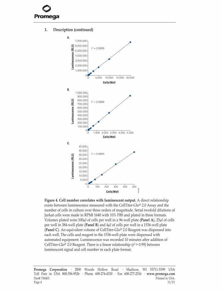
Figure 4. Cell number correlates with luminescent output. A direct relationshipexists between luminescence measured with the CellTiter-Glo® 2.0 Assay and thenumber of cells in culture over three orders of magnitude. Serial twofold dilutions ofJurkat cells were made in RPMI 1640 with 10% FBS and plated in three formats.Volumes plated were 100µl of cells per well in a 96-well plate (Panel A), 25µl of cellsper well in 384-well plate (Panel B) and 4µl of cells per well in a 1536-well plate(Panel C). An equivalent volume of CellTiter-Glo® 2.0 Reagent was dispensed intoeach well. The cells and reagent in the 1536-well plate were dispensed withautomated equipment. Luminescence was recorded 10 minutes after addition ofCellTiter-Glo® 2.0 Reagent. There is a linear relationship (r2 > 0.99) betweenluminescent signal and cell number in each plate format.
1181
5MA
r2 = 0.9999
r2 = 0.9999
r2 = 0.9993
00 5,000 15,00010,000 20,000
1,000,000
2,000,000
3,000,000
4,000,000
5,000,000
6,000,000
7,000,000
Lum
ines
cenc
e (R
LU)
Cells/Well
Lum
ines
cenc
e (R
LU)
Cells/Well
Lum
ines
cenc
e (R
LU)
Cells/Well
00 1,000 2,000 3,000 4,000 5,000
100,000200,000300,000400,000500,000600,000700,000800,000900,000
1,000,000
00 100 200 300 400 500
5,000
10,000
15,000
20,000
25,000
30,000
35,000
40,000
45,000
A.
B.
C.

Assay Advantages
• Ready-to-use reagent: No reagent preparation is required; simplyequilibrate to room temperature and “add-mix-measure”. Convenient forHTS applications.
• Flexible storage temperatures: The reagent is ready when you are. TheReagent maintains >90% activity upon storage at 4°C for 4 weeks or >85%activity upon storage at room temperature for 7 days.
• Homogeneous: The “add-mix-measure” format reduces the number of plate-handling steps.
• Fast: Data can be recorded 10 minutes after adding reagent.
• Sensitive: The assay measures cell numbers below the detection limits ofstandard colorimetric and fluorometric assays.
• Flexible: The assay can be used with various multiwell formats (96-well, regular or low-volume 384-well and 1536-well plates). Data can berecorded by luminometer, CCD camera or other imaging device capable ofreading luminescence in multiwell plates.
• Robust: Luminescent signal is stable, with a half-life >3 hours, dependingon cell type and culture medium used.
• Able to Multiplex: The assay can be used with other nonlytic assays fromPromega (2,3).
2. Product Components and Storage Conditions
Product Size Cat.#CellTiter-Glo® 2.0 Assay 10ml G9241For in vitro Research Use Only. CellTiter-Glo® 2.0 Reagent is sufficient for100 assays at 100µl/assay in 96-well plates or 400 assays at 25µl/assay in 384-wellplates.
Product Size Cat.#CellTiter-Glo® 2.0 Assay 100ml G9242For in vitro Research Use Only. CellTiter-Glo® 2.0 Reagent is sufficient for1,000 assays at 100µl/assay in 96-well plates or 4,000 assays at 25µl/assay in 384-well plates.
Promega Corporation · 2800 Woods Hollow Road · Madison, WI 53711-5399 USA Toll Free in USA 800-356-9526 · Phone 608-274-4330 · Fax 608-277-2516 · www.promega.comPrinted in USA. Part# TM40311/13 Page 5

2. Product Components and Storage Conditions (continued)
Product Size Cat.#CellTiter-Glo® 2.0 Assay 500ml G9243For in vitro Research Use Only. CellTiter-Glo® 2.0 Reagent is sufficient for 5,000 assaysat 100µl/assay in 96-well plates or 20,000 assays at 25µl/assay in 384-well plates.
Storage Conditions: The CellTiter-Glo® 2.0 Assay is shipped frozen and can bestored at –30 to –10°C through the expiration date of the reagent. The CellTiter-Glo® 2.0 Assay can maintain >90% activity upon storage at 4°C for 4 weeks or>85% activity upon storage at 22–25°C for 7 days. Do not refreeze the thawedreagent after extended storage above the label storage temperature (–30°C to–10°C). When storing the reagent at -30 to -10C, the Reagent can withstand fouradditional freeze-thaw cycles after the first thaw, with no loss of activity. We donot recommend dispensing the CellTiter-Glo® 2.0 Reagent into aliquots due tothe risk of ATP contamination.
The CellTiter-Glo® 2.0 Reagent is light sensitive and should be stored in anopaque container.
3. Performing the CellTiter-Glo® 2.0 Assay
Materials to Be Supplied by the User• 22°C water bath• opaque-walled multiwell plates adequate for cell culture• multichannel pipette or automated pipetting station• device (plate shaker) for mixing multiwell plates• luminometer, CCD camera or imaging device capable of reading luminescence in
multiwell plates• optional: ATP for use in generating a standard curve in Section 3.C (Cat.# P1132,
Sigma Cat.# A7699 or GE Healthcare Cat.# 27-1006)
3.A. Reagent Preparation
1. If frozen, thaw CellTiter-Glo® 2.0 Reagent at 4°C overnight.Reagent may also be thawed in a 22°C water bath. Do not expose thereagent to temperatures above 25°C.
2. If not at room temperature, equilibrate the CellTiter-Glo® 2.0 Reagent toroom temperature by placing the reagent in a 22°C water bath prior to use.Note: In a 22°C water bath, 100ml of the thawed reagent (4°C) requiresapproximately 30 minutes to equilibrate and 500ml requires approximately100 minutes to equilibrate to 22°C.
3. Mix gently by inverting the contents to obtain a homogeneous solution.Note: Use caution when removing the seal of the CellTiter-Glo® 2.0Reagent bottle to avoid introducing ATP contamination.
Promega Corporation · 2800 Woods Hollow Road · Madison, WI 53711-5399 USA Toll Free in USA 800-356-9526 · Phone 608-274-4330 · Fax 608-277-2516 · www.promega.comPart# TM403 Printed in USA.Page 6 11/13
!
!
3.B. Protocol for the Cell Viability Assay
Prepare and equilibrate the CellTiter-Glo® 2.0 Reagent as described inSection 3.A prior to performing the assay.
1. Prepare opaque-walled multiwell plates with mammalian cells in culturemedium. Volumes and cell number should be optimized for experimentalconditions.Multiwell plates must be compatible with the luminometer used.
2. If desired, prepare control wells containing medium without cells todetermine background luminescence.
3. Add test compound to experimental wells, and incubate according to yourculture protocol.
4. Equilibrate the plate and its contents to room temperature forapproximately 30 minutes.
5. Add a volume of CellTiter-Glo® 2.0 Reagent equal to the volume of cellculture medium present in each well (e.g., for a 96-well plate, add 100µl ofCellTiter-Glo® 2.0 Reagent to 100µl of medium containing cells).
6. Mix the contents for 2 minutes on an orbital shaker to induce cell lysis (seeAppendix for more information on mixing).
7. Allow the plate to incubate at room temperature for 10 minutes to stabilizeluminescent signal.
8. Record luminescence.
Notes:1. Instrument settings depend on the manufacturer. Use an integration time
of 0.25–1 second per well as a guideline.2. Uneven luminescent signal within plates can be caused by temperature
gradients, uneven seeding of cells or edge effects in multiwell plates.
Promega Corporation · 2800 Woods Hollow Road · Madison, WI 53711-5399 USA Toll Free in USA 800-356-9526 · Phone 608-274-4330 · Fax 608-277-2516 · www.promega.comPrinted in USA. Part# TM40311/13 Page 7
!
3.C. Protocol for Generating an ATP Standard Curve (optional)
It is a good practice to generate a standard curve using the same plate onwhich samples are assayed. Because of endogenous ATPase enzymes found inserum, the ATP standard curve should be generated immediately prior toadding the CellTiter-Glo® 2.0 Reagent. If the amount of ATPase enzymes issufficiently high, it may be necessary to omit serum from the media used togenerate the standard curve. We recommend ATP disodium salt (Cat.# P1132,Sigma Cat.# A7699 or GE Healthcare Cat.# 27-1006).
1. Prepare 1µM ATP in culture medium (100µl of 1µM ATP solution contains10–10 moles of ATP).
2. Prepare serial tenfold dilutions of ATP in culture medium (1µM to 10nM;100µl contains 10–10 to 10–12 moles of ATP, respectively).
3. Prepare a multiwell plate with varying concentrations of ATP standard in100µl of medium (25µl for a 384-well plate).
4. Add a volume of CellTiter-Glo® 2.0 Reagent equal to the volume of ATPstandard present in each well.
5. Mix contents for 2 minutes on an orbital shaker.
6. Allow the plate to incubate at room temperature for 10 minutes to stabilizethe luminescent signal.
7. Record luminescence.
3.D. Sequential Multiplexing of CellTox™ Green Cytotoxicity Assay and CellTiter-Glo® 2.0 Assay
1. Completely thaw the CellTox™ Green Dye in a 37°C water bath. Mix theCellTox™ Green Dye using a vortex mixer to ensure homogeneity. A briefcentrifugation may be necessary for complete recovery of the CellTox™Green Dye. Although performance of CellTox Green is optimal in blackplates, white plates are optimal for sequential multiplexing formats thatinclude a luminescent second measure.
2. Add the CellTox™ Green Dye at seeding or dosing so the final CellTox™Green Dye concentration is 1X (stock = 1000X) and incubate under desiredconditions.
3. Equilibrate plate to room temperature. Measure fluorescence intensity at485–500nmEx/520–530nmEm. Depending upon instrument manufacturerand optical detection configuration, replicate wells may benefit from 1-minute of shaking on an orbital shaker before measuring fluorescence.
4. Equilibrate the CellTiter-Glo® 2.0 Reagent to room temperature. After thefinal fluorescence measurement, add an equal volume of CellTiter-Glo® 2.0Reagent to each well.
5. Place on an orbital shake for 2 minutes and measure luminescence after10 minutes.
Promega Corporation · 2800 Woods Hollow Road · Madison, WI 53711-5399 USA Toll Free in USA 800-356-9526 · Phone 608-274-4330 · Fax 608-277-2516 · www.promega.comPart# TM403 Printed in USA.Page 8 11/13
4. Appendix
4.A. Overview of the CellTiter-Glo® 2.0 Assay
The CellTiter-Glo® 2.0 Assay takes advantage of the properties of a proprietarythermostable luciferase, Ultra-Glo™ Recombinant Luciferase, to enable reactionconditions that generate a stable “glow-type” luminescent signal whilesimultaneously inhibiting endogenous enzymes released during cell lysis (e.g.,ATPases). Release of ATPases will interfere with accurate ATP measurement.Historically, firefly luciferase purified from Photinus pyralis (LucPpy) has beenused in reagents for ATP assays (1,4–7). However, LucPpy has only moderatestability in vitro and is sensitive to its chemical environment, including factorssuch as pH and detergents, limiting its usefulness for developing a robusthomogeneous ATP assay. Promega has successfully developed a stable form ofluciferase based on the gene from another firefly, Photuris pennsylvanica(LucPpe2) using an approach to select characteristics that improved performancein ATP assays. The unique characteristics of this mutant (LucPpe2m), Ultra-Glo™Recombinant Luciferase, enabled design of a homogeneous single-reagent-addition approach for performing ATP assays on cultured cells. Properties ofCellTiter-Glo® 2.0 Assay overcome problems caused by factors such as ATPasesthat interfere with the measurement of ATP in cell extracts. The reagent isphysically robust and provides a sensitive and stable luminescent output.
Sensitivity and Linearity: The ATP-based detection of cells is more sensitivethan other methods (8–10). There is a linear relationship between luminescentsignal and cell number in different plate formats. The luminescence valuesshown in Figures 1 and 4 were recorded after 10 minutes of incubation at roomtemperature to stabilize the luminescent signal as described in Section 3.B. 4.A.
Promega Corporation · 2800 Woods Hollow Road · Madison, WI 53711-5399 USA Toll Free in USA 800-356-9526 · Phone 608-274-4330 · Fax 608-277-2516 · www.promega.comPrinted in USA. Part# TM40311/13 Page 9
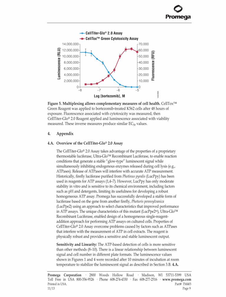
Figure 5. Multiplexing allows complementary measures of cell health. CellTox™Green Reagent was applied to bortezomib-treated K562 cells after 48 hours ofexposure. Fluorescence associated with cytotoxicity was measured, then CellTiter-Glo® 2.0 Reagent applied and luminescence associated with viabilitymeasured. These inverse measures produce similar EC50 values.
1202
3MA
0
10,000
20,000
30,000
40,000
50,000
60,000
70,000
0
2,000,000
4,000,000
6,000,000
8,000,000
10,000,000
12,000,000
14,000,000
–8 –7 –6 –5
Fluo
resc
ence
(RFU
)
Lum
ines
cenc
e (R
LU)
Log [bortezomib], M
CellTiter-Glo® 2.0 AssayCellTox™ Green Cytotoxicity Assay
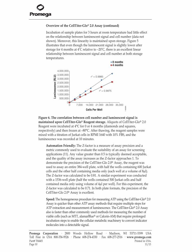
Overview of the CellTiter-Glo® 2.0 Assay (continued)
Incubation of sample plates for 3 hours at room temperature had little effecton the relationship between luminescent signal and cell number (data notshown). Moreover, this linearity is maintained upon storage. Figure 5illustrates that even though the luminescent signal is slightly lower afterstorage for 4 months at 4°C relative to –20°C, there is an excellent linearrelationship between luminescent signal and cell number at both storagetemperatures.
Automation Friendly: The Z-factor is a measure of assay precision and ametric commonly used to evaluate the suitability of an assay for screeningapplications (11). Any value greater than 0.5 is typically deemed acceptable,and the quality of the assay increases as the Z-factor approaches 1. Todemonstrate the precision of the CellTiter-Glo 2.0® Assay, the reagent wasused to assay an entire 384-well plate, with half the wells containing 600 Jurkatcells and the other half containing media only (each well at a volume of 8µl).The Z-factor was calculated to be 0.81. A similar experiment was conductedwith a 1536-well plate (half the wells contained 500 Jurkat cells and halfcontained media only using volume of 4µl per well). For this experiment, theZ-factor was calculated to be 0.71. In both plate formats, the precision of theCellTiter-Glo 2.0® Assay is excellent.
Speed: The homogeneous procedure for measuring ATP using the CellTiter-Glo® 2.0Assay is quicker than other ATP assay methods that require multiple steps forATP extraction and measurement of luminescence. The CellTiter-Glo® 2.0 Assayalso is faster than other commonly used methods for measuring the number ofviable cells (such as MTT, alamarBlue® or Calcein-AM) that require prolongedincubation steps to enable the cellular metabolic machinery to convert indicatormolecules into a detectable signal.
Promega Corporation · 2800 Woods Hollow Road · Madison, WI 53711-5399 USA Toll Free in USA 800-356-9526 · Phone 608-274-4330 · Fax 608-277-2516 · www.promega.comPart# TM403 Printed in USA.Page 10 11/13
1196
2MA
r2 = 0.9985
r2 = 0.9975
00 7,000 14,000 21,000 28,000 35,000
500,000
1,000,000
1,500,000
2,000,000
2,500,000
3,000,000
3,500,000
4,000,000
Lum
ines
cenc
e (R
LU)
Cells Per Well
0 months
4 months
Figure 6. The correlation between cell number and luminescent signal ismaintained upon CellTiter-Glo® Reagent storage. Aliquots of CellTiter-Glo® 2.0Reagent were incubated at 4°C for 0 or 4 months (diamonds and squares,respectively) and then frozen at –80°C. After thawing, the reagent samples weremixed with a titration of Jurkat cells in RPMI 1640 with 10% FBS, and theluminescence was recorded at 10 minutes.
4.B. Additional Considerations
Temperature: The intensity and rate of decay of the luminescent signal fromthe CellTiter-Glo® 2.0 Assay depends on the luciferase reaction rate.Environmental factors that affect the luciferase reaction rate will change theintensity of light output and stability of the luminescent signal. Temperature isone factor that affects the rate of this enzymatic assay and thus the light output.For consistent results, equilibrate assay plates to a constant temperature beforeperforming the assay. Transferring eukaryotic cells from 37°C to roomtemperature has little effect on ATP content (5). We have demonstrated thatremoving cultured cells from a 37°C incubator and allowing them toequilibrate to 22°C for 1–2 hours had little effect on ATP content. For batch-mode processing of multiple assay plates, take precautions to ensure completetemperature equilibration. Plates removed from a 37°C incubator and placedin tall stacks at room temperature will require a longer equilibration time thanplates arranged in a single layer. Insufficient equilibration may result in atemperature gradient between wells in the center and on the edge of theplates. The temperature gradient pattern also may depend on the position ofthe plate in the stack.
Chemicals: The chemical environment of the luciferase reaction affects theenzymatic rate and thus luminescence intensity. Differences in luminescenceintensity have been observed using different types of culture media and sera.The presence of phenol red in culture medium should have little effect onluminescence output. Solvents for the test compounds may interfere with theluciferase reaction and thus light output. To test for luciferase inhibition,assemble two reactions, one with equal volumes of CellTiter-Glo® 2.0 Reagentand 1µM ATP, and a second reaction with equal volumes of CellTiter-Glo® 2.0Reagent and 1µM ATP plus the test compound. Incubate reactions for10 minutes at 22–25°C, then measure luminescence. A decrease inluminescence in the presence of test compound indicates luciferase inhibition.
Plate Recommendations: We recommend using standard opaque-walledmultiwell plates suitable for luminescence measurements. Opaque-walledplates with clear bottoms, which allow microscopic visualization of cells, alsomay be used; however, assays in these plates will have diminished signalintensity and greater cross-talk between wells. Opaque white tape may beused to decrease luminescence loss and cross-talk.
Promega Corporation · 2800 Woods Hollow Road · Madison, WI 53711-5399 USA Toll Free in USA 800-356-9526 · Phone 608-274-4330 · Fax 608-277-2516 · www.promega.comPrinted in USA. Part# TM40311/13 Page 11

4.B. Additional Considerations
Cellular ATP Content: Different cell types have different amounts of ATP,and values reported for the ATP level in a particular cell type varyconsiderably (1,4,12–14). Factors that affect the ATP content of cells may affectthe relationship between cell number and luminescence. Anchorage-dependentcells that undergo contact inhibition at high densities may show a change inATP content per cell at high densities, resulting in a nonlinear relationshipbetween cell number and luminescence. Factors that affect cytoplasmic volumeor cell physiology also will have an effect on ATP content. For example,oxygen depletion is one factor known to cause a rapid decrease in ATP (1).
Mixing: Optimum assay performance is achieved when the CellTiter-Glo® 2.0Reagent is completely mixed with the cultured cells. Suspension cell lines (e.g.,Jurkat cells) generally require less mixing to achieve lysis and extraction ofATP than adherent cells (e.g., L929 cells).
Several additional parameters related to reagent mixing include: the force ofdelivery of the CellTiter-Glo® 2.0 Reagent, sample volume and dimensions of thewell. All of these factors may affect assay performance. The degree of reagentmixing required may be affected by the method used to add CellTiter-Glo® 2.0Reagent to the assay plates. Automated pipetting devices that use a greater orlesser force of fluid delivery may affect the degree of subsequent mixingrequired. Complete reagent mixing in 96-well plates should be achieved usingorbital plate shaking devices built into many luminometers and therecommended 2-minute shaking time. Special electromagnetic shaking devicesusing a radius smaller than the well diameter may be required to efficientlymix the contents of 384-well plates. The depth of medium and geometry of themultiwell plates may have an effect on mixing efficiency. We recommend thatyou consider these factors when performing the assay to determine whether amixing step is necessary for your application.
ATP Contamination: Strict aseptic technique is essential to prevent ATPcontamination of the CellTiter-Glo® 2.0 Reagent. Wear gloves and avoidcontact with potentially contaminated surfaces and equipment. Clean gloves,lab surfaces and equipment with a 10% bleach solution, then pat dry with labwipes (e.g., Kimwipes® tissues). Use individually wrapped or designated ATP-free pipettes and pipette tips whenever possible, and avoid inserting pipettesor pipette tips into the CellTiter-Glo® 2.0 Reagent bottle multiple times.Discard any unused, dispensed reagent; do not return it to the original bottle.
Light Sensitivity: CellTiter-Glo® 2.0 is light sensitive and will decay morerapidly if exposed to light during storage. If the reagent is aliquotted ortransferred from the original container, please be sure to protect the reagentfrom light.
Promega Corporation · 2800 Woods Hollow Road · Madison, WI 53711-5399 USA Toll Free in USA 800-356-9526 · Phone 608-274-4330 · Fax 608-277-2516 · www.promega.comPart# TM403 Printed in USA.Page 12 11/13

4.C. References
1. Crouch, S.P.M. et al. (1993) The use of ATP bioluminescence as a measure of cellproliferation and cytotoxicity. J. Immunol. Methods 160, 81–8.
2. Farfan, A. et al. (2004) Multiplexing homogeneous cell-based assays. Cell Notes 10, 2–5.
3. Riss, T., Moravec, R. and Niles, A. (2005) Selecting cell-based assays for drugdiscovery screening. Cell Notes 13, 16–21.
4. Kangas, L., Grönroos, M. and Nieminen, A.L. (1984) Bioluminescence of cellular ATP:A new method for evaluating cytotoxic agents in vitro. Med. Biol. 62, 338–43.
5. Lundin, A. et al. (1986) Estimation of biomass in growing cell lines by adenosinetriphosphate assay. Methods Enzymol. 133, 27–42.
6. Sevin, B.U. et al. (1988) Application of an ATP-bioluminescence assay in humantumor chemosensitivity testing. Gynecol. Oncol. 31, 191–204.
7. Gerhardt, R.T. et al. (1991) Characterization of in vitro sensitivity of perioperativehuman ovarian malignancies by adenosine triphosphate chemosensitivity assay. Am.J. Obstet. Gynecol. 165, 245–55.
8. Petty, R.D. et al. (1995) Comparison of MTT and ATP-based assays for measurementof viable cell number. J. Biolumin. Chemilumin. 10, 29–34.
9. Cree, I.A. et al. (1995) Methotrexate chemosensitivity by ATP luminescence in humanleukemia cell lines and in breast cancer primary cultures: Comparison of the TCA-100assay with a clonogenic assay. Anticancer Drugs 6, 398–404.
10. Maehara, Y. et al. (1987) The ATP assay is more sensitive than the succinatedehydrogenase inhibition test for predicting cell viability. Eur. J. Clin. Oncol. 23,273–6.
11. Zhang, J.H., Chung, T.D., and Oldenburg, K.R. (1999) A simple statistical parameterfor use in evaluation and validation of high throughput screening assays. J. Biomol.Screen. 4, 67–73.
12. Stanley, P.E. (1986) Extraction of adenosine triphosphate from microbial and somaticcells. Methods Enzymol. 133, 14–22.
13. Beckers, B. et al. (1986) Application of intracellular ATP determination inlymphocytes for HLA-typing. J. Biolumin. Chemilumin. 1, 47–51.
14. Andreotti, P.E. et al. (1995) Chemosensitivity testing of human tumors using amicroplate adenosine triphosphate luminescence assay. Cancer Res. 55, 5276–82.
Additional References
Auld, D.S. et al. (2009) A basis for reduced chemical library inhibition of fireflyluciferase obtained from directed evolution. J. Med. Chem. 52, 1450–8.
Niles, A.L., Moravec, R.A. and Riss, T.L. (2009) In vitro viability and cytotoxicitytesting and same-well multi-parametric combinations for high throughput screening.Curr. Chem. Genomics 3, 33–41.
Cali, J.J. et al. (2008) Bioluminescent assays for ADMET. Expert Opin. Drug Metab.Toxicol. 4, 103–20.
Xia, M. et al. (2008) Compound cytotoxicity profiling using quantitative high-throughput screening. Environ. Health Perspect. 116, 284–91.
Promega Corporation · 2800 Woods Hollow Road · Madison, WI 53711-5399 USA Toll Free in USA 800-356-9526 · Phone 608-274-4330 · Fax 608-277-2516 · www.promega.comPrinted in USA. Part# TM40311/13 Page 13
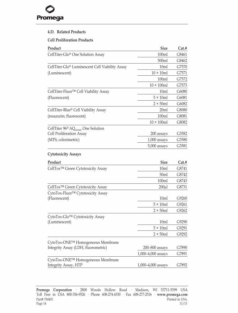
4.D. Related Products
Cell Proliferation Products
Product Size Cat.#CellTiter-Glo® One Solution Assay 100ml G8461
500ml G8462CellTiter-Glo® Luminescent Cell Viability Assay 10ml G7570(Luminescent) 10 × 10ml G7571
100ml G757210 × 100ml G7573
CellTiter-Fluor™ Cell Viability Assay 10ml G6080(Fluorescent) 5 × 10ml G6081
2 × 50ml G6082CellTiter-Blue® Cell Viability Assay 20ml G8080(resazurin; fluorescent) 100ml G8081
10 × 100ml G8082CellTiter 96® AQueous One Solution Cell Proliferation Assay 200 assays G3582(MTS; colorimetric) 1,000 assays G3580
5,000 assays G3581
Cytotoxicity Assays
Product Size Cat.#CellTox™ Green Cytotoxicity Assay 10ml G8741
50ml G8742100ml G8743
CellTox™ Green Cytotoxicity Assay 200µl G8731CytoTox-Fluor™ Cytotoxicity Assay(Fluorescent) 10ml G9260
5 × 10ml G92612 × 50ml G9262
CytoTox-Glo™ Cytotoxicity Assay(Luminescent) 10ml G9290
5 × 10ml G92912 × 50ml G9292
CytoTox-ONE™ Homogeneous MembraneIntegrity Assay (LDH, fluorometric) 200–800 assays G7890
1,000–4,000 assays G7891CytoTox-ONE™ Homogeneous MembraneIntegrity Assay, HTP 1,000–4,000 assays G7892
Promega Corporation · 2800 Woods Hollow Road · Madison, WI 53711-5399 USA Toll Free in USA 800-356-9526 · Phone 608-274-4330 · Fax 608-277-2516 · www.promega.comPart# TM403 Printed in USA.Page 14 11/13
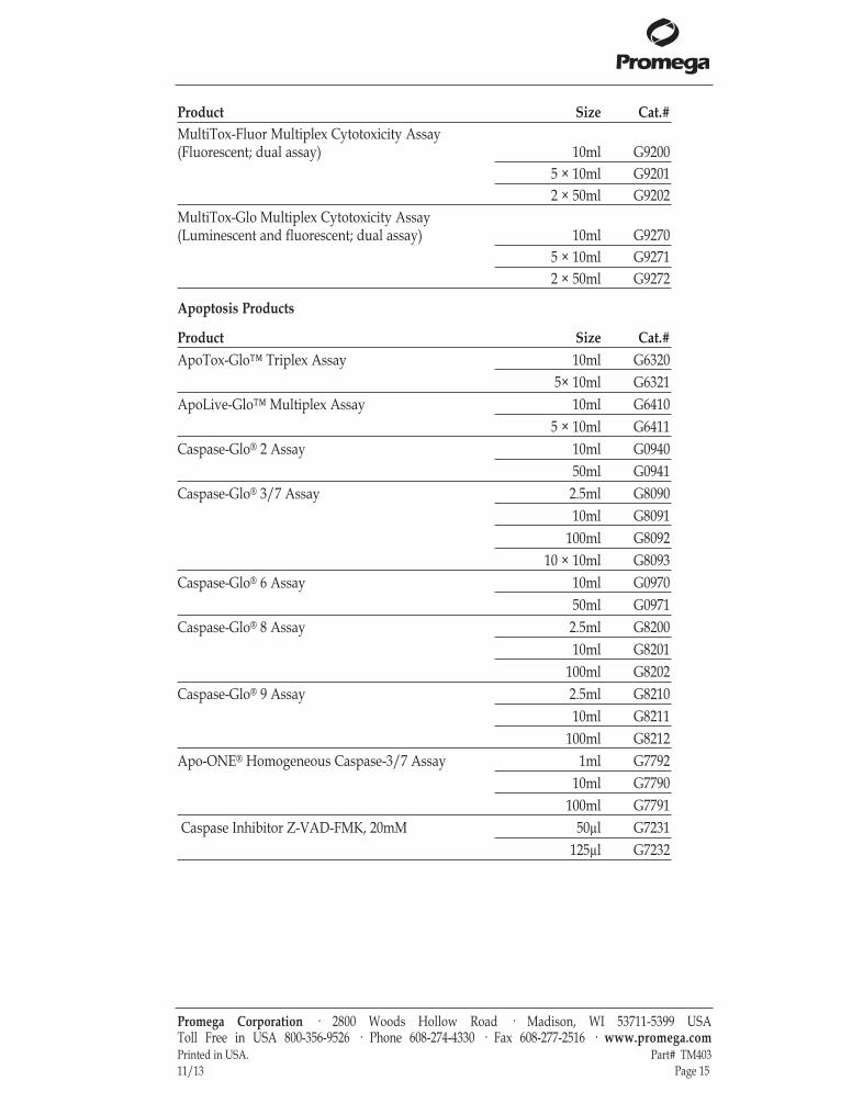
Product Size Cat.#MultiTox-Fluor Multiplex Cytotoxicity Assay(Fluorescent; dual assay) 10ml G9200
5 × 10ml G92012 × 50ml G9202
MultiTox-Glo Multiplex Cytotoxicity Assay(Luminescent and fluorescent; dual assay) 10ml G9270
5 × 10ml G92712 × 50ml G9272
Apoptosis Products
Product Size Cat.#ApoTox-Glo™ Triplex Assay 10ml G6320
5× 10ml G6321ApoLive-Glo™ Multiplex Assay 10ml G6410
5 × 10ml G6411Caspase-Glo® 2 Assay 10ml G0940
50ml G0941Caspase-Glo® 3/7 Assay 2.5ml G8090
10ml G8091100ml G8092
10 × 10ml G8093Caspase-Glo® 6 Assay 10ml G0970
50ml G0971Caspase-Glo® 8 Assay 2.5ml G8200
10ml G8201100ml G8202
Caspase-Glo® 9 Assay 2.5ml G821010ml G8211
100ml G8212Apo-ONE® Homogeneous Caspase-3/7 Assay 1ml G7792
10ml G7790100ml G7791
Caspase Inhibitor Z-VAD-FMK, 20mM 50µl G7231125µl G7232
Promega Corporation · 2800 Woods Hollow Road · Madison, WI 53711-5399 USA Toll Free in USA 800-356-9526 · Phone 608-274-4330 · Fax 608-277-2516 · www.promega.comPrinted in USA. Part# TM40311/13 Page 15
4.D. Related Products (continued)
Oxidative Stress and Metabolism Assays
Product Size Cat.#GSH-Glo™ Glutathione Assay 10ml V6911
50ml V6912GSH/GSSG-Glo™ Assay 10ml V6611
50ml V6612Mitochondrial ToxGlo™ Assay 10ml G8000
100ml G8001ROS-Glo™ H202 Assay 10ml G8820
50ml G8821NAD/NADH-Glo™ Assay 10ml G9071
50ml G9072NADP/NADPH-Glo™ Assay 10ml G9081
50ml G9082
Luminometers
Product Size Cat.#GloMax®-Multi+ Detection System with Instinct™ Software: Base Instrument with Shaking 1 each E8032GloMax®-Multi+ Detection System with Instinct™ Software: Base Instrument with Heating and Shaking 1 each E9032GloMax®-Multi+ Luminescence Module 1 each E8041
Promega Corporation · 2800 Woods Hollow Road · Madison, WI 53711-5399 USA Toll Free in USA 800-356-9526 · Phone 608-274-4330 · Fax 608-277-2516 · www.promega.comPart# TB370 Printed in USA.Page 16 Revised 11/11
(a)Patent Pending.(b)U.S. Pat. Nos. 6,602,677, 7,241,584 and 8,030,017, European Pat. No. 1131441, Japanese Pat. Nos. 4537573 and 4520084 andother patents pending(c)U.S. Pat. No. 7,741,067, Japanese Pat. No. 4485470 and other patents pending.(d)U.S. Pat. Nos 7,083,911, 7,452,663 and 7,732,128, European Pat. No. 1383914 and Japanese Pat. Nos. 4125600 and 4275715.(e)The method of recombinant expression of Coleoptera luciferase is covered by U.S. Pat. Nos. 5,583,024, 5,674,713 and 5,700,673.© 2013 Promega Corporation. All Rights Reserved.Apo-ONE, Caspase-Glo, CellTiter 96, CellTiter-Blue, CellTiter-Glo and GloMax are registered trademarks of PromegaCorporation. ApoLive-Glo, ApoTox-Glo, CellTiter-Fluor, CellTox, CytoTox-Fluor, CytoTox-Glo, CytoTox-ONE, GSH-Glo, GSH/GSSG-Glo, Instinct, NAD/NADH-Glo, NAD/NADPH-Glo, ToxGlo and Ultra-Glo are trademarks of PromegaCorporation.alamarBlue is a registered trademark of Trek Diagnostic Systems, Inc. Kimwipes is a registered trademark of Kimberly-ClarkCorporation.Products may be covered by pending or issued patents or may have certain limitations. Please visit our Web site for moreinformation.All prices and specifications are subject to change without prior notice.Product claims are subject to change. Please contact Promega Technical Services or access the Promega online catalog for themost up-to-date information on Promega products.

Promega Corpora on · 2800 Woods Hollow Road · Madison, WI 53711-5399 USA · Toll Free in USA 800-356-9526 · 608-274-4330 · Fax 608-277-2516 www.promega.com TM403 Printed 11/13