cells, tissues, organs, and systems [catch figure...
TRANSCRIPT

Cells, Tissues, Organs, and Systems
[CATCH FIGURE PUO10A: Photo of a humpback whale, ideally a shot taken in the waters off the coast of Newfoundland.]
Humpback whales are a common sight in the early spring along the south coast of Placentia Bay and Hermitage Bay. Maybe you have seen “nets” of bubbles the whales make from their blowholes to trap tiny krill, plankton, and small fish that the whales consume. Believe it or not, you have much in common with a humpback whale as well as all other living things, both large and small.
▲
384
NL8Unit4CH10.indd384 11/5/084:11:09PM

385
Key Ideas
10
12
11
The cell is the basic unit of life.
10.1 Characteristics of Life10.2 Focussing on Cells
Human body cells are organized as tissues, organs, and systems.
11.1 Cell Organization11.2 Introducing Human
Body Systems
The health of the human body depends on the health of its interdependent systems.
12.1 How Body Systems Are Connected
12.2 Body Systems and Health
NL8Unit4CH10.indd385 11/5/084:11:15PM
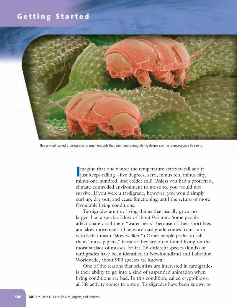
Imagine that one winter the temperature starts to fall and it just keeps falling—five degrees, zero, minus ten, minus fifty,
minus one hundred, and colder still! Unless you had a protected, climate-controlled environment to move to, you would not survive. If you were a tardigrade, however, you would simply curl up, dry out, and cease functioning until the return of more favourable living conditions.
Tardigrades are tiny living things that usually grow no larger than a speck of dust of about 0.5 mm. Some people affectionately call them “water bears” because of their short legs and slow movement. (The word tardigrade comes from Latin words that mean “slow walker.”) Other people prefer to call them “moss piglets,” because they are often found living on the moist surface of mosses. So far, 26 different species (kinds) of tardigrades have been identified in Newfoundland and Labrador. Worldwide, about 900 species are known.
One of the reasons that scientists are interested in tardigrades is their ability to go into a kind of suspended animation when living conditions are bad. In this condition, called cryptobiosis, all life activity comes to a stop. Tardigrades have been known to
386 MHR•Unit 4 Cells, Tissues, Organs, and Systems
This animal, called a tardigrade, is small enough that you need a magnifying device such as a microscope to see it.
NL8Unit4CH10.indd386 11/5/084:11:24PM

survive temperatures as high as 150°C and as low as –273°C. They can also survive powerfully lethal doses of X rays, poisonous chemicals, the airless vacuum of outer space, and pressures greater than six times that of the deepest ocean bottom. Studying cryptobiosis helps scientists to gain a better understanding of life, death, and what distinguishes one from the other. Such an understanding might some day help us travel to and live on distant planets or cure deadly diseases.
Living or Non-living? Find Out ACTIVITY
Go to Science Skill 6 for information about conducting a fair test.
Science SkillsWhat makes one thing, such as a tardigrade, living and another thing, such as a stone, not living (non-living)? In this activity, you will discuss ideas with your partners to help you find out whether two similar-looking things are living or non-living.
Materials• 2 samples in separate containers• magnifying glass• ruler• 2 bowls• warm sugar water
What to Do1. Your teacher will give you two containers
of similar-looking samples. Your task is to determine which characteristics these samples have in common and which characteristics they do not have in common. Then you will decide if one or both of these samples is a living thing.
2. Examine both samples using the magnifying glass. Use any other equipment available to help you add to your observations. Record your observations in a chart.
3. Place a small amount of each sample in separate bowls. Add equal amounts of warm sugar water to both bowls. Observe and record any changes.
4. Perform one more test you think will provide evidence that one of these samples is living. Record your observations.
5. Clean up and put away the equipment you have used.
What Did You Find Out?1. Discuss your results with your class.
2. Decide which observations suggest that one or both of the samples is living. Record these observations in a list.
3. Based on your evidence and your experiences in this activity, write a sentence that clearly explains what distinguishes a living thing from a non-living thing.
Word Connect
Cryptobiosis comes from two Greek words that mean “hidden life condition,” referring to the unknown way in which tardigrades can stay alive in otherwise deadly living conditions.
Unit 4 Getting Started • MHR 387
NL8Unit4CH10.indd387 11/5/084:11:31PM

T he stentor, which is about 2 mm in length, lives in many freshwater ditches and lakes throughout the world. The trumpet shape of stentor’s
head and neck is like a bullhorn used by people to broadcast a message to a large crowd. The tiny stentor gets its name from a character named Stentor, who was a loud-voiced messenger in an ancient poem called The Iliad.
Take a close look at the cup-shaped head and long-narrow neck. Do you see the tiny hairs around the top of the head? These hairs all wave together to create a small water current that pulls food into the bottom of the head. The long neck can slowly move up in search of food and snap back down if danger approaches.
A stentor is a living thing composed of one cell. All the life processes that keep the stentor alive take place inside this single cell. This is why the cell is called the basic unit of life. You will learn more about cells and the technologies we use to study them in this chapter.
388 MHR•Unit 4 Cells, Tissues, Organs, and Systems
NL8Unit4CH10.indd388 11/5/084:11:37PM

Chapter 10 The cell is the basic unit of life.•MHR 389
What You Will Learn
In this chapter, you will• identify the characteristics of living
things• identify and state the major functions
of the parts of a compound light microscope
• identify common structures of plant and animal cells, and explain their functions
Why It Is Important
Understanding how cells function can help you understand how your body and other living systems function.
Skills You Will Use
In this chapter, you will• learn the safe use of a microscope• use a light microscope to produce a clear
image of cells• model the structures and functions of a
cell
FOLDABLES TM
Reading & StudySkills
Make the following Foldable to take notes on what you will learn in Chapter 10.
STEP 1 Collect 2 sheets of letter size paper and layer them 2.5 cm apart vertically. (Hint: from the tip of your index finger to your first knuckle is about 2.5 cm.) Keep the edges level.
STEP 2 Fold up the bottom edges of the paper to form 4 tabs.
STEP 3 Fold the papers and crease well to hold the tabs in place. Staple along the fold.
STEP 4 Label the tabs as shown. (Note: the first tab will be larger than shown here.)
Summarize As you read the chapter, summarize what you learn under the appropriate tabs.
The cell is the basicunit of life.
Living and Non-livingThings
Cells
The Microscope
NL8Unit4CH10.indd389 11/5/084:11:43PM

Imagine that you are a scientist in the early days of civilization. You observe your surroundings, such as those in Figure 10.1, and you start to wonder about how all the objects in the world around you are similar and how they are different. For instance, you wonder how these objects are different from you. You know that you are alive, but what does this mean? What is it that makes you a living thing, while a stone, a puddle, or a candle is not a living thing?
Movement is one of the signs that something is alive. Is it always a sign of life, though? A rock rolling down a hill moves. So does rain falling from the sky. Growth is another sign that something is alive. However, a mound of rice gets bigger as you pour it, and you have certainly seen icicles get bigger and longer in the winter. These types of “growth” are different from growth in living things, however. What makes rice, ice, and stones different from you and other living things such as cats, grass, and stentor?
Key Termsarmbasecellcoarse adjustment knobcompound light microscopeeyepiecefine adjustment knobiris diaphragmlight sourcemagnification powerobjective lensesresolving powerrevolving nosepiecestagetotal magnificationtube
The cell is the basic functional unit of life. All living things have characteristics that demonstrate they are alive. These include the ability to grow, to move, to reproduce, and to respond to stimuli. Some living things are very small and can be observed only with a microscope. To study tiny living things on prepared or wet mount slides, you must handle a compound light microscope carefully and learn how to operate it correctly.
Characteristics of Life10.1
390 MHR•Unit 4 Cells, Tissues, Organs, and Systems
Figure 10.1 Which objects in this scene are living? Which ones are non-living? What ideas are you using to help you decide?
NL8Unit4CH10.indd390 11/5/084:11:46PM

The Smallest Unit of Life: The CellFor scientists, one feature separates all forms of life from everything else. All living things are made up of one or more cells. The cell is the smallest, most basic functional system of any living thing. A functional system is any system that exhibits all of the characteristics of life outlined in Table 10.1. Something must have all of these characteristics to be considered a living thing.
Chapter 10 The cell is the basic unit of life. • MHR 391
Table 10.1 Four Characteristics of Living Things
All living things grow.As you continue to grow as a teenager, you get taller and the mass of your bones and muscles increases. Your growth is the result of the cells in your body increasing in number. Even when you stop growing, your body will grow new cells as old ones die.
All living things move.Movement involves changes to the shape, position, or location of the body or body parts. For instance, animals might have legs, wings, or fins to move from one location to another. Plants might have stems that change position with the Sun. Many single-celled living things (unicellular organisms) change their shape or have hair-like body parts that help them move or sweep in food.
All living things respond to stimuli in their environment.A cat may hiss when it feels threatened by something in its external environment. Hissing is the cat’s response to a stimulus. A stimulus (plural: stimuli) is anything that causes a living thing to respond. Living things also respond to stimuli in their internal environment. Think of the last time you were hungry or thirsty. Hunger and thirst are stimuli that cause you to respond by eating or drinking.
All living things reproduce.Through reproduction, living things produce more of their own kind (offspring). Some living things (such as bacteria and some kinds plants) produce offspring that are identical to themselves, while other living things (such as most animals) produce offspring that are similar to themselves.
Examining Very Small Living ThingsIt might surprise you to know that there are many more living things that you cannot see with the unaided eye than ones that you can. The human eye can see objects that are larger than 0.1 mm. To see anything smaller, you have to use a microscope.
There are different types of microscopes, some of which you may have used to look at a leaf or an ant. A compound light microscope is the type of microscope that you will use in this unit to investigate living things. Observing living things through a microscope is one of science’s most exciting and rewarding experiences. You will be introduced to this important study tool in this section.
NL8Unit4CH10.indd391 11/5/084:11:49PM

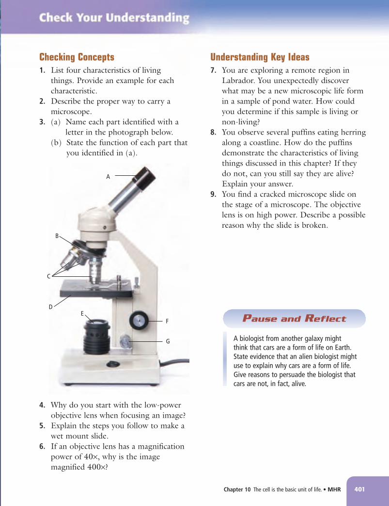
The Compound Light MicroscopeFigure 10.2 shows the correct way to hold and carry a compound light microscope. This is the type of microscope usually used in science classes and medical laboratories. Figure 10.3 shows the parts of the compound light microscope, and Table 10.2 outlines their functions.
392 MHR•Unit 4 Cells, Tissues, Organs, and Systems
Figure 10.2 Always carry a microscope with one hand on the arm and one hand on the base.
eyepiece
coarse adjustment knob
fine adjustment knob
light source
base
revolving nosepiece
objective lenses
stage
tube (or barrel)
arm
iris diaphragm
Figure 10.3 A compound light microscope
NL8Unit4CH10.indd392 11/5/084:11:52PM

How a compound light microscope works
Two sets of lenses work together to magnify and focus an image. When you look through this microscope, you see an image that is magnified (made larger), inverted (upside down), and reversed (backwards). See Figure 10.4.
Magnification
The magnification power of a lens is the number of times larger an image looks under the lens. Each objective lens has a number that states its magnification power (see Figure 10.5). Most school microscopes have these magnification powers:• low-power objective lens (4×)• medium-power objective lens (10×)• high-power objective lens (40×)
Usually the eyepiece lens has a magnification power of 10X. To find the total magnification of the microscope for each objective lens, you multiply the power of the objective lens by the power of the eyepiece. For example:
low-power objective lens × eyepiece lens
total magnificationor
4 × 10 = 40The total magnification of a medium-power lens is 100× and
a high-power lens is 400×.
Chapter 10 The cell is the basic unit of life. • MHR 393
Table 10.2 The Parts of a Compound Light Microscope
Part Function
Eyepiece Is used for viewing and contains a lens that magnifies Tube Holds the eyepiece and objective lenses at proper distance from each other Arm Supports the eyepiece Coarse adjustment knob Brings an object into focus at low or medium power Fine adjustment knob Brings an object into focus at high power Objective lenses Magnify the image. Most microscopes have three or four lenses. Revolving nosepiece Holds the three objective lenses Stage Supports the slide. Some microscopes have stage clips to hold the slide in place. Iris diaphragm controls the amount of light reaching the specimen Light source Supplies the light needed to view the slide Base Supports the entire microscope
Section 6.3 has more information about how a compound light microscope works.
Connection
Figure 10.4 The letter “e” seen through the lens of a microscope will appear like this.
objective lenses
high power
medium power
low power
Figure 10.5 Magnification power of the three objective lenses
Conduct an Investigation 10.1A on page 394
Find Out Activity 10.1B on page 397
Suggested Activities
NL8Unit4CH10.indd393 11/5/084:11:53PM

394 MHR•Unit 4 Cells, Tissues, Organs, and Systems
Safety• Microscopes, slides, and
cover slips can break, especially when using the high-power objective lens. Handle with care.
• Be careful when using sharp objects such as tweezers.
• Wash your hands thoroughly after doing this investigation.
Materials• microscope• prepared microscope
slides• see-through plastic ruler• lens paper• microscope slides• cover slips• medicine droppers• tweezers• water• live specimens
Using a microscope can open up an exciting new world of discovery. In this activity, you will practise using a compound light microscope carefully and accurately. You will also examine some living and non-living things and learn how to prepare your own slides.
Procedure
Part 1 Focus the Image
1. Pick up your microscope and bring it back to your work table. Check that it is set to the low-power objective lens.
2. Select a prepared slide from the ones provided by your teacher. Place the slide on the stage of the microscope. If your microscope has stage clips, use them to hold your slide in place.
3. Turn the coarse adjustment knob carefully to bring your image into focus. Draw and label what you observe.
4. Move your slide to the right. Which way does the image move?
5. Move your slide up. Which way does the image move?
6. Change the lens to medium power and focus the image. You may need to turn the fine adjustment knob to make minor adjustments so that you can bring the image into focus. Draw and label what you observe.
Part 2 Determine the Field of View
1. Place a see-through ruler on the stage and focus on the ruler at low power.
2. Record the length of the ruler you can see at low power. This is called the field of view.
3. You can use the field of view to estimate the size of an object you are viewing at low power. For example, the field of view at low power is usually 4.2 mm. If an object takes up half the field of view at low power, this would mean that its approximate size would be 2.1 mm. Another way to estimate the size of an object your are viewing is to estimate how many of that object could fit end-to-end across the field of view. To do this, divide the field of view by the number of objects. The answer is the approximate size of one object. (Note: Millimetre markings on the ruler are too far apart to permit direct measurement of the field of view for lenses with a magnification higher than 10×.)
4. Select another prepared slide and determine the approximate size of an object at low power. Record your answers.
Setting Up and Using a Microscope10-1A
SkillCheck• Observing
• Measuring
• Communicating
• Working co-operatively
Core Lab
NL8Unit4CH10.indd394 11/5/084:11:58PM

Chapter 10 The cell is the basic unit of life. • MHR 395
Part 3 Make a Wet Mount Slide
1. You can make your own slide rather than use a prepared one. This type of slide is called a wet mount slide. To prepare a wet mount slide, follow the instructions in the photos and captions below. (Make sure your slide is clean before you begin. If it is not, use lens paper to wipe it off.)
2. Prepare a wet mount slide of a strand of hair. Place the slide on the stage of the microscope. Observe and draw the hair at low power and then at medium power. Label your drawings.
3. Your teacher will provide you with a live specimen to observe. Prepare a wet mount slide. After placing the slide on the stage, determine which power would be best for observing it. Make your observations, then draw the specimen and label your drawing.
4. When you have completed Procedure steps 1–3, make sure your microscope is set at low power.
5. Clean up and put away the equipment you have used.
Analyze1. Compare the drawings you made in this activity. Describe how your images changed when you increased
the power of magnification. For example, did you see more or less of an image, or was it easier or harder to focus on the whole image?
2. To view the letter "e" through your microscope the right way up, how would you position the slide on the stage?
3. You are looking at an image at low power. You see a round object in the top left corner of the image that you would like to see more clearly. You switch to medium power. List the steps you would have to take to get the round object in the middle of your view.
Conclude and Apply1. A classmate has missed this lab activity and has asked you to explain how to use a microscope properly.
Write a step-by-step set of directions. Include labelled diagrams where needed.
Conduct an InVesTIgATIOn
Inquiry Focus
Place a drop of water on the slide. Use tweezers to place your specimen in the drop of water.
Hold the cover slip at a 45° angle and gently lower it onto the slide. There should be no air bubbles under the cover slip. If there is any excess water on the slide, dab a piece of tissue paper on the slide.
A B C
NL8Unit4CH10.indd395 11/5/084:12:04PM

Resolving power
Examine the four circles in Figure 10.6. All the dots in circle A are 1 mm in diameter. They are also 1 mm apart. Now look at circles B, C, and D. Can you see the dots clearly in each picture?
Most people cannot see the dots in picture D. This is normal. Average human eyesight means people can see only dots that are separated by a distance of 0.l mm or more. The microscope extends human vision by allowing you to view objects that are smaller and closer together. The ability to distinguish between two dots or objects that are very close together is called resolving power. The resolving power of a compound light microscope is about 0.2 microns (μm), which means that the microscope cannot separate two images that are less than 0.2 μm apart. A micron is one-millionth of one metre. If you were to observe a living thing that was one micron in size, you could fit one million of them side by side on a metre stick.
396 MHR•Unit 4 Cells, Tissues, Organs, and Systems
Figure 10.6 In which picture are you unable to make out individual dots?
A B
C D
NL8Unit4CH10.indd396 11/5/084:12:07PM

Observing Organisms in Pond Water
10-1B Find Out ACTIVITY
In this activity, you will use a compound light microscope to observe a variety of organisms that live in pond water. As you are looking at these organisms, try to determine how each one demonstrates the characteristics of living things. For example, observe the different ways they move or respond to stimuli.
Materials• microscope• microscope slide• cover slips• medicine dropper• tweezers• pond water
SafetyHandle microscope slides and cover slips very carefully to avoid breaking them or cutting yourself. Treat the pond organisms respectfully. They are alive.
What to Do1. Obtain a sample of pond water from your
teacher. Make a wet mount slide with this sample. (See Conduct an Investigation 10.1A)
2. Examine the slide under low and medium power, looking for different organisms in the pond water.
3. Draw what you see for at least two different organisms. Include as much detail as possible.
4. For each organism, record which characteristics of living things you observe.
5. Clean up and put away the equipment you have used.
What Did You Find Out?1. Summarize the evidence you collected that
demonstrates the organisms you observed were living. Use a chart or write a paragraph to help you organize your summary.
Chapter 10 The cell is the basic unit of life. • MHR 397
Reading Check1. What is a cell?2. Name all the parts of a compound light microscope that
are directly responsible for making a magnified image of an object. (Hint: There are three.)
3. Draw how the letter “G” would appear when viewed through a compound light microscope.
4. What is the total magnification for the medium-power objective lens?
NL8Unit4CH10.indd397 11/5/084:12:08PM

M icroscopes give us a glimpse into a previously invisible world. Improvements have vastly increased their range of visibility, allowing
researchers to study life at the molecular level. A selection of these powerful tools—and their magnification power—is shown here.
BRIGHTFIELD / DARKFIELD MICROSCOPE The light microscope is often called the brightfield microscope because the image is viewed against a bright background. A brightfield microscope is the tool most often used in laboratories to study cells. Placing a thin metal disc beneath the stage, between the light source and the objective lenses, converts a brightfield microscope to a darkfield microscope. The image seen using a darkfield microscope is bright against
a dark background. This makes details more visible than with a brightfield microscope. Below are images of a paramecium as seen using both processes.
▼
LEEUWENHOEK MICROSCOPE Held by a modern researcher, this early microscope allowed its user to view stentor and other single-celled organisms for the first time.
Up to 300▼
FLUORESCENCE MICROSCOPE This type of microscope requires that the specimen be treated with special fluorescent stains. When viewed through this microscope, certain cell structures or types of substances glow, as seen in the image of a paramecium above.
Up to 1500
▼darkfield
brightfield
Visualizing MiCRosCopes
Up to 2000
398 MHR•Unit 4 Cells, Tissues, Organs, and Systems
NL8Unit4CH10.indd398 11/5/084:12:23PM

PHASE-CONTRAST MICROSCOPE A phase-contrast microscope emphasizes slight
differences in a specimen’s capacity to bend light waves, thereby enhancing light and dark regions without the use of stains. This type of microscope is especially good for viewing living cells, like the paramecium above left. The images from a phase- contrast microscope can only be seen when the specimen is photographed or shown on a monitor.
Up to 1500
▼
SCANNING ELECTRON MICROSCOPE An SEM sweeps a beam of electrons over a specimen’s surface, causing other electrons to be emitted from the specimen. SEMs produce realistic, three-dimensional images, which can only be viewed as photographs or on a monitor, as in the image of the paramecium at right. Here a researcher compares an SEM picture to a computer monitor showing an enhanced image.
Up to 200 000▼
TRANSMISSION ELECTRON MICROSCOPE A TEM aims a beam of electrons through a specimen. Denser portions of the specimen allow fewer electrons to pass through and appear darker in the image. Organisms, such as the paramecium at right, can only be seen when the image is photographed or shown on a monitor. A TEM can magnify hundreds of thousands of times.
▼ Up to 1 000 000
Chapter 10 The cell is the basic unit of life. • MHR 399
NL8Unit4CH10.indd399 11/5/084:12:35PM

400 MHR•Unit 4 Cells, Tissues, Organs, and Systems
Size and ScaleThe table below summarizes some of the units of measurement you will use in this textbook and future science courses. The table includes the prefix, the symbol, and the mathematical measurement. Three of these units are commonly used to describe living and non-living things that are observed through a microscope. Notice that each of these mathematical measurements increases by a factor (power) of 1000 (103). Use the scale in the diagram below to help you understand what this means.
Prefix Symbol Mathematical Measurement
nano n 10–9 (billionth) micro μ 10–6 (millionth) milli m 10–3 (thousandth) kilo k 103 (thousand) mega M 106 (million)
atoms amino acids
viruses
proteins
chloroplast
most bacteria
human egg
frog egg
ant
mouse
childblue whale
human eye
compound light microscope
electron microscope
plant and animal cells
1.0 nm 10.0 nm 100.0 nm 1.0 µm 10.0 µm 100.0 µm 1.0 mm 1.0 cm 0.1 m 1.0 m 10.0 m 100.0 m 1000.0 m (1.0 km) 0.1 nm
The scale in this diagram increases by a factor, or power, of 10. Using this type of scale makes it mathematically easier to work with very small and very large numbers.
Question
1. If the thickness of one sheet of paper in this textbook is 0.2 mm, how high would a pile of paper be for:
(a) 1000 sheets (thousand) (b) 100 000 sheets (hundred thousand) (c) 1 000 000 sheets (million) (d) 1 000 000 000 sheets (billion)
NL8Unit4CH10.indd400 11/5/084:12:39PM
Chapter 10 The cell is the basic unit of life. • MHR 401
Checking Concepts1. List four characteristics of living
things. Provide an example for each characteristic.
2. Describe the proper way to carry a microscope.
3. (a) Name each part identified with a letter in the photograph below.
(b) State the function of each part that you identified in (a).
4. Why do you start with the low-power objective lens when focusing an image?
5. Explain the steps you follow to make a wet mount slide.
6. If an objective lens has a magnification power of 40×, why is the image magnified 400×?
Understanding Key Ideas7. You are exploring a remote region in
Labrador. You unexpectedly discover what may be a new microscopic life form in a sample of pond water. How could you determine if this sample is living or non-living?
8. You observe several puffins eating herring along a coastline. How do the puffins demonstrate the characteristics of living things discussed in this chapter? If they do not, can you still say they are alive? Explain your answer.
9. You find a cracked microscope slide on the stage of a microscope. The objective lens is on high power. Describe a possible reason why the slide is broken.
A
G
E
B
C
D
F
A biologist from another galaxy might think that cars are a form of life on Earth. State evidence that an alien biologist might use to explain why cars are a form of life. Give reasons to persuade the biologist that cars are not, in fact, alive.
Pause and Reflect
NL8Unit4CH10.indd401 11/5/084:12:43PM

Imagine a planet in a nearby galaxy. There is a star like our Sun that provides heat and light. Although smaller than Earth, the planet supports many different life forms. One life form is the Icthos (pronounced ICK-THOSS). They are an advanced life form and have developed a variety of technologies, such as the Protection Dome, inside which they live (see Figure 10.7). Unlike life forms on Earth, the Icthos live in a liquid, not an air atmosphere.
Over time, the Icthos have spread across the planet and settled in groups known as colonies. One of these colonies is called Newo (pronounced KNEW-OH). Newo is thriving, and the population has increased rapidly. The residents of Newo go about their daily activities, such as ingesting food packets and nutrient fluids (eating and drinking) and dreamdozing (sleeping).
Newo, however, has some serious problems, including pollution and a need for a new energy source. Management groups are responsible for performing different functions within the colony. You are a specialist on colony operations. The management groups have sent you their reports about their biggest problems. Your first task is to read these reports carefully.
Key Termscell membranecell theorycell wallchloroplastcytoplasmmitochondriamitosisnucleusorganellevacuole
Using an analogy can help you understand science concepts, such as how the parts of a cell function. All cells have similar structures and organelles. Each structure and organelle carries out a specific task to help support the life functions of a cell. Cellular respiration is the process that produces energy for the cell. Cell theory states that the cell is the basic unit of life; all living things are composed of one or more cells; all cells come from other living cells.
Focussing on Cells10.2
402 MHR•Unit 4 Cells, Tissues, Organs, and Systems
closed gate
open gate
Figure 10.7 The Newo colony is an analogy of the structure and function of the cell.
NL8Unit4CH10.indd402 11/5/084:12:46PM
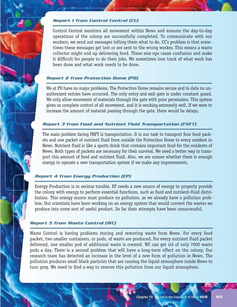
Report 1 from Control Central (CC)
Control Central monitors all movement within Newo and ensures the day-to-day operations of the colony are successfully completed. To communicate with our workers, we send out messages telling them what to do. CC’s problem is that some-times these messages get lost or are sent to the wrong worker. This means a waste collector might end up delivering food. These mix-ups cause confusion and make it difficult for people to do their jobs. We sometimes lose track of what work has been done and what work needs to be done.
Report 2 from Protection Dome (PD)
We at PD have no major problems. The Protection Dome remains secure and to date no un-authorized entries have occurred. The only entry and exit gate is under constant guard. We only allow movement of materials through the gate with prior permission. This system gives us complete control of all movement, and it is working extremely well. If we were to increase the amount of material passing through the gate, there would be delays.
Report 3 from Food and Nutrient Fluid Transportation (FNFT)
The main problem facing FNFT is transportation. It is our task to transport four food pack-ets and one packet of nutrient fluid from outside the Protection Dome to every resident in Newo. Nutrient fluid is like a sports drink that contains important food for the residents of Newo. Both types of packets are necessary for their survival. We need a better way to trans-port this amount of food and nutrient fluid. Also, we are unsure whether there is enough energy to operate a new transportation system if we make any improvements.
Report 4 from Energy Production (EP)
Energy Production is in serious trouble. EP needs a new source of energy to properly provide the colony with energy to perform essential functions, such as food and nutrient-fluid distri-bution. This energy source must produce no pollution, as we already have a pollution prob-lem. Our scientists have been working on an energy system that would convert the wastes we produce into some sort of useful product. So far their attempts have been unsuccessful.
Report 5 from Waste Control (WC)
Waste Control is having problems storing and removing waste from Newo. For every food packet, two smaller containers, or pods, of waste are produced. For every nutrient fluid packet delivered, one smaller pod of additional waste is created. WC can get rid of only 7000 waste pods a day. There is a second problem that will have a long-term effect on the colony. Our research team has detected an increase in the level of a new form of pollution in Newo. The pollution produces small black particles that are causing the liquid atmosphere inside Newo to turn grey. We need to find a way to remove this pollution from our liquid atmosphere.
Chapter 10 The cell is the basic unit of life. • MHR 403
NL8Unit4CH10.indd403 11/5/084:12:48PM

Using an Analogy to Understand a CellThe colony of Newo is an analogy for a cell. An analogy is a way to understand new ideas by comparing the new ideas to something that is more familiar. For example, you have learned that each management group in Newo carries out a specific task. You have also seen how these groups work together to ensure the survival of the colony. Similarly, cells have different structures that carry out specific functions. In working together, all the structures of the cell help to keep it alive. The structures of a cell that perform a specific function are called organelles. Organelles occupy up to 30 percent of a cell. The rest of the cell consists of water.
Each organelle has a role to play in the activities that are necessary for the life of the cell. Table 10.3 on the next page lists some of the major organelles that are found in animal cells and in plant cells. Look closely at the diagrams and the information listed in the table. Take note of which organelles are found in both animal and plant cells and which organelles are found only in plant cells.
404 MHR•Unit 4 Cells, Tissues, Organs, and Systems
Finding Solutions for Problems in the Newo Colony
10-2A Find Out ACTIVITY
In this activity, your task is to work with your classmates to figure out a possible solution for each of the problems facing the management groups of Newo. Recall that the management groups are:
• Control Central• Protection Dome• Food and Nutrient Fluid Transportation• Energy Production• Waste Control
What to Do1. Record each problem the management groups
identified.
2. Brainstorm solutions for each problem.
3. Select the best solution for each problem. Each solution must also work with the other solutions you select.
4. Make a drawing of Newo that shows how you solved each problem. Use labels and descriptions to help explain your solutions.
What Did You Find Out?1. Post your drawing on the wall.
2. Walk around and look at the drawings of other classmates. Make notes on what you observe in these drawings. Find an example of a drawing that:
(a) Shows a solution different from yours. Record what is different.
(b) Shows a solution the same as yours. Record what is the same.
3. Return to your drawing. Based on your observations, what would you change about one of your solutions so that it works better? Make this change on your drawing.
4. Share your own drawing with the class and discuss which solutions would probably work best for solving Newo’s problems.
Create a table with two columns and seven rows. In the first column, list the cell organelles. In the second column, list the corresponding management group from the Newo colony. Invent a management group for any organelle that does not have one.
NL8Unit4CH10.indd404 11/5/084:12:51PM

Chapter 10 The cell is the basic unit of life. • MHR 405
Table 10.3 Common Organelles of Animal and Plant Cells
Cell Organelle Structure and Function
[A] cell membrane • found in animal cells and plant cells • surrounds and protects the contents of the cell • helps to control the movement of foods, wastes, and other substances into the cell and out of the cell
[B] cytoplasm • found in animal cells and plant cells • jelly-like, watery fluid in which internal organelles float • helps to distribute materials such as food and oxygen to different parts of the cell [C] cell wall • found in plant cells but not animal cells • tough, rigid structure that surrounds the cell membrane and gives plant cells a regular, box-like shape • made mostly of a material called cellulose (which you might know better as dietary fibre) [D] nucleus • found in animal cells and plant cells • large, round structure often clearly visible in most cells • contains the chromosomes—the structures that are made of genetic material that control a cell’s growth, reproduction, and other life-sustaining activities [E] vacuole • balloon-like spaces within the cytoplasm • provide space to store extra food, wastes, and other substances that the cell cannot use right away • found in animal cells and plant cells (they are smaller and more numerous in animal cells) [F] mitochondria • oval, bean-shaped structures (singular: mitochondrion) • produce energy for the cell by breaking down food particles to release their stored energy • found in animal cells and plant cells [G] chloroplast • green structures that contain a green pigment (coloured substance) called chlorophyll • capture energy from the Sun, which is used to produce food (sugars) in the leaves and green stems of plants (this process is called photosynthesis) • found in plant cells but not animal cells
Animal Cell Plant Cell
A
E
D
B
A
E
D
B
F
C
G
F
NL8Unit4CH10.indd405 11/5/084:12:58PM

The Importance of the Cell MembraneThink about the Newo colony. One management group that had a problem was the Protection Dome—the barrier around the colony. It had only one entrance and exit. The Food and Nutrient Fluid Transportation group had to find other ways to get materials into the colony. The Waste Control group had to find new ways to get wastes out. A cell would have problems like these if the cell membrane had just one entrance and exit.
The cell membrane has many openings, but they are selective. They let some substances enter and leave the cell, but they also prevent other substances from entering and leaving.Because only some substances can cross it, the cell membrane is said to be selectively permeable. This is like a coffee filter—hot water can move through the filter, but the coffee grounds cannot.
The Cell TheoryDuring the 1800s, many scientists around the world were using compound light microscopes to study cells. The cell nucleus was first described in 1802, and it was studied in more detail about 30 years later. By 1846, the importance of the cytoplasm to the life of the cell was appreciated. A year later, the cell membrane was described. As time went on, more organelles were discovered, and their functions were investigated and described.
By the mid 1850s, scientists had recorded thousands of observations about the cells of plants, animals, and other living things. Based on their studies, scientists agreed on three important facts about cells and their connection with living things. Together, these facts are called the cell theory. The cell theory is one of the key ideas of biology. It helps scientists to describe and explain their observations of living things.
406 MHR•Unit 4 Cells, Tissues, Organs, and Systems
Word Connect
The word permeable comes from a Latin word that means “to pass through.” You might know another word that is related: permeate. For instance, the smell of frying onions permeates (moves or spreads through) the kitchen.
The Cell Theory• The cell is the basic unit of life.• All living things are made up of one or more cells.• All cells come from other living cells.
Reading Check1. What are organelles?2. Why is the cell membrane said to be selectively permeable?3. Why is the cell theory valuable to scientists?
NL8Unit4CH10.indd406 11/5/084:13:01PM
Chapter 10 The cell is the basic unit of life. • MHR 407
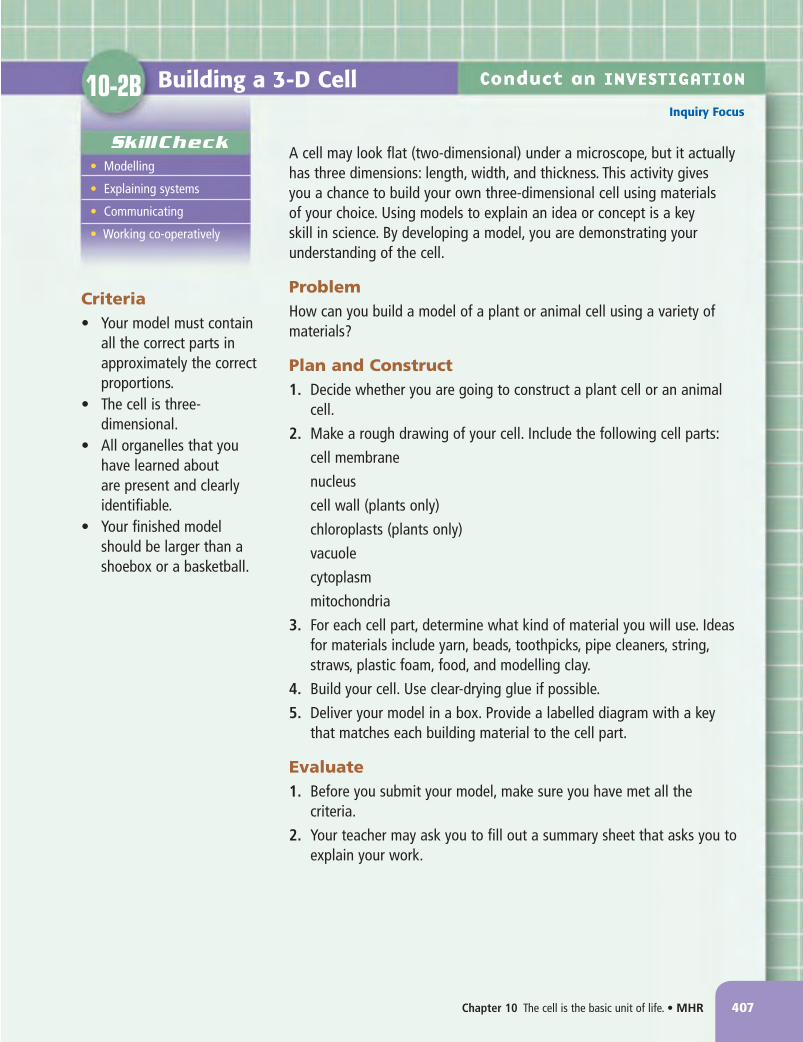
Building a 3-D Cell10-2B
SkillCheck• Modelling
• Explaining systems
• Communicating
• Working co-operatively
Conduct an InVesTIgATIOn
Inquiry Focus
A cell may look flat (two-dimensional) under a microscope, but it actually has three dimensions: length, width, and thickness. This activity gives you a chance to build your own three-dimensional cell using materials of your choice. Using models to explain an idea or concept is a key skill in science. By developing a model, you are demonstrating your understanding of the cell.
ProblemHow can you build a model of a plant or animal cell using a variety of materials?
Plan and Construct1. Decide whether you are going to construct a plant cell or an animal
cell.
2. Make a rough drawing of your cell. Include the following cell parts:
cell membrane
nucleus
cell wall (plants only)
chloroplasts (plants only)
vacuole
cytoplasm
mitochondria
3. For each cell part, determine what kind of material you will use. Ideas for materials include yarn, beads, toothpicks, pipe cleaners, string, straws, plastic foam, food, and modelling clay.
4. Build your cell. Use clear-drying glue if possible.
5. Deliver your model in a box. Provide a labelled diagram with a key that matches each building material to the cell part.
Evaluate1. Before you submit your model, make sure you have met all the
criteria.
2. Your teacher may ask you to fill out a summary sheet that asks you to explain your work.
Criteria• Your model must contain
all the correct parts in approximately the correct proportions.
• The cell is three-dimensional.
• All organelles that you have learned about are present and clearly identifiable.
• Your finished model should be larger than a shoebox or a basketball.
NL8Unit4CH10.indd407 11/5/084:13:02PM

408 MHR•Unit 4 Cells, Tissues, Organs, and Systems
Safety• Microscopes, slides, and
cover slips can break. Handle with care.
• Be careful when using sharp objects such as tweezers.
• Wash your hands thoroughly after doing this activity.
MaterialsPart 1• microscope• microscope slides• cover slips• lens paper• tweezers• medicine droppers• water• onion• iodine solution• paper towel
Part 2• microscope• prepared slide of human
skin cells• lens paper
In this investigation, you will continue to develop your microscope skills by preparing and observing a wet mount of onion skin cells and observing a prepared slide of human skin cells. You will also learn about a process called staining. Scientists stain cells to help them see organelles that are not visible in a standard wet mount slide.
QuestionWhat do plant and animal cells look like through a microscope?
Procedure
Part 1: Observing Plant Cells
1. Obtain your microscope and the materials you will need to make a wet mount slide.
2. Clean your slides with lens paper before you begin. Prepare a wet mount slide by putting a drop of water on the slide. Review Part 3 of Conduct an Investigation 10-1A if you want to review how to prepare a wet mount slide.
3. Take a piece of onion from the outer layer and carefully break it in half. As you separate the two sections, use tweezers to pull the top layer of the onion sideways as shown below. This should give you a sample of translucent onion skin.
4. Place the onion skin in the drop of water on the slide. Finish making your wet mount slide.
5. Place your slide on the stage of the microscope and focus at low power. Select one cell and draw it. Include all the organelles you recognize, and label them.
6. Place a drop of iodine solution on one side of your slide. On the other side, place a small piece of paper towel as shown below. The paper towel will soak up the water under the cover slip and draw the iodine solution under the slide and into the cells. This process is called staining the cells.
Observing Plant and Animal Cells10-2C
SkillCheck• Observing
• Communicating
• Evaluating information
Pull the top layer of the onion skin sideways to make a thin section.
NL8Unit4CH10.indd408 11/5/084:13:08PM

Chapter 10 The cell is the basic unit of life. • MHR 409
7. Estimate the size of one cell under low power. First, recall the diameter of the field of view from Investigation 10-1A. Next, estimate how many cells could fit end to end across the field of view. Then divide the field of view by the number of cells you estimated. For example, if the field of view is 1.5 mm, and if you estimated 10 cells, then 1.5 ÷ 10 = 0.15. Each cell would be about 0.15 mm in diameter.
8. Observe the onion cell under medium and high power. Add any more organelles you observe to your drawing and label them.
9. Your teacher either will have you continue on to Part 2 or will ask you to clean up and put away your equipment.
Part 2: Observing Animal Cells
1. Obtain a prepared slide of human skin cells.
2. Set up the slide on your microscope, and examine the skin cells under low power. Select one cell and draw it. Include all the organelles you recognize, and label them.
3. Estimate the size of one skin cell.
4. Observe the skin cell under medium power. Make a new diagram, showing and labelling the organelles.
Analyze1. Which organelles became more visible after you stained the cell with iodine?
2. Vacuoles tend to be larger in plant cells than in animal cells. Why do you think they are larger?
3. Compare your drawings and cell sizes with those of your classmates. Explain any differences in details shown or sizes estimated.
Conclude and Apply1. Make a comparison chart to summarize the differences and similarities between onion skin cells and
human skin cells.
2. One function of skin is to protect and support the parts underneath it. How might the structure and arrangement of cells in the onion skin and human skin help do this?
Conduct an InVesTIgATIOn
Inquiry Focus
Place a drop of iodine on one side of your slide, and hold a small piece of paper towel on the other.
NL8Unit4CH10.indd409 11/5/084:13:13PM
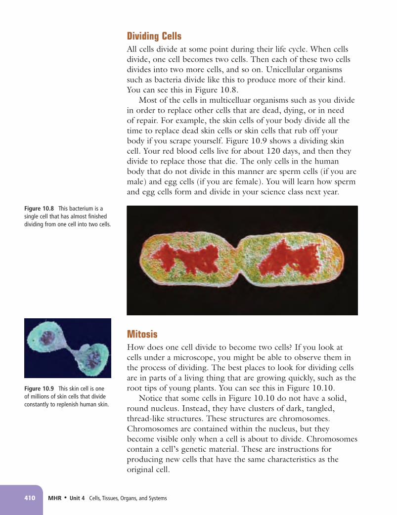
Dividing CellsAll cells divide at some point during their life cycle. When cells divide, one cell becomes two cells. Then each of these two cells divides into two more cells, and so on. Unicellular organisms such as bacteria divide like this to produce more of their kind. You can see this in Figure 10.8.
Most of the cells in multicelluar organisms such as you divide in order to replace other cells that are dead, dying, or in need of repair. For example, the skin cells of your body divide all the time to replace dead skin cells or skin cells that rub off your body if you scrape yourself. Figure 10.9 shows a dividing skin cell. Your red blood cells live for about 120 days, and then they divide to replace those that die. The only cells in the human body that do not divide in this manner are sperm cells (if you are male) and egg cells (if you are female). You will learn how sperm and egg cells form and divide in your science class next year.
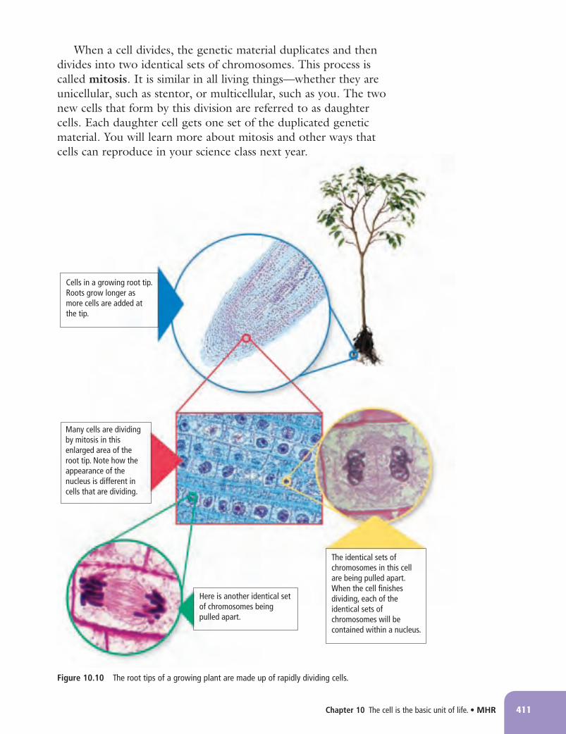
MitosisHow does one cell divide to become two cells? If you look at cells under a microscope, you might be able to observe them in the process of dividing. The best places to look for dividing cells are in parts of a living thing that are growing quickly, such as the root tips of young plants. You can see this in Figure 10.10.
Notice that some cells in Figure 10.10 do not have a solid, round nucleus. Instead, they have clusters of dark, tangled, thread-like structures. These structures are chromosomes. Chromosomes are contained within the nucleus, but they become visible only when a cell is about to divide. Chromosomes contain a cell’s genetic material. These are instructions for producing new cells that have the same characteristics as the original cell.
410 MHR•Unit 4 Cells, Tissues, Organs, and Systems
Figure 10.8 This bacterium is a single cell that has almost finished dividing from one cell into two cells.
Figure 10.9 This skin cell is one of millions of skin cells that divide constantly to replenish human skin.
NL8Unit4CH10.indd410 11/5/084:13:14PM
Chapter 10 The cell is the basic unit of life. • MHR 411
Cells in a growing root tip. Roots grow longer as more cells are added at the tip.
Many cells are dividing by mitosis in this enlarged area of the root tip. Note how the appearance of the nucleus is different in cells that are dividing.
The identical sets of chromosomes in this cell are being pulled apart. When the cell finishes dividing, each of the identical sets of chromosomes will be contained within a nucleus.
Here is another identical set of chromosomes being pulled apart.
Figure 10.10 The root tips of a growing plant are made up of rapidly dividing cells.
When a cell divides, the genetic material duplicates and then divides into two identical sets of chromosomes. This process is called mitosis. It is similar in all living things—whether they are unicellular, such as stentor, or multicellular, such as you. The two new cells that form by this division are referred to as daughter cells. Each daughter cell gets one set of the duplicated genetic material. You will learn more about mitosis and other ways that cells can reproduce in your science class next year.
NL8Unit4CH10.indd411 11/5/084:13:19PM

412 MHR•Unit 4 Cells, Tissues, Organs, and Systems
Observing Root Tip Cells10-2D
In this activity, you will look for evidence of cells dividing in a prepared slide of a root tip from an onion.
Materials• compound microscope• prepared slide of onion root tip
What to Do1. Put the prepared slide of the onion root tip on
the stage of the microscope.
2. View the cells using low power.
3. Look for the part of the root tip where cells appear to be dividing most actively. Reposition the slide so that these cells occupy most or all of your field of view.
4. Change to medium power and refocus.
5. Sketch the cells that are visible in your field of view.
What Did You Find Out?1. On the sketch you made, identify the cell or
cells in which you can see:
(a) a cell dividing to become two cells
(b) a cell in which chromosomes are visible (refer to page 410 to review what chromosomes are and when they are visible)
(c) a cell in which chromosomes are not visible
2. Compare your sketch with sketches made by other members of your class. As a class, discuss and identify similarities and differences in your sketches.
3. (a) Why would you look at a root tip to find evidence of cells dividing?
(b) Suggest one other part of a plant that you think would show evidence of cells dividing if you viewed it with a microscope. Explain your reasoning.
Find Out ACTIVITY
NL8Unit4CH10.indd412 11/5/084:13:21PM
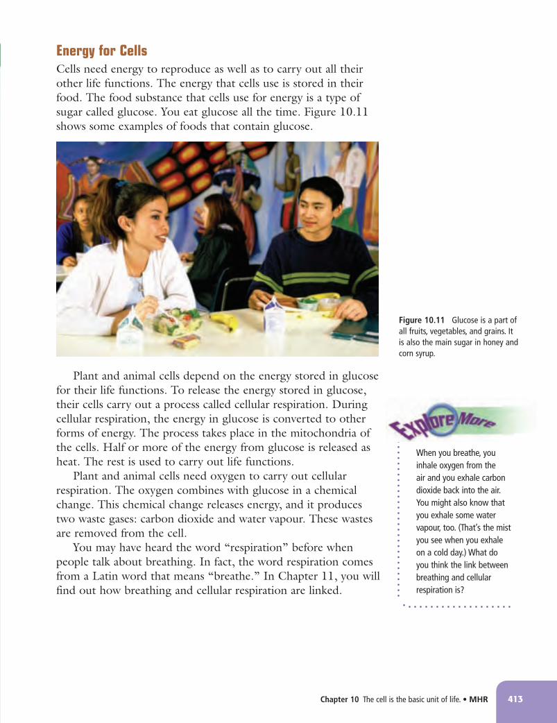
Energy for CellsCells need energy to reproduce as well as to carry out all their other life functions. The energy that cells use is stored in their food. The food substance that cells use for energy is a type of sugar called glucose. You eat glucose all the time. Figure 10.11 shows some examples of foods that contain glucose.
Plant and animal cells depend on the energy stored in glucose for their life functions. To release the energy stored in glucose, their cells carry out a process called cellular respiration. During cellular respiration, the energy in glucose is converted to other forms of energy. The process takes place in the mitochondria of the cells. Half or more of the energy from glucose is released as heat. The rest is used to carry out life functions.
Plant and animal cells need oxygen to carry out cellular respiration. The oxygen combines with glucose in a chemical change. This chemical change releases energy, and it produces two waste gases: carbon dioxide and water vapour. These wastes are removed from the cell.
You may have heard the word “respiration” before when people talk about breathing. In fact, the word respiration comes from a Latin word that means “breathe.” In Chapter 11, you will find out how breathing and cellular respiration are linked.
Chapter 10 The cell is the basic unit of life. • MHR 413
Figure 10.11 Glucose is a part of all fruits, vegetables, and grains. It is also the main sugar in honey and corn syrup.
[CATCH FIGURE P10.24: Photo of grade 8-aged students eating lunch together. We have to be able to see some of their foods, which must include breads (e.g., sandwiches), fruits (e.g., apple, orange, or banana visible), and if possible vegetables (e.g., carrot sticks.
When you breathe, you inhale oxygen from the air and you exhale carbon dioxide back into the air. You might also know that you exhale some water vapour, too. (That’s the mist you see when you exhale on a cold day.) What do you think the link between breathing and cellular respiration is?
NL8Unit4CH10.indd413 11/5/084:13:30PM

414 MHR•Unit 4 Cells, Tissues, Organs, and Systems
Observing Evidence of Cellular Respiration
10-2E Find Out ACTIVITY
Yeasts are unicellular organisms that are part of the same kingdom as mushrooms and other fungi. In this activity, you will look for evidence that yeast cells are carrying out cellular respiration.
Materials• warm water • 2 stirring rods• 2 beakers (500 mL) • 2 plastic pop bottles (600 mL or 1 L)• 2 scoopulas or measuring spoons • 2 balloons• 2 samples of white sugar (5 mL each) • tape• 2 samples of active dry yeast (15 mL each)
What to Do1. Read through all the instructions, and prepare a table to record your observations.
2. Work with a partner. Each of you should carry out the following steps at the same time.
3. Observe the bottles every 15 minutes for the next few hours. Each time you observe, also feel the bottom of each bottle and swirl the contents of the bottles gently.
What Did You Find Out?1. In which bottle did you observe evidence of a gas being given off? Describe this evidence.
2. In which bottle did you observe evidence of energy being released? Describe this evidence.
3. (a) How did the changes you observed in Partner 1’s bottle compare with Partner 2’s bottle?
(b) What is the function of Partner 2’s bottle?
4. What evidence did you observe that cellular respiration was taking place?
Partner 1 Partner 2
(a) Pour 250 mL of warm water into the beaker. (a) Pour 250 mL of warm water into the beaker. (b) Add 5 mL of sugar. (b) Add 15 mL of yeast. Use the stirring rod to stir the mixture. (c) Add 15 mL of yeast. Use the stirring rod to stir (c) Pour the mixture into the pop bottle. the mixture. (d) Pour the mixture into the pop bottle. (d) Blow up a balloon, and then release the air. Fit the open end of the balloon over the neck of the bottle, and tape it securely. (e) Blow up a balloon, and then release the air. (e) Cup the sides of the bottle at the bottom with both Fit the open end of the balloon over the neck of the hands. Take note of how cool or warm it feels. bottle, and tape it securely. (f) Cup the sides of the bottle at the bottom with (f) Label the bottle with the contents. both hands. Take note of how cool or warm it feels. (g) Label the bottle with the contents.
NL8Unit4CH10.indd414 11/5/084:13:33PM

Chapter 10 The cell is the basic unit of life. • MHR 415
Checking Concepts 1. What is the role of the nucleus in a cell? 2. Describe the function of the cell
membrane. 3. Which cell organelle produces the
energy that the cell needs to carry out its life activities?
4. Which organelle is like a storage container?
5. Predict what would happen to a plant cell if the chloroplasts stopped functioning.
6. Correctly identify the labelled organelles in the illustrations below.
7. Which cell in question 6 is a plant cell? Support your answer.
8. Describe the composition of cytoplasm. 9. List the key points of the cell theory. 10. Why do scientists consider the cell
theory to be a main idea of modern biology?
Understanding Key Ideas 11. Recall the Protection Dome of Newo.
Explain why a cell membrane could not be like the Protection Dome, which had a solid wall and just one opening.
12. Draw a Venn diagram like the one below. Fill in each section with the correct organelles.
13. Why would you not find chloroplasts in an onion root cell?
14. Explain why animal cells do not have chloroplasts.
15. Why do animal cells have different shapes while plant cells have a more regular, box-like shape?
16. (a) Which point of the cell theory is related to cell division?
(b) The cells of some living things divide to produce new living things. The cells of other living things divide to replace or repair older cells. Name two examples for each of these statements.
C
D
A
B
E
G F
Plant organelles
Animal organelles
Over 2000 years ago, many Greek philosophers (thinkers) believed that organisms and all other objects in the world were made of four basic things that they called elements: air, earth, fire, and water. Although the ancient Greeks did not know about cells, they would have believed that cells are made of air, earth, fire, and water, too. How is this idea about the make-up of cells and other organisms different from the ideas about cells stated in the cell theory?
Pause and Reflect
NL8Unit4CH10.indd415 11/5/084:13:37PM
C h a p t e r10
416 MHR•Unit 4 Cells, Tissues, Organs, and Systems
Prepare Your Own SummaryIn this chapter, you investigated the cell as the basic unit of life. Create your own summary of the key ideas from this chapter. You may include graphic organizers or illustrations with your notes. (See Science Skill 10 for help with using graphic organizers.) Use the following headings to organize your notes: 1. Characteristics of Living Things 2. The Microscope 3. Cell Theory 4. Cell Organelles
Checking Concepts 1. How are living things different from
non-living things? 2. What do unicellular organisms and
multicellular organisms have in common?
3. List four characteristics of living things. 4. Why must living things reproduce? 5. The coarse adjustment knob on a
microscope should be used with which objective lens(es)?
6. A slide on a microscope is moved toward you. In which direction does the object you are viewing through the eyepiece move?
7. Why should an objective lens never touch the slide?
8. What is a wet mount slide?
9. What organelles do plants have that animals do not?
10. Which part of the cell stores food and waste materials?
11. Which organelle controls the movement of substances in and out of the cell?
12. In which organelle would you find the genetic material in a cell?
Understanding Key Ideas 13. What is the difference between the cell
wall and the cell membrane? 14. A friend tells you that all living things
grow larger by increasing the size of their cells. Write one or two sentences explaining to your friend why this is not correct.
15. Why do the cells of a multicellular organism continue to reproduce even after it is fully grown?
16. How can you tell if a cell is undergoing mitosis?
17. Suppose that you are studying a slide of plant cells. You count 40 cells in a row across the diameter of the field of view. Describe a technique that you can use to estimate the average size of each cell.
NL8Unit4CH10.indd416 11/5/084:13:42PM

Chapter 10 The cell is the basic unit of life. • MHR 417
18. You have been given the responsibility of teaching some new classmates how to hold, carry, and use a microscope safely and properly.
(a) Explain how the students should bring the microscope from a storage cart to their desks.
(b) List the steps the students should follow to set up a wet mount slide.
(c) List the steps the students should follow to observe the slide under low power and then under medium power.
19. Examine the photograph of cells shown below.
(a) State whether these are plant cells or an animal cells and explain how you decided.
(b) Identify two organelles that are visible in this photograph.
You set up a slide on the stage of a compound light microscope. When you look into the eyepiece, you see only darkness. Give three reasons that could account for this problem.
Pause and Reflect
NL8Unit4CH10.indd417 11/5/084:13:47PM