cell, vol. 123, 249–263, october 21, 2005, copyright ©2005 by...
TRANSCRIPT
Cell, Vol. 123, 249–263, October 21, 2005, Copyright ©2005 by Elsevier Inc. DOI 10.1016/j.cell.2005.08.033
Regulating Gene Expressionthrough RNA Nuclear Retention
Kannanganattu V. Prasanth,1 Supriya G. Prasanth,1
Zhenyu Xuan,1 Stephen Hearn,1 Susan M. Freier,2
C. Frank Bennett,2 Michael Q. Zhang,1
and David L. Spector1,*1Cold Spring Harbor Laboratory1 Bungtown RoadCold Spring Harbor, New York 117242 ISIS Pharmaceuticals2280 Faraday AvenueCarlsbad, California 92008
Summary
Multiple mechanisms have evolved to regulate theeukaryotic genome. We have identified CTN-RNA, amouse tissue-specific w8 kb nuclear-retained poly(A)+
RNA that regulates the level of its protein-coding part-ner. CTN-RNA is transcribed from the protein-codingmouse cationic amino acid transporter 2 (mCAT2)gene through alternative promoter and poly(A) siteusage. CTN-RNA is diffusely distributed in nuclei andis also localized to paraspeckles. The 3�UTR of CTN-RNA contains elements for adenosine-to-inosine edit-ing, involved in its nuclear retention. Interestingly,knockdown of CTN-RNA also downregulates mCAT2mRNA. Under stress, CTN-RNA is posttranscription-ally cleaved to produce protein-coding mCAT2 mRNA.Our findings reveal a role of the cell nucleus in har-boring RNA molecules that are not immediately neededto produce proteins but whose cytoplasmic presenceis rapidly required upon physiologic stress. This mech-anism of action highlights an important paradigm forthe role of a nuclear-retained stable RNA transcript inregulating gene expression.
Introduction
In addition to harboring the cell’s genome, the nucleuscontains distinct substructures in the form of special-ized domains/compartments that are characterized bythe absence of delineating membranes but that containdefining sets of proteins (reviewed, Spector, 2001). Adynamic interplay between these domains and/or theirconstituents and the genome is thought to foster theefficient progression of gene expression. Gene expres-sion in eukaryotes is carried out through a series ofcomplex processes that includes transcription, pre-mRNA processing, and export of mature mRNA to thecytoplasm for translation. Recent evidence has indi-cated that these processes are coordinated and can beregulated at each level (reviewed, Maniatis and Reed,2002). Changes in chromatin structure as well as in-teractions between cis-DNA sequences and trans-act-ing factors are required for the activation or silencingof genes. In addition to constitutive gene expression,transcription of specific genes can be induced in re-
*Correspondence: [email protected]
sponse to cell-cycle stage as well as several stimuli,including stress and hormonal induction. Cotranscrip-tional gene regulation can occur at the levels of 5#cap-ping; constitutive and alternative pre-mRNA splicing;polyadenylation; and, in some cases, RNA editing (re-viewed, Bass, 2002; Orphanides and Reinberg, 2002).Furthermore, translation can be tightly regulated ateither a global level or the level of specific mRNAs (i.e,via miRNAs) (reviewed, Gebauer and Hentze, 2004; Heand Hannon, 2004).
The central dogma of molecular biology holds thatgenetic information normally flows from DNA to RNA toprotein. As a consequence, it has been assumed thatgenes generally encode for proteins and that proteinsfulfill not only structural and catalytic but also most reg-ulatory functions in cells. However, a subset of RNAsexecute functions without being translated into protein.Such RNAs include housekeeping RNAs (ribosomalRNA, transfer RNA, and uridine-rich small nuclear andnucleolar RNAs) and nuclear-retained RNAs that exe-cute regulatory roles in various aspects of gene expres-sion (Xist, Tsix, and Rian in mammals and hsr-ω-n androXs in Drosophila) (reviewed, Szymanski and Bar-ciszewski, 2003). Interestingly, the percentage of non-coding DNA in prokaryotes is <25% of the total ge-nome compared to w98% in humans, most of whichcontains active transcription units (Mattick, 2004). Theincreased number of noncoding genes in multicellularorganisms is likely to provide more complexity to thetranscriptome.
An earlier study indicated that, in many cell types, aconsiderable fraction of the poly(A)+ RNA (as much as30%) is nuclear retained and undetectable in the cyto-plasm (Herman et al., 1976). More recently, RNA fluores-cence in situ hybridization (RNA-FISH) analysis using anoligo dT probe in mammalian cells has shown that apopulation of poly(A)+ RNA is enriched in nuclearspeckles, also known as interchromatin granule clus-ters (IGCs) (Carter et al., 1991; Huang et al., 1994; Visaet al., 1993). However, IGCs are not transcription sites;rather, they are thought to be involved in the assembly/modification and/or storage of the pre-mRNA process-ing machinery (reviewed, Lamond and Spector, 2003).The poly(A)+ RNA in the IGCs does not appear to betransported to the cytoplasm (Huang et al., 1994), aswould be the case if they represented protein-codingmRNAs, suggesting that this RNA or these RNAs con-stitute new members of the nuclear regulatory RNA(nrRNA) family.
In the present study, we biochemically purified IGCsand associated nuclear domains from mouse liver nu-clei and constructed a cDNA library from the poly(A)+
RNA. One of the cDNA clones localized in a micropunc-tate nuclear distribution, and, in a population of cells,it also localized to a subnuclear domain, paraspeckles(Fox et al., 2002). Northern blot and genomic sequenceanalysis revealed that this clone encodes a w8 kbnuclear-retained poly(A)+ RNA, transcribed from themouse cationic amino acid transporter 2 (mCAT2) gene,hence named CAT2 transcribed nuclear RNA (CTN-
Cell250
RNA). The mCAT2 gene also encodes for the protein-coding mCAT2 mRNA. However, each transcript pre-dominantly utilizes a different promoter and has uniqueuntranslated regions (UTRs). The 3#UTR of CTN-RNA isessential for its nuclear retention. Interestingly, CTN-RNA is adenosine-to-inosine edited (A-to-I) in its 3#UTR.Depleting CTN-RNA from cells using antisense oligonu-cleotides resulted in downregulation of both CTN-RNAand mCAT2 mRNA. Most interestingly, our results dem-onstrated that, under stress conditions, CTN-RNA getsposttranscriptionally cleaved at its 3#UTR, producingan mRNA containing the mCAT2 coding region and aunique 5#UTR, suggesting that CTN-RNA regulates thelevels of its protein-coding partner. This mechanism ofaction brings to light an important paradigm for the roleof a nuclear-retained stable RNA transcript in regulatinggene expression.
Results
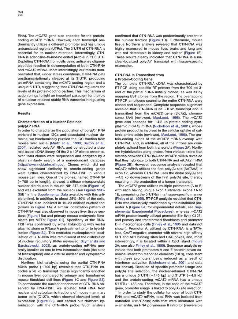
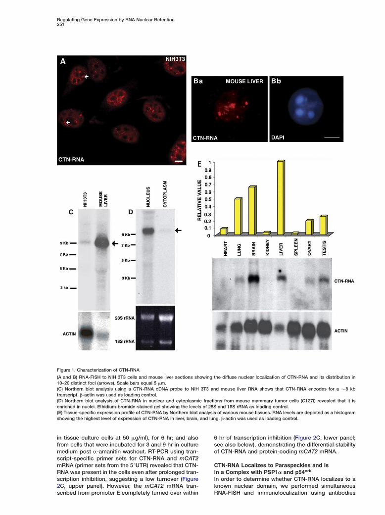
Characterization of a Nuclear-Retainedpoly(A)+ RNAIn order to characterize the population of poly(A)+ RNAenriched in nuclear IGCs and associated nuclear do-mains, we biochemically purified the IGC fraction frommouse liver nuclei (Mintz et al., 1999; Saitoh et al.,2004), isolated poly(A)+ RNA, and constructed a plas-mid-based cDNA library. Of the 2 × 10³ clones screened,over 1500 clones were sequenced and analyzed by ablast similarity search of a nonredundant database(http://www.ncbi.nlm.nih.gov). Clones which did notshow significant similarity to protein-coding geneswere further characterized by RNA-FISH in variousmouse cell lines. One of the clones, named CTN-RNA(w700 bp in length), showed a diffuse micropunctatenuclear distribution in mouse NIH 3T3 cells (Figure 1A)and was excluded from the nucleoli (see Figures S5B–S5B$ in the Supplemental Data available with this arti-cle online). In addition, in about 25%–30% of the cells,CTN-RNA also localized in 10–20 distinct nuclear foci(arrows in Figure 1A). A similar localization pattern ofCTN-RNA was also observed in mouse liver tissue sec-tions (Figure 1Ba) and primary mouse embryonic fibro-blasts (wt MEFs; Figure S1). Specificity of the RNA-FISH was confirmed by either hybridizing with vectorplasmid alone or RNase A pretreatment prior to hybrid-ization (Figure S2). This restricted nucleoplasmic local-ization of CTN-RNA was reminiscent of the distributionof nuclear regulatory RNAs (reviewed, Szymanski andBarciszewski, 2003), as protein-coding mRNAs gen-erally localize as one to two intranuclear dots (the sitesof transcription) and a diffuse nuclear and cytoplasmicdistribution.
Northern blot analysis using the partial CTN-RNAcDNA probe (w700 bp) revealed that CTN-RNA en-codes a >8 kb transcript that is significantly enrichedin mouse liver compared to primary and transformedmouse fibroblast cell lines (Figure 1C and Figure S3).To corroborate the nuclear enrichment of CTN-RNA ob-served by RNA-FISH, we isolated total RNA fromnuclear and cytoplasmic fractions of mouse mammarytumor cells (C127I), which showed elevated levels ofexpression (Figure S3), and carried out Northern hy-
ctthwTce
CaTRemRcrtsgppitCpeot(mewr
w1(RmpmaDslSi2vnwinpha5g
Ru
bridization with the CTN-RNA probe. Such analysis
onfirmed that CTN-RNA was predominantly present inhe nuclear fraction (Figure 1D). Furthermore, mouseissue Northern analysis revealed that CTN-RNA wasighly expressed in mouse liver, brain, and lung andas not detectable in kidney and spleen (Figure 1E).hese results clearly indicated that CTN-RNA is a nu-lear-localized poly(A)+ transcript with tissue-specificxpression.
TN-RNA Is Transcribed fromProtein-Coding Gene
he complete CTN-RNA cDNA was characterized byT-PCR using specific RT primers from the 700 bp 3#nd of the partial cDNA initially cloned, as well as byapping EST clones from the region. The overlappingT-PCR amplicons spanning the entire CTN-RNA wereloned and sequenced. Complete sequence alignmentevealed that CTN-RNA is an w8 kb transcript and isranscribed from the mCAT2 gene (Slc7a2; chromo-ome 8A4) (reviewed, MacLeod, 1996). The mCAT2ene also encodes for w4.2 kb protein-coding cyto-lasmic mCAT2 mRNA (Nicholson et al., 2001), whoserotein product is involved in the cellular uptake of cat-
onic amino acids (reviewed, MacLeod, 1996). The pro-ein-coding exons of the mCAT2 mRNA overlap withTN-RNA, and, in addition, all of the introns are com-letely spliced from both transcripts (Figure 2A). North-rn hybridization using probes from various exons thatverlap between CTN-RNA and mCAT2 mRNA revealed
hat they hybridize to both CTN-RNA and mCAT2 mRNAFigure 2B). However, sequence analysis revealed that
CAT2 mRNA utilizes the first poly(A) site (AATAAA) inxon 12, whereas CTN-RNA uses the distal poly(A) site4.5 kb downstream of the first poly(A) site, thereby
esulting in the production of a longer transcript.The mCAT2 gene utilizes multiple promoters (A to E,ith each having unique exon 1 variants: exons 1A toE, comprising the 5#UTRs) in a tissue-specific mannerFinley et al., 1995). RT-PCR analysis revealed that CTN-NA was exclusively transcribed by the distalmost pro-oter A (Figure S4; for experimental details, see Sup-lemental Experimental Procedures), whereas mCAT2RNA predominantly utilized promoter E in liver, C127I,
nd primary and transformed fibroblasts and promoterin macrophage cells (Finley et al., 1995 and data not
hown). Promoter A, utilized by CTN-RNA, is a TATA-ess, CAAT-negative promoter with several high-affinityP1 and AP1 binding sites and CAC boxes, and, most
nterestingly, it is located within a CpG island (FigureA; see also Finley et al., 1995). Sequence analysis re-ealed that both promoters A and E contain seven ca-onical interferon response elements (IREs), consistentith these promoters’ being induced as a result of
nterferon activation (Nicholson et al., 2001 and dataot shown). Because of specific promoter usage andoly(A) site selection, the nuclear-retained CTN-RNAas a unique 5#UTR (w145 bp) and 3#UTR (w4.5 kb)nd the protein-coding mCAT2 mRNA has a unique#UTR (w483 bp). Therefore, in the case of the mCAT2ene, promoter usage is linked to poly(A) site selection.In order to study the cellular turnover of both CTN-
NA and mCAT2 mRNA, total RNA was isolated fromntreated C127I cells; cells that were incubated with
α-amanitin, an RNA polymerase II inhibitor (irreversible
Regulating Gene Expression by RNA Nuclear Retention251
Figure 1. Characterization of CTN-RNA
(A and B) RNA-FISH to NIH 3T3 cells and mouse liver sections showing the diffuse nuclear localization of CTN-RNA and its distribution in10–20 distinct foci (arrows). Scale bars equal 5 �m.(C) Northern blot analysis using a CTN-RNA cDNA probe to NIH 3T3 and mouse liver RNA shows that CTN-RNA encodes for a w8 kbtranscript. β-actin was used as loading control.(D) Northern blot analysis of CTN-RNA in nuclear and cytoplasmic fractions from mouse mammary tumor cells (C127I) revealed that it isenriched in nuclei. Ethidium-bromide-stained gel showing the levels of 28S and 18S rRNA as loading control.(E) Tissue-specific expression profile of CTN-RNA by Northern blot analysis of various mouse tissues. RNA levels are depicted as a histogramshowing the highest level of expression of CTN-RNA in liver, brain, and lung. β-actin was used as loading control.
in tissue culture cells at 50 �g/ml), for 6 hr; and alsofrom cells that were incubated for 3 and 9 hr in culturemedium post α-amanitin washout. RT-PCR using tran-script-specific primer sets for CTN-RNA and mCAT2mRNA (primer sets from the 5#UTR) revealed that CTN-RNA was present in the cells even after prolonged tran-scription inhibition, suggesting a low turnover (Figure2C, upper panel). However, the mCAT2 mRNA tran-
scribed from promoter E completely turned over within6 hr of transcription inhibition (Figure 2C, lower panel;see also below), demonstrating the differential stabilityof CTN-RNA and protein-coding mCAT2 mRNA.
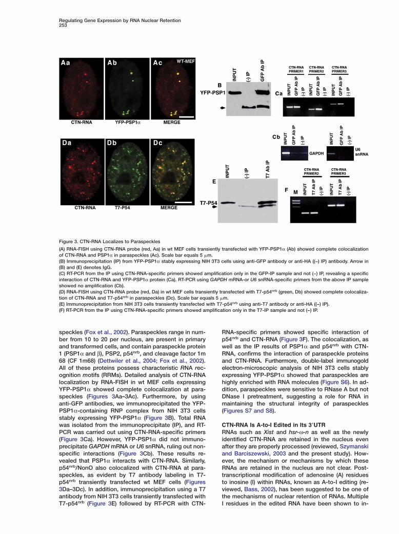
CTN-RNA Localizes to Paraspeckles and Isin a Complex with PSP1� and p54nrb
In order to determine whether CTN-RNA localizes to aknown nuclear domain, we performed simultaneous
RNA-FISH and immunolocalization using antibodies
Cell252
Figure 2. CTN-RNA and mCAT2 mRNA Are Transcribed from the mCAT2 Gene
(A) Diagram of the genomic region of the mCAT2 gene on mouse chromosome 8A4. CTN-RNA is transcribed from the mCAT2 gene, whichalso encodes the protein-coding mCAT2 mRNA. The light gray boxes (1A to 1E) represent the multiple promoters of the mCAT2 gene, eachcontaining a unique 5#UTR (exon 1). CTN-RNA is transcribed from promoter A, has unique 5# and 3#UTRs, and utilizes the distalmost poly(A)site (AATAAA). mCAT2 mRNA is predominantly transcribed from promoter D in macrophages and E in mouse liver and utilizes the proximalpoly(A) site. The black boxes represent exons, and the lines between the black boxes depict intronic regions. The hatched boxes representCpG islands. The dark gray boxes in each of the RNAs represent the unique 5# and 3#UTRs.(B) Northern blot analysis of total RNA from C127I cells using an exon 11 probe detects both the w8 kb CTN-RNA and the w4.2 kbmCAT2 mRNA.(C) CTN-RNA is a stable RNA. cDNA was prepared from RNA of untreated (control) and α-amanitin-treated cells (α-amanitin, 3 hr postwashout, and 9 hr post washout; see Experimental Procedures for details). RT-PCR was carried out with specific primers from the 5#UTRs ofCTN-RNA or mCAT2 mRNA.
against protein members of known nuclear domains inwt MEFs (Figure S5). Dual labeling of CTN-RNA (red;Figure S5E) and the nuclear speckle protein SF2/ASF(green; Figure S5E#) revealed that some of the CTN-RNA foci were located at the periphery of the SF2/ASF-positive speckles and showed partial overlap (Figure
SsfR
d
5E$; see yellow signal in the higher-magnification in-et in Figure S5E%). This finding is consistent with theact that our cDNA library was generated from theNAs of IGCs and associated nuclear compartments.Recently, Lamond and colleagues identified a nuclear
omain, paraspeckles, localized adjacent to nuclear
Regulating Gene Expression by RNA Nuclear Retention253
speckles (Fox et al., 2002). Paraspeckles range in num-ber from 10 to 20 per nucleus, are present in primaryand transformed cells, and contain paraspeckle protein1 (PSP1α and β), PSP2, p54nrb, and cleavage factor 1m68 (CF 1m68) (Dettwiler et al., 2004; Fox et al., 2002).All of these proteins possess characteristic RNA rec-ognition motifs (RRMs). Detailed analysis of CTN-RNAlocalization by RNA-FISH in wt MEF cells expressingYFP-PSP1α showed complete colocalization at para-speckles (Figures 3Aa–3Ac). Furthermore, by usinganti-GFP antibodies, we immunoprecipitated the YFP-PSP1α-containing RNP complex from NIH 3T3 cellsstably expressing YFP-PSP1α (Figure 3B). Total RNAwas isolated from the immunoprecipitate (IP), and RT-PCR was carried out using CTN-RNA-specific primers(Figure 3Ca). However, YFP-PSP1α did not immuno-precipitate GAPDH mRNA or U6 snRNA, ruling out non-specific interactions (Figure 3Cb). These results re-vealed that PSP1α interacts with CTN-RNA. Similarly,p54nrb/NonO also colocalized with CTN-RNA at para-speckles, as evident by T7 antibody labeling in T7-p54nrb transiently transfected wt MEF cells (Figures3Da–3Dc). In addition, immunoprecipitation using a T7antibody from NIH 3T3 cells transiently transfected withT7-p54nrb (Figure 3E) followed by RT-PCR with CTN-
Figure 3. CTN-RNA Localizes to Paraspeckles
(A) RNA-FISH using CTN-RNA probe (red, Aa) in wt MEF cells transiently transfected with YFP-PSP1α (Ab) showed complete colocalizationof CTN-RNA and PSP1α in paraspeckles (Ac). Scale bar equals 5 �m.(B) Immunoprecipitation (IP) from YFP-PSP1α stably expressing NIH 3T3 cells using anti-GFP antibody or anti-HA ((−) IP) antibody. Arrow in(B) and (E) denotes IgG.(C) RT-PCR from the IP using CTN-RNA-specific primers showed amplification only in the GFP-IP sample and not (−) IP, revealing a specificinteraction of CTN-RNA and YFP-PSP1α protein (Ca). RT-PCR using GAPDH mRNA-or U6 snRNA-specific primers from the above IP sampleshowed no amplification (Cb).(D) RNA-FISH using CTN-RNA probe (red, Da) in wt MEF cells transiently transfected with T7-p54nrb (green, Db) showed complete colocaliza-tion of CTN-RNA and T7-p54nrb in paraspeckles (Dc). Scale bar equals 5 �m.(E) Immunoprecipitation from NIH 3T3 cells transiently transfected with T7-p54nrb using anti-T7 antibody or anti-HA ((−) IP).(F) RT-PCR from the IP using CTN-RNA-specific primers showed amplification only in the T7-IP sample and not (−) IP.
RNA-specific primers showed specific interaction ofp54nrb and CTN-RNA (Figure 3F). The colocalization, aswell as the IP results of PSP1α and p54nrb with CTN-RNA, confirms the interaction of paraspeckle proteinsand CTN-RNA. Furthermore, double-label immunogoldelectron-microscopic analysis of NIH 3T3 cells stablyexpressing YFP-PSP1α showed that paraspeckles arehighly enriched with RNA molecules (Figure S6). In ad-dition, paraspeckles were sensitive to RNase A but notDNase I pretreatment, suggesting a role for RNA inmaintaining the structural integrity of paraspeckles(Figures S7 and S8).
CTN-RNA Is A-to-I Edited in Its 3�UTRRNAs such as Xist and hsr-ω-n as well as the newlyidentified CTN-RNA are retained in the nucleus evenafter they are properly processed (reviewed, Szymanskiand Barciszewski, 2003 and the present study). How-ever, the mechanism or mechanisms by which theseRNAs are retained in the nucleus are not clear. Post-transcriptional modification of adenosine (A) residuesto inosine (I) within RNAs, known as A-to-I editing (re-viewed, Bass, 2002), has been suggested to be one ofthe mechanisms of nuclear retention of RNAs. MultipleI residues in the edited RNA have been shown to in-
Cell254
teract with a protein complex comprised of p54nrb, PSF,and matrin 3, resulting in the retention of this RNP com-plex in the nucleus (Zhang and Carmichael, 2001).Adenosine deaminase acting on RNA (ADAR) enzymescatalyze the A-to-I editing of RNA and recognize thedouble-stranded regions of the RNA for their activity(reviewed, Bass, 2002). The finding that p54nrb is pres-ent in paraspeckles and is involved in retaining A-to-Iedited RNAs in the nucleus and our finding that CTN-RNA interacts with p54nrb raised the possibility thatCTN-RNA might be a target for RNA editing. Consistentwith this possibility, immunolocalization studies in wtMEFs using an antibody against PSF revealed that itcolocalized with YFP-PSP1α in paraspeckles, furtherstrengthening the link between RNA editing and para-speckles (Figure S9).
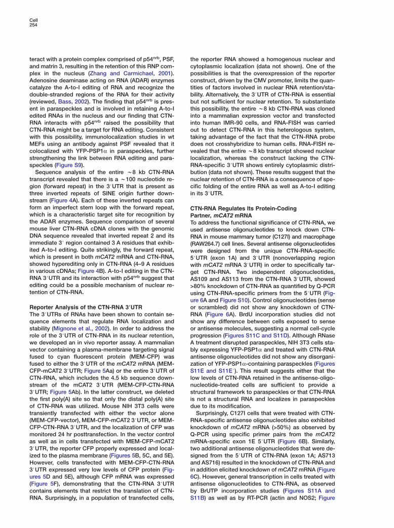
Sequence analysis of the entire w8 kb CTN-RNAtranscript revealed that there is a w100 nucleotide re-gion (forward repeat) in the 3#UTR that is present asthree inverted repeats of SINE origin further down-stream (Figure 4A). Each of these inverted repeats canform an imperfect stem loop with the forward repeat,which is a characteristic target site for recognition bythe ADAR enzymes. Sequence comparison of severalmouse liver CTN-RNA cDNA clones with the genomicDNA sequence revealed that inverted repeat 2 and itsimmediate 3# region contained 3 A residues that exhib-ited A-to-I editing. Quite strikingly, the forward repeat,which is present in both mCAT2 mRNA and CTN-RNA,showed hyperediting only in CTN-RNA (4–9 A residuesin various cDNAs; Figure 4B). A-to-I editing in the CTN-RNA 3#UTR and its interaction with p54nrb suggest thatediting could be a possible mechanism of nuclear re-tention of CTN-RNA.
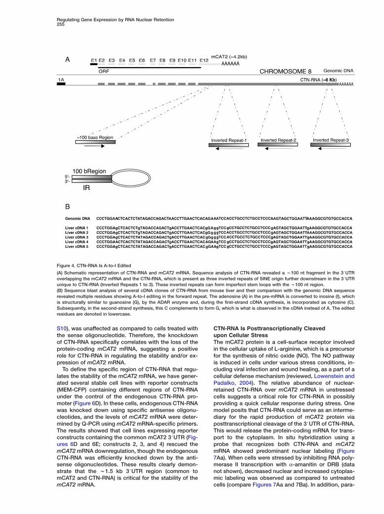
Reporter Analysis of the CTN-RNA 3�UTRThe 3#UTRs of RNAs have been shown to contain se-quence elements that regulate RNA localization andstability (Mignone et al., 2002). In order to address therole of the 3#UTR of CTN-RNA in its nuclear retention,we developed an in vivo reporter assay. A mammalianvector containing a plasma-membrane targeting signalfused to cyan fluorescent protein (MEM-CFP) wasfused to either the 3#UTR of the mCAT2 mRNA (MEM-CFP-mCAT2 3#UTR; Figure 5Aa) or the entire 3#UTR ofCTN-RNA, which includes the 4.5 kb sequence down-stream of the mCAT2 3#UTR (MEM-CFP-CTN-RNA3#UTR; Figure 5Ab). In the latter construct, we deletedthe first poly(A) site so that only the distal poly(A) siteof CTN-RNA was utilized. Mouse NIH 3T3 cells weretransiently transfected with either the vector alone(MEM-CFP-vector), MEM-CFP-mCAT2 3#UTR, or MEM-CFP-CTN-RNA 3#UTR, and the localization of CFP wasmonitored 24 hr posttransfection. In the vector controlas well as in cells transfected with MEM-CFP-mCAT23#UTR, the reporter CFP properly expressed and local-ized to the plasma membrane (Figures 5B, 5C, and 5E).However, cells transfected with MEM-CFP-CTN-RNA3#UTR expressed very low levels of CFP protein (Fig-ures 5D and 5E), although CFP mRNA was expressed(Figure 5F), demonstrating that the CTN-RNA 3#UTRcontains elements that restrict the translation of CTN-RNA. Surprisingly, in a population of transfected cells,
tcpctbbtiiotdvlRbnci
CPTuR(w5wgA>uuoRsopAbazSlnsid
RkQmtsai6abS
he reporter RNA showed a homogenous nuclear andytoplasmic localization (data not shown). One of theossibilities is that the overexpression of the reporteronstruct, driven by the CMV promoter, limits the quan-ities of factors involved in nuclear RNA retention/sta-ility. Alternatively, the 3#UTR of CTN-RNA is essentialut not sufficient for nuclear retention. To substantiatehis possibility, the entire w8 kb CTN-RNA was clonednto a mammalian expression vector and transfectednto human IMR-90 cells, and RNA-FISH was carriedut to detect CTN-RNA in this heterologous system,aking advantage of the fact that the CTN-RNA probeoes not crosshybridize to human cells. RNA-FISH re-ealed that the entire w8 kb transcript showed nuclearocalization, whereas the construct lacking the CTN-NA-specific 3#UTR shows entirely cytoplasmic distri-ution (data not shown). These results suggest that theuclear retention of CTN-RNA is a consequence of spe-ific folding of the entire RNA as well as A-to-I editing
n its 3#UTR.
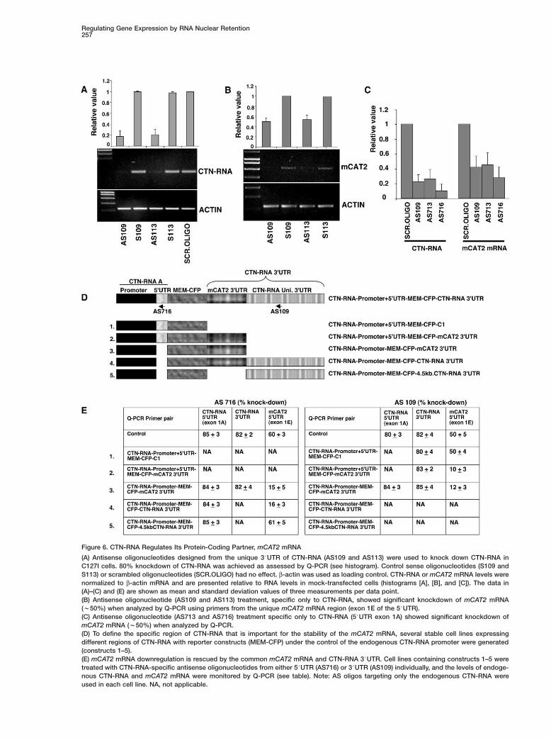
TN-RNA Regulates Its Protein-Codingartner, mCAT2 mRNAo address the functional significance of CTN-RNA, wesed antisense oligonucleotides to knock down CTN-NA in mouse mammary tumor (C127I) and macrophage
RAW264.7) cell lines. Several antisense oligonucleotidesere designed from the unique CTN-RNA-specific#UTR (exon 1A) and 3#UTR (nonoverlapping regionith mCAT2 mRNA 3#UTR) in order to specifically tar-et CTN-RNA. Two independent oligonucleotides,S109 and AS113 from the CTN-RNA 3#UTR, showed80% knockdown of CTN-RNA as quantified by Q-PCRsing CTN-RNA-specific primers from the 5#UTR (Fig-re 6A and Figure S10). Control oligonucleotides (senser scrambled) did not show any knockdown of CTN-NA (Figure 6A). BrdU incorporation studies did nothow any difference between cells exposed to senser antisense molecules, suggesting a normal cell-cyclerogression (Figures S11C and S11D). Although RNasetreatment disrupted paraspeckles, NIH 3T3 cells sta-
ly expressing YFP-PSP1α and treated with CTN-RNAntisense oligonucleotides did not show any disorgani-ation of YFP-PSP1α-containing paraspeckles (Figures11E and S11E#). This result suggests either that the
ow levels of CTN-RNA retained in the antisense-oligo-ucleotide-treated cells are sufficient to provide atructural framework to paraspeckles or that CTN-RNAs not a structural RNA and localizes in paraspecklesue to its modification.Surprisingly, C127I cells that were treated with CTN-
NA-specific antisense oligonucleotides also exhibitednockdown of mCAT2 mRNA (>50%) as observed by-PCR using specific primer pairs from the mCAT2RNA-specific exon 1E 5#UTR (Figure 6B). Similarly,
wo additional antisense oligonucleotides that were de-igned from the 5#UTR of CTN-RNA (exon 1A; AS713nd AS716) resulted in the knockdown of CTN-RNA and
n addition elicited knockdown of mCAT2 mRNA (FigureC). However, general transcription in cells treated withntisense oligonucleotides to CTN-RNA, as observedy BrUTP incorporation studies (Figures S11A and11B) as well as by RT-PCR (actin and NOS2; Figure
Regulating Gene Expression by RNA Nuclear Retention255
Figure 4. CTN-RNA Is A-to-I Edited
(A) Schematic representation of CTN-RNA and mCAT2 mRNA. Sequence analysis of CTN-RNA revealed a w100 nt fragment in the 3#UTRoverlapping the mCAT2 mRNA and the CTN-RNA, which is present as three inverted repeats of SINE origin further downstream in the 3#UTRunique to CTN-RNA (Inverted Repeats 1 to 3). These inverted repeats can form imperfect stem loops with the w100 nt region.(B) Sequence blast analysis of several cDNA clones of CTN-RNA from mouse liver and their comparison with the genomic DNA sequencerevealed multiple residues showing A-to-I editing in the forward repeat. The adenosine (A) in the pre-mRNA is converted to inosine (I), whichis structurally similar to guanosine (G), by the ADAR enzyme and, during the first-strand cDNA synthesis, is incorporated as cytosine (C).Subsequently, in the second-strand synthesis, this C complements to form G, which is what is observed in the cDNA instead of A. The editedresidues are denoted in lowercase.
S10), was unaffected as compared to cells treated withthe sense oligonucleotide. Therefore, the knockdownof CTN-RNA specifically correlates with the loss of theprotein-coding mCAT2 mRNA, suggesting a positiverole for CTN-RNA in regulating the stability and/or ex-pression of mCAT2 mRNA.
To define the specific region of CTN-RNA that regu-lates the stability of the mCAT2 mRNA, we have gener-ated several stable cell lines with reporter constructs(MEM-CFP) containing different regions of CTN-RNAunder the control of the endogenous CTN-RNA pro-moter (Figure 6D). In these cells, endogenous CTN-RNAwas knocked down using specific antisense oligonu-cleotides, and the levels of mCAT2 mRNA were deter-mined by Q-PCR using mCAT2 mRNA-specific primers.The results showed that cell lines expressing reporterconstructs containing the common mCAT2 3#UTR (Fig-ures 6D and 6E; constructs 2, 3, and 4) rescued themCAT2 mRNA downregulation, though the endogenousCTN-RNA was efficiently knocked down by the anti-sense oligonucleotides. These results clearly demon-strate that the w1.5 kb 3#UTR region (common tomCAT2 and CTN-RNA) is critical for the stability of themCAT2 mRNA.
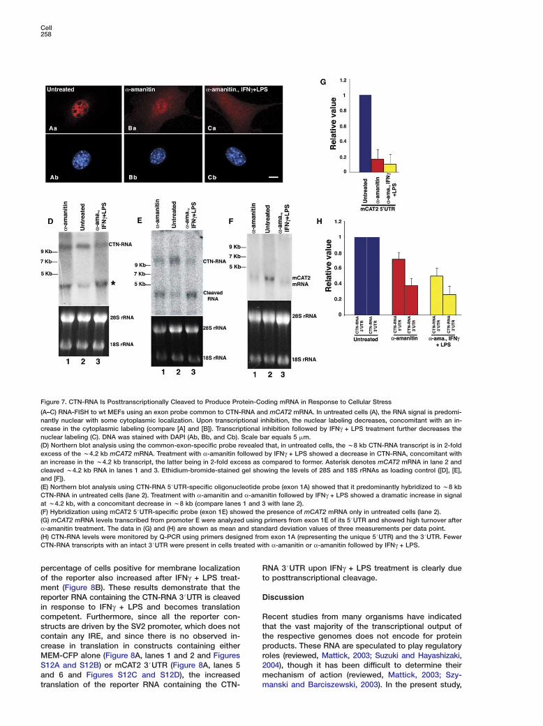
CTN-RNA Is Posttranscriptionally Cleavedupon Cellular StressThe mCAT2 protein is a cell-surface receptor involvedin the cellular uptake of L-arginine, which is a precursorfor the synthesis of nitric oxide (NO). The NO pathwayis induced in cells under various stress conditions, in-cluding viral infection and wound healing, as a part of acellular defense mechanism (reviewed, Lowenstein andPadalko, 2004). The relative abundance of nuclear-retained CTN-RNA over mCAT2 mRNA in unstressedcells suggests a critical role for CTN-RNA in possiblyproviding a quick cellular response during stress. Onemodel posits that CTN-RNA could serve as an interme-diary for the rapid production of mCAT2 protein viaposttranscriptional cleavage of the 3#UTR of CTN-RNA.This would release the protein-coding mRNA for trans-port to the cytoplasm. In situ hybridization using aprobe that recognizes both CTN-RNA and mCAT2mRNA showed predominant nuclear labeling (Figure7Aa). When cells were stressed by inhibiting RNA poly-merase II transcription with α-amanitin or DRB (datanot shown), decreased nuclear and increased cytoplas-mic labeling was observed as compared to untreatedcells (compare Figures 7Aa and 7Ba). In addition, para-

Cell256
Figure 5. Reporter Analysis Using the CTN-RNA 3#UTR
(A) A mammalian expression vector encoding a membrane targeting signal at the amino terminus of CFP was fused to either the 3#UTR ofthe mCAT2 mRNA (Aa) or the 3#UTR of the CTN-RNA (Ab), which includes a w4.5 kb sequence downstream of the mCAT2 3#UTR.(B–D) Mouse NIH 3T3 cells were transiently transfected with either the MEM-CFP-vector alone (B), MEM-CFP-mCAT2 3#UTR (C), or MEM-CFP-CTN-RNA 3#UTR (D). Expression and localization were monitored 24 hr posttransfection. DNA is stained with 7-AAD. Scale bar equals5 �m.(E) Immunoblot analysis using anti-GFP antibody in NIH 3T3 cells transiently transfected (24 hr) with the above constructs. The vector aloneand mCAT2 3#UTR-containing constructs showed expression of the GFP reporter, whereas the construct containing the CTN-RNA 3#UTRshowed extremely low levels of GFP protein. α-tubulin was used as loading control.(F) Total RNA was isolated from NIH 3T3 cells transiently transfected with the above plasmid constructs and RT-PCR carried out usingCFP primers.
speckle labeling of CTN-RNA was abolished. Additionof IFNγ + LPS post α-amanitin treatment further in-creased the cytoplasmic labeling, concomitant with re-duced nuclear labeling (Figure 7Ca). Northern analysisusing an exon probe (hybridizing to both CTN-RNA andmCAT2 mRNA) further demonstrated that in untreatedcells there is w2-fold more CTN-RNA than mCAT2mRNA (Figure 7D, lane 2). Furthermore, cells treatedwith α-amanitin followed by IFNγ + LPS showed a sig-nificant decrease in the level of w8 kb CTN-RNA, witha concomitant increase in the w4.2 kb mRNA (Figure7D, lane 3). Similarly, Northern blot hybridization usinga CTN-RNA 5#UTR oligonucleotide probe predomi-nantly hybridized to the w8 kb band in unstressed cells(Figure 7E, lane 2). In α-amanitin-treated cells as wellas in cells treated with α-amanitin followed by IFNγ +LPS, the CTN-RNA 5#UTR probe now showed an in-creased hybridization signal at w4.2 kb, with a con-comitant decrease in w8 kb signal (Figure 7E, lanes 1and 3). Furthermore, the mCAT2-specific 5#UTR probehybridized to a w4.2 kb band only in untreated cellsand not in either α-amanitin-treated cells or cellstreated with α-amanitin followed by IFNγ + LPS, corrob-
oQcICtotdCrt
ptTdcCtFrl
rating the Q-PCR results (Figures 7F and 7G). Finally,-PCR analysis to evaluate the levels of CTN-RNA inells treated with α-amanitin or α-amanitin followed byFNγ + LPS, using two independent primer pairs fromTN-RNA (5#UTR exon 1A and unique 3#UTR), de-
ected fewer transcripts containing the 3#UTR, corrob-rating the cleavage of CTN-RNA to release the pro-ein-coding mRNA (Figure 7H). These observationsemonstrate that, under stress conditions, the w8 kbTN-RNA is posttranscriptionally cleaved at its 3#UTR,
eleasing a protein-coding message for transport tohe cytoplasm.
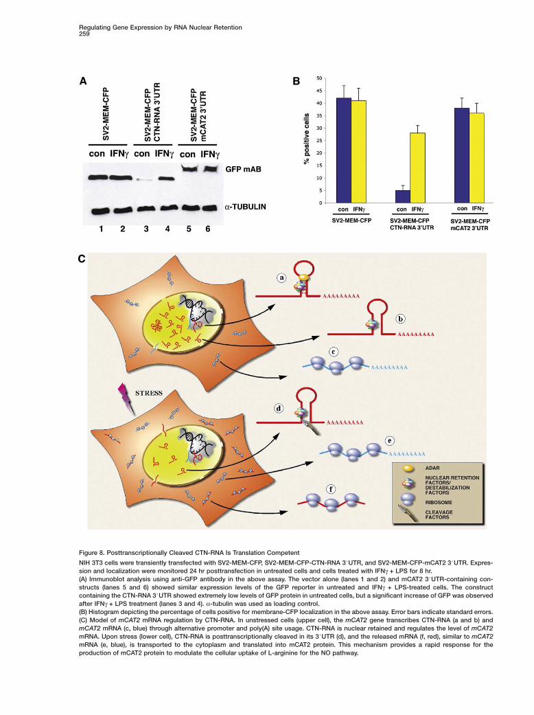
In order to address whether the cleaved CTN-RNAroduct is translation competent, we carried out repor-er analysis since there is no mCAT2 antibody available.he CTN-RNA or mCAT2 mRNA 3#UTRs were clonedownstream of a MEM-CFP reporter, which is under theontrol of the SV2 promoter. In unstressed cells, theTN-RNA 3#UTR reporter did not get translated even
hough it is efficiently transcribed (Figure 8A, lane 3 andigure S12E). However, upon addition of IFNγ + LPS,eporter protein was detected (Figure 8A, lane 4) andocalized to the plasma membrane (Figure S12F). The
Regulating Gene Expression by RNA Nuclear Retention257
Figure 6. CTN-RNA Regulates Its Protein-Coding Partner, mCAT2 mRNA
(A) Antisense oligonucleotides designed from the unique 3#UTR of CTN-RNA (AS109 and AS113) were used to knock down CTN-RNA inC127I cells. 80% knockdown of CTN-RNA was achieved as assessed by Q-PCR (see histogram). Control sense oligonucleotides (S109 andS113) or scrambled oligonucleotides (SCR.OLIGO) had no effect. β-actin was used as loading control. CTN-RNA or mCAT2 mRNA levels werenormalized to β-actin mRNA and are presented relative to RNA levels in mock-transfected cells (histograms [A], [B], and [C]). The data in(A)–(C) and (E) are shown as mean and standard deviation values of three measurements per data point.(B) Antisense oligonucleotide (AS109 and AS113) treatment, specific only to CTN-RNA, showed significant knockdown of mCAT2 mRNA(w50%) when analyzed by Q-PCR using primers from the unique mCAT2 mRNA region (exon 1E of the 5#UTR).(C) Antisense oligonucleotide (AS713 and AS716) treatment specific only to CTN-RNA (5#UTR exon 1A) showed significant knockdown ofmCAT2 mRNA (w50%) when analyzed by Q-PCR.(D) To define the specific region of CTN-RNA that is important for the stability of the mCAT2 mRNA, several stable cell lines expressingdifferent regions of CTN-RNA with reporter constructs (MEM-CFP) under the control of the endogenous CTN-RNA promoter were generated(constructs 1–5).(E) mCAT2 mRNA downregulation is rescued by the common mCAT2 mRNA and CTN-RNA 3#UTR. Cell lines containing constructs 1–5 weretreated with CTN-RNA-specific antisense oligonucleotides from either 5#UTR (AS716) or 3#UTR (AS109) individually, and the levels of endoge-nous CTN-RNA and mCAT2 mRNA were monitored by Q-PCR (see table). Note: AS oligos targeting only the endogenous CTN-RNA wereused in each cell line. NA, not applicable.
Cell258
Figure 7. CTN-RNA Is Posttranscriptionally Cleaved to Produce Protein-Coding mRNA in Response to Cellular Stress
(A–C) RNA-FISH to wt MEFs using an exon probe common to CTN-RNA and mCAT2 mRNA. In untreated cells (A), the RNA signal is predomi-nantly nuclear with some cytoplasmic localization. Upon transcriptional inhibition, the nuclear labeling decreases, concomitant with an in-crease in the cytoplasmic labeling (compare [A] and [B]). Transcriptional inhibition followed by IFNγ + LPS treatment further decreases thenuclear labeling (C). DNA was stained with DAPI (Ab, Bb, and Cb). Scale bar equals 5 �m.(D) Northern blot analysis using the common-exon-specific probe revealed that, in untreated cells, the w8 kb CTN-RNA transcript is in 2-foldexcess of the w4.2 kb mCAT2 mRNA. Treatment with α-amanitin followed by IFNγ + LPS showed a decrease in CTN-RNA, concomitant withan increase in the w4.2 kb transcript, the latter being in 2-fold excess as compared to former. Asterisk denotes mCAT2 mRNA in lane 2 andcleaved w4.2 kb RNA in lanes 1 and 3. Ethidium-bromide-stained gel showing the levels of 28S and 18S rRNAs as loading control ([D], [E],and [F]).(E) Northern blot analysis using CTN-RNA 5#UTR-specific oligonucleotide probe (exon 1A) showed that it predominantly hybridized to w8 kbCTN-RNA in untreated cells (lane 2). Treatment with α-amanitin and α-amanitin followed by IFNγ + LPS showed a dramatic increase in signalat w4.2 kb, with a concomitant decrease in w8 kb (compare lanes 1 and 3 with lane 2).(F) Hybridization using mCAT2 5#UTR-specific probe (exon 1E) showed the presence of mCAT2 mRNA only in untreated cells (lane 2).(G) mCAT2 mRNA levels transcribed from promoter E were analyzed using primers from exon 1E of its 5#UTR and showed high turnover afterα-amanitin treatment. The data in (G) and (H) are shown as mean and standard deviation values of three measurements per data point.(H) CTN-RNA levels were monitored by Q-PCR using primers designed from exon 1A (representing the unique 5#UTR) and the 3#UTR. FewerCTN-RNA transcripts with an intact 3#UTR were present in cells treated with α-amanitin or α-amanitin followed by IFNγ + LPS.
percentage of cells positive for membrane localizationof the reporter also increased after IFNγ + LPS treat-ment (Figure 8B). These results demonstrate that thereporter RNA containing the CTN-RNA 3#UTR is cleavedin response to IFNγ + LPS and becomes translationcompetent. Furthermore, since all the reporter con-structs are driven by the SV2 promoter, which does notcontain any IRE, and since there is no observed in-crease in translation in constructs containing eitherMEM-CFP alone (Figure 8A, lanes 1 and 2 and FiguresS12A and S12B) or mCAT2 3#UTR (Figure 8A, lanes 5and 6 and Figures S12C and S12D), the increasedtranslation of the reporter RNA containing the CTN-
Rt
D
Rttpr2mm
NA 3#UTR upon IFNγ + LPS treatment is clearly dueo posttranscriptional cleavage.
iscussion
ecent studies from many organisms have indicatedhat the vast majority of the transcriptional output ofhe respective genomes does not encode for proteinroducts. These RNA are speculated to play regulatory
oles (reviewed, Mattick, 2003; Suzuki and Hayashizaki,004), though it has been difficult to determine theirechanism of action (reviewed, Mattick, 2003; Szy-anski and Barciszewski, 2003). In the present study,
Regulating Gene Expression by RNA Nuclear Retention259
Figure 8. Posttranscriptionally Cleaved CTN-RNA Is Translation Competent
NIH 3T3 cells were transiently transfected with SV2-MEM-CFP, SV2-MEM-CFP-CTN-RNA 3#UTR, and SV2-MEM-CFP-mCAT2 3#UTR. Expres-sion and localization were monitored 24 hr posttransfection in untreated cells and cells treated with IFNγ + LPS for 8 hr.(A) Immunoblot analysis using anti-GFP antibody in the above assay. The vector alone (lanes 1 and 2) and mCAT2 3#UTR-containing con-structs (lanes 5 and 6) showed similar expression levels of the GFP reporter in untreated and IFNγ + LPS-treated cells. The constructcontaining the CTN-RNA 3#UTR showed extremely low levels of GFP protein in untreated cells, but a significant increase of GFP was observedafter IFNγ + LPS treatment (lanes 3 and 4). α-tubulin was used as loading control.(B) Histogram depicting the percentage of cells positive for membrane-CFP localization in the above assay. Error bars indicate standard errors.(C) Model of mCAT2 mRNA regulation by CTN-RNA. In unstressed cells (upper cell), the mCAT2 gene transcribes CTN-RNA (a and b) andmCAT2 mRNA (c, blue) through alternative promoter and poly(A) site usage. CTN-RNA is nuclear retained and regulates the level of mCAT2mRNA. Upon stress (lower cell), CTN-RNA is posttranscriptionally cleaved in its 3#UTR (d), and the released mRNA (f, red), similar to mCAT2mRNA (e, blue), is transported to the cytoplasm and translated into mCAT2 protein. This mechanism provides a rapid response for theproduction of mCAT2 protein to modulate the cellular uptake of L-arginine for the NO pathway.
Cell260
we identified CTN-RNA, a nuclear-retained poly(A)+
RNA that is transcribed from the protein-coding mCAT2gene. The 3#UTR of CTN-RNA contains elements thatretain this RNA in the nucleus, in part through A-to-Iediting, thereby inhibiting its translation. Knockdownof CTN-RNA results in the downregulation of bothCTN-RNA and mCAT2 mRNA. Rescue experimentsdemonstrated that a common region of the 3#UTR ofCTN-RNA and mCAT2 mRNA contains an element orelements that govern mCAT2 mRNA stability. Most in-terestingly, upon cellular stress, CTN-RNA is posttran-scriptionally cleaved to release a translation-competentmRNA, thereby revealing an important cellular mecha-nism for the rapid production of protein-coding mRNAs.
CTN-RNA Is a Member of the Nuclear RegulatoryRNA (nrRNA) FamilyTypically, nascent pre-mRNAs of protein-coding genesare processed at the site of transcription and are trans-ported to the cytoplasm for translation. However, sev-eral studies have shown that a significant population ofRNAs are retained within the nucleus and are sug-gested to play structural roles or act as riboregulators(Herman et al., 1976; Huang et al., 1994). The X chromo-some-encoded nuclear-retained RNA, Xist (reviewed,Plath et al., 2002), and its anti-sense transcript, Tsix(Lee et al., 1999), are among the best studied nuclearregulatory RNAs (nrRNAs) in mammalian cells.
Similar to the mCAT2 locus, the stress-inducible anddevelopmentally regulated hsr-ω locus in Drosophila isan example of a gene that transcribes two independenttranscripts from the same DNA strand (Hogan et al.,1994). A longer transcript, hsr-ω-n RNA (w10–14 kb), isan unspliced, polyadenylated nuclear-retained tran-script suggested to regulate the intranuclear traffickingof various hnRNPs (Prasanth et al., 2000). A shorter 1.2kb transcript, hsr-ω-c RNA, is transcribed from thesame promoter as hsr-ω-n RNA but uses the proximalpoly(A) site. The hsr-ω-c RNA is spliced and polyadeny-lated and, although it localizes in the cytoplasm, doesnot encode for a protein (Fini et al., 1989). The nucleardistribution of CTN-RNA suggests that it is a previouslyunidentified member of the nrRNA family.
The 3�UTR of CTN-RNA Is Essentialfor Nuclear RetentionThe mechanisms underlying nuclear retention of RNAare not well understood. However, A-to-I editing of RNAis postulated to be one such mechanism (Zhang andCarmichael, 2001). Biochemical purification studiesusing in vitro-transcribed RNAs (containing viral se-quences) that are A-to-I edited revealed that they co-purify with p54nrb, PSF, and matrin 3 as a complex, andit has been suggested that this p54nrb complex is in-volved in the nuclear retention of hyperedited RNAs(Zhang and Carmichael, 2001). Sequence comparisonof CTN-RNA cDNAs from mouse liver against genomicDNA showed A-to-I editing at multiple residues in theCTN-RNA 3#UTR. The interaction of CTN-RNA with ap54nrb-containing RNP complex further suggests thatA-to-I editing of CTN-RNA facilitates its nuclear reten-tion. Our finding that p54nrb and PSF localize to para-speckles suggests that paraspeckles may serve as a
dijIcp
atALttsvt3trRiiiedmlnd
RIoomCcmmbtttmcimaoamempfmrucpclt
epot for sequestering A-to-I-edited nuclear RNAs. Thentense labeling of paraspeckles by colloidal-gold-con-ugated RNase T1 (RNase T1 cleaves RNA 5# to G orwith maximum efficiency; Morse and Bass, 1997) isonsistent with the possibility of edited RNAs beingresent in paraspeckles.Recently, several studies using computational as well
s experimental approaches have shown that morehan w1700 new candidate human RNAs are potentially-to-I edited (Athanasiadis et al., 2004; Kim et al., 2004;evanon et al., 2004; Morse et al., 2002). In most cases,he editing was confined to the noncoding region ofhe mRNA (5#UTR, 3#UTR, or introns) and the editedtructures were moderately repetitive sequences, in-erted repeats predominantly of Alu origin. Our descrip-ion of A-to-I editing in the Alu-like SINE repeats in the#UTR of CTN-RNA demonstrates that like in humans,he editing machinery shows a similar preference forepeats in the UTRs of mice. Our results elucidate CTN-NA as the first endogenous nuclear-retained RNA that
s hyperedited in its 3#UTR. This nuclear retention isnvolved in the regulation of mCAT2 gene expression,n contrast to previous studies suggesting that hyper-dited viral RNAs are retained in the nucleus for degra-ation (Kumar and Carmichael, 1997; Zhang and Car-ichael, 2001). It would be critical to evaluate whether,
ike CTN-RNA, the above-mentioned edited RNAs areuclear retained and whether paraspeckles serve as aepot for such RNAs.
egulation of mCAT2 mRNA by CTN-RNAnterestingly, knockdown of CTN-RNA using antisenseligonucleotides designed from the unique 5# or 3#UTRf CTN-RNA also resulted in the downregulation ofCAT2 transcripts, suggesting that the nuclear-retainedTN-RNA positively regulates the levels of the protein-oding mCAT2 mRNA. Furthermore, rescue experi-ents revealed that the 3#UTR sequence common toCAT2 mRNA and CTN-RNA was regulating the sta-ility of mCAT2 mRNA. This regulation may occurhrough the sequestration of a certain factor or factorshat are involved in the stability of mCAT2 mRNA byhis 1.5 kb region of the CTN-RNA 3#UTR. Since mCAT2RNA and CTN-RNA share this region, they could
ompete for binding of such a factor or factors. Also, its important to note in this context that CTN-RNA is
ore abundant than mCAT2 mRNA in unstressed cellsnd thus may more efficiently sequester such a factorr factors. Thereby, the knockdown of CTN-RNA mayllow access of this factor or these factors to theCAT2 transcripts, resulting in their degradation. An
xample of such regulation has been reported for theouse Makorin1 gene (Hirotsune et al., 2003). Makorin1seudogene transcripts were proposed to be essential
or the stability of Makorin1 mRNA by providing aeans of sequestering destabilizing factors through di-
ect competition. Similar to the mCAT2 locus, studiessing high-density oligonucleotide arrays of humanhromosomes 21 and 22 revealed the existence of sub-opulations of coordinately regulated coding and non-oding transcripts from the same genomic region (Caw-
ey et al., 2004). Future studies will determine whetherhese RNAs are regulated in a similar manner and will
Regulating Gene Expression by RNA Nuclear Retention261
elucidate the factor or factors that are involved in thecoordinate regulation of these transcripts.
The protein product of mCAT2 mRNA is a member ofthe cationic amino acid transporter family of proteinsinvolved in the uptake of extracellular arginine, whichis essential for sustained NO production via nitric oxidesynthase 2 (NOS2) (Nicholson et al., 2001; Stevens etal., 1996). Overproduction of NO has been shown toplay a pivotal role in inflammatory responses (Hier-holzer et al., 1998). Persistent activation of NOS2 canlead to the production of toxic levels of NO, resulting inseveral diseases, including multiple sclerosis, Hunting-ton’s disease, and Parkinson’s disease (Lee et al.,2003). It was previously reported that interleukin 10,which is an anti-inflammatory cytokine, attenuated theinduction of NO by inhibiting mCAT2 expression (Huanget al., 2002). Furthermore, lack of L-arginine in cells re-sults in the production of superoxide via the NOS2pathway, resulting in cell death (Rosen et al., 2002). Thishighlights the importance of cellular uptake of L-argi-nine as a method of regulating inducible NO biosynthe-sis. In addition, the tight regulation of the cellular up-take of L-arginine is also governed by the CAT1 orSlc7a1 gene (another cationic amino acid transporter).CAT1 translational control is governed by the activity ofan internal ribosomal entry site (IRES) within the mRNAleader (Yaman et al., 2003). mCAT1 is expressed in tis-sues in a manner mutually exclusive of mCAT2. In cellsexpressing mCAT2, the mCAT1 transcript is degradedby miRNAs (miR-122), suggesting that a critical balanceis required for maintaining cellular homeostasis (Changet al., 2004). The present study has revealed a cell-autonomous level of regulation whereby CTN-RNA dic-tates the level of mCAT2 mRNA, thereby controlling thecellular level of mCAT2 protein.
Posttranscriptional Cleavage of CTN-RNA: A RapidResponse for Gene ExpressionWe have shown by RNA-FISH, Northern, and Q-PCRanalysis a precursor/product relationship between CTN-RNA and mCAT2 mRNA when cells are subjected tocellular stress. Our data demonstrate that the large ex-cess of the nuclear-retained w8 kb CTN-RNA may actas a storage form of the transcript until the cell en-counters stress, upon which it gets cleaved posttran-scriptionally to release a protein-coding mRNA (Figure8C). As the cleaved transcript would not contain theextended 3#UTR of CTN-RNA, which is involved innuclear retention and translation inhibition, it can beexported to the cytoplasm and translated to producethe mCAT2 protein. If the response were to start withthe initiation of transcription of mCAT2 upon stress, itwould take w25 min for RNA polymerase II to tran-scribe the 24.5 kb mCAT2 gene. The posttranscripionalcleavage of CTN-RNA upon stress ensures that the cellcan act rapidly by producing mCAT2 protein that isessential for NO production.
Although cleavage and polyadenylation of nascentpre-mRNA is generally believed to commence co-transcriptionally (reviewed, Colgan and Manley, 1997;Proudfoot, 2004), our results suggest that some tran-scripts such as CTN-RNA can be posttranscriptionallycleaved upon stress. Interestingly, a recent study showed
that CF 1m68, involved in the early stages of pre-mRNAcleavage, localizes to paraspeckles, suggesting a pos-sible involvement of paraspeckles in this process (Dett-wiler et al., 2004). Though the exact site of cleavage isnot defined, based upon the size of the cleaved tran-script, it is likely that cleavage occurs in the common3#UTR. Longer exposure of the Northern blot reveals asmeared signal below the 4.2 kb region in stressedcells, suggesting that there is not a single cleavagepoint and therefore that cleavage may occur at multipleclosely positioned points. In light of our results fromunstressed cells, we would predict that cleavage oc-curs upstream of the potential binding site of a destabi-lization factor or factors.
The cell nucleus is usually thought of as an activeenvironment involved in processes such as transcrip-tion, RNA processing, and DNA replication. We haverevealed a novel role of the cell nucleus in harboringRNA molecules that are not immediately needed to pro-duce proteins but whose cytoplasmic presence is rap-idly required upon physiologic stress or other cellularsignals (Figure 8C). Such a role is analogous to thepresence of some transcription factors in the cyto-plasm, such as the glucocorticoid receptor, which israpidly imported into the nucleus to initiate transcrip-tion upon a cellular signal (reviewed, Hager et al., 2004).The rapid response mechanism of nuclear RNA releasefor protein synthesis elucidated in our study may be ageneral paradigm for the production of some criticalregulatory proteins.
Experimental Procedures
cDNA ConstructsMouse liver IGCs were isolated (Mintz et al., 1999) and poly(A)+
RNA was purified by passing total IGC RNA (isolated using Tri re-agent; Molecular Research Center, Inc., Cincinnati) through oligodT columns (Stratagene, La Jolla, California). A plasmid-basedcDNA library was constructed from poly(A)+ RNA using the pBlue-script II XR cDNA library kit as per the manufacturer’s instructions(Stratagene). The complete CTN-RNA cDNA was identified with thehelp of RT-PCR (RT primers designed from the original w700 bpCTN-RNA 3#UTR) as well as by analyzing EST clones upstream ofthe 700 bp 3#UTR region. Finally, the complete CTN-RNA wascloned and sequenced (NCBI Accesion Number DQ086834). Re-porter constructs containing various regions of CTN-RNA were de-signed as described in Supplemental Experimental Procedures.
Cell Culture and Drug Treatmentswt MEF, C127I, and RAW264.7 cells were grown in DMEM contain-ing high glucose (Invitrogen, Carlsbad, California) supplementedwith penicillin-streptomycin and 10% fetal bovine serum (FBS;Hyclone Laboratories, Logan, Utah). NIH 3T3 cells were grown inDMEM plus 10% calf serum. Cells were electroporated using 2 �gof plasmid DNA plus 40 �g of salmon-sperm DNA (Amresco, Solon,Ohio) and were seeded onto acid-washed coverslips and pro-cessed for immunofluorescence localization or RNA in situ hybrid-ization (Spector et al., 1998).
To inhibit RNA polymerase II transcription, murine cells were in-cubated with α-amanitin (50 �g/ml; Sigma, St. Louis) for 6 hr at37°C.
Antisense-Oligonucleotide TreatmentSynthesis and purification of phosphorothioate-modified oligo-deoxynucleotides (AS109, GTTTGCTCGCTAGTCAAAT; AS113, TCTTGGTGATGTACTGCTC; AS713, CAGTGCCCTGGCCGCGCGAC;AS716, CGCAGACAGAAGCTCCCGCC) were performed using anApplied Biosystems 380B automated DNA synthesizer as described
Cell262
previously (McKay et al., 1999). Oligonucleotides were adminis-tered to C127I and RAW264.7 cells using Lipofectamine 2000 re-agent as per the manufacturer’s instructions (Invitrogen). OptimalLipofectamine 2000/oligonucleotide ratios were empirically deter-mined. Cells were incubated with a mixture of Lipofectamine 2000and oligonucleotide in OptiMEM medium (Invitrogen) at 37°C, 5%CO2. After 5 hr, the transfection mixture was aspirated from thecells and replaced with fresh DMEM plus 10% FBS and incubatedat 37°C, 5% CO2 for 16–18 hr.
To induce mCAT2 mRNA and CTN-RNA expression, untreated oroligonucleotide-treated cells were incubated with IFNγ (50 units/ml;Sigma) + LPS (100 ng/ml, Escherichia coli 055:B5; Sigma) for vari-ous time points prior to the harvest of cells for RNA analysis.
RNA Fluorescence In Situ HybridizationTo detect CTN-RNA, cells were rinsed briefly in PBS and then fixedin 2% formaldehyde in PBS (pH 7.4) for 15 min at RT. Cells werepermeabilized in PBS containing 0.5% Triton X-100 and 5 mM VRC(New England Biolabs, Inc., Beverly, Massachusetts) on ice for 10min. Cells were washed in PBS 3× 10 min and rinsed once in 2×SSC prior to hybridization. Hybridization was carried out usingnick-translated cDNA probes (nick-translation kit; VYSIS Inc.,Downers Grove, Illinois) in a moist chamber at 37°C for 12–16 hr asdescribed earlier (Spector et al., 1998). For colocalization studies,after RNA-FISH, cells were again fixed for 5 min in 2% formalde-hyde, and IF and imaging were performed as described elsewhere(Prasanth et al., 2003).
Cellular Fractionation and RNA-Protein-ComplexImmunoprecipitationNuclear and cytoplasmic fractionation and RNA isolation were de-scribed previously (Topisirovic et al., 2003). NIH 3T3 cells tran-siently transfected with a T7-p54nrb construct or stably expressingYFP-PSP1α were used for immunoprecipitation. Lysates were pre-pared as described previously (Platani et al., 2000). Following ly-sate centrifugation, the supernatant was precleared and used forIP using either HA, T7, or GFP antibodies. IP was carried out for4 hr at 4°C. This was followed by 1 hr incubation with γ bind GSepharose beads (Amersham Biosciences Corp., Piscataway, NewJersey). Beads were washed five times in buffer B (Platani et al.,2000). Half of the IP material was used for immunoblot analysis andthe other half for extraction of RNA from the IP using Tri reagent(Molecular Research Center, Inc.). For RT-PCR, RNA isolated fromthe IPs was DNase I treated and reverse transcribed using randomhexamers, and the resulting material was used for PCR amplifica-tion using CTN-RNA-specific primer pairs.
Northern blot hybridization using random-labeled probes and im-munoblotting were performed according to previously publishedprocedures (Janicki et al., 2004). Northern hybridization using oli-gonucleotide probes (exon 1A) was performed as per manufactur-er’s instructions (Ambion Inc., Austin, Texas).
RT-PCR and Q-PCRRNA was isolated from cultured cells or mouse tissues using Trireagent solution according to the manufacturer’s instructions (Mo-lecular Research Center, Inc.). For RNA analysis, equivalent amountsof total RNA served as template for cDNA synthesis using reversetranscriptase, followed by PCR using specific primers (Invitrogen).
For real-time quantitative PCR (Q-PCR), total RNA was isolatedusing Tri reagent, treated with RNase-free DNase I (Invitrogen), andconverted to cDNA using the TaqMan Reverse Transcription Re-agents (Applied Biosystems, Foster City, California). Gene-specificTaqMan primer sets were designed using Primer 3 software (se-quences are available from the authors upon request). Q-PCR wascarried out in triplicate using SYBR Green PCR Master Mix (AppliedBiosystems) on an ABI Prism 7700 Sequence Detector (AppliedBiosystems), and β-actin served as an endogenous normalizationcontrol. Sequence Detector software (version 1.7) was utilized fordata analysis, and relative fold induction was determined by thecomparative threshold cycle method. The data are shown as mean
ap
SSaw
A
WCtbCkvKlaf1t
RRAP
R
ARP
Bo
Cnt
CKAsr
CM1mt
Cm
DSct3
Fvsp
Fau
FMC
Gt
H
nd standard deviation values of three measurements per dataoint.
upplemental Dataupplemental Data include Supplemental Experimental Proceduresnd 12 figures and can be found with this article online at http://ww.cell.com/cgi/content/full/123/2/249/DC1/.
cknowledgments
e would like to thank Adrian Krainer, Angus Lamond, Scott Lowe,arol MacLeod, James Patton, Bruce Stillman, and Zuo Zhang for
heir gifts of reagents; Ravi Sachidanandam for designing an ESTrowser for the CTN-RNA genomic region; and Katherine Borden,athie York-Defalco, Grisha Enikolopov, Ileng Kumaran, Naoki Na-aya, Masashi Narita, Philip Sharp, and Mona Spector for their in-aluable suggestions and help. We would also like to thank Adrianrainer, Marie Öhman, Mona Spector, and members of the Spector
aboratory for critical review of the manuscript and Jim Duffy forrtistic services. Supported by grant GM42694 (NIH) and a grantrom the Louis Morin Charitable Trust to D.L.S., grant DAMD17-00--0209 (USAMRMC) to K.V.P., grants HG02600 and HG01696 (NIH)o M.Q.Z., and grant CA13016 (NCI) to S.G.P.
eceived: February 26, 2005evised: June 8, 2005ccepted: August 9, 2005ublished: October 20, 2005
eferences
thanasiadis, A., Rich, A., and Maas, S. (2004). Widespread A-to-INA editing of Alu-containing mRNAs in the human transcriptome.LoS Biol. 2, e391. 10.1371/journal.pbio.0020391.
ass, B.L. (2002). RNA editing by adenosine deaminases that actn RNA. Annu. Rev. Biochem. 71, 817–846.
arter, K.C., Taneja, K.L., and Lawrence, J.B. (1991). Discreteuclear domains of poly(A) RNA and their relationship to the func-ional organization of the nucleus. J. Cell Biol. 115, 1191–1202.
awley, S., Bekiranov, S., Ng, H.H., Kapranov, P., Sekinger, E.A.,ampa, D., Piccolboni, A., Sementchenko, V., Cheng, J., Williams,.J., et al. (2004). Unbiased mapping of transcription factor bindingites along human chromosomes 21 and 22 points to widespreadegulation of noncoding RNAs. Cell 116, 499–509.
hang, J., Nicolas, E., Marks, D., Sander, C., Lerro, A., Buendia,.A., Xu, C., Mason, W.S., Moloshok, T., Bort, R., et al. (2004). miR-
22, a mammalian liver specific microRNA, is processed from hcrRNA and may downregulate the high affinity cationic amino acid
ransporter CAT-1. RNA Biology 1, 106–113.
olgan, D.F., and Manley, J.L. (1997). Mechanism and regulation ofRNA polyadenylation. Genes Dev. 11, 2755–2766.
ettwiler, S., Aringhieri, C., Cardinale, S., Keller, W., and Barabino,.M. (2004). Distinct sequence motifs within the 68-kDa subunit ofleavage factor Im mediate RNA binding, protein-protein interac-ions, and subcellular localization. J. Biol. Chem. 279, 35788–5797.
ini, M.E., Bendena, W.G., and Pardue, M.L. (1989). Unusual beha-ior of the cytoplasmic transcript of hsr omega: an abundant,tress-inducible RNA that is translated but yields no detectablerotein product. J. Cell Biol. 108, 2045–2057.
inley, K.D., Kakuda, D.K., Barrieux, A., Kleeman, J., Huynh, P.D.,nd MacLeod, C.L. (1995). A mammalian arginine/lysine transporterses multiple promoters. Proc. Natl. Acad. Sci. USA 92, 9378–9382.
ox, A.H., Lam, Y.W., Leung, A.K., Lyon, C.E., Andersen, J., Mann,., and Lamond, A.I. (2002). Paraspeckles: a novel nuclear domain.urr. Biol. 12, 13–25.
ebauer, F., and Hentze, M.W. (2004). Molecular mechanisms ofranslational control. Nat. Rev. Mol. Cell Biol. 5, 827–835.
ager, G.L., Nagaich, A.K., Johnson, T.A., Walker, D.A., and John, S.
Regulating Gene Expression by RNA Nuclear Retention263
(2004). Dynamics of nuclear receptor movement and transcription.Biochim. Biophys. Acta 1677, 46–51.
He, L., and Hannon, G.J. (2004). MicroRNAs: small RNAs with a bigrole in gene regulation. Nat. Rev. Genet. 5, 522–531.
Herman, R.C., Williams, J.G., and Penman, S. (1976). Message andnon-message sequences adjacent to poly(A) in steady stateheterogeneous nuclear RNA of HeLa cells. Cell 7, 429–437.
Hierholzer, C., Harbrecht, B., Menezes, J.M., Kane, J., MacMicking,J., Nathan, C.F., Peitzman, A.B., Billiar, T.R., and Tweardy, D.J.(1998). Essential role of induced nitric oxide in the initiation of theinflammatory response after hemorrhagic shock. J. Exp. Med. 187,917–928.
Hirotsune, S., Yoshida, N., Chen, A., Garrett, L., Sugiyama, F., Taka-hashi, S., Yagami, K., Wynshaw-Boris, A., and Yoshiki, A. (2003). Anexpressed pseudogene regulates the messenger-RNA stability ofits homologous coding gene. Nature 423, 91–96.
Hogan, N.C., Traverse, K.L., Sullivan, D.E., and Pardue, M.L. (1994).The nucleus-limited Hsr-omega-n transcript is a polyadenylatedRNA with a regulated intranuclear turnover. J. Cell Biol. 125, 21–30.
Huang, C.J., Stevens, B.R., Nielsen, R.B., Slovin, P.N., Fang, X.,Nelson, D.R., and Skimming, J.W. (2002). Interleukin-10 inhibitionof nitric oxide biosynthesis involves suppression of CAT-2 tran-scription. Nitric Oxide 6, 79–84.
Huang, S., Deerinck, T.J., Ellisman, M.H., and Spector, D.L. (1994).In vivo analysis of the stability and transport of nuclear poly(A)+RNA. J. Cell Biol. 126, 877–899.
Janicki, S.M., Tsukamoto, T., Salghetti, S.E., Tansey, W.P., Sachida-nandam, R., Prasanth, K.V., Ried, T., Shav-Tal, Y., Bertrand, E.,Singer, R.H., and Spector, D.L. (2004). From silencing to gene ex-pression: real-time analysis in single cells. Cell 116, 683–698.
Kim, D.D., Kim, T.T., Walsh, T., Kobayashi, Y., Matise, T.C., Buyske,S., and Gabriel, A. (2004). Widespread RNA editing of embeddedalu elements in the human transcriptome. Genome Res. 14, 1719–1725.
Kumar, M., and Carmichael, G.G. (1997). Nuclear antisense RNAinduces extensive adenosine modifications and nuclear retentionof target transcripts. Proc. Natl. Acad. Sci. USA 94, 3542–3547.
Lamond, A.I., and Spector, D.L. (2003). Nuclear speckles: a modelfor nuclear organelles. Nat. Rev. Mol. Cell Biol. 4, 605–612.
Lee, J., Ryu, H., Ferrante, R.J., Morris, S.M., Jr., and Ratan, R.R.(2003). Translational control of inducible nitric oxide synthase ex-pression by arginine can explain the arginine paradox. Proc. Natl.Acad. Sci. USA 100, 4843–4848.
Lee, J.T., Davidow, L.S., and Warshawsky, D. (1999). Tsix, a geneantisense to Xist at the X-inactivation centre. Nat. Genet. 21,400–404.
Levanon, E.Y., Eisenberg, E., Yelin, R., Nemzer, S., Hallegger, M.,Shemesh, R., Fligelman, Z.Y., Shoshan, A., Pollock, S.R., Sztybel,D., et al. (2004). Systematic identification of abundant A-to-I editingsites in the human transcriptome. Nat. Biotechnol. 22, 1001–1005.
Lowenstein, C.J., and Padalko, E. (2004). iNOS (NOS2) at a glance.J. Cell Sci. 117, 2865–2867.
MacLeod, C.L. (1996). Regulation of cationic amino acid transpor-ter (CAT) gene expression. Biochem. Soc. Trans. 24, 846–852.
Maniatis, T., and Reed, R. (2002). An extensive network of couplingamong gene expression machines. Nature 416, 499–506.
Mattick, J.S. (2003). Challenging the dogma: the hidden layer ofnon-protein-coding RNAs in complex organisms. Bioessays 25,930–939.
Mattick, J.S. (2004). RNA regulation: a new genetics? Nat. Rev.Genet. 5, 316–323.
McKay, R.A., Miraglia, L.J., Cummins, L.L., Owens, S.R., Sasmor,H., and Dean, N.M. (1999). Characterization of a potent and specificclass of antisense oligonucleotide inhibitor of human protein kinaseC-alpha expression. J. Biol. Chem. 274, 1715–1722.
Mignone, F., Gissi, C., Liuni, S., and Pesole, G. (2002). Untranslatedregions of mRNAs. Genome Biol. 3, REVIEWS0004. Published on-line February 28, 2002. 10.1186/gb-2002-3-3-reviews0004.
Mintz, P.J., Patterson, S.D., Neuwald, A.F., Spahr, C.S., and Spector,D.L. (1999). Purification and biochemical characterization of inter-chromatin granule clusters. EMBO J. 18, 4308–4320.
Morse, D.P., and Bass, B.L. (1997). Detection of inosine in messen-ger RNA by inosine-specific cleavage. Biochemistry 36, 8429–8434.
Morse, D.P., Aruscavage, P.J., and Bass, B.L. (2002). RNA hairpinsin noncoding regions of human brain and Caenorhabditis elegansmRNA are edited by adenosine deaminases that act on RNA. Proc.Natl. Acad. Sci. USA 99, 7906–7911.
Nicholson, B., Manner, C.K., Kleeman, J., and MacLeod, C.L.(2001). Sustained nitric oxide production in macrophages requiresthe arginine transporter CAT2. J. Biol. Chem. 276, 15881–15885.
Orphanides, G., and Reinberg, D. (2002). A unified theory of geneexpression. Cell 108, 439–451.
Platani, M., Goldberg, I., Swedlow, J.R., and Lamond, A.I. (2000).In vivo analysis of Cajal body movement, separation, and joining inlive human cells. J. Cell Biol. 151, 1561–1574.
Plath, K., Mlynarczyk-Evans, S., Nusinow, D.A., and Panning, B.(2002). Xist RNA and the mechanism of X chromosome inactivation.Annu. Rev. Genet. 36, 233–278.
Prasanth, K.V., Rajendra, T.K., Lal, A.K., and Lakhotia, S.C. (2000).Omega speckles - a novel class of nuclear speckles containinghnRNPs associated with noncoding hsr-omega RNA in Drosophila.J. Cell Sci. 113, 3485–3497.
Prasanth, K.V., Sacco-Bubulya, P.A., Prasanth, S.G., and Spector,D.L. (2003). Sequential entry of components of the gene expressionmachinery into daughter nuclei. Mol. Biol. Cell 14, 1043–1057.
Proudfoot, N. (2004). New perspectives on connecting messengerRNA 3# end formation to transcription. Curr. Opin. Cell Biol. 16,272–278.
Rosen, G.M., Tsai, P., and Pou, S. (2002). Mechanism of free-radicalgeneration by nitric oxide synthase. Chem. Rev. 102, 1191–1200.
Saitoh, N., Spahr, C.S., Patterson, S.D., Bubulya, P., Neuwald, A.F.,and Spector, D.L. (2004). Proteomic analysis of interchromatingranule clusters. Mol. Biol. Cell 15, 3876–3890.
Spector, D.L. (2001). Nuclear domains. J. Cell Sci. 114, 2891–2893.
Spector, D.L., Goldman, R.D., and Leinwand, L.A. (1998). Cells: ALaboratory Manual (Cold Spring Harbor, NY: Cold Spring HarborLaboratory Press).
Stevens, B.R., Kakuda, D.K., Yu, K., Waters, M., Vo, C.B., and Rai-zada, M.K. (1996). Induced nitric oxide synthesis is dependent oninduced alternatively spliced CAT-2 encoding L-arginine transportin brain astrocytes. J. Biol. Chem. 271, 24017–24022.
Suzuki, M., and Hayashizaki, Y. (2004). Mouse-centric comparativetranscriptomics of protein coding and non-coding RNAs. Bio-essays 26, 833–843.
Szymanski, M., and Barciszewski, J. (2003). Regulation by RNA. Int.Rev. Cytol. 231, 197–258.
Topisirovic, I., Culjkovic, B., Cohen, N., Perez, J.M., Skrabanek, L.,and Borden, K.L. (2003). The proline-rich homeodomain protein,PRH, is a tissue-specific inhibitor of eIF4E-dependent cyclin D1mRNA transport and growth. EMBO J. 22, 689–703.
Visa, N., Puvion-Dutilleul, F., Harper, F., Bachellerie, J.P., and Puv-ion, E. (1993). Intranuclear distribution of poly(A) RNA determinedby electron microscope in situ hybridization. Exp. Cell Res. 208,19–34.
Yaman, I., Fernandez, J., Liu, H., Caprara, M., Komar, A.A., Koromi-las, A.E., Zhou, L., Snider, M.D., Scheuner, D., Kaufman, R.J., andHatzoglou, M. (2003). The zipper model of translational control: asmall upstream ORF is the switch that controls structural remodel-ing of an mRNA leader. Cell 113, 519–531.
Zhang, Z., and Carmichael, G.G. (2001). The fate of dsRNA in thenucleus: a p54(nrb)-containing complex mediates the nuclear re-tention of promiscuously A-to-I edited RNAs. Cell 106, 465–475.