cell magazine β-catenin
DESCRIPTION
Cell Magazine β-cateninTRANSCRIPT
Cell Stem Cell
Article
Distinct Functions for Wnt/b-Catenin in HairFollicle Stem Cell Proliferation and Survivaland Interfollicular Epidermal HomeostasisYeon Sook Choi,1,8 Yuhang Zhang,1,2,8 Mingang Xu,1 Yongguang Yang,2 Mayumi Ito,1,9 Tien Peng,3 Zheng Cui,3
Andras Nagy,4 Anna-Katerina Hadjantonakis,5 Richard A. Lang,6 George Cotsarelis,1 Thomas Andl,1,10
Edward E. Morrisey,3,* and Sarah E. Millar1,7,*1Department of Dermatology, University of Pennsylvania Perelman School of Medicine, Philadelphia, PA 19104, USA2Division of Pharmaceutical Sciences, College of Pharmacy, University of Cincinnati, Cincinnati, OH 45267, USA3Cardiology Division, Department of Medicine, University of Pennsylvania Perelman School of Medicine, Philadelphia, PA 19104, USA4Samuel Lunenfeld Research Institute, Mount Sinai Hospital, Toronto M5G 1X5, ON, Canada5Developmental Biology Program, Sloan-Kettering Institute, New York, NY 10065, USA6Division of Pediatric Ophthalmology and Division of Developmental Biology, Cincinnati Children’s Hospital Medical Center, Cincinnati,
OH 45229, USA7Department of Cell and Developmental Biology, University of Pennsylvania Perelman School of Medicine, Philadelphia, PA 19104, USA8These authors contributed equally to this work9Present address: Departments of Dermatology and Cell Biology, NYU School of Medicine, New York, NY 10016, USA10Present address: Division of Dermatology, Department of Medicine, Vanderbilt University, Nashville, TN 37232-2600, USA
*Correspondence: [email protected] (E.E.M.), [email protected] (S.E.M.)
http://dx.doi.org/10.1016/j.stem.2013.10.003
SUMMARY
Wnt/b-catenin signaling is a central regulator of adultstem cells. Variable sensitivity of Wnt reportertransgenes, b-catenin’s dual roles in adhesion andsignaling, and hair follicle degradation and inflamma-tion resulting from broad deletion of epithelialb-catenin have precluded clear understanding ofWnt/b-catenin’s functions in adult skin stem cells.By inducibly deleting b-catenin globally in skinepithelia, only in hair follicle stem cells, or only ininterfollicular epidermis and comparing the pheno-types with those caused by ectopic expression ofthe Wnt/b-catenin inhibitor Dkk1, we show that thispathway is necessary for hair follicle stem cell prolif-eration. However, b-catenin is not required withinhair follicle stem cells for their maintenance, and fol-licles resume proliferating after ectopic Dkk1 hasbeen removed, indicating persistence of functionalprogenitors. We further unexpectedly discovered abroader role for Wnt/b-catenin signaling in contrib-uting to progenitor cell proliferation in nonhairyepithelia and interfollicular epidermis under homeo-static, but not inflammatory, conditions.
INTRODUCTION
The Wnt/b-catenin signaling pathway is broadly utilized in devel-
opment and controls the activity of embryonic and adult stem
cell populations. Signaling is initiated by Wnt ligands that bind
Frizzled (FZD) receptors and LRP5/6 coreceptors, resulting in
inactivation of a complex of proteins that targets cytoplasmic
720 Cell Stem Cell 13, 720–733, December 5, 2013 ª2013 Elsevier In
b-catenin for degradation. As a consequence, b-catenin accu-
mulates in the cytoplasm and translocates to the nucleus where
it partners with members of the LEF/TCF family of DNA binding
factors to activate target gene expression (McNeill and Wood-
gett, 2010). Members of the Dickkopf (DKK) family of secreted
Wnt inhibitors specifically inhibit Wnt/LRP signaling by forming
a complexwith LRP andKremen (KRM) receptors that is internal-
ized, removing LRP from the membrane (Mao et al., 2002).
Certain Wnt ligands and Wnt-FZD pairings initiate signaling
through noncanonical pathways that are independent of both
LRP and b-catenin. Furthermore, the b-catenin pathway can
be activated by non-Wnt ligands (McNeill and Woodgett,
2010). Adding to the complexity of its biological functions,
b-catenin is a component of adherens junctions and plays dual
roles in adhesion and signaling (Incassati et al., 2010). Thus
manipulation of b-catenin may lead to phenotypes that are
independent of Wnt ligands and LRP.
Hair follicles (HFs) regenerate periodically throughout life,
providing an accessible system for delineating molecular mech-
anisms controlling adult stem cell proliferation andmaintenance.
In mice, HF morphogenesis is completed in the 2 weeks
following birth, when rapidly proliferating epithelial matrix cells
almost completely surround the mesenchymal component of
the HF, known as the dermal papilla (DP), and differentiate to
produce the hair shaft and an inner root sheath (IRS) that molds
the hair shaft as it emerges from the follicle. The follicle is
bounded by an outer root sheath (ORS) that is contiguous with
the epidermis and contains a population of rarely cycling epithe-
lial stem cells in a specialized niche known as the bulge (Myung
and Ito, 2012) (Figure 1A).
At the end of the growth phase (anagen), the lower two-thirds
of the follicle, including the matrix and lower part of the IRS,
undergo programmed regression (catagen) before entering a
telogen resting phase (Figure 1A). The telogen HF retains bulge
stem cells and a distinct population of secondary hair germ
c.
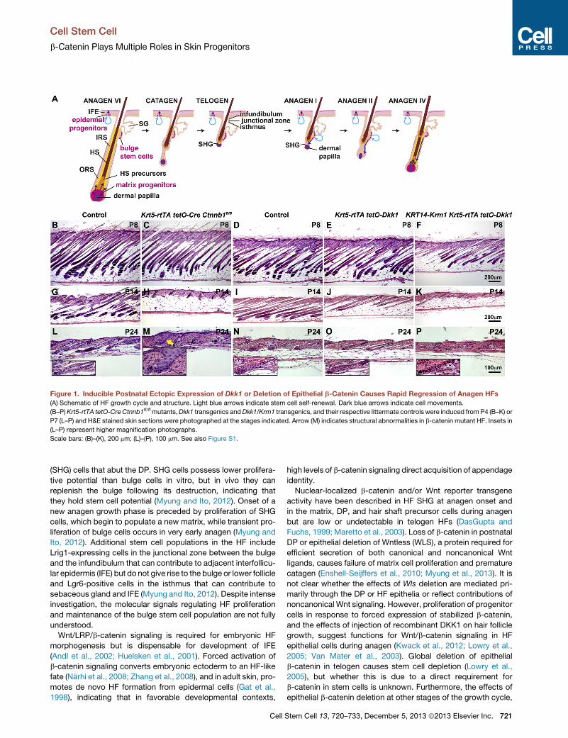
Figure 1. Inducible Postnatal Ectopic Expression of Dkk1 or Deletion of Epithelial b-Catenin Causes Rapid Regression of Anagen HFs
(A) Schematic of HF growth cycle and structure. Light blue arrows indicate stem cell self-renewal. Dark blue arrows indicate cell movements.
(B–P)Krt5-rtTA tetO-CreCtnnb1fl/flmutants,Dkk1 transgenics andDkk1/Krm1 transgenics, and their respective littermate controls were induced fromP4 (B–K) or
P7 (L–P) and H&E stained skin sections were photographed at the stages indicated. Arrow (M) indicates structural abnormalities in b-catenin mutant HF. Insets in
(L–P) represent higher magnification photographs.
Scale bars: (B)–(K), 200 mm; (L)–(P), 100 mm. See also Figure S1.
Cell Stem Cell
b-Catenin Plays Multiple Roles in Skin Progenitors
(SHG) cells that abut the DP. SHG cells possess lower prolifera-
tive potential than bulge cells in vitro, but in vivo they can
replenish the bulge following its destruction, indicating that
they hold stem cell potential (Myung and Ito, 2012). Onset of a
new anagen growth phase is preceded by proliferation of SHG
cells, which begin to populate a new matrix, while transient pro-
liferation of bulge cells occurs in very early anagen (Myung and
Ito, 2012). Additional stem cell populations in the HF include
Lrig1-expressing cells in the junctional zone between the bulge
and the infundibulum that can contribute to adjacent interfollicu-
lar epidermis (IFE) but do not give rise to the bulge or lower follicle
and Lgr6-positive cells in the isthmus that can contribute to
sebaceous gland and IFE (Myung and Ito, 2012). Despite intense
investigation, the molecular signals regulating HF proliferation
and maintenance of the bulge stem cell population are not fully
understood.
Wnt/LRP/b-catenin signaling is required for embryonic HF
morphogenesis but is dispensable for development of IFE
(Andl et al., 2002; Huelsken et al., 2001). Forced activation of
b-catenin signaling converts embryonic ectoderm to an HF-like
fate (Narhi et al., 2008; Zhang et al., 2008), and in adult skin, pro-
motes de novo HF formation from epidermal cells (Gat et al.,
1998), indicating that in favorable developmental contexts,
Cell
high levels of b-catenin signaling direct acquisition of appendage
identity.
Nuclear-localized b-catenin and/or Wnt reporter transgene
activity have been described in HF SHG at anagen onset and
in the matrix, DP, and hair shaft precursor cells during anagen
but are low or undetectable in telogen HFs (DasGupta and
Fuchs, 1999; Maretto et al., 2003). Loss of b-catenin in postnatal
DP or epithelial deletion of Wntless (WLS), a protein required for
efficient secretion of both canonical and noncanonical Wnt
ligands, causes failure of matrix cell proliferation and premature
catagen (Enshell-Seijffers et al., 2010; Myung et al., 2013). It is
not clear whether the effects of Wls deletion are mediated pri-
marily through the DP or HF epithelia or reflect contributions of
noncanonical Wnt signaling. However, proliferation of progenitor
cells in response to forced expression of stabilized b-catenin,
and the effects of injection of recombinant DKK1 on hair follicle
growth, suggest functions for Wnt/b-catenin signaling in HF
epithelial cells during anagen (Kwack et al., 2012; Lowry et al.,
2005; Van Mater et al., 2003). Global deletion of epithelial
b-catenin in telogen causes stem cell depletion (Lowry et al.,
2005), but whether this is due to a direct requirement for
b-catenin in stem cells is unknown. Furthermore, the effects of
epithelial b-catenin deletion at other stages of the growth cycle,
Stem Cell 13, 720–733, December 5, 2013 ª2013 Elsevier Inc. 721
Cell Stem Cell
b-Catenin Plays Multiple Roles in Skin Progenitors
and the consequences of specifically inhibiting canonical Wnt
signaling upstream of b-catenin, have not been systematically
investigated.
Unlike the HF, which proliferates periodically, basal IFE is
active throughout life, both renewing itself and generating cells
that differentiate to form a cornified layer that is continuously
shed. While expression of the TOPGAL Wnt reporter transgene
is undetectable in the IFE (DasGupta and Fuchs, 1999), expres-
sion of other, more sensitive reporters, and possible functions of
b-catenin signaling in adult IFE in vivo, have not been examined.
Here we show, using two independent, sensitive in vivo
reporters, that Wnt/b-catenin signaling is active in IFE and
specialized nonhairy epithelia as well as in anagen HFs. Using
multiple genetic approaches to manipulate signaling in specific
cell types, we demonstrate that epithelial b-catenin signaling is
required for maintenance of proliferation in anagen HFs and con-
tributes to proliferation of footpad and tongue but is not required
within the HF bulge and SHG for stem cell survival. Consistent
with this, hair regrowth occurs spontaneously after removal
of Wnt/b-catenin signaling inhibition. To analyze the role of
b-catenin in the IFE of hairy skin, we developed a system that
permits gene deletion specifically in IFE while sparing the hair
follicle bulge, SHG, and DP, allowing analysis of IFE phenotypes
in the absence of inflammatory reactions associated with HF
degradation. These experiments revealed a previously unknown
role for b-catenin signaling in contributing to proliferation of IFE
in vivo.
RESULTS
Wnt/b-Catenin Signaling Is Active in Basal IFE, NonhairyEpithelia, and Anagen HFsTo assay for Wnt/b-catenin signaling in postnatal skin, we
utilized Axin2lacZ, in which lacZ is inserted into the endogenous
Axin2 locus, a ubiquitous Wnt target (Lustig et al., 2002; Yu
et al., 2005), and TCF/Lef:H2B-GFP (TL-GFP), in which six
copies of a TCF/LEF responsive element and an hsp68 minimal
promoter drive expression of an H2B-GFP fusion protein
(Ferrer-Vaquer et al., 2010). Sensitivity of the TL-GFP reporter
is supported by its activity at sites of Wnt/b-catenin signaling
not documented with other reporters but confirmed through
genetic analysis (Ferrer-Vaquer et al., 2010). Expression of
Axin2lacZ and TL-GFP was low or undetectable in the bulge,
SHG, and DP of telogen HFs (Figures S1D and S1H available
online). Both reporters were expressed in the DP, SHG, and
matrix in early and mid-anagen and very strongly in hair shaft
precursor cells at mid-anagen (Figures S1A–S1C, S1E, and
S1G). Unexpectedly, bothAxin2lacZ and TL-GFPwere expressed
at low levels in IFE (Figures S1B, S1D, S1E, S1F, and S1H). TL-
GFP-expressing IFE cells were observed in basal and supra-
basal layers.Axin2lacZ-positive cells were confined to basal cells,
suggesting that suprabasal expression of TL-GFPmay be due to
perdurance of H2B-GFP. The number of positive cells and the
intensity of reporter expression in IFE increased with age (Fig-
ures S1F and S1H). Axin2lacZ and TL-GFP were also expressed
in stratified tongue epithelia (Figures S1I and S1K). Axin2lacZ,
but not TL-GFP, was expressed in adult footpad epidermis (Fig-
ures S1J and S1L), perhaps reflecting greater sensitivity of the
Axin2lacZ reporter in the footpad or increased signaling in mice
722 Cell Stem Cell 13, 720–733, December 5, 2013 ª2013 Elsevier In
heterozygous for loss of Axin2 function. Consistent with Wnt/
b-catenin signaling in IFE, several Wnt ligands and FZD recep-
tors are expressed in embryonic and adult IFE as well as in
HFs (Reddy et al., 2001, 2004) (Figures S1M and S1N).
Epithelial b-Catenin Deletion or Ectopic Dkk1
Expression Induced during Embryonic Anagen CausesRapid HF RegressionTo delineate the requirements for Wnt/b-catenin signaling at
successive stages of the embryonic hair growth cycle, we
compared the effects of doxycycline-inducible deletion of
b-catenin and inducible ectopic expression of DKK1, which in-
hibits signaling at the level of the LRP coreceptor. Mice carrying
a Krt5-rtTA transgene (Diamond et al., 2000) in which a reverse
tet transactivator is expressed in basal epidermis and HF ORS
including bulge stem cells were mated either to mice carrying
tetO-Cre (Mucenski et al., 2003) and a conditional null allele of
Ctnnb1 (Brault et al., 2001) or to tetO-Dkk1 mice (Chu et al.,
2004) (Figures S1O–S1S). Inducible Dkk1 was expressed at
higher levels in HFs than in IFE (Figure S1S). In contrast with pub-
lished data (Kwack et al., 2012), we were not able to detect
significant levels of expression of endogenous Dkk1 by in situ
hybridization at any stage of the adult HF growth cycle. The
Wnt/LRP inhibitory actions of DKK1 require interaction with
KRM (Mao et al., 2002), which is expressed at low levels in post-
natal skin (Figures S1T and S1U). Because limiting levels of KRM
may restrict the effectiveness of DKK1-mediated inhibition, we
generated KRT14-Krm1 mice that constitutively expressed
high levels of Krm1 in epithelial cells (Figure S1V) and assayed
the effects of coexpressed Krm1 and Dkk1 on hair growth.
KRT14-Krm1 mice did not display detectable abnormalities in
skin histology or in timing of the hair growth cycle in the absence
of coexpressed Dkk1 (Figures S1W and S1X).
ExperimentalKrt5-rtTA tetO-Cre Ctnnb1fl/fl (b-cateninmutant),
Krt5-rtTA tetO-Dkk1 (Dkk1 transgenic), or KRT14-Krm1 Krt5-
rtTA tetO-Dkk1 (Dkk1/Krm1 transgenic) mice and their respec-
tive control littermates were doxycycline treated from postnatal
day (P) 4 (Figures 1B–1K) or 7 (Figures 1L–1P), and dorsal skin
was harvested at P8, P14, and P24. Loss of epithelial b-catenin
or forced expression of Dkk1 caused rapid cessation of anagen
and entry into a premature regression phase comparedwith con-
trols (Figures 1B–1E, 1G–1J, and 1L–1O). The effects of Dkk1
were enhanced by coexpression with Krm1 (Figures 1F, 1K,
and 1P). As HF regression is caused by either epithelial b-catenin
deletion or LRP signaling inhibition, these data indicate that
canonical Wnt/LRP/b-catenin pathway activity is required within
epithelial cells to maintain anagen. At P24, HFs in epithelial-
b-catenin-deleted, but not Dkk1- or Dkk1/Krm1-expressing
skin, displayed structural defects including a widened infundib-
ulum and loss of a clearly distinguishable SHG (Figure 1M, arrow;
see inset), suggesting additional, LRP-independent roles for
b-catenin in telogen (Figures 1L–1P, see insets).
Rapid Regression of Mutant HFs Is Associated withInhibition of Cell Proliferation, but Bulge Stem CellMarkers Are MaintainedTo determine the underlying cause of rapid regression of b-cate-
nin-deficient andDkk1-expressing HFs, we assayed for changes
in cell proliferation and apoptosis. Ki67 immunofluorescence at
c.
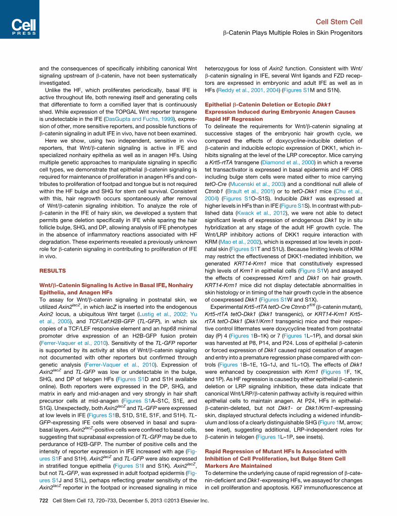
Figure 2. Inducible b-Catenin Deletion or Dkk1 Expression in Anagen Inhibits HF Matrix Proliferation without Loss of Stem Cells
Krt5-rtTA tetO-Cre Ctnnb1fl/flmutants,Dkk1 transgenics andDkk1/Krm1 transgenics, and their respective littermate controls mice were induced from P4 (A–T) or
P7 (U–Y0 ) and skin samples were harvested at the stages indicated. (A–J) Ki67 immunofluorescence (A, B, red; C–J, green). (K–O) TUNEL staining (green) (yellow
arrows). (P–Y) KRT15 immunofluorescence (P, Q, red; R–Y, green) (white arrows). Insets in (P)–(T) represent highermagnification photographs of regions indicated
by arrows. (U0–Y0) CD34 immunofluorescence (U0, V0, red;W0–Y0, green) (white arrows). Scale bars: (A)–(E), (K)–(O), (U)–(Y0), 50 mm; (F)–(J), (P)–(T), 100 mm. See also
Figure S2.
Cell Stem Cell
b-Catenin Plays Multiple Roles in Skin Progenitors
P8or P14 revealed greatly reduced proliferation in both b-catenin
mutant (Figures 2B and 2G) and Dkk1 or Dkk1/Krm1 transgenic
(Figures 2D, 2E, 2I, and 2J) follicles compared with controls (Fig-
ures 2A, 2C, 2F, and 2H). Similarly, Dkk1-expressing hair bulb
sections contained a mean of 16% ± 3% BrdU-positive nuclei
after 4 days of induction at P8, compared with 36% ± 5% in
controls (n = 17 HFs from two control mice and 13 HFs from
two transgenic mice) (Figures S2A–S2C). b-catenin-deleted and
Dkk1-expressing HFs showed greatly diminished expression of
cyclin D1, a direct Wnt/b-catenin target gene that helps initiate
transition from late G1 to S phase of the cell cycle (Kobielak
et al., 2003; Tetsu and McCormick, 1999), likely contributing to
decreased HF matrix proliferation (Figures S2D–S2G).
Consistent with accelerated entry into catagen, P8 Dkk1- and
Dkk1/Krm1-expressing follicles contained slightly increased
numbers of apoptotic cells compared with controls (Figures
2M–2O, yellow arrows). Although the timing of regression was
similar in b-catenin-deleted and Dkk1/Krm1-expressing follicles,
Cell
many more TUNEL-positive cells were observed in b-catenin
mutant HF matrix (Figures 2L and 2O).
Interestingly, expression of both KRT15, a marker for epithelial
stem cells in the HF bulge and SHG (Morris et al., 2004), and
CD34, which specifically marks bulge stem cells (Trempus
et al., 2003), was readily detected at P14 and P24 in b-catenin
mutants and Dkk1 transgenics induced from P4 and P7, respec-
tively (Figures 2P–2Y0), indicating that cessation of anagen was
not due to immediate loss of the stem cell compartment.
b-Catenin Deletion or Ectopic Dkk1 Block BothPlucking-Induced and Spontaneous AnagenTo determine whether adult anagen onset requires b-catenin or
signaling through LRP, we doxycycline-treated Krt5-rtTA tetO-
Cre Ctnnb1fl/fl and Dkk1 or Dkk1/Krm1 transgenic mice and their
respective littermate controls from P50 (telogen) and plucked
hair at P54 to induce a newgrowth phase. At 3 days postplucking
(3DPP), SHG cells in control follicles gave rise to a primitive
Stem Cell 13, 720–733, December 5, 2013 ª2013 Elsevier Inc. 723
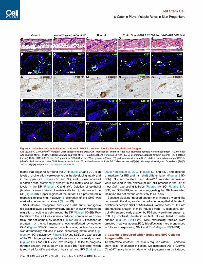
Figure 3. Inducible b-Catenin Deletion or Ectopic Dkk1 Expression Blocks Plucking-Induced Anagen
Krt5-rtTA tetO-Cre Ctnnb1fl/fl mutants, Dkk1 transgenics and Dkk1/Krm1 transgenics, and their respective littermate controls were induced from P50, their hair
was plucked at P54, and their dorsal skin was analyzed at P57. Paraffin sections were stained with H&E (A–E) or immunostained for Ki67 (green) (F–J), b-catenin
(brown) (K–O), KRT15 (P, Q, red; R–T, green), or CD34 (U, V, red; W–Y, green). In (F) and (G), yellow arrows indicate SHG; white arrows indicate upper ORS. In
(M)–(O), black arrow indicates SHG, blue arrows indicate IFE, and red arrows indicate DP. Yellow arrows in (P)–(Y) indicate positive signals. Scale bars: (A)–(E),
100 mm; (F)–(Y), 50 mm. See also Figures S2 and S3.
Cell Stem Cell
b-Catenin Plays Multiple Roles in Skin Progenitors
matrix that began to surround the DP (Figures 3A and 3C). High
levels of proliferationwere observed in the developingmatrix and
in the upper ORS (Figures 3F and 3H), and nuclear localized
b-catenin was prominently present in the matrix and at lower
levels in the DP (Figures 3K and 3M). Deletion of epithelial
b-catenin caused failure of matrix cells to migrate around the
DP (Figure 3B). Upper regions of the mutant HFs proliferated in
response to plucking; however, proliferation of the SHG was
markedly decreased or absent (Figure 3G).
Dkk1 double transgenic and Dkk1/Krm1 triple transgenic
follicles displayed signs of very early anagen at 3DPPwith limited
migration of epithelial cells around the DP (Figures 3C–3E). Pro-
liferation of the SHG was severely reduced compared with con-
trols, but not completely absent (Figures 3H–3J). Presence of
b-catenin at the cell membrane was unaffected by ectopic
Dkk1 (Figures 3M–3O, blue arrows); however, nuclear b-catenin
was dramatically reduced in Dkk1-expressing matrix cells (Fig-
ures 3M–3O, black arrow; Figures S3A and S3B), and expression
of the Wnt target gene cyclin D1 was almost completely absent
(Figures S3C and S3D). Dkk1-expressing HF failed to progress
through anagen, indicated by decreased BMP signaling, which
is required for differentiation of hair shaft and IRS (Andl et al.,
724 Cell Stem Cell 13, 720–733, December 5, 2013 ª2013 Elsevier In
2004; Kobielak et al., 2003) (Figures S3I and S3J), and absence
of markers for IRS and hair shaft differentiation (Figures S3K–
S3N). Nuclear b-catenin and Axin2lacZ reporter expression
were reduced in the epithelium but still present in the DP of
most Dkk1-expressing follicles (Figures 3M–3O; Figures S3A,
S3B, and S3E–S3H, red arrows), suggesting thatDkk1-mediated
inhibition did not extend effectively to DP cells.
Because plucking-induced anagen may induce a wound-like
response in the skin, we also tested whether epithelial b-catenin
deletion or ectopic Dkk1 or Dkk1/Krm1 blocked entry of HFs into
spontaneous anagen. In mice induced from P17 (catagen), con-
trol HFs entered early anagen by P25 and were in full anagen at
P30. By contrast, b-catenin mutant follicles failed to enter
anagen (Figures S2H–S2K). Dkk1-expressing follicles were
arrested in early anagen at P30; this effect wasmore pronounced
in follicles coexpressing Dkk1 and Krm1 (Figures S3O–S3T).
b-Catenin Is Required within Bulge and SHG Cells forAnagen InitiationTo determine whether b-catenin is required within HF epithelial
stem cells for anagen initiation, we generated Krt15-CrePR1
Ctnnb1fl/fl mice in which deletion of b-catenin can be induced
c.
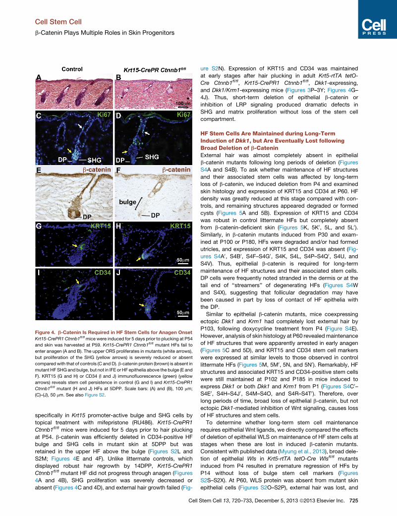
Figure 4. b-Catenin Is Required in HF Stem Cells for Anagen Onset
Krt15-CrePR1 Ctnnb1fl/flmice were induced for 5 days prior to plucking at P54
and skin was harvested at P59. Krt15-CrePR1 Ctnnb1fl/fl mutant HFs fail to
enter anagen (A and B). The upper ORS proliferates in mutants (white arrows),
but proliferation of the SHG (yellow arrows) is severely reduced or absent
compared with that of controls (C and D). b-catenin protein (brown) is absent in
mutant HF SHG and bulge, but not in IFE or HF epithelia above the bulge (E and
F). KRT15 (G and H) or CD34 (I and J) immunofluorescence (green) (yellow
arrows) reveals stem cell persistence in control (G and I) and Krt15-CrePR1
Ctnnb1fl/fl mutant (H and J) HFs at 5DPP. Scale bars: (A) and (B), 100 mm;
(C)–(J), 50 mm. See also Figure S2.
Cell Stem Cell
b-Catenin Plays Multiple Roles in Skin Progenitors
specifically in Krt15 promoter-active bulge and SHG cells by
topical treatment with mifepristone (RU486). Krt15-CrePR1
Ctnnb1fl/fl mice were induced for 5 days prior to hair plucking
at P54. b-catenin was efficiently deleted in CD34-positive HF
bulge and SHG cells in mutant skin at 5DPP but was
retained in the upper HF above the bulge (Figures S2L and
S2M; Figures 4E and 4F). Unlike littermate controls, which
displayed robust hair regrowth by 14DPP, Krt15-CrePR1
Ctnnb1fl/fl mutant HF did not progress through anagen (Figures
4A and 4B), SHG proliferation was severely decreased or
absent (Figures 4C and 4D), and external hair growth failed (Fig-
Cell
ure S2N). Expression of KRT15 and CD34 was maintained
at early stages after hair plucking in adult Krt5-rtTA tetO-
Cre Ctnnb1fl/fl, Krt15-CrePR1 Ctnnb1fl/fl, Dkk1-expressing,
and Dkk1/Krm1-expressing mice (Figures 3P–3Y; Figures 4G–
4J). Thus, short-term deletion of epithelial b-catenin or
inhibition of LRP signaling produced dramatic defects in
SHG and matrix proliferation without loss of the stem cell
compartment.
HF Stem Cells Are Maintained during Long-TermInduction of Dkk1, but Are Eventually Lost followingBroad Deletion of b-CateninExternal hair was almost completely absent in epithelial
b-catenin mutants following long periods of deletion (Figures
S4A and S4B). To ask whether maintenance of HF structures
and their associated stem cells was affected by long-term
loss of b-catenin, we induced deletion from P4 and examined
skin histology and expression of KRT15 and CD34 at P60. HF
density was greatly reduced at this stage compared with con-
trols, and remaining structures appeared degraded or formed
cysts (Figures 5A and 5B). Expression of KRT15 and CD34
was robust in control littermate HFs but completely absent
from b-catenin-deficient skin (Figures 5K, 5K0, 5L, and 5L0).Similarly, in b-catenin mutants induced from P30 and exam-
ined at P100 or P180, HFs were degraded and/or had formed
utricles, and expression of KRT15 and CD34 was absent (Fig-
ures S4A0, S4B0, S4F–S4G0, S4K, S4L, S4P–S4Q0, S4U, and
S4V). Thus, epithelial b-catenin is required for long-term
maintenance of HF structures and their associated stem cells.
DP cells were frequently noted stranded in the dermis or at the
tail end of ‘‘streamers’’ of degenerating HFs (Figures S4W
and S4X), suggesting that follicular degradation may have
been caused in part by loss of contact of HF epithelia with
the DP.
Similar to epithelial b-catenin mutants, mice coexpressing
ectopic Dkk1 and Krm1 had completely lost external hair by
P103, following doxycycline treatment from P4 (Figure S4E).
However, analysis of skin histology at P60 revealedmaintenance
of HF structures that were apparently arrested in early anagen
(Figures 5C and 5D), and KRT15 and CD34 stem cell markers
were expressed at similar levels to those observed in control
littermate HFs (Figures 5M, 5M0, 5N, and 5N0). Remarkably, HF
structures and associated KRT15 and CD34-positive stem cells
were still maintained at P102 and P185 in mice induced to
express Dkk1 or both Dkk1 and Krm1 from P1 (Figures S4C0–S4E0, S4H–S4J0, S4M–S4O, and S4R–S4T0). Therefore, over
long periods of time, broad loss of epithelial b-catenin, but not
ectopic Dkk1-mediated inhibition of Wnt signaling, causes loss
of HF structures and stem cells.
To determine whether long-term stem cell maintenance
requires epithelial Wnt ligands, we directly compared the effects
of deletion of epithelial WLS on maintenance of HF stem cells at
stages when these are lost in induced b-catenin mutants.
Consistent with published data (Myung et al., 2013), broad dele-
tion of epithelial Wls in Krt5-rtTA tetO-Cre Wlsfl/fl mutants
induced from P4 resulted in premature regression of HFs by
P14 without loss of bulge stem cell markers (Figures
S2S–S2X). At P60, WLS protein was absent from mutant skin
epithelial cells (Figures S2O–S2P), external hair was lost, and
Stem Cell 13, 720–733, December 5, 2013 ª2013 Elsevier Inc. 725

Figure 5. HF StemCells Disappear after Long-Term Broad Deletion of Epithelial b-Catenin but Persist when b-Catenin Deletion Is Restricted
to the Bulge and SHG
Krt5-rtTA tetO-Cre Ctnnb1fl/fl mutants, Dkk1/Krm1 triple transgenics, and control littermates were doxycycline treated from P4 to P60 and Krt5-rtTA tetO-Cre
Wlsfl/fl and control mice, from P4 to P18. Krt15-CrePR1Wlsfl/fl and control littermates were treated topically with 1%mifepristone from P19 to P25. Krt15-CrePR1
Ctnnb1fl/fl mice and control littermates were treated topically with 1% mifepristone from P20 to P27. All skin samples were analyzed at P60 (telogen) by H&E
staining (A–J) or immunofluorescence for KRT15 (K–P, S, T, green; Q0, R0, red), CD34 (K0–N0, Q0 0, R0 0, S0, T0, green; O0, P0, red), b-catenin (Q, R, Q0 0, R0 0, red), orWLS
(S, T, S0, T0, red). Yellow arrows indicate positive staining for KRT15 or CD34. Scale bars: (A)–(J), 120 mm; (K)–(T0), (Q0 0 ), and (R0 0 ), 60 mm. See also Figures S2, S4,
S6, and S7.
Cell Stem Cell
b-Catenin Plays Multiple Roles in Skin Progenitors
HF structures were retained but showed marked abnormalities
including the formation of cysts (Figures 5E and 5F). Despite
these defects, strong expression of KRT15 and CD34 was asso-
ciatedwith follicle structures inWlsmutant skin (Figures 5O–5P0).KRT15 and CD34 were also detected at P60 following specific
726 Cell Stem Cell 13, 720–733, December 5, 2013 ª2013 Elsevier In
deletion of WLS in the bulge and SHG of Krt15-CrePR1 Wlsfl/fl
mutant HFs (Figures S2Q and S2R; Figures 5S, 5S0, 5T, and5T0). Thus, long-term broad deletion of b-catenin, but not inhibi-
tion of LRP function or deletion of epithelial Wls, prevents stem
cell maintenance.
c.
Cell Stem Cell
b-Catenin Plays Multiple Roles in Skin Progenitors
StemCells AreMaintained following SpecificDeletion ofb-Catenin in the Bulge and SHGTo determine whether eventual loss of HF stem cell markers in
epithelial b-catenin mutants was due to a requirement for
b-catenin within bulge stem cells and SHG, we examined their
expression in P60 Krt15-CrePR1 Ctnnb1fl/fl mice, following
topical 1% mifepristone treatment from P20–27. Double immu-
nofluorescence for b-catenin and CD34 revealed maintenance
of CD34 immunoreactivity in b-catenin-deleted bulge cells at
this stage (Figures 5Q0 0 and 5R0 0). Staining of serial sections for
b-catenin and KRT15 indicated that expression of KRT15
persisted in b-catenin-deleted HFs (Figures 5Q, Q0, R, and R0).Similar results were obtained with another bulge stem cell
marker, S100A4 (data not shown). Despite persistence of bulge
stem cell marker expression, the SHG of Krt15-CrePR1
Ctnnb1fl/fl HF appeared abnormal or even absent in some HFs;
however, the upper follicles and sebaceous glands (SGs) re-
mained intact (Figures 5G and 5H). These data, and the slow
time course of bulge stem cell disappearance in Krt5-rtTA
tetO-Cre Ctnnb1fl/fl mice, suggested that bulge cell loss in
Krt5-rtTA tetO-Cre Ctnnb1fl/fl mutants occurred secondary to
follicular degradation resulting from combined absence of
b-catenin in the HF bulge/SHG and KRT15-negative cell popula-
tions, such as those in the junctional zone and isthmus (Myung
and Ito, 2012).
Long-Term Dkk1-Mediated Inhibition of HF Growth IsReversibleOur observation that KRT15andCD34expressionpersisted even
following very longperiodsofDkk1 induction raised thepossibility
that functional stem cells were maintained in Dkk1-expressing
follicles. To test this,weaskedwhether hair growthwas reversible
following removal of the inhibitor. Control and Dkk1 transgenic
mice weremaintained on doxycycline from postnatal day 21 until
15.5 months of age. At this stage, the experimental mice
completely lacked external hair (Figures 6A and 6B), but main-
tained HF structures (Figures 6C and 6D) with associated stem
cells (Figures 6E–6H). Ectopic expression of Dkk1 was readily
detected in induceddouble transgenicskinby in situhybridization
(Figure6I).Micewere sacrificed for analysis 14daysafter doxycy-
cline withdrawal. At this time point, expression of Dkk1 was no
longer detected (Figure 6J) and, remarkably, growth of numerous
follicles had occurred spontaneously (Figures 6K and 6L). HF
growth following doxycycline withdrawal was accompanied by
resumption of Wnt/b-catenin signaling, indicated by accumula-
tion of nuclear b-catenin in the HF matrix and precortical regions
(Figures 6Mand 6N), expression of cyclin D1 (Figures 6O and 6P),
and high levels of matrix cell proliferation (Figures 6Q and 6R). To
avoid multiple survival biopsies, we did not test whether external
hair growth occurred following removal of the inhibitor. However,
the reactivated matrix cells were capable of differentiating, as
indicated by positive staining for hair shaft precursor cells with
the hair keratin marker AE13 (Figures 6S and 6T). These data
suggested persistence in Dkk1-inhibited follicles of Wnt ligands
capable of reactivating HF growth following removal of the
inhibitor. Consistent with this, Wnt10b, a ligand specifically ex-
pressed in anagen HFs within the skin (Reddy et al., 2001), was
expressed in double transgenic HFs both before and after doxy-
cycline withdrawal (Figures 6U and 6V).
Cell
To test whether HFs coexpressing Dkk1 and Krm1 were also
capable of reinitiating follicular growth following removal of the
inhibitor, control littermate, Dkk1 transgenic, and Dkk1/Krm1
transgenic mice were induced from birth until P100. Analysis of
histology and immunostaining for KRT15, CD34, b-catenin,
cyclin D1, Ki67, and AE13 confirmed maintenance of HF struc-
tures and stem cell marker expression in Dkk1/Krm1 and Dkk1
transgenic and control mice at this stage, with lack of nuclear
b-catenin or cyclin D1 expression, absent or low levels of prolif-
eration, and lack of hair shaft differentiation (Figures S5A–S5Q).
Following 15 days of doxycycline withdrawal, both Dkk1 and
Dkk1/Krm1 skin contained numerous HFs that had spontane-
ously entered anagen, compared with control littermate follicles
that remained in telogen (Figures S5A0–S5C0). Hair follicle
regrowth was less synchronous in Dkk1/Krm1 skin than in skin
that had expressed Dkk1 alone, consistent with the stronger
inhibitory effects of Dkk1/Krm1. Dkk1 and Dkk1/Krm1 transgenic
follicles displayed an extended pattern of KRT15 and CD34
staining that is characteristic of anagen, rather than the compact
staining pattern seen in control telogen stage follicles (Figures
S5D0–S5I0). This was distinct from the staining observed in
Dkk1 and Dkk1/Krm1 transgenics prior to doxycycline removal,
which was intermediate between telogen and full anagen pat-
terns, consistent with arrest in early anagen (Figures S5E, S5F,
S5H, and S5I). Both Dkk1 and Dkk1/Krm1 transgenic samples
displayed accumulation of nuclear b-catenin and expression of
cyclin D1 in HF matrix regions following doxycycline withdrawal
(Figures S5J0–S5M0), as well as robust matrix proliferation (Fig-
ures S5N0 and S5O0) and expression of hair shaft keratins (Fig-
ures S5P0 and S5Q0).These data indicated functional persistence of HF bulge and/
or SHG cells and reversibility of the effects of LRP inhibition even
following very long-term Dkk1 induction, with or without coex-
pressed Krm1. Histological analyses, the intermediate expres-
sion pattern of KRT15, persistent expression of Wnt10b, and
the ability of HFs to spontaneously resume growth immediately
following removal of the inhibitor supported the interpretation
that Dkk1-inhibited HFs were arrested in early anagen and
primed for hair regrowth rather than beingmaintained in a normal
telogen resting phase.
b-Catenin Contributes to Proliferation of the IFE duringHomeostasisThe epidermis of mice with long-term broad deletion of epithelial
b-catenin orWls displayed marked thickening and hyperprolifer-
ation, expansion of p63-positive basal and KRT10-positive
suprabasal layers, and ectopic expression of hyperproliferation
markers including KRT6 and KRT17 (Figures 5A, 5B, 5E, and
5F; Figures S4A0, S4B0, S6A, and S6C). Induced Krt14-CreERT2
Ctnnb1fl/fl and Krt5-rtTA tetO-Cre Ctnnb1fl/fl mutants did not
display overt blistering, and a-catenin and E-cadherin were pre-
sent at cell membranes in b-catenin mutant as well as control
epidermis (Figures S7A–S7D). Transmission electron microgra-
phy (TEM) revealed the presence of enlarged intercellular spaces
in b-catenin mutant epidermis following long periods of broad
depletion of epithelial b-catenin. In some cases, mutant keratino-
cytes displayed long membranous protrusions (Figures S7E
and S7F) that were not seen in control skin or in induced
KRT14-Krm1/Dkk1 triple transgenics (Figures S7G and S7H) or
Stem Cell 13, 720–733, December 5, 2013 ª2013 Elsevier Inc. 727
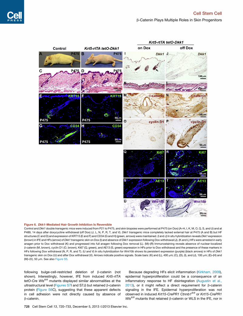
Figure 6. Dkk1-Mediated Hair Growth Inhibition Is Reversible
Control andDkk1 double transgenic mice were induced from P21 to P475, and skin biopsies were performed at P475 (on Dox) (A–H, I, K, M, O, Q, S, and U) and at
P489, 14 days after doxycycline withdrawal (off Dox) (J, L, N, P, R, T, and V). Dkk1 transgenic mice completely lacked external hair at P475 (A and B) but HF
structures (C andD) and expression of KRT15 (E and F) and CD34 (G andH) (green, arrows) weremaintained. (I and J) In situ hybridization revealsDkk1 expression
(brown) in IFE andHFs (arrow) ofDkk1 transgenic skin on Dox (I) and absence ofDkk1 expression following Doxwithdrawal (J). (K and L) HFswere arrested in early
anagen prior to Dox withdrawal (K) and progressed into full anagen following Dox removal (L). (M)–(R) Immunostaining reveals absence of nuclear-localized
b-catenin (M, brown), cyclin D1 (O, brown), Ki67 (Q, green), and AE13 (S, green) expression in HFs prior to Dox withdrawal and the presence of these markers in
HFs following Dox withdrawal (N, P, R, and T). (U and V) In situ hybridization for Wnt10b shows its persistent expression (purple) (black arrows) in HFs of Dkk1
transgenic skin on Dox (U) and after Dox withdrawal (V). Arrows indicate positive signals. Scale bars: (K) and (L), 400 mm; (C), (D), (I), and (J), 100 mm; (E)–(H) and
(M)–(V), 50 mm. See also Figure S5.
Cell Stem Cell
b-Catenin Plays Multiple Roles in Skin Progenitors
following bulge-cell-restricted deletion of b-catenin (not
shown). Interestingly, however, IFE from induced Krt5-rtTA
tetO-Cre Wlsfl/fl mutants displayed similar abnormalities at the
ultrastructural level (Figures S7I and S7J) but retained b-catenin
protein (Figure S6C), suggesting that these apparent defects
in cell adhesion were not directly caused by absence of
b-catenin.
728 Cell Stem Cell 13, 720–733, December 5, 2013 ª2013 Elsevier In
Because degrading HFs elicit inflammation (Kirkham, 2009),
epidermal hyperproliferation could be a consequence of an
inflammatory response to HF disintegration (Augustin et al.,
2013), or it might reflect a direct requirement for b-catenin
signaling in the IFE. Epidermal hyperproliferation was not
observed in induced Krt15-CrePR1 Ctnnb1fl/fl or Krt15-CrePR1
Wlsfl/fl mutants that retained b-catenin or WLS in the IFE, nor in
c.
Cell Stem Cell
b-Catenin Plays Multiple Roles in Skin Progenitors
KRT14-Krm1 Krt5-rtTA tetO-Dkk1 mice, which express lower
levels of Dkk1 in IFE than in HFs (Figures S6B, S6D, S6E, and
S1S). However, these mutants also lacked overt HF structural
defects and displayed lower levels of inflammatory cells
than those seen in Krt5-rtTA tetO-Cre-driven mutants (Figures
S7K–S7T).
To test directly whether b-catenin is required for IFE homeo-
stasis, we generated a Axin2CreERT2/tdT knockin line by insert-
ing a CreERT2/tdTomato fusion cDNA downstream of the first
codon of the endogenous mouse Axin2 gene (Figure S7U). This
line expresses cytoplasmic tandem dimer Tomato (tdT) and
tamoxifen-inducible Cre-recombinase (CreERT2) in Axin2-
promoter-active cells (not shown). To assess the efficiency of
inducible deletion, we bred Axin2CreERT2/tdT mice with the
R26RmTmG Cre reporter line that expresses membrane-targeted
tandem dimer Tomato (mT) prior to Cre-mediated excision and
membrane-targeted GFP (mG) after excision (Muzumdar et al.,
2007). Following induction of Cre activity by treatment with
tamoxifen during telogen, GFP-positive cells were present in
the IFE and HF infundibulum, but no excision was observed in
the HF bulge, SHG, or DP (Figure 7A). Clones of GFP-positive
deleted cells were also observed in tongue and footpad
epidermis (Figures 7B and 7C), consistent with Axin2lacZ expres-
sion in basal progenitor cells of these tissues. To determine
whether Axin2-promoter-active cells give rise to clones in the
IFE, we crossed Axin2CreERT2/tdT mice with R26R-Confetti
mice that function as a stochastic multicolor Cre reporter,
permitting clonal analysis of cells expressing GFP, YFP, RFP,
or CFP under the control of a CAG promoter (Snippert et al.,
2010). RFP was expressed much more strongly than Axin2-pro-
moter-driven tdT, allowing us to easily identify RFP-positive and
other fluorescently marked clones. Following tamoxifen induc-
tion in telogen, we observed clones of fluorescently marked
cells originating in the basal progenitor layer of the IFE (Figures
7D and 7D0).Because efficient Axin2CreERT2/tdT-mediated deletion of
b-catenin may cause systemic defects, we used conditions
under which mice exhibited mosaic deletion. Axin2CreERT2/
tdT Ctnnb1fl/fl mice and Axin2CreERT2/tdT Ctnnb1fl/+ control
littermates were injected with 200 mg/kg tamoxifen daily at
P43, P44, and P45 and tissue was harvested 4 weeks later.
Immunofluorescence for b-catenin revealed mosaic absence of
b-catenin in IFE, but not in HF bulge or SHG (Figures 7E–7H).
Histological analysis did not reveal gross HF structural defects
in these animals (not shown). We quantified proliferation by
assaying for Ki67 immunofluorescence in nonserial sections of
IFE, choosing regions where b-catenin was either efficiently
deleted in continuous strips of at least 20 cells (n = 75 nonadja-
cent strips of 22–67 cells, total of 2,965 cells analyzed) or re-
tained in continuous strips of at least 20 cells (n = 23 nonadjacent
strips of 20–65 cells, total of 903 cells analyzed), and compared
proliferation rates with those seen in littermate control epidermis
(n = 52 nonadjacent strips of 38–85 cells, total of 2,780 cells
analyzed). Surprisingly, rather than exhibiting hyperproliferation,
regions of b-catenin-deleted epidermis showed a statistically
significant decrease in proliferation rate of more than 40%
when compared with either nondeleted mutant epidermis (p =
5.4 3 10�13) or control epidermis (p = 1.9 3 10�15) (Figures
7E–7H and 7Y). The proliferation rate of nondeleted mutant
Cell
epidermis was similar to that of control epidermis, indicating
that decreased proliferation of deleted cells was not due to sys-
temic defects (Figure 7Y).
Nonhairy epithelia of the footpad and tongue lacked obvious
inflammation; however, filiform papillae were reduced or absent
in induced Krt5-rtTA tetO-Cre Ctnnbfl/fl and Krt5-rtTA tetO-Dkk1
KRT14-Krm1 mice compared with controls (Figures 7I, 7J, 7Q,
and 7R). We assayed for epithelial proliferation in n = 10 non-
adjacent samples of footpad and dorsal tongue from each
littermate control and mutant or transgenic, with each sample
containing 60 DAPI-stained basal nuclei. Proliferation was signif-
icantly reduced in footpad and tongue of Krt5-rtTA tetO-Cre
Ctnnbfl/fl mutants (Figures 7K–7P and 7Y); similar though less
pronounced, defects were observed in the footpad and tongue
of induced Krt5-rtTA tetO-Dkk1 KRT14-Krm1 mice (Figures
7S–7Y), indicating that they were caused by decreased Wnt/
LRP/b-catenin signaling, rather than being directly due to loss
of b-catenin’s functions in adhesion. Thus, in addition to control-
ling HF matrix proliferation, Wnt/b-catenin signaling contributes
to proliferation of IFE and specialized nonhairy epithelia.
In summary, our data suggest a model in which high levels of
Wnt/b-catenin signaling in the HF matrix drive proliferation. As
cells exit the matrix compartment, a further elevation of Wnt/
b-catenin signaling levels causes them to terminally differentiate
(Zhang et al., 2008; Zhou et al., 1995). Ectopic Dkk1, or deletion
of Wls or b-catenin, inhibits these high levels of signaling, pre-
venting matrix proliferation and differentiation. Unlike broad
loss of epithelial b-catenin, bulge/SHG-restricted b-catenin dele-
tion does not cause loss of stem cells, indicating that b-catenin is
not required within bulge/SHG cells for their maintenance. Unex-
pectedly, our data also reveal low level Wnt/b-catenin signaling
in IFE that is required to maintain normal levels of proliferation
under homeostatic conditions (Figure 7Z) and demonstrate that
Wnt/b-catenin signaling contributes to proliferation in special-
ized nonhairy epithelia. Despite these functions, complete loss
of epithelial b-catenin does not prevent long-term maintenance
of IFE or footpad and tongue epithelia, and b-catenin-deleted
IFE can still mount a hyperproliferative response to inflammation
associated with HF degradation.
DISCUSSION
Here we describe the sites and levels of Wnt/b-catenin signaling
in postnatal skin using two independent, sensitive in vivo
reporter assays. High levels of signaling were observed in the
HF DP, SHG, and matrix at early stages of anagen. As matrix
cells differentiated, they showed a further increase in signaling
levels. Reporter gene expression was not observed in telogen
HF bulge, SHG, or DP, but it was detected at low levels in the
infundibulum and IFE. Reporter expression was also detected
in nonhairy basal epithelia of footpad and tongue.
To delineate the functions of high- and low-level signaling in
different epithelial cell types, we directly compared the effects
of three independent genetic manipulations: deletion of epithelial
b-catenin, which abolishes all signaling through the Wnt/
b-catenin pathway; deletion of epithelialWls, which dramatically
decreases the availability of ligands for paracrine signaling
(Ching and Nusse, 2006); and ectopic expression of the secreted
Wnt/LRP inhibitor DKK1, which inhibits signaling depending on
Stem Cell 13, 720–733, December 5, 2013 ª2013 Elsevier Inc. 729
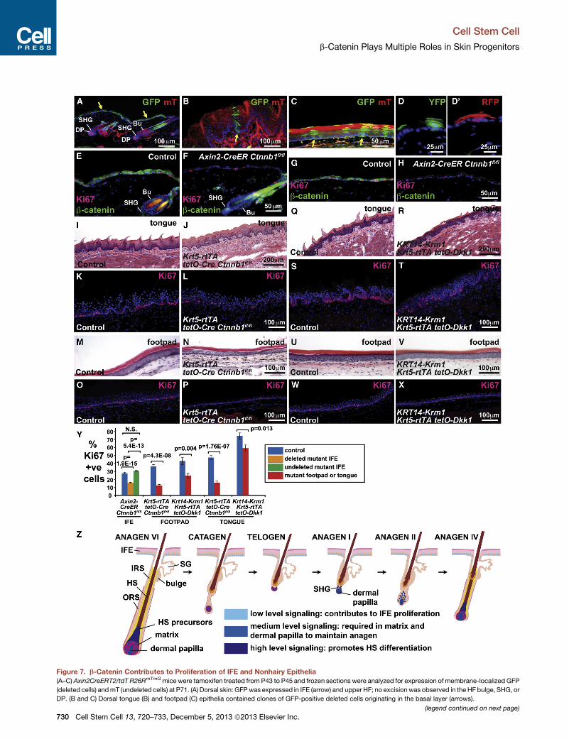
Figure 7. b-Catenin Contributes to Proliferation of IFE and Nonhairy Epithelia
(A–C) Axin2CreERT2/tdT R26RmTmGmice were tamoxifen treated from P43 to P45 and frozen sections were analyzed for expression of membrane-localized GFP
(deleted cells) andmT (undeleted cells) at P71. (A) Dorsal skin: GFPwas expressed in IFE (arrow) and upper HF; no excision was observed in the HF bulge, SHG, or
DP. (B and C) Dorsal tongue (B) and footpad (C) epithelia contained clones of GFP-positive deleted cells originating in the basal layer (arrows).
(legend continued on next page)
Cell Stem Cell
b-Catenin Plays Multiple Roles in Skin Progenitors
730 Cell Stem Cell 13, 720–733, December 5, 2013 ª2013 Elsevier Inc.
Cell Stem Cell
b-Catenin Plays Multiple Roles in Skin Progenitors
the levels of DKK1 expression and availability of the DKK1
receptor KRM.
During an established anagen phase, either deletion of epithe-
lial b-catenin or expression of Dkk1 caused HFs to rapidly cease
proliferating and enter premature catagen. The similar pheno-
types resulting from these two independent manipulations indi-
cate that maintenance of proliferation during anagen requires
signaling within epithelial cells through the canonical Wnt/LRP/
b-catenin pathway. The KRT15-positive stem cell compartment
was maintained during short-term deletion of b-catenin or
expression of Dkk1. This observation, together with the strong
localization of nuclear b-catenin and Wnt reporter gene expres-
sion in normal matrix cells, suggests that the immediate effects
of Wnt pathway inhibition are manifest directly in the matrix,
rather than via acute loss of bulge stem cells.
Because b-catenin is cell autonomous, the block to prolifera-
tion caused by deletion of epithelial b-catenin in anagen was
caused by its loss within epithelial cells. Interestingly, however,
the observed effects on proliferation and catagen induction are
similar to those noted when b-catenin is specifically deleted in
the anagen DP (Enshell-Seijffers et al., 2010). Thus b-catenin is
required in both these HF compartments for continued hair
growth. Our results further suggest that antagonism of Wnt
signaling in the epithelium and DP could constitute part of the
normal mechanism of catagen induction and prompt study of
endogenous Wnt inhibitors that might promote the anagen-
catagen transition.
Epithelial b-catenin mutants and mice expressing Dkk1 dis-
played subtle differences in response to hair plucking: in
b-catenin mutants cells proliferated in the ORS of plucked HFs,
but not in the SHG. By contrast, expression of ectopic Dkk1,
even when concomitant with forced expression of the DKK1
receptor Kremen1, permitted some degree of proliferation of
SHG as well as ORS cells. Similarly, following deletion of epithe-
lial Wls, many follicles enter anagen but are arrested in anagen I
(Myung et al., 2013). These observations provide circumstantial
evidence that the initial burst of b-catenin-dependent prolifera-
tion at anagen onset is controlled by mechanisms other than
Wnt/LRP signaling, consistent with prior data indicating that (1)
HF stem cells are initially activated by downregulation of dermal
BMP signaling (Plikus et al., 2008) and (2) impaired BMP
signaling stabilizes b-catenin through a Wnt-ligand-independent
pathway (Kobielak et al., 2007).
(D and D0) Cytoplasmic YFP- and RFP-marked clones originating in whisker pad b
at 16 weeks and analyzed 5 weeks later.
(E–H) Axin2CreERT2/tdT Ctnnb1fl/fl (F and H) and control littermate Axin2CreERT
sections were analyzed at P71 by immunofluorescence for b-catenin (green) and
was mosaically deleted in mutant IFE (E–H) and upper HF (E and F) but was retain
cells than controls.
(I–P) Absent tongue filiform papillae (I and J), decreased footpad epidermal thick
(O and P) epithelia in P100 Krt5-rtTA tetO-Cre Ctnnbfl/fl mice doxycycline-treated
(Q–X) Defective tongue filiform papillae (Q and R), mildly decreased footpad epide
and footpad (W and X) epithelia in P360 KRT14-Krm1 Krt5-rtTA tetO-Dkk1 mice
(Y) Quantification reveals statistically significant reductions in proliferation of b-
epithelia. Blue bars represent littermate controls; orange and green bars repre
Axin2CreERT2/tdT Ctnnb1fl/fl mutants; red bars represent Krt5-rtTA tetO-Cre Ct
shown as mean ± SEM. p values were calculated using a two-tailed Student’s t
(Z) Model depicting functions of low-, medium-, and high-level Wnt/b-catenin sig
Scale bars: (I), (J), (Q), and (R), 200 mm; (A), (B), (K)–(P), and (S)–(X), 100 mm; (C) a
Cell
In mice expressing epithelial Dkk1, with or without forced
expression of Krm1, HFs and their associated stem cells were
maintained for more than 1 year of Dkk1 expression. Stem cells
were also maintained following long-term deletion of epithelial
Wls, either throughout skin epithelia or specifically in the bulge/
SHG stem cell compartment, and following specific deletion of
b-catenin in bulge/SHG stem cells. These observations suggest
that the degradation of HF structures and complete loss of bulge
stem cells seen following broad deletion of b-catenin results from
combined absence of b-catenin in the HF bulge/SHG, junctional
zone, isthmus, and/or infundibulum. Furthermore, long-term
maintenance of follicular structures and their associated stem
cells occurs via a mechanism distinct from that controlling
proliferation and either requires very low levels of Wnt/LRP/
b-catenin signaling that persist in the presence of ectopic Dkk1
or absence of epithelialWls or is independent of LRP and epithe-
lial Wnt ligands.
Remarkably, removal of the inducing agent in aged Dkk1
transgenic mice following long periods of Dkk1 expression lead
to spontaneous resumption of hair germ proliferation, formation
of a new matrix, and differentiation of hair shaft and IRS cells,
providing evidence that functional bulge/SHG stem cells were
maintained in the absence of Wnt/b-catenin signaling. These
data suggest graded inhibition of Wnt/LRP signaling as a
promising method for reversibly inhibiting the growth of
unwanted hair without causing permanent follicle destruction
and skin damage. Small molecule Wnt inhibitors have been
identified (Voronkov and Krauss, 2013) and could potentially
be tested as topical agents for prevention of growth of unwanted
hair in humans. Use of such approaches would require tight
control of the levels of applied Wnt inhibitor to avoid causing
hair follicle abnormalities.
Unexpectedly, we detected activity of Wnt reporter genes in
adult IFE and in specialized nonhairy epithelia. We discovered
that specific loss of b-catenin in the IFE of hairy skin, sparing
the HF bulge and SHG, caused significantly reduced IFE prolifer-
ation. Similarly, b-catenin deletion or forced expression of Dkk1/
Krm1 decreased the thickness and proliferation of specialized
nonhairy epithelia of the footpad and dorsal tongue. In line with
these observations, human patients with mutations in the
canonical Wnt geneWNT10A display epidermal defects, smooth
tongues, and palmoplantar keratoderma in addition to hair
growth defects (Adaimy et al., 2007; Petrof et al., 2011).
asal IFE of Axin2CreERT2/tdT R26R-Confettimice tamoxifen-treated for 3 days
2/tdT Ctnnb1fl/+ (E and G) mice were tamoxifen treated at P43–P45 and skin
Ki67 (red) after microwave pretreatment to remove tdT fluorescence. b-catenin
ed in HF bulge and SHG. b-catenin-deleted IFE displayed fewer Ki67-positive
ness (M and N), and decreased proliferation of tongue (K and L) and footpad
from P20 compared with control littermates.
rmal thickness (U and V), and decreased proliferation of dorsal tongue (S and T)
doxycycline-treated from P20 compared with control littermates.
catenin-deleted and Dkk1/Krm1-expressing IFE, footpad, and dorsal tongue
sent regions of b-catenin-deleted and -undeleted epidermis, respectively, in
nnbfl/fl or KRT14-Krm1 Krt5-rtTA tetO-Dkk1 samples as indicated. Results are
test. N.S., not significant.
naling in skin epithelia.
nd (E)–(H), 50 mm; (D and D0), 25 mm. See also Figures S6 and S7.
Stem Cell 13, 720–733, December 5, 2013 ª2013 Elsevier Inc. 731
Cell Stem Cell
b-Catenin Plays Multiple Roles in Skin Progenitors
Furthermore, inducible deletion of epithelial b-catenin causes
regression of squamous cell carcinoma (SCC) in mice (Malanchi
et al., 2008). By contrast, broad epithelial deletion of b-catenin in
IFE and HFs was associated with HF disintegration, an inflam-
matory response, and IFE hyperproliferation. Taken together,
these data indicate that Wnt/b-catenin signaling contributes to
adult IFE proliferation under homeostatic conditions and in
SCC but is not required for long-term maintenance of the IFE
and is bypassed during hyperproliferative responses to inflam-
mation. The approaches described here will be generally useful
for determining the effects of gene deletion in adult IFE in cases
where HF degradation and inflammation resulting from broad
epithelial deletion complicate analysis of IFE phenotypes.
EXPERIMENTAL PROCEDURES
Mouse Strains, Transgene Induction, Depilation, Skin Biopsies, and
Genotyping
Krt5-rtTA tetO-Dkk1 mice were generated as described previously (Chu et al.,
2004). AKRT14-Krm1 transgenewas constructed by cloning full-lengthmouse
Krm1 cDNA (NM_032396) into KRT14 promoter vector (Saitou et al.,
1995). Ctnnb1fl/fl, Wlsfl/fl, Krt15-CrePR1, Krt14-CreER, R26R-Confetti, and
R26RmTmG mice were obtained from Jackson Labs (Bar Harbor, ME).
Axin2CreERT2/tdT mice were generated by insertion of a CreERT2/tdTomato
fusion cDNA downstream of the first ATG of the mouse Axin2 gene using
homologous recombination in mouse ESCs. Transgene induction was per-
formed as described previously (Chu et al., 2004; Ito et al., 2005). Skin biopsies
were taken from euthanized mice or under anesthesia. Detailed methods are
provided in the Supplemental Information. The IACUC committees of the
University of Pennsylvania and the University of Cincinnati approved all exper-
imental procedures involving mice.
Histology, Staining Procedures, and TEM
Preparation of paraffin sectioned skin, histological analysis, BrdU incorpora-
tion assays, TUNEL assays, immunostaining, in situ hybridization, X-gal
staining, alkaline phosphatase staining, and TEM were performed according
to published protocols (Andl et al., 2006; Zhang et al., 2009). Details are
provided in the Supplemental Information.
SUPPLEMENTAL INFORMATION
Supplemental Information for this article includes Supplemental Experimental
Procedures and seven figures and can be foundwith this article online at http://
dx.doi.org/10.1016/j.stem.2013.10.003.
ACKNOWLEDGMENTS
We thank AdamGlick forKrt5-rtTAmice, Boris Jerchow andWalter Birchmeier
for Axin2lacZ mice, Pierre Coulombe for anti-KRT17, and Jean Richa for
production of transgenic mice. This work was supported by NIAMS
R37AR47709 and the Penn SDRC P30AR057217 (S.E.M.); University of
Cincinnati NIEHS P30-ES006096 and UL1RR026314 (Y.Z.); and NHLBI
HL087825, HL100405, and HL110942 (E.E.M.).
Received: October 21, 2011
Revised: August 23, 2013
Accepted: October 4, 2013
Published: December 7, 2013
REFERENCES
Adaimy, L., Chouery, E., Megarbane, H., Mroueh, S., Delague, V., Nicolas, E.,
Belguith, H., de Mazancourt, P., and Megarbane, A. (2007). Mutation in
WNT10A is associated with an autosomal recessive ectodermal dysplasia:
the odonto-onycho-dermal dysplasia. Am. J. Hum. Genet. 81, 821–828.
732 Cell Stem Cell 13, 720–733, December 5, 2013 ª2013 Elsevier In
Andl, T., Reddy, S.T., Gaddapara, T., and Millar, S.E. (2002). WNT signals are
required for the initiation of hair follicle development. Dev. Cell 2, 643–653.
Andl, T., Ahn, K., Kairo, A., Chu, E.Y., Wine-Lee, L., Reddy, S.T., Croft, N.J.,
Cebra-Thomas, J.A., Metzger, D., Chambon, P., et al. (2004). Epithelial
Bmpr1a regulates differentiation and proliferation in postnatal hair follicles
and is essential for tooth development. Development 131, 2257–2268.
Andl, T., Murchison, E.P., Liu, F., Zhang, Y., Yunta-Gonzalez, M., Tobias, J.W.,
Andl, C.D., Seykora, J.T., Hannon, G.J., and Millar, S.E. (2006). The miRNA-
processing enzyme dicer is essential for the morphogenesis and maintenance
of hair follicles. Curr. Biol. 16, 1041–1049.
Augustin, I., Gross, J., Baumann, D., Korn, C., Kerr, G., Grigoryan, T., Mauch,
C., Birchmeier,W., and Boutros,M. (2013). Loss of epidermal Evi/Wls results in
a phenotype resembling psoriasiform dermatitis. J. Exp. Med. 210, 1761–
1777.
Brault, V., Moore, R., Kutsch, S., Ishibashi, M., Rowitch, D.H., McMahon, A.P.,
Sommer, L., Boussadia, O., and Kemler, R. (2001). Inactivation of the beta-
catenin gene by Wnt1-Cre-mediated deletion results in dramatic brain malfor-
mation and failure of craniofacial development. Development 128, 1253–1264.
Ching, W., and Nusse, R. (2006). A dedicated Wnt secretion factor. Cell 125,
432–433.
Chu, E.Y., Hens, J., Andl, T., Kairo, A., Yamaguchi, T.P., Brisken, C., Glick, A.,
Wysolmerski, J.J., and Millar, S.E. (2004). Canonical WNT signaling promotes
mammary placode development and is essential for initiation of mammary
gland morphogenesis. Development 131, 4819–4829.
DasGupta, R., and Fuchs, E. (1999). Multiple roles for activated LEF/TCF tran-
scription complexes during hair follicle development and differentiation.
Development 126, 4557–4568.
Diamond, I., Owolabi, T., Marco, M., Lam, C., and Glick, A. (2000). Conditional
gene expression in the epidermis of transgenic mice using the tetracycline-
regulated transactivators tTA and rTA linked to the keratin 5 promoter.
J. Invest. Dermatol. 115, 788–794.
Enshell-Seijffers, D., Lindon, C., Kashiwagi, M., andMorgan, B.A. (2010). beta-
catenin activity in the dermal papilla regulates morphogenesis and regenera-
tion of hair. Dev. Cell 18, 633–642.
Ferrer-Vaquer, A., Piliszek, A., Tian, G., Aho, R.J., Dufort, D., and
Hadjantonakis, A.K. (2010). A sensitive and bright single-cell resolution live
imaging reporter of Wnt/ß-catenin signaling in the mouse. BMC Dev. Biol.
10, 121.
Gat, U., DasGupta, R., Degenstein, L., and Fuchs, E. (1998). De Novo hair
follicle morphogenesis and hair tumors in mice expressing a truncated beta-
catenin in skin. Cell 95, 605–614.
Huelsken, J., Vogel, R., Erdmann, B., Cotsarelis, G., andBirchmeier,W. (2001).
beta-Catenin controls hair follicle morphogenesis and stem cell differentiation
in the skin. Cell 105, 533–545.
Incassati, A., Chandramouli, A., Eelkema, R., and Cowin, P. (2010). Key
signaling nodes in mammary gland development and cancer: b-catenin.
Breast Cancer Res. 12, 213.
Ito, M., Liu, Y., Yang, Z., Nguyen, J., Liang, F., Morris, R.J., and Cotsarelis, G.
(2005). Stem cells in the hair follicle bulge contribute to wound repair but not to
homeostasis of the epidermis. Nat. Med. 11, 1351–1354.
Kirkham, N. (2009). Tumors and Cysts of the Epidermis. In Lever’s
Histopathology of the Skin, D.E. Elder, R. Elenitsas, B.L. Johnson, G.F.
Murphy, and X. Xu, eds. (Philadelphia: Lippincott Williams & Wilkins),
pp. 791–849.
Kobielak, K., Pasolli, H.A., Alonso, L., Polak, L., and Fuchs, E. (2003). Defining
BMP functions in the hair follicle by conditional ablation of BMP receptor IA.
J. Cell Biol. 163, 609–623.
Kobielak, K., Stokes, N., de la Cruz, J., Polak, L., and Fuchs, E. (2007). Loss of
a quiescent niche but not follicle stem cells in the absence of bone morphoge-
netic protein signaling. Proc. Natl. Acad. Sci. USA 104, 10063–10068.
Kwack, M.H., Kim, M.K., Kim, J.C., and Sung, Y.K. (2012). Dickkopf 1
promotes regression of hair follicles. J. Invest. Dermatol. 132, 1554–1560.
c.

Cell Stem Cell
b-Catenin Plays Multiple Roles in Skin Progenitors
Lowry, W.E., Blanpain, C., Nowak, J.A., Guasch, G., Lewis, L., and Fuchs, E.
(2005). Defining the impact of beta-catenin/Tcf transactivation on epithelial
stem cells. Genes Dev. 19, 1596–1611.
Lustig, B., Jerchow, B., Sachs, M., Weiler, S., Pietsch, T., Karsten, U., van de
Wetering, M., Clevers, H., Schlag, P.M., Birchmeier, W., and Behrens, J.
(2002). Negative feedback loop of Wnt signaling through upregulation of con-
ductin/axin2 in colorectal and liver tumors. Mol. Cell. Biol. 22, 1184–1193.
Malanchi, I., Peinado, H., Kassen, D., Hussenet, T., Metzger, D., Chambon, P.,
Huber, M., Hohl, D., Cano, A., Birchmeier, W., and Huelsken, J. (2008).
Cutaneous cancer stem cell maintenance is dependent on beta-catenin sig-
nalling. Nature 452, 650–653.
Mao, B., Wu, W., Davidson, G., Marhold, J., Li, M., Mechler, B.M., Delius, H.,
Hoppe, D., Stannek, P., Walter, C., et al. (2002). Kremen proteins are Dickkopf
receptors that regulate Wnt/beta-catenin signalling. Nature 417, 664–667.
Maretto, S., Cordenonsi, M., Dupont, S., Braghetta, P., Broccoli, V., Hassan,
A.B., Volpin, D., Bressan, G.M., and Piccolo, S. (2003). Mapping Wnt/beta-
catenin signaling during mouse development and in colorectal tumors. Proc.
Natl. Acad. Sci. USA 100, 3299–3304.
McNeill, H., and Woodgett, J.R. (2010). When pathways collide: collaboration
and connivance among signalling proteins in development. Nat. Rev. Mol. Cell
Biol. 11, 404–413.
Morris, R.J., Liu, Y., Marles, L., Yang, Z., Trempus, C., Li, S., Lin, J.S., Sawicki,
J.A., and Cotsarelis, G. (2004). Capturing and profiling adult hair follicle stem
cells. Nat. Biotechnol. 22, 411–417.
Mucenski, M.L., Wert, S.E., Nation, J.M., Loudy, D.E., Huelsken, J.,
Birchmeier, W., Morrisey, E.E., and Whitsett, J.A. (2003). beta-Catenin is
required for specification of proximal/distal cell fate during lung morphogen-
esis. J. Biol. Chem. 278, 40231–40238.
Muzumdar, M.D., Tasic, B., Miyamichi, K., Li, L., and Luo, L. (2007). A global
double-fluorescent Cre reporter mouse. Genesis 45, 593–605.
Myung, P., and Ito, M. (2012). Dissecting the bulge in hair regeneration. J. Clin.
Invest. 122, 448–454.
Myung, P.S., Takeo, M., Ito, M., and Atit, R.P. (2013). Epithelial Wnt Ligand
Secretion Is Required for Adult Hair Follicle Growth and Regeneration.
J. Invest. Dermatol. 133, 31–41.
Narhi, K., Jarvinen, E., Birchmeier, W., Taketo, M.M., Mikkola, M.L., and
Thesleff, I. (2008). Sustained epithelial beta-catenin activity induces preco-
cious hair development but disrupts hair follicle down-growth and hair shaft
formation. Development 135, 1019–1028.
Petrof, G., Fong, K., Lai-Cheong, J.E., Cockayne, S.E., and McGrath, J.A.
(2011). Schopf-Schulz-Passarge syndrome resulting from a homozygous
nonsense mutation, p.Cys107X, in WNT10A. Australas. J. Dermatol. 52,
224–226.
Cell
Plikus, M.V., Mayer, J.A., de la Cruz, D., Baker, R.E., Maini, P.K., Maxson, R.,
and Chuong, C.M. (2008). Cyclic dermal BMP signalling regulates stem cell
activation during hair regeneration. Nature 451, 340–344.
Reddy, S., Andl, T., Bagasra, A., Lu, M.M., Epstein, D.J., Morrisey, E.E., and
Millar, S.E. (2001). Characterization of Wnt gene expression in developing
and postnatal hair follicles and identification of Wnt5a as a target of Sonic
hedgehog in hair follicle morphogenesis. Mech. Dev. 107, 69–82.
Reddy, S.T., Andl, T., Lu, M.M., Morrisey, E.E., and Millar, S.E. (2004).
Expression of Frizzled genes in developing and postnatal hair follicles.
J. Invest. Dermatol. 123, 275–282.
Saitou, M., Sugai, S., Tanaka, T., Shimouchi, K., Fuchs, E., Narumiya, S., and
Kakizuka, A. (1995). Inhibition of skin development by targeted expression of a
dominant-negative retinoic acid receptor. Nature 374, 159–162.
Snippert, H.J., van der Flier, L.G., Sato, T., van Es, J.H., van den Born, M.,
Kroon-Veenboer, C., Barker, N., Klein, A.M., van Rheenen, J., Simons, B.D.,
and Clevers, H. (2010). Intestinal crypt homeostasis results from neutral
competition between symmetrically dividing Lgr5 stem cells. Cell 143,
134–144.
Tetsu, O., and McCormick, F. (1999). Beta-catenin regulates expression of
cyclin D1 in colon carcinoma cells. Nature 398, 422–426.
Trempus, C.S., Morris, R.J., Bortner, C.D., Cotsarelis, G., Faircloth, R.S.,
Reece, J.M., and Tennant, R.W. (2003). Enrichment for living murine keratino-
cytes from the hair follicle bulge with the cell surface marker CD34. J. Invest.
Dermatol. 120, 501–511.
Van Mater, D., Kolligs, F.T., Dlugosz, A.A., and Fearon, E.R. (2003). Transient
activation of beta -catenin signaling in cutaneous keratinocytes is sufficient
to trigger the active growth phase of the hair cycle in mice. Genes Dev. 17,
1219–1224.
Voronkov, A., and Krauss, S. (2013). Wnt/beta-catenin signaling and small
molecule inhibitors. Curr. Pharm. Des. 19, 634–664.
Yu, H.M., Jerchow, B., Sheu, T.J., Liu, B., Costantini, F., Puzas, J.E.,
Birchmeier, W., and Hsu, W. (2005). The role of Axin2 in calvarial morphogen-
esis and craniosynostosis. Development 132, 1995–2005.
Zhang, Y., Andl, T., Yang, S.H., Teta, M., Liu, F., Seykora, J.T., Tobias, J.W.,
Piccolo, S., Schmidt-Ullrich, R., Nagy, A., et al. (2008). Activation of beta-cat-
enin signaling programs embryonic epidermis to hair follicle fate. Development
135, 2161–2172.
Zhang, Y., Tomann, P., Andl, T., Gallant, N.M., Huelsken, J., Jerchow, B.,
Birchmeier, W., Paus, R., Piccolo, S., Mikkola, M.L., et al. (2009). Reciprocal
requirements for EDA/EDAR/NF-kappaB and Wnt/beta-catenin signaling
pathways in hair follicle induction. Dev. Cell 17, 49–61.
Zhou, P., Byrne, C., Jacobs, J., and Fuchs, E. (1995). Lymphoid enhancer
factor 1 directs hair follicle patterning and epithelial cell fate. Genes Dev. 9,
700–713.
Stem Cell 13, 720–733, December 5, 2013 ª2013 Elsevier Inc. 733