cell-laden assembled cell- ecm microtissues in soft pectin
TRANSCRIPT

Cell-laden micropatterns using self-assembled cell-ECM microtissues in soft pectin hydrogels, for skin regeneration
Fábio Jorge OliveiraRangel
Mestrado em Biologia Molecular e Celular
Departamento de Biologia
2014-2015
Orientador
Pedro Granja, Ph.D, Professor Auxiliar, FEUP
Coorientador
Aureliana Sousa, Ph.D, INEB

Todas as correções determinadas
pelo júri, e só essas, foram efetuadas.
O Presidente do Júri,
Porto, ______/______/_________

FCUP Cell-laden micropatterns using self-assembled cell-ECM microtissues in soft pectin hydrogels
i
Dissertação de candidatura ao grau de
Mestre em Biologia Celular e Molecular
submetida à Faculdade de Ciências da
Universidade do Porto.
O presente trabalho foi desenvolvido
sob a orientação científica do Professor
Doutor Pedro Granja, com co-
orientação pela Doutora Aureliana
Sousa, no INEB (Instituto Nacional de
Engenharia Biomédica), I3S (Instituto
de Investigação e Inovação em Saúde).
Dissertation for applying to a Master’s
Degree in Cell and Molecular Biology,
submitted to the Faculty of Sciences of
the University of Porto.
The present work was developed under
the scientific supervision of Professor
Bruno Silva-Santos, co-supervised by
Doutora Auraliana Filipa and was done
at INEB (Instituto Nacional de
Engenharia Biomédica), I3S (Instituto
de Investigação e Inovação em Saúde

FCUP Cell-laden micropatterns using self-assembled cell-ECM microtissues in soft pectin hydrogels
ii
Aknowledgements
First of all, I would like to thank Professor Doutor Pedro Granja. His passion for this work drew
me to the field with the feeling that I always belonged there. Thank you for pushing me to do
my best.
To the person that contributed the most to my growth, Doutora Aureliana Sousa, thank you for
all the knowledge you passed on to me on both the scientifical and personal fields. Thank you
for allowing me to fall on my face.
To my collegues in INEB, who welcomed me into the family and helped me whenever I needed.
To my friends, Helena Brigas and Miguel Rocha, who stayed with me and shared their joys and
sorows. To Romeu Catarino, Daniela Gonzáles, André Resende, Bárbara Andrade, João
Teixeira, André Silva, André Resende e Paulo Neves, who got me out of my house so I can clear
my head.
Finally, and foremost, to my family. Thanks to my parents Fatima and Jorge Rangel to always
supported me at the economic, emotional and intelectual levels.To my girlfriend, Marta
Monteiro who, even when away, gave me strenght to carry on. To my sister Catarina and
Daniel who bugged me when I needed to. To Sr. José and Dona Isabel who offered me home
and distraction when I needed.

FCUP Cell-laden micropatterns using self-assembled cell-ECM microtissues in soft pectin hydrogels
iii
Abstract
Advanced skin regeneration therapies can combine biomaterials, cells, growth factors
and advanced biomanufacturing techniques for the fabrication of constructs that will
ultimately mimic native skin anatomy. Regardless of the specific tissue-engineering
approach for in vitro artificial skin substitute production, to engineer functional skin, the
formation of an efficient vascular network is required.
Aiming to develop a strategy to improve constructs microvascularization with fibroblasts
support endothelial cells in the formation of self-assembled vascular structures, this
study allowed the dissection of human umbilical vein endothelial cells (HUVEC) and
neonatal human dermal fibroblasts (NHDFs) behavior in a 3D microenvironment. We
addressed for the first time the effect of several culture parameters on cells behavior
when embedded on RGD-grafted soft pectin hydrogels. Conditions such as media
composition, cell density, cell type to type ratio and polymer concentration were
optimized on standard 2D culture conditions. The results obtained allowed us to
choose the best conditions to proceed into a 3D experimental setup.
A 3:1 ratio of M199 to DMEM media was selected for HUVEC:NHDFs co-cultures and
we also determined that low HUVEC to NHDFs ratios, in 2D environments led to
NHDFs spreading in detriment of HUVEC proliferation while higher ratios sustained a
controlled environment where HUVECs were able to grow and assemble in spider web-
like structures. In a three dimensional context, Cell behavior parameters displayed
better outcomes for lower hydrogel formulations (1.5% w/v) and higher cell densities
(1.5x10⁷ cells.mL-1).. Fibroblasts formed spheroidss and contracted the matrix, while
maintaining the metabolic activity, in a matrix and cell density-dependent way, with
1.5% (w/v) pectin hydrogels embedded with 1x10⁷cells.mL-1 demonstrating
microtissues formation.
Based on combination of NHDFs and HUVECs, a cocuture systems were developed in
soft pectin hydrogel matrices. Within these, HUVEC survival was increased, and
fibroiblast spheroids formation was observed. Although further investigation is needed,
we developed a a three-dimensional co-culture system in RGD-grafted soft pectin
hydrogel in which fibroblasts support endothelial cells, and established this techniques
as a promising strategy for in vitro microvascularization towards skin regeneration
therapies.

FCUP Cell-laden micropatterns using self-assembled cell-ECM microtissues in soft pectin hydrogels
iv
Resumo
As terapias avançadas de regeneração da pele combinam biomateriais, células,
fatores de crescimento e técnicas avançadas de biofabrico de estruturas que, em
última análise, visam mimetizar a anatomia da pele. Independentemente da
abordagem in vitro usada em engenharia de tecidos para regeneração de pele
artificial, para produzir uma pele funcional, é necessária a formação de uma rede
vascular eficiente.
Com o objetivo de desenvolver uma estratégia para melhorar a microvascularização in
vitro, este estudo visou dissecar o comportamento de células endoteliais da veia
umbilical humana (HUVECs) e fibroblastos dérmicos humanos neonatais (HDFns) num
microambiente 3D. Abordamos, pela primeira vez, os efeitos de vários parâmetros de
cultura no comportamento das células de em cultura em matrizes macias de hidrogéis
de pectina modificados com RGD. Condições como a composição do meio, a
densidade celular e a proporção entre os tipos de células foram optimizadas em
condições de cultura 2D padrão. Os resultados obtidos permitiram-nos escolher as
melhores condições para proceder às experiencias em ambientes 3D.
Um meio composto por um rácio de 3:1 de M199 para DMEM, foi selecionado para a a
cocultura de HUVEC:HDFns. Determinamos também que, em condições de cultura
2D, um baixo rácio de HUVEC para HDFns levou à proliferação de HDFns em
detrimento do crescimento das HUVEC enquanto rácios mais elevados sustentaram
um ambiente onde as HUVECs foram capazes de crescer e estabelecer estruturas
numa formação semelhante a teias de aranha. Os parâmetros de comportamento
celular sobre os quais nos debruçamos exibiram melhores resultados para
formulações de hidrogéis com concentrações de pectina menores (1.5% w/v) e
concentrações altas de células (1.5x10⁷ celulas.mL-1). Os fibroblastos, demonstraram-
se capazes de formar esferóides e contrair a matriz, mantendo a atividade metabólica,
de uma forma dependente da densidade celular e da matriz, verificando-se que,
aquando do aprisionamento de 1x10⁷ celulas.mL-1 em hidrogéis de pectina com uma
concentração de 1.5% (w/v), ocorreu a formação de microtecidos.
Com base na combinação de NHDFs e HUVECs, foram desenvolvidos dois sistemas
de cocultura em hidrogéis de pectina macia. Nestes sistemas, a sobrevivência das
HUVECs foi aumentada e a formação de esferóides foi observada nos fibroblastos.
Embora seja necessária uma investigação mais aprofundada, desenvolvemos um
sistema de cocultura tridimensional em hidrogéis macios de pectina transformada com
RGD no qual os fibroblastos suportam as células endoteliais. A técnica neste trabalho

FCUP Cell-laden micropatterns using self-assembled cell-ECM microtissues in soft pectin hydrogels
v
estabelecida apresenta-se assim como uma estratégia promissora para a a
microvascularização in vitro tendo em vista terapias de regeneração da pele.

FCUP Cell-laden micropatterns using self-assembled cell-ECM microtissues in soft pectin hydrogels
vi
Table of contents
Aknowledgements......................................................................................................................... ii
Abstract ........................................................................................................................................ iii
Resumo ..........................................................................................................................................iv
List of Abbreviations ..................................................................................................................... xii
1. Introduction .......................................................................................................................... 1
1.1. Skin ................................................................................................................................ 2
1.1.1 Skin lesions and regenerative medicine ................................................................ 3
1.2. Vascularization .............................................................................................................. 6
1.2.1 Endothelial cells............................................................................................................ 7
1.2.2 Vascularization strategies ............................................................................................ 8
1.3. Extracellular matrix ..................................................................................................... 11
1.3.1 Hydrogels ............................................................................................................. 12
1.4. Main Goals................................................................................................................... 17
2. Materials and Methods ........................................................................................................... 18
2.1. Cell Culture .................................................................................................................. 19
2.1.1 Routine passaging ............................................................................................... 19
2.1.2 Cell thawing ......................................................................................................... 20
2.1.3. Co-culture media selection ....................................................................................... 20
2.1.4. HUVECs and FBs density optimization ..................................................................... 21
2.2. Pectin hydrogel............................................................................................................ 21
2.2.1. Pectin purification ..................................................................................................... 21
2.2.2. Carbodiimide RGD-grafting ....................................................................................... 22
2.3. 3D in vitro cell characterization .................................................................................. 23
2.3.1 Characterization of HUVECs and FBs monocultures behavior within 3D RGD-grafted
soft pectin hydrogels ........................................................................................................... 23
2.3.2. HUVEC and Fibroblasts 3D monocultures performance under different culture
media ................................................................................................................................... 24
2.3.3. 3D HUVEC:FB co-culture in soft pectin hydrogels ..................................................... 24
2.4. Phenotype characterization ........................................................................................ 26
2.4.1. Cell metabolic activity ............................................................................................... 26
2.4.2. Total dsDNA quantification ....................................................................................... 27

FCUP Cell-laden micropatterns using self-assembled cell-ECM microtissues in soft pectin hydrogels
vii
2.4.3 2D co-culture readouts........................................................................................ 27
2.4.4. HUVECs and FBs 3D monocultures and co-culture morphology and spatial
distribution .......................................................................................................................... 28
2.5. Data treatment ............................................................................................................ 29
2.5.1. Statistical analysis ...................................................................................................... 29
2.5.2. Image treatment ....................................................................................................... 30
3. Results ..................................................................................................................................... 31
3.1 Preparation of 3D biofuncional RGD-grafted pectin ......................................................... 32
3.2. Determination of 2D optimal HUVEC/FB culture media composition ............................. 33
3.3. Determination of 2D optimal in vitro HUVEC/FB ratio .................................................... 36
3.4. Analysis of HUVEC and FB monocultures’ behavior in 3D-culture ................................... 40
3.4.1 HUVEC behavioral analysis on 3D soft pectin hydrogels............................................ 41
3.4.2. FBs behavioral analysis on 3D soft pectin hydrogels FBs .......................................... 42
3.5 HUVEC:FB co-culture establishment in 3D soft pectin hydrogels ..................................... 53
3.5.1. Characterization of the influence of M 3:1 supplementation on HUVECs or FB
monocultures in 3D soft pectin hydrogels .......................................................................... 53
3.5.2. Characterization of HUVEC:FB co-culture behavior in a 3D soft pectin hydrogel ..... 54
3.5.3 Micropatterning ......................................................................................................... 57
4. Discussion ................................................................................................................................ 59
4.1. 2D characterization of an HUVEC:FB co-culture .............................................................. 60
4.1.1. Characterization of HUVEC and FB 2D monocultures under under different
supplementation conditions ............................................................................................... 61
4.1.2. Characterization of HUVEC:FB co-culture behavior in a 2D environment, under
different seeding ratios ....................................................................................................... 62
4.2 HUVEC and FB monocultures’ behaviour in 3D-culture .................................................... 64
4.3 HUVEC:FB co-culture establishment in 3D soft pectin hydrogels ..................................... 70
4.4 3D HUVEC:FB co-culture spatial patterning: Microinjected HUVEC-laden soft pectin on a
FB-ladden soft pectin bed ....................................................................................................... 73
5. Conclusions and Future Remarks ............................................................................................ 75
6. References ............................................................................................................................... 78
7. Annexes ................................................................................................................................... 97
List of Figures

FCUP Cell-laden micropatterns using self-assembled cell-ECM microtissues in soft pectin hydrogels
viii
Figure 1. A schematic of the structure of skin. Image from Naturally Healthy Skin
(http://www.naturallyhealthyskin.org/anatomy-of-the-skin/the-dermis/dermis-anatomy-of-
the-skin/ .................................................................................................................................... 2
Figure 2. Chronological representation of the phases of wound healing. Adapted from
Häggström et al., 2010. ............................................................................................................. 4
Figure 3. Schematic representation of the dynamics of a co-culture system. Adapted from
Battiston et al., 2014 ............................................................................................................... 10
Figure 4. Representation of pectin structure. Adapted from Munarin et al., 2012 ............... 15
Figure 5. Schematic representation of an “egg box” structure formation in the presence of
Ca2+. Adapted from Coimbra et al., 2011 ............................................................................... 16
Figure 6. Schematic representation of the 3D HUVEC:FB co-culture spatial patterning
embedding process. ................................................................................................................ 26
Figure 7. UV spectra of RGD-pectin, soluble RGD peptide and serial dilutions of RGD in a 1%
pectin solution ......................................................................................................................... 32
Figure 8. Effect of cell density and medium composition on metabolic activity and
proliferation of HUVECs in 2D during 5 days in culture a) and b) total dsDNA (PicoGreen
assay), c) and d) metabolic activity (resazurin assay) and e) and f) metabolic activity per
nanogram of dsDNA of HUVECs. * denotes statistically significant differences (p < 0.05). ... 34
Figure 9. Effect of cell density and medium composition on metabolic activity and
proliferation of FBs in 2D within a 5 days culture period. a) and b) total dsDNA (PicoGreen
assay), c) and d) metabolic activity (resazurin assay) and e) and f) metabolic activity per
nanogram of dsDNA of NHDFs. * denotes statistically significant differences (p < 0.05). ..... 35
Figure 10. Effect of cell density and cell ratio on metabolic activity and proliferation of
HUVEC:FB co-culture in 2D within a 5 days culture period. a) and b) total dsDNA (PicoGreen
assay), c) and d) metabolic activity (resazurin assay) and e) and f) metabolic activity per
nanogram of dsDNA of HUVEC: * denotes statistically significant differences (p < 0.05). ..... 37
Figure 11. Pictures of HUVEC:FB co-cultures prepared at 6.08x10⁴ cells/well at the different
ratios of 1:1, 2:1, 3:1 and 5:1. Images were obtained at the first and last day of 5-days. Scale
bars, 200 µm............................................................................................................................ 38
Figure 12. Pictures of HUVEC:FB co-cultures prepared at 6.08x10⁴ cells/well at the different
ratios of 1:1, 2:1, 3:1 and 5:1. Images were obtained at the first and last day of a 5. Scale
bars, 200 µm............................................................................................................................ 39
Figure 13. Effect of initial cell entrapment density and pectin concentration on metabolic
activity and proliferation of HUVEC in a 3D soft pectin hydrogel within a 6 days culture
period. a) and b) total dsDNA (PicoGreen assay), c) and d) metabolic activity (resazurin
assay) and e) and f) metabolic activity per nanogram of dsDNA of HUVEC * denotes
statistically significant differences (p < 0.05). ......................................................................... 42

FCUP Cell-laden micropatterns using self-assembled cell-ECM microtissues in soft pectin hydrogels
ix
Figure 14. Effect of initial cell entrapping density and pectin concentration on the 3D
HUVECs’ spatial distribution within a 6 days culture period on a soft pectin hydrogel.
HUVECs were stained for F-actin (Green) and nuclei (Blue). Scale bars, 100 µm. .................. 43
Figure 15. Effect of initial cell entrapping density and pectin concentration on the 3D
HUVECs’ conformation within a 6 days culture period on a soft pectin hydrogel. HUVECs
were stained for F-actin (Green) and nuclei (Blue). Scale bars, 100 µm................................. 44
Figure 16. Effect of initial cell entrapping density and pectin concentration on metabolic
activity and proliferation of NHDFs in a 3D soft pectin hydrogel within a 6 days culture
period. a) and b) total dsDNA (PicoGreen assay), c) and d) metabolic activity (resazurin
assay) and e) and f) metabolic activity per nanogram of dsDNA of NHDFs * denotes
statistically significant differences (p < 0.05) . ........................................................................ 45
Figure 17. Effect of the initial entrapping density on a 1.5% pectin 3D hydrogel on FBs’
spatial distribution within a 6 days culture period. FBs were stained for F-actin (Green) and
nuclei (Blue). Scale bars, 100 µm. ........................................................................................... 46
Figure 18. Effect of the initial entrapping density on a 1.5% pectin 3D hydrogel on FBs’
conformation within a 6 days culture period. FBs were stained for F-actin (Green) and nuclei
(Blue). Scale bars, 100 µm. ...................................................................................................... 47
Figure 19. Effect of the initial entrapping density on a 2.5% pectin 3D hydrogel on FBs’
spatial distribution within a 6 days culture period. FBs were stained for F-actin (Green) and
nuclei (Blue). Scale bars, 100 µm. ........................................................................................... 48
Figure 20. Effect of the initial entrapping density on a 2.5% pectin 3D hydrogel on FBs’
conformation within a 6 days culture period. FBs were stained for F-actin (Green) and nuclei
(Blue). Scale bars, 100 µm. ...................................................................................................... 49
Figure 21. Effect of Initial cell entrapping and pectin concentration over FBs spheroid size. a)
and b) spheroids average size throughout the 6 days of culture. c).and d) relative frequency
of spheroid size at day 6. * denotes statistically significant differences (p < 0.05) ) between
different entrapping densities on different pectin concentrations. ....................................... 50
Figure 22. Effect of pectin concentration over the ability of FBs to contract the matrix.
Macroscopic differences of 1.5% and 2.5% pectin hydrogels seeded with 1x10⁷ FBs.mL-1.
Images were obtained at the first and last day of a 6-days culture using an inverted
microscope using a magnification of 16.3 xs. a) and b) correspond to 1.5% (w/v) pectin
hydrogels at day 1 and 6 respectively. c) and d) correspond to 2.5% (w/v) pectin hydrogels at
day 1 and 6 respectively. e) represents the relative size of the pectin matrices when
compared to day 1 .................................................................................................................. 51
Figure 23. Effect medium composition on metabolic activity and proliferation of HUVECs and
FBs in a 3D pectin hydrogel within a 6 days culture period. a) total dsDNA (PicoGreen assay),
b) metabolic activity (resazurin assay) and c) metabolic activity per nanogram of dsDNA. *
denotes statistically significant differences (p < 0.05). ........................................................... 54
Figure 24. Co-culture of HUVECs and FBs in a ratio of 3:1 (HUVEC:FB) in a 3D pectin hydrogel
within a 6 days culture period. a) total dsDNA (PicoGreen assay), b) metabolic activity
(resazurin assay) and c) metabolic activity per nanogram of dsDNA ..................................... 55

FCUP Cell-laden micropatterns using self-assembled cell-ECM microtissues in soft pectin hydrogels
x
Figure 25. Effect of cell type to type 3:1 ratio on HUVEC:FB Co-culture cell morphology and
spatial distribution for a 6 days culture period. Cells were stained against vWF (Red) and α-
SMA (Gray), for F-actin (Green) and nuclei (Blue). Scale bars, 100 µm .................................. 56
Figure 26. Effect of cell type to type 1:3 ratio on HUVEC:FB co-culture cell morphology and
spatial distribution for a 6 days culture period. Cells were stained against vWF (Red) and α-
SMA (Gray), for F-actin (Green) and nuclei (Blue). Scale bars, 100 µm .................................. 58
Figure 27. Effect of the initial seeding density on a 2.5% pectin 3D hydrogel on FBs’ spatial
distibution within a 6 days culture period. Pectin surface view. FBs were stained for F-actin
with Alexa Fluor 488 phalloidin (Green) and nuclei were counterstained with DAPI (Blue).
Scale bars, 100 µm. ................................................................................................................. 98

FCUP Cell-laden micropatterns using self-assembled cell-ECM microtissues in soft pectin hydrogels
xi
List of Tables
Table 1. Effect of initial cell entrapping densities and pectin concentration over FBs
spheroid size and number. Area means and standard deviation is presented in
micrometers. N stands for total number of spheroids. * denotes statistically significant
differences (p < 0.05) between the same entrapping density on different pectin
concentrations. α denotes statistically significant differences (p < 0.05) between
different entrapping densities on different pectin concentrations. ................................ 51
Table 2. Effect of pectin concentration over the ability of FBs to contract the matrix.
Macroscopic differences of 1.5% and 2.5% pectin hydrogels entrapped with
1x10⁷FB.mL-1. Areas means and standard deviation is measured in milimeters .......... 52

FCUP Cell-laden micropatterns using self-assembled cell-ECM microtissues in soft pectin hydrogels
xii
List of Abbreviations
2D – Two-dimensional
3D – Three-dimensional
bFGF - Basic fibroblast growth factor
Ca2+ - Calcium ions
CaCO3 – Calcium carbonate
CD31 - Cluster of differentiation 31
DAPI - 4',6-diamidino-2-phenylindole
DM - Degree of methylation
DMEM - Dulbecco’s Modified Eagle Medium
DNA – Deoxyribonucleic acid
dsDNA – Double-stranded Deoxyribonucleic acid
ECGS - Endothelial cell growth supplement
ECM - Extracellular matrix
ECs - Endothelial cells
EDC - (N-(3-dimethylaminopropyl)-N’-ethylcarbodiimide)
EDTA - Ethylenediaminetetraacetic acid
Em - Emission
Ex - Exitation
FBS - Fetal bovine serum
FBs - Fibroblasts
FN - Fibronectin
G4RGDSP - (Glycine)4-Arginine-Glycine-Aspartic acid-Serine-Proline
GAGs - Glycosaminoglycans
GalA - (1–4)-linked-α-D-galacturonic acid
GDL- D-glucono-d-lactone
NHDFs - Neonatal human dermal fibroblasts
HGA - Homogalacturonan
HM - High methoxyl
HUVECs - Human umbilical vein endothelial cells
LM - Low methoxyl
MES - 2-(N-morpholino) ethanesulfonic acid buffer
PBS - Phosphate-buffered saline
PCL - Poly(e-carpolactone)
PEG - Poly(ethylene glycol)

FCUP Cell-laden micropatterns using self-assembled cell-ECM microtissues in soft pectin hydrogels
xiii
Pen - Penicillin
PFA – Paraformaldehyde
PGA - Poly(glycolic acid)
pHEMA - Poly(2-hydroxyethyl methacrylate)
PLA - Poly(lactic acid)
PLGA - Poly(lactic-co-glycolic acid)
PMMA - Poly(methyl methacrylate)
PVGLIG - Proline-valine-glycine-leucine-isoleucine-glycine
RFUs - Relative fluorescence units
RGD - Arginine-Glycine-Aspartic acid
RG-I - Rhamnogalacturonan-I
RG-II - Rhamnogalacturonan-II
RPM – Rotations per minute
RT – Room temperature
Strep - Streptomycin
sulfo-NHS - N-hydroxy-sulfosuccinimide
TBS - Tris-buffered saline
TE – Tris-EDTA buffer
UV –Ultraviolet
VE-cadherin (CD144) – Vascular endothelial cadherin
VEGF - Vascular endothelial growth factor
VSMCs - Vascular smooth muscle cells
vWF - von Willebrand factor
α-SMA - α-smooth muscle actin

FCUP Cell-laden micropatterns using self-assembled cell-ECM microtissues in soft pectin hydrogels
1
1. Introduction
Introduction

FCUP Cell-laden micropatterns using self-assembled cell-ECM microtissues in soft pectin hydrogels
2
1.1. Skin
Skin is the largest organ of the human body, representing roughly one tenth of the body
mass (Metcalfe & Ferguson 2006; Groeber et al., 2011) performing very important
functions besides its obvious aesthetical function. Skin performs several functions: acts
as a protective barrier, preventing dehydration, limiting organism invasion by potentially
noxious agents (e.g. toxins, virus, UV radiation) also by impermeabilizing the body,
helps in the thermoregulation of the body, works as a cushion, among others (Metcalfe
& Ferguson 2007; Yildirimer et al., 2012; Pereira et al., 2013).
Figure 1. A schematic of the structure of skin. Image from Naturally Healthy Skin (http://www.naturallyhealthyskin.org/anatomy-of-the-skin/the-dermis/dermis-anatomy-of-the-skin/
The skin is composed of three layers: epidermis, dermis and hypodermis (Figure 1)
(hypodermis (Groeber et al., 2011; Pereira et al., 2013). The epidermis is thin and
totally cellular, mainly composed of keratinocytes but also containing other cell types,
such as Langerhans cells and melanocytes. Due to the constant exposure,
homeostasis is achieved by constant substitution of the environment-exposed cells by
cell migration from the basal layers, which, in turn, are composed of epidermal stem
cells able of self-renewal and repair (Alonso et al., 2003; Chunmeng & Tianmin, 2004;
Metcalfe & Ferguson 2007; Pereira et al., 2013). In addition, the skin appendages (e.g
hair, nails, sweat glands and sebaceous glands) are derived from and linked to the

FCUP Cell-laden micropatterns using self-assembled cell-ECM microtissues in soft pectin hydrogels
3
epidermal layer presenting however deep projections into the dermal layer (Martin,
1997). Situated directly below the epidermis is the dermis. This layer constitutes the
bulk of the skin, providing support and nourishment. It contains vascularized
extracellular matrix (ECM) rich in collagen, elastin and glycosaminoglycans (GAGs),
being responsible for the elasticity and mechanical integrity (Jones et al., 2002;
Metcalfe & Ferguson 2007; Groeber et al., 2011). These properties are modulated by
fibroblasts, the main cell type in the dermal layer and the the main source of ECM
(Berthod et al., 2006). Furthermore, fibroblasts also produce remodeling enzymes,
such as proteases and collagenases, playing an important role in wound healing
(Ratner et al., 2004). Present in this layer, but in lesser amounts, are also endothelial
cells and smooth muscle cells, composing a vascular system, mast cells, which are
part of the immune system being responsible for the early recognition of pathogens and
cutaneous sensory nerves that pass through dermis into the epidermal layer (Metcalfe
& Ferguson 2007; Urb & Sheppard 2012; Pereira et al., 2013). The third layer, the
hypodermis, is a well vascularized area mostly composed of adipose tissue,
contributing for the mechanical and thermoregulatory properties of the skin as well as
acting as an energy source (Metcalfe & Ferguson 2007; Yildirimer et al., 2012; Pereira
et al., 2013).
1.1.1 Skin lesions and regenerative medicine
Skin lesions, whether caused by physical/chemical factors (e.g. burns, lacerations,
ulcers, acute wounds, surgery, among others) or by chronicle diseases are fairly
common (Martin, 1997; Groeber et al., 2011). Upon injury that leads to the disruption of
the structure and function of natural tissue, under certain physiological circumstances,
skin displays a complex and continuous natural process, overlapping events of
hemostasis, inflammation, migration, proliferation and differentiation. These occur due
to a constant environmental change that exposes cells to complex molecular patterns
which sets off a series of metabolic cascades, propelling the wound through the phases
of healing, overlapping events of hemostasis, inflammation, migration, proliferation and
differentiation (Figure 2) (Mutsaers et al., 1997; Martin, 1997; Guo & DiPietro, 2010;
Häggström et al., 2010).

FCUP Cell-laden micropatterns using self-assembled cell-ECM microtissues in soft pectin hydrogels
4
Figure 2. Chronological representation of the phases of wound healing. Adapted from Häggström et al., 2010.
However, depending on the lesion extent, wound environmental exposure can pose a
high infection risk that can lead to deeper skin damage, tissue necrosis or, ultimately,
death. As such, skin lesions must be treated as a critical issue in healthcare (Zöller et
al., 2014). In current medical treatments, clinical strategies rely on the use of closure
materials that may act solely as a barrier while natural wound healing occurs or actively
contribute for the restoration of the epidermal function while becoming incorporated into
the healing wound. Nowadays, it is possible to find several solutions for skin wound
treatment (Guo & DiPietro, 2010). Depending on the wound type, depth, extension and
the patient, several strategies can be applied. For superficial lesions (mainly affecting
the epidermis), creams and ointments are used for disinfection, cleaning, debridement
or to help the wound healing process. Although still used, due to their properties their
limited permanency in the human body, these solutions have been substituted for more
advanced strategies (Boateng et al., 2008). Wound dressings have been widely used
due to their low cost and effectiveness. This medical strategy consists in the application
of natural or synthetic material over the wound protecting it from the environment.
Traditional wound dressings (e.g. bandages, cotton wool, lint and gauzes), covered the
wound, keeping the wound dry and preventing the entry of pathogens into the wound
(Boateng et al., 2008; Pereira et al., 2013).
Nowadays, accompanying the evolution in the science and technological fields, wound
dressings present more advanced solutions for wound healing. Obtained from natural
or synthetic sources, modern wound dressings are available as films, foams or gels
(Boateng et al., 2008). Based on the concept of creating an optimal environment, which
includes an exudation control allowing a moist, non-detrimental environment, effective
oxygen circulation aiding the regeneration process, good adhesion to the lesion surface
and low pathogen penetration, while minimizing maceration and scar formation,
(Stephen-Haynes et al., 2014), several modern wound dressings were developed, as
reviewed by Pereira et al. (2013). Moreover, some dressings can even act as drug
delivery systems, incorporating the therapeutic agent releasing it in the wound bed
(Elsner & Zilberman, 2010; Pereira et al., 2013; Boateng et al., 2015; Momoh et al.,
2015). However, due to the complexity of the healing process and the wide variety of

FCUP Cell-laden micropatterns using self-assembled cell-ECM microtissues in soft pectin hydrogels
5
skin wounds existent, no single dress is able to fulfill the requirements for full skin
recovery. Notwithstanding the importance of the referred methods for skin regeneration
therapies, in cases of severe lesions in the dermis or hypodermis, a complex treatment
is required. At the present day, autografts, surgical reconstruction using the patient own
skin, are the ―gold standard‖ procedure (Goldberg, 1992; MacNeil, 2007). This strategy
however presents limitations depending on the lesion extension and due to the creation
of additional surgical sites (Goldberg, 1992). Another solution is the use of allografts,
surgical reconstruction using another patient skin. This, however, can pose
complication at both ethical and medical levels, as another patient is exposed to a risk
situation while also subjecting the wounded patient to a graft that can potentially carry a
disease or suffer immunological rejection (Goldberg, 1992).
A potential solution to this problem is to approach this from a tissue engineering–based
standpoint for de novo organogenesis, using biomaterial scaffolds and a person’s own
cells to grow or fabricate skin substitutes (Cuono et al., 1986; Zöller et al., 2014). To
date, there are several clinically available skin substitutes, with these being divided into
epidermal, dermal, and dermo-epidermal tissue-engineered constructs. As mimicking
the extracellular matrix (ECM) structural integrity and function is of key importance,
several strategies are revolving around collagen-based matrices (Boyd et al., 2007;
Johnen et al., 2008; Cen et al., 2008). Other skin substitute biomaterials used as
matrices are chitosan (Mao et al., 2003; Mohd et al., 2013), hyaluronic acid (Park et al.,
2004; Wang et al., 2006), among others. Despite recent developments wound healing,
the techniques and biomaterials available present significant limitation for skin
regeneration. To our knowledge, at the present time, there are no models of skin
substitutes that fulfill all the criteria, replicating the anatomical and physiological
requirements for biological stability at epidermal and/or dermal. Additionally, available
skin substitutes suffer from poor integration, scarring and lack of differentiated
structures (e.g. hair and sebaceous glands), contrasting with the aesthetics of
uninjured skin (Boateng et al., 2008). Advanced skin regeneration therapies already
combine biomaterials, cells, growth factors and advanced biomanufacturing techniques
for the fabrication of constructs that mimic skin anatomy. Recently, several methods
have been developed to spatially encode local properties to 3D materials-based culture
systems. These biofabrication techniques are capable of constructing micropatterned
materials, with a high degree of control, by finely tuning and defining material
geometries, localization of biomolecular cues, and other mechanical properties,
enabling a precise control over the bulk material properties (Nichol & Khademhosseini,
2009; Nikkhah et al., 2012; Pataky et al., 2012; Culver et al., 2012). These are
designated bottom up approaches and consist on the formulation of tissue building

FCUP Cell-laden micropatterns using self-assembled cell-ECM microtissues in soft pectin hydrogels
6
blocks with specific microarchitectural features for modular assembly, in an attempt to
replicate the heterogeneous nature of endogenous tissues and organs. Another used
approach to is to use tissue engineering strategies typically that employs a ―top-down‖
These consist on seeding cells into biomaterial matrices capable of recreating
biomimetic structures, exploiting the innate abilities of cells to sense their local
environment through cell–cell and cell–extracellular matrix (ECM), self-assembling into
complex networks (Dean et al., 2007; Seidlits et al., 2011; Maia et al., 2014). This
strategy relies on the ability of the cells to reconstruct the intricate microarchitectural
and functional features of natural microenvironments to achieve the desired biological
effect. However, for a given skin substitute to attach promptly, a vascularized wound
bed is required. Deep wounds that affect the dermal layer constitute a problem. If the
skin substitute surpasses a certain thickness nutrient diffusion is limited and the
vascularization process is too slow, resulting in necrosis and graft loss. As such, any
tissue-engineering constructs that aims to mimic natural tissues and, ultimately,
organs, must ideally conjugate all the key components – cells, extracellular matrix
(ECM), and vasculature – in precise geometries (Auger et al., 2013; Battiston et al,
2014).
1.2. Vascularization
From the various obstacles for tissue engineered skin substitutes, the inability of the
grafts to acquire proper vascularization has been proposed as the most likely reason
for deleterious effect on epidermal survival human tissue-engineered skin constructs.
The inability to properly assemble a vascular structure within the graft, leads to
necrosis at the tissue core, and poor survival due to ischemic injury (Rivron et al.,
2008; Auger et al., 2013). Regardless of the specific tissue-engineering approach to
create artificial skin any construct that involves living cells needs to fulfill the conditions
in which cells are able survive and redeem their biological functions. Reconstructed
tissues need to be able to access to oxygen and nutrients, as well as elimination of
carbon dioxide and other cellular waste products (Folkman & Hochberg, 1973; Novosel
et al., 2011; Auger et al., 2013). It is, therefore, paramount, for the successful
transplantation of human tissue-engineered constructs, the formation of a vascular
network. At both the stage of in vitro growth and assemble and after the patient
implantation of the graft (Rivron et al., 2008; Novosel et al., 2011; Auger et al., 2013).

FCUP Cell-laden micropatterns using self-assembled cell-ECM microtissues in soft pectin hydrogels
7
1.2.1 Endothelial cells
Blood vessels are a multi-cellular system composed of vascular smooth muscle cells
(VSMCs), fibroblasts and endothelial cells (ECs) (Ratner et al., 2004). Vascular
networks, ranging from large sized vessels, as are arteries and veins, to the micro-
sized vasculature networks formed within organs, are lined with a single layer of
endothelial cells (ECs), on which one part of the surface defines the lumen while the
other is in contact with a highly specialized EC, the basement membrane. In order to
sustain their tubular architecture and allow a contractile behavior in these structures,
ECs are enveloped by mural cells (e.g. pericytes, VSMCs). EC formation occurs mainly
through mesodermal precursor’s differentiation of hemangioblasts and/or angioblasts, a
critical process in embryogenesis and tumor formation (Augustin et al., 1994; Mani et
al., 2008). These cells form a barrier that, due to their capacity of extravasation and
high surface-to-volume ratio are capable of actively transport small molecules,
macromolecules and hormones, while also performing multiple functions depending on
the location and size of the blood vessel that they are lining (Ruoslahti & Rajotte, 2000,
Bouis et al., 2001; Pinkney et al., 1997). As such, ECs play an important role mediating
many physiological functions such as hemostasis maintenance, vasomotor tone, blood
cell trafficking, permeability, proliferation, survival, and innate and adaptive immunity
(Aird, 2007).
There are two processes from which neovascularization can take place: angiogenesis,
a process through which new blood vessels are formed from preexisting ones, and
vasculogenesis, the generation of a new vascular network from endothelial progenitor
cells (EPCs) in the absence of preexisting blood vessels (Luttun et al., 2002). These
capillary generation events involve a complex sequence of events, which cell adhesion,
migration, alignment, protease secretion, and tubule formation. Throughout these, ECs
must be exposed to growth factors interaction and mechanical cues as well as cell-cell
and cell-EMC interactions all of which must be precisely timed and with the correct
concentrations (Yamamoto et al., 2003; Lokmic et al., 2008; Arnaoutova et al., 2009).
ECs can be isolated from different endothelium. With proper specific medium
supplementation, several ECs population like human umbilical vein endothelial cells
(HUVECs) or human dermal microvascular endothelial cells (HDMECs) can be isolated
and cultured in vitro. Although they possess several common characteristics like cell-
cell contact inhibition when confluent, similar morphology and identical expression of
cellular markers, choosing the source of ECs is of critical issue. Due to the endothelium

FCUP Cell-laden micropatterns using self-assembled cell-ECM microtissues in soft pectin hydrogels
8
heterogeneity, site specific properties of the ECs could be translated in vitro, originating
different outcomes when exposed to the same factors. Throughout the years, the
phenotypic heterogeneity of the endothelium has been characterized and described
recurring lectin staining, immunohistochemistry, in situ hybridization and real-time
intravital microscopy, being, nowadays possible to select the most appropriate EC type
for each design (Boius, 2001; Aird, 2003; Aird, 2012).
Among these, human umbilical vein endothelial cells (HUVECs) have been of critical
importance, largely contributing for scientific knowledge breakthroughs in molecular
medicine providing insights over ECs embryogenesis, angiogenesis, vasculogenesis
and pathology, at both cellular and molecular levels (Nakatsu et al., 2003; Poliseno et
al., 2006; Anand et al., 2010). HUVECs are easily available, free from any pathological
process and they are physiologically more relevant than many established cell lines
(Cooper & Sefton, 2011).Initial passages of these cells, maintain nearly all of the
features of native vascular endothelial cells expressing several endothelial cell specific
markers such as: von Willebrand factor a large adhesive glycoprotein that, in the blood,
serves as a stabilizing factor for Factor VIII (Zanetta et al., 2000); platelet endothelial
cell adhesion molecule-1 (PECAM or CD31), an endothelial specific adhesion molecule
(Goldberger et al., 1994); VE-cadherin (CD144), a cadherin expressed in the tight
junctions (Esser et al., 1998); and specific signaling pathways receptors markers for
vascular endothelial growth factor (VEGF) and fibroblast growth factor (FGF) (Esser et
al., 1998; Salcedo et al., 1999). HUVECs have an average life span of 10 serial
passages, time after which the cells enter senescence, tending to stop proliferation,
form giant multicellular aggregates and dye (Jaffe et al.,1973). Although recovered
from a major vessel, HUVECs have been proven capable of forming microvascular
structures (Kenneth et al., 2006; Sorrell et al., 2007; Zheng et al., 2012). All summed
up, HUVECs 3D culture presents itself as a promising strategy for in vitro
microvasculature formation and characterization.
To recover functional endothelial cell self-assembled into microvascular structures
could presents itself as a major advance in biomanufacturing techniques forthcoming
the construction of functional grafts for patient transplantation.
1.2.2 Vascularization strategies
New vessel formation is essential for wound healing. As such, to culture cells under 3D
conditions using a material that can mimic the ECM, and recapitulate some key
aspects of the native cellular microenvironment is paramount. Although several 2D

FCUP Cell-laden micropatterns using self-assembled cell-ECM microtissues in soft pectin hydrogels
9
strategies were conducted, in 1983, Montesano et al. (1983) evidenced the importance
of culture ECs in a three dimensional environment. Bidimensional environments fail to
mimic several cues necessary for the creation of a specific cellular organization.
Nowadays, 3D cell cultures stand as essential models for the study of cell biology, as
well as support matrices that can incorporate mechanical and biochemical stimuli
directly conveyed by the ECM. As such, in vitro 3D microvascularization is highly
dependent on the composition and properties of biomaterial matrix along with the
presence of precisely timed delivery of angiogenic growth factors (Montesano et al.,
1983; Nakatsu et al., 2003; Sieminski et al., 2004; Ghajar et al., 2008), being
necessary for any attempt that intends to mimic this process, a fine tune of the
conditions to which ECs will be exposed. Since the perception that angiogenesis could
be achieved, several in vitro (Folkman & Haudenschild, 1980), several studies
attempted to mimic the natural conditions necessary for this process to occur. Although
some single component matrices (e.g. collagen, Matrigel and fibrin), when coupled with
specialized growth factors, were able to support tube formation (Montesano et al.,
1983; Montesano et al., 1986; Chalupowicz et al., 1995; Bach et al., 1998; Dai et al.,
2004; Kleinman & Martin, 2005), attempts to monoculturing ECs on biomaterial
matrices for microvasculature formation has not been an effective strategy. In
monocultures, ECs seem unable to survive and proliferate and, subsequently, self-
assembly into tube-like structures is not archived (Janvier et al., 1997). In addition, it is
important, that newly formed structures mature and form stable structures. This implies
that the interconnected capillary structures are self-sustained after the initial conditions
are not present, which is hard to achieve in monoculture as capillary structural
sustainability is dependent on the formation of highly specific bonds between ECs and
ECM as well as the envelopment of these cells by mural cells (Ribatti et al., 2011).
Although during angiogenesis ECs migrate and make sprouts without mural cells’
perivascular cells (PCs) are among the first cells responsible for the invasion of newly
vascularized tissues, determining the location of sprout formation and guiding newly
formed vessels by interaction with EC via paracrine communication (Ribatti et al.,
2011).

FCUP Cell-laden micropatterns using self-assembled cell-ECM microtissues in soft pectin hydrogels
10
Figure 3. Schematic representation of the dynamics of a co-culture system. Adapted from Battiston et al., 2014
To reproduce this complexity, co-culture systems can be developed to mimic the
natural conditions. These involve the culture of two or more types of cells within the
same matrix (Battiston et al., 2014). This strategy takes advantage of both the natural
cell-ECM interaction and the natural crosstalk between cells, through soluble factors
and/or cell-cell interaction and cell-cell contact (Figure 3) (Seghezzi et al., 1998;
Grinnel et al., 2000; Saito et al., 2005; Wenger et al., 2005). Co-culture systems are
often used with the intent of using one cell type to provide a desired stimulus to a
second cell type, presenting a natural, cost-effective strategy for tissue regeneration.
This strategy as proven itself effective for ECs tube-like structures formation, as
coculturing ECs with fibroblasts (Wenger et al., 2005; Sorrell et al., 2005; Li et al.,
2013; Guerreiro et al., 2014; Costa-Almeida et al, 2015), osteoblasts (Hoffman et al.,
2008; Grellier et al., 2009; Ghanaati et al., 2011), mesenchymal cells (Wu et al., 2007;
Kolbe et al., 2011) and smooth muscle cells (Melero-Martin et al., 2007; Foubert et al.,
2008) provides the necessary stimulation for increased ECs survival, proliferation and
capillary-like structures assembly that resemble the normal ECs alignment. Human
fibroblasts are abundant in the dermis, being the main source of ECM components
(e.g. collagen, fibronectin and proteoglycans) and, therefore, modulating mechanical
extracellular microenvironment which is critical for vasculogenesis (Berthod et al.,
2006). Furthermore, these cells are strongly related to angiogenesis as they infer over
the EC behavior through fibroblast-derived proteins (e.g. fibroblast growth factor-2
(FGF-2) and vascular endothelial growth factor (VEGF), the latter a key modulator of
normal vessel generation (Seghezzi et al., 1998; Saito et al., 2005), cell-cell dynamics
(Wenger et al., 2005) and mechanical extracellular microenvironment contraction
(Grinnel et al., 2000), all of which are necessary to modulate EC sprouting and the
expansion of capillary-like network (Neufeld et al., 1999; Velazquez et al., 2002;
Yamamoto et al., 2003).
However, to build a co-culture system, the physico-chemical properties must be
carefully considered as biomaterial will serve as support in the initial stages of the

FCUP Cell-laden micropatterns using self-assembled cell-ECM microtissues in soft pectin hydrogels
11
culture. Matrix dimensionality plays a key role in cell signaling event, affecting, in
particular, the way cells experience mechanical stresses and strains (Cukierman et al.,
2001; Cukierman et al., 2002; Reilly et al., 2010.), which was proven to have a direct
effect on cells’ self-patterning (Sieminski et al., 2004; Palama et al., 2012). To engineer
a functional tissue, compliant hydrogel matrices with a storage modulus, G’ inferior to
1000 Pa (hereafter designated as soft matrices), facilitate different cellular activities,
including spreading, proliferation and migration (Bott et al., 2010; Ehrbar et al., 2011;
Maia et al., 2014). As Reinhart-King et al. (2011) described endothelial cells
communicate through mechanical signals in a stiffness-dependent manner, reacting to
strains created by the traction stresses of neighboring cells. In addition, Bott et al.
(2010) demonstrated that softer hydrogels matrixes increase fibroblasts spreading and
proliferation.
All together, decreasing the substrate stiffness and, therefore, creating a more
compliant matrix, while coculturing EC with fibroblasts, presents itself as a promising
strategy for the self-assembly of endothelial cells into network-like structures.
1.3. Extracellular matrix
Multicellular organisms are governed by cohesion mechanisms. Among these
mechanisms, the matrix adhesiveness is known to be a potent modulator of the
architecture and organization of the tissue, playing a key role in cell survival,
proliferation, migration and differentiation (Wang et al., 2010; Bowers et al., 2010). The
extracellular matrix (ECM) consists in network of proteins and proteoglycans secreted
locally and assembled into an organized meshwork. Among the macromolecules that
compose the ECM, special attention is given to glycosaminoglycans (GAGs),
negatively charged unbranched polysaccharide chains composed of repeating
disaccharide units, collagens, which are fibrous proteins, and fibronectin (FN), a
glycoprotein (Labat-Robert et al., 1990; Bowers et al., 2010). Different types of collagen
provide unique properties the ECM, modeling tensile strength and fibril formation. As
such, alterations in the biochemical composition of collagens impose different
mechanical properties to the microenvironment (Daley et al, 2008). On the other hand,
FN plays crucial role in cell-matrix interactions, serving as a substrate for different
adhesion molecules, namely integrins (Romer et al., 2006; Daley et al, 2008). More
precisely, FN has been shown to interact with αvβ3 through a small sequence of amino
acids, Arginine-Glycine-Aspartate or RGD, mediating cell survival, migration and
invasion (Stupack et al., 2003; Yu et al., 2009). This ECM-integrin interaction plays a

FCUP Cell-laden micropatterns using self-assembled cell-ECM microtissues in soft pectin hydrogels
12
key role in cellular fate, providing not only anchorage, but also information concerning
their microenvironment (Stupack et al., 2003). Variations in the relative amounts of
these macromolecules, coupled with modifications in their organization, provide
different patterns of cell adhesion to matrix and growth behavior, leading to in situ
specific cellular response (Discher et al., 2005; Engler et al., 2006; Li et al., 2010). It is
therefore imperative for progress in developmental biology, regenerative medicine, and
tissue engineering to provide to the cells the matrix cues necessary for a driven
response to the desired effect.
1.3.1 Hydrogels
Biomaterials play a critical role in tissue engineering as they can modulate cell
response via different material properties such as surface chemistry and topography,
spatial patterning, roughness, mechanical compliance, porosity, isotropy, surface
wettability, among others (Ratner et al., 1996; Battiston et al., 2014). Cell-biomaterial
interactions affect cell-cell interactions in 3D culture systems, promoting unique
behaviors upon interaction with different biomaterials. An ideal biomaterial should able
to mimic functionality and complexity of native tissues, providing biospecific cellular
adhesion and the subsequent control of cellular functions. Three-dimensional (3D)
hydrogels matrixes offer an exciting possibility, capturing many important features of
the ECM (Pereira et al., 2013; Drury et al., 2003). Hydrogel matrices are water-swollen
crosslinked polymeric networks. These provide a highly hydrated and mechanically
compliant environment, permeable to oxygen, nutrients, wastes and water-soluble
metabolites (Tibbitt et al., 2009). The microenvironment profile, however, is not only
dependent on the biomaterial’s properties. By altering the crosslinking reaction
scheme, which can be achieved by physical or chemical methods, the gelation reaction
kinetics can be tuned and the subsequent hydrogels properties, altered (Yu & Ding,
2008; Neves et al., 2015). Moreover, hydrogels can often be formed under mild
conditions, creating the adequate conditions for cytocompatible cell entrapment (Drury
et al., 2003). Their delivery can be performed in a minimally invasive manner as
several hydrogel matrices can be prepared from soluble precursor’s solutions that
crosslink in situ (Hall, 2007). As such, hydrogels have been proposed for a myriad
functions in the field of tissue engineering, ranging from as space cling agents (Yao &
Swords, 2001; Drury & Mooney, 2003; Koran et al., 2007), drug/bioactive molecule
delivery (Ribera et al., 2004; Green et al., 2006; Qiu & Kinam, 2012), cell/tissue
delivery vehicles (Bidarra et al, 2011; Munarin et al., 2012; Fonseca et al., 2013;

FCUP Cell-laden micropatterns using self-assembled cell-ECM microtissues in soft pectin hydrogels
13
Bidarra et al., 2014) and 3D cellular microenvironments (Seidlits et al., 2011; Fonseca
et al., 2011; Neves et al., 2015).
As previously described, hydrogels can be adjusted to fit the demands of each
construct. By tuning the biochemical and viscoelastic profile of the hydrogels, it is
possible to effectively modulate the process of mechanosensing, promoting, for
example, the proliferation and spreading of fibroblasts and favoring endothelial cells
network assembly and tubulogenesis (Grinnell & Petroll, 2010; Bott et al., 2010; Bidarra
et al., 2011). Naturally derived polymers include components of the extracellular matrix
(e.g. collagen, fibronectin, and fibrinogen) or present a chemical structure similar to
natural glycosaminoglycans (GAGs) (e.g. alginate, hyaluronic acid, chitosan). Due to
this, natural polymers present intrinsic advantages over synthetic ones (e.g.
Poly(ethylene glycol) (PEG), Poly(glycolic acid) (PGA), Poly(lactic acid) (PLA);
Poly(lactic-co-glycolic acid) (PLGA), Poly(methyl methacrylate) (PMMA), Poly(e-
carpolactone) (PCL) (Hoffman, 2012; DeVolder & Kong, 2012). Although some contain
cellular binding domains due to their derivation from natural sources, thus allowing cell
adhesion, others constitute permissive hydrogels. Notwithstanding that the latter
provides a 3D environment for cell culturing, it lacks the ability to promote the specific
cell-matrix interactions necessary for cell adhesion and the subsequent physiologic
events of anchorage-dependent cells (Munarin et al, 2011). This occurs due to the
presence of negatively charged carboxyl groups. To overcome this problem non-
adhesive hydrogels can be modified to have a bioactive role by grafting a small
oligopeptide sequence that is known to be present in FN, namely, RGD (Stupack et al.,
2003; Yu et al., 2009). Incorporating this cell-adhesive peptide (RGD) into the non-
adhesive polymer has been shown to significantly improve cell adhesion, growth and
differentiation (Rowley & Mooney, 1999; Rowley et al., 2002; Grellier et al., 2009;
Bidarra et al., 2011). Furthermore, hydrogels can also be modified with protease-
sensitive peptides (e.g. PVGLIG). This allows the matrixes to mimic two key features of
the natural ECM: cell-matrix adhesion and cell-driven matrix proteolytic degradation
(Raeber et al., 2005; Fonseca et al., 2011).
Three dimensional matrices for cell culture are no longer thought only a structural
support to maintain tissue and organ configuration. Nowadays, it is widely accepted
that the highly dynamic interactions between cells and the ECM are of key importance
in the cellular fate (Berrier & Yamada, 2007). As such, the success of matrices in these
roles hinges on finding an appropriate material to address the variables inherent to the
desired application. Different biomaterials should be explored to develop new
approaches for tissue regeneration therapies, thus providing an insight on the best
possible design for each situation.

FCUP Cell-laden micropatterns using self-assembled cell-ECM microtissues in soft pectin hydrogels
14
1.3.1.1. Pectin
A multitude of natural biomaterials has been explored to form hydrogels. Natural
polymers possess highly organized structure, being are frequently used in tissue
engineering applications as they are either components of or have macromolecular
properties similar to the natural ECM (Drury et al., 2003). Due to their tunable
characteristics, by grafting of the desired peptides into the biopolymer structure or by
controlling their viscoelastic profile through the control of the gelation kinetics (e.g.
variations in pH, gelation time, and crosslinking divalent cation), specific tissue
engineering matrices can be constructed. Among these, pectin, a complex structural
polysaccharide present in the cell walls of higher plants, stand out as an attractive cell
carrier. Pectin is a biocompatible anionic polysaccharide that constitutes 30% of the
cell wall of plants (Harholt et al., 2010) widely used as thickener, gelling agent,
stabilizer, and emulsifier in several food products (Tho et al., 2003). As depicted by
Munarin et al. (2012), pectin is mainly extracted from waste products of juice, apples
and cider industries through chemical or enzymatic methods. Due to the number of
sources and extraction processes that pectin can be obtained from, a wide range of
pectin degrees of esterification can be obtained. As such, each batch must be
thoroughly characterized for an adequate microenvironment construction and results
interpretation (Munarin et al., 1012). Furthermore, the interest in pectin as spread into
the pharmaceutical and medical fields (Maxwell et al., 2012) as it has been reported to
have multiple positive effects on human health, including lowering cholesterol and
serum glucose levels (Mohnen et al., 2008) reducing cancer (Jackson et al., 2007) and
stimulating the immune response (Inngjerdingen et al., 2007).
FCUP Cell-laden micropatterns using self-assembled cell-ECM microtissues in soft pectin hydrogels
15
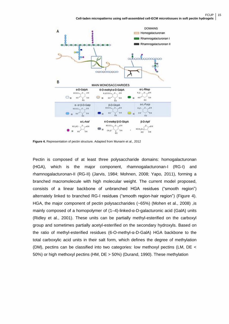
Figure 4. Representation of pectin structure. Adapted from Munarin et al., 2012
Pectin is composed of at least three polysaccharide domains: homogalacturonan
(HGA), which is the major component, rhamnogalacturonan-I (RG-I) and
rhamnogalacturonan-II (RG-II) (Jarvis, 1984; Mohnen, 2008; Yapo, 2011), forming a
branched macromolecule with high molecular weight. The current model proposed,
consists of a linear backbone of unbranched HGA residues (―smooth region‖)
alternately linked to branched RG-I residues (―smooth region-hair region‖) (Figure 4).
HGA, the major component of pectin polysaccharides (~65%) (Mohen et al., 2008) ,is
mainly composed of a homopolymer of (1–4)-linked-α-D-galacturonic acid (GalA) units
(Ridley et al., 2001). These units can be partially methyl-esterified on the carboxyl
group and sometimes partially acetyl-esterified on the secondary hydroxyls. Based on
the ratio of methyl-esterified residues (6-O-methyl-α-D-GalA) HGA backbone to the
total carboxylic acid units in their salt form, which defines the degree of methylation
(DM), pectins can be classified into two categories: low methoxyl pectins (LM, DE <
50%) or high methoxyl pectins (HM, DE > 50%) (Durand, 1990). These methylation

FCUP Cell-laden micropatterns using self-assembled cell-ECM microtissues in soft pectin hydrogels
16
Figure 5. Schematic representation of an ―egg box‖ structure formation in the presence of Ca2+
. Adapted from Coimbra et al., 2011
differences provide different properties to the pectin, significantly affecting the
properties of the formed gels.
LM pectins, in the presence of strong, positive, divalent metal ions, such as Ca2+ ions,
establish strong bonds between the carboxyl groups of the HGA pectin backbone
leading to the formation of an ―egg box‖ structure. This mechanism involves side-by-
side associations of specific sequences of GalA monomer in parallel or adjacent
chains linked through electrostatic and ionic bonding of carboxyl groups using the
divalent ions, forming a flexible network of polymer chains that can swell but does not
dissolve in water (Pérez et al., 2001; Fang et al., 2008) (Figure 5). Furthermore, van
der Waals interactions and hydrogen bonds are established within the polymer,
stabilizing the egg-boxes formed between neighbored chains (Braccini et al., 1999;
Fraeye et al., 2010).The promising aspects of pectin gels for biomedical applications,
namely, its easily tunable physical properties, high water content and ability to
homogeneously immobilize cells, genes, proteins, drugs or growth factors (Munarin et
al., 2010 a; Munarin et al., 2010 b; Munarin et al., 2011; Munarin et al., 2012; Neves e
al., 2015), led to a renewed interest on this polymer. Moreover, this biopolymer’s
solubility can be controlled by quickly displacing the Ca2+ ions by monovalent
counterions such as Na+ or K+ (Munarin et al., 2011). Pectin fulfils all of the
requirements for hydrogel formation, presenting itself as particularly appealing
biomimetic systems providing an adequate microenvironment by simulating the ECM-
cell dynamics. Nonetheless, as other natural polysaccharides (e.g. alginate), due to the
presence of negatively charged carboxyl groups, pectin presents a hydrophobic nature,
resisting to protein adsorption and cell adhesion. To bypass this issue, RGD-containing
oligopeptides must be grafted into the pectin backbone, granting the minimal peptide
sequence required for the adhesion of integrins to the ECM components
(Pierschbacher & Ruoslahti, 1987; Ruoslahti & Pierschbacher, 1987; Yamada, 1997;
Giancotti & Ruoslahti, 1999). As for other polymers (e.g. alginate (Bidarra et al, 2011;
Fonseca et al., 2011; Fonseca et al., 2013; Bidarra et al., 2014)) RGD-containing

FCUP Cell-laden micropatterns using self-assembled cell-ECM microtissues in soft pectin hydrogels
17
pectin gels present a higher cytocompatibility, cell adhesion and proliferation, improving
al the subsequent cellular functions (Munarin et al., 2011; Munarin et al., 2012; Neves
e al., 2015). Furthermore, in addition to the structural resemblance between pectin and
alginate, allowing into present the same numerous benefits of alginate, pectin stands
out as it presents an interesting degradation profile under simulated physiological
conditions (Munarin et al., 2012). Finally, more recently, our group explored the
potential of the pectin hydrogels crosslinking by internal ionotropic gelation using the
slow-gelling calcium carbonate/D-glucono-d-lactone (CaCO3/GDL) system. Neves e al.,
(2015) addressed, for the first time, the use of in situ-forming pectin hydrogels as skin
cell carriers for tissue engineering, providing an ionotropic internal gelation scheme
suitable for in situ gelling systems.
Although much still remains to be elucidated about this polymer as a biomaterial, the
studies found about the easy tunability of this biomaterial for tissue regeneration (Morra
et al., 2004; Bussy et al., 2008; Nagel et al., 2008; Munarin et al., 2011; Munarin et al.,
2012; Neves et al., 2015), evidence the promising capabilities of pectin hydrogels as a
powerful material system for cell delivery, tissue engineering and regenerative
medicine applications.
1.4. Main Goals
The incorporation of microvascular networks within the in vitro tissue-engineered skin
before its transplantation into a patient would be a major contribution, surpassing the
need of relying only on the host’s system ability to promote vascularization. Although
encouraging developments have been made in the field (Rivron et al., 2008; Place et
al., 2009), in vitro vascularization remains a challenge. In this work, we intend to use a
combined approach using the tunable characteristics of soft pectin hydrogel, cells and
growth factors to mimic the natural mechanisms involved in the formation of a
microvascular network. We aim to construct a three-dimensional, RGD-grafted, soft
pectin hydrogel in which fibroblasts support endothelial cells in the formation of self-
assembled vascular structures for skin regeneration therapies, while also providing
new insights on the biomimetic properties of soft pectin hydrogel’s for future tissue-
engineering strategies.

FCUP Cell-laden micropatterns using self-assembled cell-ECM microtissues in soft pectin hydrogels
18
2. Materials and Methods
Materials and Methods

FCUP Cell-laden micropatterns using self-assembled cell-ECM microtissues in soft pectin hydrogels
19
2.1. Cell Culture
2.1.1 Routine maintenance
Commercial human umbilical vein endothelial cells (HUVECs) (LONZA) and neonatal
human dermal fibroblasts (NHDFs, hereafter referred as FBs) (Corriel Institute) were
used. HUVECs were cultured in T75 culture flasks, coated with 0.2% (w/v) gelatin from
porcine skin (30 minutes at 37 °C, Fluka), with M199 medium (Sigma) supplemented
with 10% v/v of inactivated fetal bovine serum (FBS), 1% of antibiotic solution
composed of penicillin and streptomycin (Pen/Strep, Gibco) and 0.1 mg.mL-1 of heparin
(Sigma-Aldrich) with every-other-day medium exchange. Fibroblasts were cultured in
T75 culture flasks with Dulbecco’s Modified Eagle Medium (DMEM; Gibco)
supplemented with 10% (v/v) non heat inactivated FBS (Gibco), 1% (v/v) 1% pen/strep
(Gibco) and 1% of antimycotic Amphotericin B solution (Sigma) with no medium
changes necessary. The cells were incubated at 37 °C, under a humidified atmosphere
of 5% v/v CO2 in air. Entrapped cells in pectin discs, when in monoculture, were also
cultured in the same conditions, with the media being renewed every three days.
After reaching confluence, the cells were trypsinized. For HUVEC trypsinization, the
culture medium was removed and the T75s were washed with 5 mL of PBS (NaCl 137
mM, KCl 2.7 mM, NaHPO4.2H2O 10 mM, KH2PO4 1.8 mM, pH 7.4). The HUVECs were
incubated with 2 mL of Trypsin/EDTA in PBS (Trypsin 0.05 % w/v, Sigma; EDTA 0.5
mM, Sigma; pH 7.5) for 5 minutes at 37 °C. The T75s were gently tapped to loosen the
cells and 2 mL of M199 were added to inactivate the enzyme. The cells were recovered
into a single T75, resuspended to avoid aggregates and 10 µL of the solution were
loaded into a Neubauer chamber, where the cells were counted under a microscope.
HUVECs where seeded in 0.2% (w/v) gelatin-coated T75 at a density of 6x10⁵
cells/T75 and supplemented with 12 mL M199 with 0.03 mg.mL-1 of ECGS. For FBs
trypsinization the culture medium was removed and the T75s were washed with 5 mL
of PBS. The cells were incubated with 1 mL of Trypsin/EDTA in PBS (Trypsin 0.25%
w/v, Sigma; EDTA 2.21 mM, Sigma; pH 7.5) for 5 minutes at 37 °C, after which the
flasks were gently tapped to loosen the cells. Neutralization of the trypsin was archived
by adding 1 mL of DMEM to each T75 and cells were recovered to a single flask. FBs
were resuspended, and cells were counted under the microscope using a Neubauer
chamber. FBs were seeded at 5 x 10⁵ cells/T75 and supplemented with 8 mL of
DMEM. Both cell types were incubated in the previously described conditions.

FCUP Cell-laden micropatterns using self-assembled cell-ECM microtissues in soft pectin hydrogels
20
For each experiment, HUVECs were used at passages 6-10 and fibroblasts were used
at passages 5-10.
2.1.2 Cell thawing
HUVECs and FBs cryovials containing 1x10⁶ cells.mL-1 in 10% v/v DMSO in medium,
stored in liquid nitrogen, were thawed by immediately placing them in a 37 °C water
bath for 1 minute. To the cryovials containing HUVECs or FBs, 1 mL of, respectively,
M199 or DMEM was added and a mild up and down was carried out to resuspend the
cells. HUVECs were seeded at 6x10⁵ cells/T75 and on supplemented with 12 mL of
M199 with 0.03 mg.mL-1 of endothelial cell growth supplement (ECGS, Corning),
whereas FBs were seeded at 5x10⁵ cells/T75 and supplemented with 8 mL of DMEM.
The cells were incubated at 37 °C under a humidified atmosphere of 5% v/v CO2 in air.
Each medium was the next day, for DMSO removal.
2.1.3. Co-culture media selection
In order to select a medium that provides a performance close to the ideal for both cell
types, HUVECs and FBs behavior was evaluated in several media, including M199,
DMEM (both supplemented as previously described) and a combination of the two in
three different ratios of M199:DMEM: 3:1 (M3:1), 1:1 (M1:1) and 1:3 (M1:3).
Monocultures of both HUVECs and FBs were carried out on 12-well plates with
seeding densities of 3.0x10⁴ (D1) and 6.1x10⁴ (D2), which corresponds, respectively, to
the relative seeding density per surface area of a T75 and twice as much cells per
surface area. For each medium composition, HUVECs were seeded on 0.2% (w/v)
gelatin-coated 12-well plates, whereas 1 mL FBs were seeded in uncoated 12-well
plates. For HUVECs, each medium composition was supplemented with 0.03 mg.mL-1
of ECGS. To evaluate the effect of the different media on cell behavior, three time
points were selected (24h, 72h and 120h) and metabolic activity and total double-
stranded DNA quantification assays were carried out. Three replicates were conducted
for each time point.

FCUP Cell-laden micropatterns using self-assembled cell-ECM microtissues in soft pectin hydrogels
21
2.1.4. HUVECs and FBs density optimization
FBs and HUVECs co-cultures were established with four different cell ratios of 1:1 (R
1:1), 2:1 (R 2:1), 3:1 (R 3:1) and 5:1 (R 5:1) (HUVECs:FBs), at the two seeding
densities of D1 and D2. Cells were obtained from T75s cultures following the previously
described trypsinization methods for each cell type (Section 2.1.1.). After cell count, the
different cell ratios were established. Cells were seeded on 0.2% (w/v) gelatin-coated
12-well plates with M 3:1 supplemented with 0.03 mg.mL-1 of ECGS. To evaluate the
effect of the different ratios on cell behavior, at 24h, 72h and 120h, metabolic activity
and total double-stranded DNA quantification assays were carried out. Three replicates
were conducted for each time point. At each time point, cells were fixed with 1 mL of
4% (v/v) paraformaldehyde (Merk) in PBS for 20 minutes at room temperature (RT) for
phenotype characterization.
2.2. Pectin hydrogel
2.2.1. Pectin purification
Low methoxyl (LM) citrus pectin (Classic CU701), 86% and a DM of 37%, kindly
provided by Herbstreith & Fox (Neuenbürg, Germany), hereafter known as RawPec,
was purified based on the protocol as described in Neves et al. (2015). A 1% (w/v)
RawPec solution was prepared in ultrapure water (18 MU, Milli-Q UltraPure Water
System, Millipore). Following complete pectin dissolution, the pH of the solution was
measured and adjusted to 6. The 1% (w/v) RawPec solution was submitted to a
sequential filtration through decreasing pore diameter filters, namely, 0.80 µm, 0.45
µm, and 0.22 µm filter membranes (mixed cellulose esters, MCE, Millipore). After
filtration, activated charcoal (Norit, Sigma-Aldrich, 2% (w/w) were added to the solution,
which was stirred for 1 hour at RT. The suspension was centrifuged for 1h at 27 000 rcf
at RT. The supernatant was carefully recovered, and submitted to a new centrifugation
with the same parameters, to remove the activated charcoal. The supernatant was
recovered and submitted to filtration through a 0.22 µm filter membranes. Pectin was
then lyophilized and stored at -20 °C until further use.
FCUP Cell-laden micropatterns using self-assembled cell-ECM microtissues in soft pectin hydrogels
22
2.2.2. Carbodiimide RGD-grafting
To surpass the cell-anchorage difficulties imposed by the hydrophobic nature of the
hydrogels, biofuncional chemically modified pectin has to be obtained. Pectin was
covalently modified with the oligopeptide (Glycine)4-Arginine-Glycine-Aspartic acid-
Serine-Proline (G4RGDSP) (GenScript), using aqueous carbodiimide chemistry
(Rowley et al. 1998), based on the methods previously described for pectin (Munarin et
al. 2011; Munarin et al. 2012; Neves et al. 2015). To minimize carbodiimide chemistry
side reactions and provide maximum reaction efficiency, a purified pectin solution (1%
(w/v)) was prepared in 2-(N-morpholino) ethanesulfonic (MES) acid buffer (0.1 M MES
buffering salt, Sigma, and 0.3 M NaCl), with the pH adjusted to 6.5 using 1 M NaOH at
RT, overnight. The solution was divided in two in order to obtain RGD-grafted purified
pectin (RGDPec) and a control of unmodified pectin (BLKPec). The covalent pectin-
RGD bond is then achieved by adding, water-soluble carbodiimide, 1-ethyl-
(dimethylaminopropyl) carbodiimide (EDC), which is used to form amide linkages
between amine containing molecules and the carboxylate moieties on the polymer
backbone, N-hydroxy-sulfosuccinimide (sulfo-NHS), a co-reactant which stabilizes the
reactive EDC-intermediate form against a competing hydrolysis reaction and
G4RGDSP, our RGD-containing oligopeptide. To increase efficiency of the amide bond
formation, by minimizing COOH on the RGD reaction, these components were added
quickly and following the order: sulfo-NHS (N-Hydroxysulfosuccinimide) (Pierce
Chemical, 27.40 mg) and EDC (N-(3-dimethylaminopropyl)-N’-ethylcarbodiimide)
(Sigma, 48.42 mg), at a molar ratio of 1:2, followed by addition of the oligopeptide
(G4RGDSP) (16.70 mg) to the RGDPec, and both solutions were allowed to react for
20 h under constant stirring. The reactions were quenched with hydroxylamine
hydrochloride (Sigma, 18 mg per gram of pectin), and dialyzed against decreasing
concentrations of NaCl (30 g, 25 g, 20 g, 15 g, 10 g, 5 g) in ultrapure water for the first
2 days and against ultrapure water with 0 g of NaCl on the last day. The membranes
stayed in each solution at least 4 hours, remaining at least one night in the ultrapure
water. Both RGDPec and BLKPec were treated with activated charcoal (Norit, Sigma-
Aldrich, 2% (w/w)) for 1h, at RT with stirring. The suspension was centrifuged for 1h at
27 000 rcf at RT. The supernatant was carefully recovered and submitted to filtration
through a 0.22 µm filter membranes. Pectin was then lyophilized and stored at -20 °C
until further use.
The success of the immobilization was further confirmed via UV spectra analysis of
RGD-pectin. The amount of covalently modified peptide was estimated by successive

FCUP Cell-laden micropatterns using self-assembled cell-ECM microtissues in soft pectin hydrogels
23
dilutions of RGD in 1% (w/v) pectin solutions, measured in a microplate reader (Biotek
Synergy MX) with Ex/Em at 530/590 nm
2.3. 3D in vitro cell characterization
2.3.1 Characterization of HUVECs and FBs monocultures behavior
within 3D RGD-grafted soft pectin hydrogels
In order to verify the optimal set up for HUVECs and FBs monocultures on soft pectin
matrices, six experimental set-ups were tested, consisting on three different cell
densities (D3 = 5 x 10⁶ cells.mL-1; D4 = 1 x 10⁷ cells.mL-1; D5 = 1.5 x 10⁷ cells.mL-1) and
two RGD-grafted pectin concentrations (1.5% and 2.5% (w/v)), adjusted to 200 µM of
RGD).
2.3.1.1. Cell entrapment
The 3D matrices will be prepared by calcium-induced gelation, following the previously
described method by Neves et al. (2015).
To obtain the final pectin concentrations of 1.5% and 2.5% (w/v), lyophilized sterile-
filtered (0.22 mm) RGDPec was dissolved in 0.9 wt% NaCl (in ultrapure water) at 3%
and 4% (w/v), respectively. These precursor solutions were adjusted with sterile-filtered
(0.22 mm) BLKPec at the same concentrations in order to obtain a final RGD
concentration of 200 µM. To trigger hydrogel formation, based on the stoichiometric
considerations from Neves et al. (2015), CaCO3 dissolved in 0.9 wt% NaCl (in ultrapure
water) was added and carefully mixed with the pectin solution, followed by the addition
of D-glucono-d-lactone (GDL, Sigma). Each type of cell, previously cultured in T75 was
trypsinized, centrifuged and resuspended in 0.9 wt% NaCl (in ultrapure water) and, at
the considered densities, mixed with the pectin solution. For the preparation of
cylindrical pectin matrices with a height = 0.5 mm, 20 µL of the cell-laden hydrogel was
cast onto a teflon plate. 0.5 mm spacers were used and a second teflon plate was
applied over the hydrogel. A humidified chamber was prepared and gelation was
allowed to occur at 37 °C for 1 hour under a humidified atmosphere of 5% v/v CO2 in
air. After the crosslinking reaction occurred, the cell-laden matrices were transferred to
a 24-well culture plate coated with pHEMA (Folkman & Moscona, 1978) and 500 µL of
fresh medium was added. To evaluate both the effects of pectin concentration and

FCUP Cell-laden micropatterns using self-assembled cell-ECM microtissues in soft pectin hydrogels
24
entrapping density on cell behavior, four time points were selected (24h, 48h, 96 and
144h) and metabolic activity and total double-stranded DNA quantification assays were
carried out. Three replicas were casted for each formulation and time point. At each
time point, cell-laden soft pectin matrices were recovered and fixed in a 4% v/v PFA in
TBS-Ca, for a phenotype analysis.
2.3.2. HUVEC and Fibroblasts 3D monocultures performance under
different culture media
To evaluate the effect of different media on HUVECs and FBs monoculture in a RGD-
grafted soft pectin 3D hydrogels, the formulations E3 and F3 were supplemented with
the medium that presented the closest to ideal performance maintenance in a 2D
environment. The embeddings for these formulations were carried out as described in
―Characterization of HUVECs and FBs monocultures behavior within 3D RGD-grafted
soft pectin hydrogels‖ and the cell-laden matrices recovered into a 24-well culture plate
coated with pHEMA (Folkman & Moscona, 1978) and 500 µL of fresh M3:1 was added.
The HUVEC-laden matrices were also supplemented with 0.03 mg.mL-1 of ECGS. At
each time point (24h, 48h, 96 and 144h) the cell-laden pectin matrices were recovered
and fixated in a 4% v/v PFA in TBS-Ca and metabolic activity and total double-stranded
DNA quantification assays were carried out. Three replicates were conducted for each
formulation and time point.
2.3.3. 3D HUVEC:FB co-culture in soft pectin hydrogels
Co-cultures were established by entrapping the two different cell types (HUVECs and
FBs) at a cell ratio of 3:1 (HUVEC:FB) using the formulation: 1.5% (w/v) Pectin with 1.5
x 107 cells.mL-1 (hereafter described as CC1). Both cell types, cultured in T75, were
individually trypsinized and the ratio was established in a 50 mL Falcon. The cells were
centrifuged at 1 200 RPM for 5 minutes at RT and the embedding was carried out as
described in ―Characterization of HUVECs and FBs monocultures behavior within 3D
RGD-grafted soft pectin hydrogels‖. The resulting cell-laden matrices were transferred
to a 24-well culture plate coated with pHEMA (Folkman & Moscona, 1978) and 500 µL
the selected medium, supplemented with 0.03 mg.mL-1 of ECGS, was added. At each
time point (24h, 48h, 96 and 144h) the cell-laden pectin matrices were recovered and
fixated in a 4% v/v PFA in TBS-Ca and metabolic activity and total double-stranded

FCUP Cell-laden micropatterns using self-assembled cell-ECM microtissues in soft pectin hydrogels
25
DNA quantification assays were carried out. Three replicates were conducted for each
formulation and time point.
2.3.3.1. 3D HUVEC:FB co-culture spatial patterning: Microinjected
HUVEC-laden soft pectin on a FBS-laden soft pectin bed
As reviewed by Battiston et al., 2014 an initial spatial patterning of the cells onto the
matrix may lead to different outcomes in the co-culture. In this work, a micropatterning
technique was used for HUVEC:FB co-culture. Due to time limitations we were only
able to carry out a pilot assay. This micropattern was designed to incorporate a
HUVEC-laden soft pectin island in the center of an FB-laden soft pectin matrix. Two
different independent cell-laden pectin formulations, namely 1.5% (w/v) Pectin with 1.5
x 107 HUVECs.mL-1 and 1.5% (w/v) Pectin with 1 x 10⁷ FBs.mL-1 were simultaneously
carried out, using the previously described method. When ready, the spatially patterned
matrix was constructed by casting 40 µL of F2 onto a Teflon plate followed by a rapid
addition of 10 µL of E3 in the center of the F2 matrix using a 10 µL gel micropipette (See
figure 6). 0.5 mm spacers were used and a second Teflon plate was applied over the
pectin hydrogel. A humidified chamber was prepared and gelation was left to occur at
37 °C for 1 hour under a humidified atmosphere of 5% v/v CO2 in air. After the
crosslinking the cell-laden matrices were transferred to a 24-well culture plate coated
with pHEMA (Folkman & Moscona, 1978) and 500 µL of fresh M3:1, supplemented with
0.03 mg.mL-1 of ECGS, was added. Cell-laden matrices were maintained at 37 °C
under a humidified atmosphere of 5% v/v CO2 in air, with the media being substituted
by the third day.

FCUP Cell-laden micropatterns using self-assembled cell-ECM microtissues in soft pectin hydrogels
26
Figure 6. Schematic representation of the 3D HUVEC:FB co-culture spatial patterning embedding process.
2.4. Phenotype characterization
2.4.1. Cell metabolic activity
Cell metabolic activity assessment was performed using a resazurin assay. This assay
consists on the bioreduction of the resazurin, which is the oxidized form, to resofurin,
which, accordingly, increases the fluorescence. For the 2D resazurin assays, resazurin
solution was prepared by dissolution of 10 mg of resazurin (Sigma) in 100 mL of PBS
(NaCl 137 mM, KCl 2.7 mM, NaHPO4.2H2O 10 mM, KH2PO4 1.8 mM, pH 7.4), whereas
for 3D assays the solution was prepared in 100 mL of TBS (Tris-buffered saline, 50 mM
Tris-Cal, 150 mM NaCl, pH 7,5). After complete dissolution, the solution was sterilized
by filtration through a 0.22 µm filter and stored at -20 °C, protected from light until
further use. At the designated time points for each experiment, namely, 24h, 72h and
120h for 2D and 24h, 48h, 96h and 144h for 3D, the medium was carefully removed
cells were incubated with of 20% v/v of the stock resazurin solution (0.1 mg.mL-1,
Sigma) in medium for 3 h at 37 °C. Three replicates were monitored for each condition,
with three cellular-less replicates serving as control. After the incubation time, 100 µL of
the supernatant was transferred to a 96-well black plate with clear bottom (Greiner).
Three replicates per well were performed. Fluorescence measurements were carried
out using a microplate reader (Biotek Synergy MX) with Ex/Em at 530/590 nm and final
results were plotted on Graph Pad Prism 6 software (PRISM).

FCUP Cell-laden micropatterns using self-assembled cell-ECM microtissues in soft pectin hydrogels
27
2.4.2. Total dsDNA quantification
For total dsDNA quantification, the cells used for the resazurin assay were recovered.
Regarding the 2D assays, the remaining medium used for the resazurin assay was
removed and each well was washed with 1 mL of PBS. Each well was incubated with
250 µL of Trypsin/EDTA in PBS (Trypsin 0.05% w/v, Sigma; EDTA 0.5 mM, Sigma; pH
7.5) for 5 minutes at 37 °C. After incubation, 250 µL of medium was added to the cells
and the supernatant was recovered to an eppendorf. For entrapped cells, each matrix
was individually recovered to an eppendorf and dissolution was accomplished by
incubation with 100 µL of Trypsin/EDTA in PBS (Trypsin 0.25% w/v, Sigma; EDTA 50
mM, Sigma; Glucose 0,1% w/v, Sigma; pH 7.5) for 5 minutes at RT. Cells recovered by
centrifugation (10 000 rpm, 5 min), washed with 100 µL PBS, centrifuged again and the
pellet was stored at -20 °C until analyzed. Double-stranded DNA (dsDNA)
quantification was determined using the Quant-iT PicoGreen dsDNA kit (Molecular
Probes, Invitrogen), according to manufacturer’s instructions. Briefly the samples were
centrifuged (10 000 rpm, 5 min) and the supernatant was removed and cells were lysed
using 1% v/v Triton X-100 for 1 h at 400 rpm at 4 °C. Samples were then diluted 1:10 in
PBS. For each pool, 10 µL were transferred to a 96-well plate black with clear bottom
(Greiner) and in 90 µL of TE buffer was added (200 mM Tris–HCl, 20 mM EDTA, pH
7.5). 100 µL of Quant-iT PicoGreen dsDNA reagent in TE buffer was added and the
samples were incubated for 5 minutesat RT in the dark. Fluorescence was quantified
using a microplate reader with Ex/Em at 480/520 nm. RFUs were converted into
mg.mL-1 using a standard curve of DNA in the range of 1-1000 mg.mL-1. For each
condition n=3 replicates were analyzed.
2.4.3 2D co-culture readouts
After 24h, 72h and 120h, HUVECs:FBs co-cultures seeded on 12-well plates were
fixed. The medium of each well was removed and the cells were washed with 1 mL of
PBS. Cells were fixed with 1 mL of 4% (v/v) paraformaldehyde (Merk) in PBS for 20
minutes at RT. The paraformaldehyde solution was removed and the cells were
washed with PBS for 5 minutes at RT. No staining was conducted. Co-culture’s spatial
profile and morphology was evaluated using a Carl Zeiss Axiovert inverted microscope.
Bright field microscopy images were recovered using a monochromatic camera. Image
analysis was performed using the software software ImageJ64.

FCUP Cell-laden micropatterns using self-assembled cell-ECM microtissues in soft pectin hydrogels
28
2.4.4. HUVECs and FBs 3D monocultures and co-culture
morphology and spatial distribution
For HUVECs or FBs monoculture analysis, cells were directly assayed within hydrogels
(whole mounts).
2.4.4.1 Immunostaining
Cell-laden soft pectin matrices were recovered, washed with 500 µL of TBS-Ca (50 mM
Tris-Cl, 150 mM NaCl, pH 7,5 with 7.5 mM CaCl2) and fixed with 500 µL of 4% v/v PFA
in TBS-Ca for 20 minutesat RT. The PFA was removed and the matrices were washed
with 500 µL of TBS-Ca, for 5 minutes at RT. The matrices were stored at 4 °C in 500
µL of TBS-Ca until further use. For monocultures, F-acting and nuclei immunostaining
was performed. F-actin Immunostaining was carried out with Alexa Fluor 488
(Molecular Probes), a toxin with a fluorescent tag that selectively binds to F-actin,
whereas the nuclei were stained with DAPI (4’,6-diamidino-2’-phenylindole
dihydrochloride, Vectashield, Vector), a fluorescent stain that binds selectively to
double-stranded DNA. The matrices, stored at 4 °C were recovered, washed with 500
µL of TBS-Ca and permeabilized with 500 µL of 0.1% (v/v) Triton-X 100 (Sigma) for 5
minutes at RT, providing the stains the required access to the inside of the cell. The
permeabilized matrices were washed with 500 µL of TBS-Ca for 5 minutes at RT and
then blocked with 100 µL of 1% (w/v) Bovine serum albumin (BSA, nzytech) in TBS-Ca
for 30 minutes at RT, protected from light. This step is performed in order to prevent
nonspecific binging of the fluorescent molecules, providing a higher specificity. The
blocked matrices were incubated with 100 µL of Alexa Fluor 488 phalloidin (1:40) in a
1% (w/v) BSA in TBS-Ca, for 1 hour at RT, protected from light. The stained soft pectin
matrices were washed with 500 µL of TBS-Ca and stored at 4 °C until further use.
For HUVEC:FB co-cultures analysis, the whole mounts were fixed, stored
permeabilized and blocked as described above. Primary antibodies used: rabbit anti-
human vWF (1:300; Dako) and mouse anti-human α-smooth muscle actin (α-SMA,
1:100; Dako). These primary antibodies were incubated in 100 µL 1% (w/v) BSA in
TBS-Ca, for 16h at 4 °C, protected from light. Following the incubation period, the
matrices were washed with 500 µL of TBS-Ca for 5 minutes and incubated with 100 µL
of a second solution containing the secondary antibodies, goat anti-rabbit Alexafluor
594 (1:1000; abcam) and chicken anti-mouse Alexafluor 647 (1:1000; abcam), and
Alexa Fluor 488 phalloidin (1:40) in a 1% (w/v) BSA in TBS-Ca, for 1 hour at RT,

FCUP Cell-laden micropatterns using self-assembled cell-ECM microtissues in soft pectin hydrogels
29
protected from light. The matrices were washed with 500 µL of TBS-Ca and stored at 4
°C until further use.
2.4.4.2 Image acquisition
Confocal images of monocultures were acquired on a Leica SP2 confocal microscope
(LSCM, Leica SP2 AOBS SE; Leica Microsystems). Individually, each matrix was
recovered from the TBS-Ca into a support were 6 µL of Vectashield mounting media
(Vector) with DAPI was applied onto the matrix. Using the Leica Confocal Software
(LCS 2.61, Leica Microsystems), images of the matrices were taken with the objectives
HC PLAN APO CS 10x/0.40 and HC PL APO CS 40x/1.25-0.75 Oil. The software was
configured to recover the images in a 1024x1024 format, performing each plane sweep
with the parameters Line Average and a Frame Average set to 2 and 3, respectively. A
sequential scan was performed in each matrix capturing the fluorescence emitted. The
distance between each Z plane was set to 5 µm and 2.5 µm, for the 10x and 40x
objectives, respectively. Image analysis was performed using the software ImageJ64.
2.4.4.3. Evaluation of the influence of pectin concentration over the
ability of FBs monocultures to promote matrix contraction
To verify if the fibroblasts ability to remodel the matrix and microtissue formation, a
contraction assay was carried out. 1.5% (w/v) pectin with 1 x 10⁷ FBs.mL-1 and 2.5%
(w/v) Pectin with 1 x 10⁷ FBs.mL-1 embeddings were carried out as previously
described. These conditions were monitored using an inverted light microscope to
observe the any matrix diameter change that might occur. Matrices were monitored at
24, 48, and 96 and 144 hours.
2.5. Data treatment
2.5.1. Statistical analysis
Cell metabolic activity and proliferation data was analyzed using GraphPad Prism 6
software (PRISM) Mann-Whitney test, for two unpaired groups. Statistically significant
differences were considered when p values were lower than 0.05. All data is presented
as mean values with ± standard deviation.

FCUP Cell-laden micropatterns using self-assembled cell-ECM microtissues in soft pectin hydrogels
30
2.5.2. Image treatment
Image analysis was performed using the ImageJ64 software. For fibroblast-laden
matrices, fibroblast spheroids were counted in the confocal images and the average
number of structures was determined by dividing the number of structures by the area
(mm2) of the image. Finally, the area of the spheroid-like structures was also quantified
with an n=75 in all images.

FCUP Cell-laden micropatterns using self-assembled cell-ECM microtissues in soft pectin hydrogels
31
3. Results
Results

FCUP Cell-laden micropatterns using self-assembled cell-ECM microtissues in soft pectin hydrogels
32
3.1 Preparation of 3D biofuncional RGD-grafted pectin
Due to its hydrophobic nature, pectin resists to protein adsorption and cell adhesion
(Rowley et al, 1999), which is required for the survival of anchorage-dependent cell,
playing a critical role in several physiological events (Dee et al, 1999; Price et al.,
1997). The incorporation of the RGD (Arg-Gly-Asp) sequence improves cell adhesion
on non-adhesive substrates, providing the minimal peptide sequence required for the
adhesion of integrins to the ECM components (Pierschbacheret al., 1987; Ruoslahti et
al., 1987; Yamada et al., 1997). In this study, RGD-functionalized pectin was
synthesized and further used to prepare hydrogel matrices for culturing HUVECs and
FBs under 3D conditions. Based on the method previously described by Neves et al.
(2015), pectin was purified and covalently grafted with the oligopeptide (Glycine)4-
Arginine-Glycine-Aspartic acid-Serine-Proline (G4RGDSP) (GenScript), using aqueous
carbodiimide chemistry (Rowley et al. 1998). The amount of covalently modified
peptide was estimated by successive dilutions of RGD in 1% (w/v) pectin solutions.
Figure 7. UV spectra of RGD-pectin, soluble RGD peptide and serial dilutions of RGD in a 1% pectin solution

FCUP Cell-laden micropatterns using self-assembled cell-ECM microtissues in soft pectin hydrogels
33
Final approximate RGD content is 18 mg RGD per 1 g of pectin.
3.2. Determination of 2D optimal HUVEC/FB culture media
composition
As reviewed by Battiston et al. (2014), a co-culture system involves multiple
interactions, presenting several challenges. A correct selection of the base medium is
then necessary to optimize growth and cell phenotype. Endothelial cells and fibroblasts
will be cultured separately in 24-well culture plates using five different medium
compositions. These included M199, DMEM (both supplemented as previously
described) and a combination of the two in three different ratios of M199:DMEM: 3:1
(M3:1), 1:1 (M1:1) and 1:3 (M1:3). This experiment was carried out to evaluate the
effect of the different media on cell behavior, allowing the selection of the best-suited
media for the co-culture of endothelial cells and fibroblasts.
At day 1, HUVECs (Figure 8), at both initial seeding densities (C1 and C2) present
similar dsDNA yields (Figure 8, a and b). From the start it is possible to observe that
despite the dsDNA profiles are not significantly different, significant differences can be
found in the metabolic activity (Figure 8, c and d). Three distinct profiles can be
perceived whereas M3:1 presents a similar profile to the optimal medium, M199. M1:1
and M1:3 possess similar metabolic behaviors but are already significantly different
from the optimal medium. Finally, DMEM presents the lowest metabolic rate. From day
1 to day 3, an increase in the DNA content is observed, accompanied by an increase in
the total metabolic activity in all but DMEM supplemented cultures, where these profiles
seem to be maintained or decrease. Finally, from day 3 to day 5, we observe a
decrease in the total dsDNA and metabolic activity. Despite these fluctuations, the
three profiles are maintained and significantly different between them. Throughout the
experiment it is possible to observe that the metabolic activity (normalized with the total
dsDNA) of HUVECs is maintained.

FCUP Cell-laden micropatterns using self-assembled cell-ECM microtissues in soft pectin hydrogels
34
Figure 8. Effect of cell density and medium composition on metabolic activity and proliferation of HUVECs in 2D during
5 days in culture a) and b) total dsDNA (PicoGreen assay), c) and d) metabolic activity (resazurin assay) and e) and f)
metabolic activity per nanogram of dsDNA of HUVECs. * denotes statistically significant differences (p < 0.05).
Regarding FBs (Figure 9), at day 1, it is possible to see that the different seeding
densities are translated into different dsDNA yields (Figure 9, a and b). This difference
is also represented in the total metabolic activity (Figure 9, c and d) whereas C1
presents a lower total metabolic activity than C2. At day 3, it is observed an increase for
both seeding densities in the total DNA content and metabolic activity. By day 5, the
total DNA content seems to remain unaltered except for the M199, M3:1 and M1:1
media conditions of C1, where it increases. Here, we observe a metabolic activity
increase for C1 densities, whereas for C2 the values remain unaltered for M 3:1, M 1:1
and M 1:3, increasing in DMEM and M199. When normalized, these results point to a
constant metabolic activity FBs for seeded at 6.1x10⁴ cell/well throughout the culture

FCUP Cell-laden micropatterns using self-assembled cell-ECM microtissues in soft pectin hydrogels
35
time, whereas for lower seeding concentrations (namely 3.0x10⁴ cell/well) it is possible
to observe a decrease followed by a slight increase, stabilizing at similar values to C2.
Figure 9. Effect of cell density and medium composition on metabolic activity and proliferation of FBs in 2D within a 5 days culture period. a) and b) total dsDNA (PicoGreen assay), c) and d) metabolic activity (resazurin assay) and e) and f) metabolic activity per nanogram of dsDNA of NHDFs. * denotes statistically significant differences (p < 0.05).
Given that, for HUVECs, there are no significant alterations in the metabolic and
proliferation profiles alterations when using M3:1 (when compared to M199) and that
the same happens to FBs (when compared to DMEM), M3:1 is a suitable candidate for
supplementation in a co-culture system.

FCUP Cell-laden micropatterns using self-assembled cell-ECM microtissues in soft pectin hydrogels
36
3.3. Determination of 2D optimal in vitro HUVEC/FB ratio
The majority of natural tissues consist of multi-cellular systems of two or more cell
types which interact with each other to facilitate viability, proliferation and differentiation
(Nam et al., 2011; Schubert et al., 2008; Traphagen et al., 2013). Blood vessels are a
multi-cellular system composed of endothelial cells (ECs), vascular smooth muscle
cells (VSMCs), and fibroblasts (Ratner et al., 1996). In tissue engineering, co-culture
systems have been increasingly used as it provides simulation of the in vivo physical
and biological properties. Furthermore, published data indicate that co-culture of ECs
with FBs can provide the complex mixture of growth factors, ECM and cell-cell contacts
necessary to potentiate tubulogenesis (Berthod et al., 2006; Sorrell et al., 2007; Auger
et al., 2013). However, as in natural environments cell distribution is not uniform, for in
vitro optimal experimental outcomes in co-culture assays it is necessary an
optimization of the cell seeding number and ratio. In order to evaluate the effect of FBs
on HUVECs capillary-like self-assembly, four our different cell ratios were established,
namely, R 1:1, R 2:1, R 3:1 and R 5:1 (HUVECs:FBs) were tested at two seeding
densities of 3.04x10⁴ (D1) and 6.08x10⁴ (D2). Cells were seeded on a 0.2% gelatin-
coated 12-well plate with the selected medium (M 3:1) supplemented with ECGS for
five days.
At day 1, the different seeding densities present different DNA content, with higher
values for C2, result of the different densities seeded. Regarding the total metabolic
activity, for C1 all ratios present similar values whereas for C2 it is possible to observe a
pattern where R 5:1 > R 3:1 > R 2:1 > R 1:1, hence presenting higher activities in ratios
containing more HUVECs (Figure 10, d), which can also be verified when the values
are normalized (using the total dsDNA) (Figure 10, f). By day 3, there are observed
increases in the total DNA for C1 and C2. However, whereas for C1 there are no
noticeable differences between the different ratios, for C2, R 1:1 presents higher values
than the remainder ratios. This difference is translated in the total metabolic activity
where is possible to verify that R 1:1 presented significantly higher metabolic values
when compared to the other ratios. Although it is possible to see a decrease in the
metabolic activity of the cells (normalized with the total dsDNA) (Figure 10, e and f) it is
verified a maintenance of the higher values for endothelial rich ratios (except for R1:1
and R2:1 in C1 seeding conditions, which can be due to an deficient recovery of the
dsDNA, as these also present lower values for total DNA). It is however important to

FCUP Cell-laden micropatterns using self-assembled cell-ECM microtissues in soft pectin hydrogels
37
Figure 10. Effect of cell density and cell ratio on metabolic activity and proliferation of HUVEC:FB co-culture in 2D within a 5 days culture period. a) and b) total dsDNA (PicoGreen assay), c) and d) metabolic activity (resazurin assay) and e) and f) metabolic activity per nanogram of dsDNA of HUVEC: * denotes statistically significant differences (p < 0.05).
point out that despite this decrease, the cells’ metabolic activity is 5 to 6-fold higher
than any of the individual cultures. At day 5, the total DNA increases for both seeding
conditions, presenting, in all ratios, values superiors to those verified in the
monocultures. For C1 and C2, in the R 1:1, the ratio containing the highest initial
fibroblasts number (presenting however half of those verified in the monocultures), the
total DNA content is 3 and 4-fold higher, respectively, when compared to FBs
monocultures whereas for R 5:1, an endothelial rich ratio, the total DNA content is 3-
fold higher than for HUVEC monocultures. However, regarding the metabolic activity,
despite an increase for the total metabolic activity, normalized values show a decrease
in the activity of the cells, as it already occurred from day 1 to 3. Nonetheless, the
metabolic values are 2-fold higher than individual cell cultures. Putting all together, for
a 2D HUVEC:FB co-culture, lower ratios seem to favor proliferation whereas higher
ratios seem to favor higher metabolic activities per cell (metabolic activity normalized

FCUP Cell-laden micropatterns using self-assembled cell-ECM microtissues in soft pectin hydrogels
38
Figure 11. Pictures of HUVEC:FB co-cultures prepared at 6.08x10⁴ cells/well at the different ratios of 1:1, 2:1, 3:1 and
5:1. Images were obtained at the first and last day of 5-days. Scale bars, 200 µm.

FCUP Cell-laden micropatterns using self-assembled cell-ECM microtissues in soft pectin hydrogels
39
Figure 12. Pictures of HUVEC:FB co-cultures prepared at 6.08x10⁴ cells/well at the different ratios of 1:1, 2:1, 3:1 and
5:1. Images were obtained at the first and last day of a 5. Scale bars, 200 µm.

FCUP Cell-laden micropatterns using self-assembled cell-ECM microtissues in soft pectin hydrogels
40
with total dsDNA). For a 5 days culture, the different seeding densities do not seem to
influence the final outcome of the culture, affecting however the development of the
culture throughout the culture days, as it was also seen for individual cultures.
However, to our purpose, to obtain capillary like structures, metabolic and proliferation
studies are not sufficient. In order to properly choose for an adequate ratio, the cell
morphology and spatial distribution must be taken into account. To analyze this, we’ve
fixated the different conditions at the different time points and took pictures of the
structures formed using an inverted light microscope. The images reveal evident
differences between cells organization in different ratios used for HUVEC:FB co-
cultures, as can be depicted in the Figures 11 and 12. At lower HUVEC:FB ratios,
namely R 1:1 and R 2:1 both cells appeared widely distributed at the cell culture
surface throughout the five days, presenting their respective fibroblastic-like and
cobblestone-like shapes. At day 5, however, it is observed that these cultures present
similar structures to the ones observed in FB monocultures at confluence, hence
demonstrating FB dominance in the cell culture surface. At higher ratios, namely R 3:1
and R 5:1, both cell types also archive fibroblastic-like and cobblestone-like shapes.
With these ratios, at day 5, it is possible to verify that cells were rearranged in a
significantly different manner featuring web-like structures characteristics of tubular-like
cellular network formation. All together, high densities with endothelial rich ratios seem
more appropriate as 2D co-culture formulations for HUVEC development and capillary-
like self assembly.
3.4. Analysis of HUVEC and FB monocultures’ behavior in 3D-
culture
As depicted in previously, RGD-grafted pectin hydrogels present cytocompatibility, cell
adhesion and proliferation characteristics for the improvement of cellular functions
(Munarin et al., 2011; Munarin et al., 2012; Neves e al., 2015). As such, pectin stands
as a particularly appealing biomimetic material for 3D hydrogel formation for cell
culture. For hydrogels, the mechanical performance of a matrix depends on the
polymer and crosslinker characteristics, concentration, gelling conditions (e.g.
temperature, pH, and gelation time), swelling, and degradation. In turn, these will have
an impact in the biomaterial mechanical properties, including elasticity, compressibility,
viscoelastic behavior, tensile strength, and failure strain (Anseth et al., 1996). With this
in mind, we have followed an already described gelation scheme thoroughly
characterized by Neves et al (2015), varying the concentration of the pectin used in the

FCUP Cell-laden micropatterns using self-assembled cell-ECM microtissues in soft pectin hydrogels
41
hydrogels between 1.5% and 2.5% (w/v). These concentrations where used to
determine the effects matrix characteristics (e.g. mesh size, mechanical compliance)
over the ability of cell to regulate their metabolic activity, alter their morphology, migrate
and establish cell-cell contact. Furthermore, to evaluate the effect of cell density on the
behavior of cells in 3D environments, cells were entrapped within pectin matrices at
different entrapping densities, namely: D3 = 5x10⁶ cells.mL-1; D4 = 1x10⁷ cells.mL-1 and
D5 = 1.5x10⁷ cells.mL-1. Cell metabolic activity (Resazurin assay), total dsDNA
(PicoGreen assay) and morphology (Confocal microscope images) assays were
carried out at days 1, 2, 4 and 6.
3.4.1 HUVEC behavioral analysis on 3D soft pectin hydrogels
At day 1, the dsDNA content of the different formulations was proportional to their
respective original cell density, presenting however higher values for a 2.5% (w/v)
pectin concentration (Figure 13). It is also important to note that the total dsDNA for
lowest entrapping density in 1.5% (w/v) pectin hydrogels is nearly inexistent. Regarding
the metabolic activity, HUVECs presented higher total metabolic activities in agreement
with the previous observations for the total dsDNA. The highest metabolic activity
registered was for the 1.5x10⁷ HUVECs.mL-1. When normalized (with total dsDNA), the
metabolic results show that for 1.5% (w/v) pectin concentrations all entrapping
densities present the same cellular activity, whereas for 2.5% (w/v) lower densities
seem to present higher cellular activities. For all the formulations tested (except for
2.5% (w/v) hydrogels with 1x10⁷ HUVECs.mL-1, possibly due to deficient manipulation
throughout the experiment), the total dsDNA content gradually decreased along the
period of culture, being accompanied by the decrease in the metabolic activity. By day
4, the metabolic activity assay demonstrate close or equal to 0, independently of the
content of DNA, which were maintained till day 6.
Regarding the spatial behavior (Figures 14 and 15), for the lowest entrapping density,
namely, 5x10⁶ HUVECs.mL-1, the images were not presented as they did not help to
elucidate what was happening within the hydrogels. As for 1x10⁷ HUVECs.mL-1 and
1.5x10⁷ HUVECs.mL-1 entrapping densities, independently of the pectin concentration,
cells seem widely distributed within the matrix. Cells maintained a globular structure,
indicative of the lack of adhesion, throughout the experiment. As the total dsDNA and
metabolic assays shown very low DNA content and metabolic activity for day 4 and 6,
no stains were carried, hence no images are presented.

FCUP Cell-laden micropatterns using self-assembled cell-ECM microtissues in soft pectin hydrogels
42
All together, these results indicate that, independently of the pectin concentration and
entrapment densities, monoculturing HUVECs on pectin hydrogels is not an efficient
strategy for HUVEC self-assembled tubulogenesis or even maintenance.
Figure 13. Effect of initial cell entrapment density and pectin concentration on metabolic activity and proliferation of HUVEC in a 3D soft pectin hydrogel within a 6 days culture period. a) and b) total dsDNA (PicoGreen assay), c) and d) metabolic activity (resazurin assay) and e) and f) metabolic activity per nanogram of dsDNA of HUVEC * denotes statistically significant differences (p < 0.05).
3.4.2. FBs behavioral analysis on 3D soft pectin hydrogels FBs
As regards to FBs, two significantly different profiles can be observed for different
pectin concentrations (Figure 16). For 1.5% (w/v), at day 1, it is possible to verify that
the cell-loaded matrix present total dsDNA values in accordance to the initial number of
cells entrapped. By day 2, a slight increase in the total dsDNA is observed for the
entrapping densities of 5x10⁶ FBs.mL-1 and 1x10⁷ FBs.mL-1, which, attending to the
remaining profile, could be the result of the total DNA assay manipulation. At day 4,
dsDNA seems to slightly decrease, maintaining its values at day 6. Relatively to the

FCUP Cell-laden micropatterns using self-assembled cell-ECM microtissues in soft pectin hydrogels
43
Figure 14. Effect of initial cell entrapping density and pectin concentration on the 3D HUVECs’ spatial distribution within a 6 days culture period on a soft pectin hydrogel. HUVECs were stained for F-actin (Green) and nuclei (Blue). Scale bars, 100 µm.

FCUP Cell-laden micropatterns using self-assembled cell-ECM microtissues in soft pectin hydrogels
44
Figure 15. Effect of initial cell entrapping density and pectin concentration on the 3D HUVECs’ conformation within a 6 days culture period on a soft pectin hydrogel. HUVECs were stained for F-actin (Green) and nuclei (Blue). Scale bars, 100 µm
.

FCUP Cell-laden micropatterns using self-assembled cell-ECM microtissues in soft pectin hydrogels
45
metabolic activity, FBs, as HUVECs, presented lower metabolic values in 3D matrices
when compare to a 2D culture as expected. Regarding 1.5% (w/v) pectin hydrogels, at
day 1, total dsDNA contents were in accordance with the entrapping densities, with the
metabolic activity in conformity with these entrapping densities. From day 1 to day 2,
there is observed a decrease in the 1.5x10⁷ FBs.mL-1 entrapping density and an
increase in the 5x10⁶ FBs.mL-1 1x10⁷ FBs.mL-1 entrapping densities, reaching similar
values. From this point onwards, the metabolic activity seems to maintain a steady-
state. When normalizing these results, an increase in the cellular metabolic activity is
observed between day 2 and 4. To note that, for entrapping densities of 1.5x10⁷
FBs.mL-1, several discs broke from the first day, complicating the manipulation and
subsequent recovery of data (both the metabolic and dsDNA assays and image
recovery). As for 2.5% (w/v) pectin hydrogels, a different profile is observed. For all the
densities tested, the total the total dsDNA content remained constant along the time.
However, the total metabolic activity gradually decreased along the period of culture,
which, when normalized, translates in a steady decrease in the cellular metabolic
activity.
Figure 16. Effect of initial cell entrapping density and pectin concentration on metabolic activity and proliferation of NHDFs in a 3D soft pectin hydrogel within a 6 days culture period. a) and b) total dsDNA (PicoGreen assay), c) and d) metabolic activity (resazurin assay) and e) and f) metabolic activity per nanogram of dsDNA of NHDFs * denotes statistically significant differences (p < 0.05) .

FCUP Cell-laden micropatterns using self-assembled cell-ECM microtissues in soft pectin hydrogels
46
Figure 17. Effect of the initial entrapping density on a 1.5% pectin 3D hydrogel on FBs’ spatial distribution within a 6 days culture period. FBs were stained for F-actin (Green) and nuclei (Blue). Scale bars, 100 µm.

FCUP Cell-laden micropatterns using self-assembled cell-ECM microtissues in soft pectin hydrogels
47
Figure 18. Effect of the initial entrapping density on a 1.5% pectin 3D hydrogel on FBs’ conformation within a 6 days culture period. FBs were stained for F-actin (Green) and nuclei (Blue). Scale bars, 100 µm.

FCUP Cell-laden micropatterns using self-assembled cell-ECM microtissues in soft pectin hydrogels
48
Figure 19. Effect of the initial entrapping density on a 2.5% pectin 3D hydrogel on FBs’ spatial distribution within a 6 days culture period. FBs were stained for F-actin (Green) and nuclei (Blue). Scale bars, 100 µm.

FCUP Cell-laden micropatterns using self-assembled cell-ECM microtissues in soft pectin hydrogels
49
Figure 20. Effect of the initial entrapping density on a 2.5% pectin 3D hydrogel on FBs’ conformation within a 6 days culture period. FBs were stained for F-actin (Green) and nuclei (Blue). Scale bars, 100 µm.

FCUP Cell-laden micropatterns using self-assembled cell-ECM microtissues in soft pectin hydrogels
50
Regarding the spatial behavior, accompanying the tendency presented by cell
metabolic activity, 1.5% (w/v) pectin hydrogels presented better results when compared
to 2.5% (w/v) pectin hydrogels. Within these, as seen in the Figures 17 and 18, cells
were widely distributed, being able to adhere, acquiring a fibroblast-like shape.
Throughout time, these cells were able to establish cell-cell contacts, forming FB
spheroids which increased in number and size, as depicted in Figure 21 and 22 Table
1. These structures presented significantly larger spheroids when compared to the
ones formed in 2.5% (w/v) pectin hydrogels. It is also demonstrated that, depending on
the initial entrapping densities in 1.5% (w/v) pectin hydrogels, higher entrapping
densities favor a significantly larger size of the spheroids (Table 1). However, as stated
before, due to degradation of the matrices at 1.5x10⁷ FBs.mL-1 (the discs broke and
started to decompose). Regarding 2.5% (w/v) pectin hydrogels, independently of the
Figure 21. Effect of Initial cell entrapping and pectin concentration over FBs spheroid size. a) and b) spheroids average size throughout the 6 days of culture. c).and d) relative frequency of spheroid size at day 6. * denotes statistically significant differences (p < 0.05) ) between different entrapping densities on different pectin concentrations.
c) d)
Area (µm2) Area (µm2)

FCUP Cell-laden micropatterns using self-assembled cell-ECM microtissues in soft pectin hydrogels
51
Table 1. Effect of initial cell entrapping densities and pectin concentration over FBs spheroid size (µm2) and number.
Area means and standard deviation is presented in micrometers. N stands for total number of spheroids. * denotes statistically significant differences (p < 0.05) between the same entrapping density on different pectin concentrations. α denotes statistically significant differences (p < 0.05) between different entrapping densities on different pectin concentrations.
Day 1 Day 2 Day 4 Day 6
1.5% Pectin Area n Area n Area n Area n
5x10⁶
FBs.mL-1
761±288* 37 960±358* 176 1032±486* 261 1017±676* 206
1x10⁷
FBs.mL-1
927±287*α 170 1051±422* 183 1121±481* 291 1244±580*
α 251
2.5% Pectin
5x10⁶
FBs.mL-1
660±120 34 652±105 29 441±90 19 489±77 26
1x10⁷ FBs.mL
-1 705±203 87 735±212 60 500±140 53 528±150 34
1.5x10⁷
FBs.mL-1
895±232
α 153 915±294
α 83 889±235
α 51 758±136
α 59
entrapping density, cells exhibit a globular-like shape, with some cells presenting a
fibroblast-like shape, mostly observed at the surface (Figures 19 and 20). Although
there is possible to observe some spheroids, the number and size is significantly lower
than what is observed for 1.5% (w/v) pectin hydrogels. Throughout the period of the
culture, 2.5% (w/v) pectin matrices loaded with 1.5x10⁷ FBs.mL-1 present significant
larger spheroids when compared to the other entrapping densities. However, for all
densities, spheroid number and size decreases with time, accompanying the decrease
in metabolic activity previously described for these conditions.
Figure 22. Effect of pectin concentration over the ability of FBs to contract the matrix. Macroscopic differences of 1.5%
and 2.5% pectin hydrogels seeded with 1x10⁷ FBs.mL
-1. Images were obtained at the first and last day of a 6-days
culture using an inverted microscope using a magnification of 16.3 x. a) and b) correspond to 1.5% (w/v) pectin hydrogels at day 1 and 6 respectively. c) and d) correspond to 2.5% (w/v) pectin hydrogels at day 1 and 6 respectively. e) represents the relative size of the pectin matrices when compared to day 1

FCUP Cell-laden micropatterns using self-assembled cell-ECM microtissues in soft pectin hydrogels
52
Table 2. Effect of pectin concentration over the ability of FBs to contract the matrix. Macroscopic differences of 1.5% and
2.5% pectin hydrogels entrapped with 1x10⁷ FBs.mL
-1. Areas means and standard deviation is measured in square
millimeters
1x10⁷ cells.mL
-1 Day 1 Day 2 Day 4 Day 6
1.5% Pectin 31.95 ± 1.09 21.40 ± 0.04 18.81 ± 0.35 17.58 ± 0.57
2.5% Pectin 38.34 ± 1,09 33.72 ± 0.42 31.46 ± 1.32 29.81 ± 0.12
Another important feature of the gels is the mechanical compliance, which will impact
over the cells ability to exert forces to deform the matrices. To test the influence of
pectin concentration over the ability of FBs monocultures to promote matrix contraction,
macroscopic images of 1.5% and 2.5% (w/v) pectin hydrogels matrices loaded with
1x10⁷ FBs.mL-1, the highest performing comparable density (1.5x10⁷ FBs.mL-1 was
excluded as for 1.5% (w/v) pectin matrices were unsuitable to handle), were recovered
at day 1, 2, 4 and 6 (Table 2 and Figure 22). At day 1, 1.5% (w/v) pectin hydrogels
presented smaller areas than 2.5% (w/v) pectin hydrogels. As these matrices were
produced with the same volume (20 µL), this difference indicates that after the initial
swelling (verified while manipulating but not measured), from day 0 to day 1 matrices
start to contract at lower pectin concentrations. By day 2, 1.5% (w/v) pectin hydrogels
presented a 33% matrix contraction when compared to day 1 and by day 6 and 45%.
Regarding 2.5% (w/v) pectin hydrogels, for these days, we observe a matrix
contraction of 12% and 22% respectively, when compared to day 1. Furthermore, by
day 6, 2.5% (w/v) pectin hydrogels are 1.70x larger than 1.5% (w/v) pectin hydrogels.
Summing up, for both pectin concentrations, total dsDNA values remained essentially
constant throughout the period of culture, suggesting that the entrapped cells were not
able to proliferate, independently of the original cell density. The main differences were
found at the metabolic activity and cellular spatial distribution. In 1.5% (w/v) pectin
hydrogel matrices, cells were able to adhere to the matrix, acquiring fibroblast-like
shape, and establish cell-cell interactions, forming FBs spheroids, while maintaining a
steady-state of metabolic activity. In these conditions, higher entrapping densities
favored a higher spheroid formation with significantly higher areas. Moreover, for 1x10⁷
FBs.mL-1, macroscopic images of the cell-laden matrices allowed to observe alterations
of the construct size, presenting at day 6 to almost half of the size of the one observed
at day 1. However, regarding 2.5% (w/v) pectin hydrogels, cells essentially maintained

FCUP Cell-laden micropatterns using self-assembled cell-ECM microtissues in soft pectin hydrogels
53
a globular like shape, exhibiting a fibroblast-like shape mostly at the surface. These
results were in accordance to what was expected as 2.5% (w/v) pectin hydrogels are
less compliant and provide less free space for cells to spread when compared to 1.5%
(w/v) pectin hydrogels (Neves et al., 2015). FBs spheroids were also observed in this
construct but with significantly inferior numbers and size. Finally, macroscopic images
demonstrate that these matrices maintain a more robust aspect possessing not only, at
day 6, 80% of the size when compared to day 1 but also almost a 2-fold increase when
compared to 1.5% (w/v) pectin hydrogels. Results obtained by us indicate that cells
might be constrained by the polymeric network at higher pectin concentrations, favoring
the use of more compliant matrices for FBs culture. Within these (1.5% (w/v) pectin
hydrogels), higher entrapping densities seem to stimulate cell-cell contacts and the
formation spheroids.
3.5 HUVEC:FB co-culture establishment in 3D soft pectin hydrogels
As previously stated, coculturing ECs with FBs can potentiate tubulogenesis (Berthod
et al., 2006; Sorrell et al., 2007; Auger et al., 2013). Nonetheless, great differences can
be observed among different matrices, cell type’s combinations, entrapping densities
and entrapping ratios. As such, it is of utmost importance to optimize these factors for
3D cultures in each specific application design. As documented throughout this thesis,
several optimization steps were taken into account in order to choose the coculturing
conditions, namely: medium supplementation (through metabolic activity and total
dsDNA) and cell ratio (through metabolic activity, total dsDNA and optical microscopy
analyzes), in 2D experiments, and polymer concentration and cell entrapping densities
(through metabolic activity, total dsDNA and optical microscopy analyzes), in 3D
experiments. Aiming to promote the capillary self-assembly of HUVECs in a 3D pectin
hydrogel, these were co-culture with FB at a ratio of 3:1 (HUVEC:FB), with an
entrapping density of 1.5x10⁷ cells.mL-1. These were cultured in pectin hydrogels with a
1.5% pectin concentration and supplemented with M 3:1 with 0.03 mg.mL-1 of
endothelial cell growth supplement (ECGS).
3.5.1. Characterization of the influence of M 3:1 supplementation on
HUVECs or FB monocultures in 3D soft pectin hydrogels
Preceding the HUVEC:FB co-culture, in order to evaluate the medium influence on the
3D cells behavior, both HUVECs and FBs were loaded in 1.5% (w/v) pectin hydrogels

FCUP Cell-laden micropatterns using self-assembled cell-ECM microtissues in soft pectin hydrogels
54
at the chosen entrapment density namely, 1.5x10⁷ cell.mL-1. The behavior was
analyzed recurring to metabolic and total dsDNA assays and, as depicted by Figure 23.
Throughout the culture period, M3:1 does not present any significant difference
displaying very similar profiles when compared to the optimal medium for each cell
type, with exceptions made to the metabolic activity of FBs at day 1 for M 3:1
supplementation and dsDNA content for FBs in M3:1 at day 6. The first can be
explained due to a faster manipulation of the cell in the entrapment process, leading to
a slower loss of activity by the FBs whereas the second, given the metabolic results for
the same condition, should be a DNA manipulation error. With this experiment, M 3:1
was successfully tested as a potential candidate for HUVEC:FB co-culture in both 2D
and 3D environments.
Figure 23. Effect medium composition on metabolic activity and proliferation of HUVECs and FBs in a 3D pectin hydrogel within a 6 days culture period. a) total dsDNA (PicoGreen assay), b) metabolic activity (resazurin assay) and c) metabolic activity per nanogram of dsDNA. * denotes statistically significant differences (p < 0.05).
3.5.2. Characterization of HUVEC:FB co-culture behavior in a 3D
soft pectin hydrogel
HUVEC:FB co-culture were established by simultaneous entrapment of both cell types
in 3D pectin hydrogel, with a entrapping density of 1.5x10⁷ cells.mL-1, at a ratio of 3:1
(HUVEC:FB). To evaluate the behavior of the co-culture, at days 1, 2, 4 and 6, we
proceeded to measure the total dsDNA and metabolic activity. These values where
then compared to the ones obtained for FBs and HUVECs monocultures in the same
conditions.

FCUP Cell-laden micropatterns using self-assembled cell-ECM microtissues in soft pectin hydrogels
55
Figure 24. Co-culture of HUVECs and FBs in a ratio of 3:1 (HUVEC:FB) in a 3D pectin hydrogel within a 6 days culture
period. a) total dsDNA (PicoGreen assay), b) metabolic activity (resazurin assay) and c) metabolic activity per nanogram of dsDNA
As Figure 24 shows, throughout the culture period co-cultures present a steady
decrease in the total dsDNA. This decrease is accompanied by a decrease in
metabolic activity. When compared to HUVEC monocultures, at all time points, co-
cultures presented higher dsDNA and metabolic values. However, the dsDNA
decreasing profile it is similar for both conditions. This could imply that the metabolic
differences observed can be a result of a higher cell activity of the cells in a co-culture
environment, which is verified throughout the culture period when normalizing the
metabolic results with dsDNA content, and/or by the activity of FBs within the matrix.
Nonetheless, these results showed that despite the dsDNA loss with time (which
represents a cell loss) the culture was able to maintain activity until day 6, which did not
happened for HUVEC monoculture.
Cell morphology and re-arrangement within the matrices was analyzed by confocal
microscopy. Cell were stained against vWF (Red), an endothelial specific cell marker,
α-SMA (Gray) a protein expressed by cell with contractile abilities, F-actin (Green) and
nuclei were counterstained with DAPI (Blue) and the merged images were assembled
with ImageJ.
At day 1 it is possible to observe a wide distribution of vWF-positive cell, representing
HUVECs and vWF-negative cell, which represent FBs (Figure 25 a and d). At this time
point images do not show α-sma presence in the cells. Moreover, few FB spheroids are
observed, which is probably a consequence of the low number of fibroblasts entrapped

FCUP Cell-laden micropatterns using self-assembled cell-ECM microtissues in soft pectin hydrogels
56
(we previously verified that lower entrapment densities led to fewer and smaller FB
spheroids size). Due to experimental problems, as it had already occurred for FB
monocultures in 2.5% (w/v) pectin hydrogels, actin staining was not successful,
therefore not elucidatory of the shape of the cells. By day 4, it was possible to observe
an increase in the number of aggregates in the co-culture, maintaining however widely
distributed, unorganized profile (Figure 25 b and e). At this time point there is possible
to observe that vWF-negative cells start to express α-sma, which could be indicative of
a differentiation of fibroblasts into myofibroblasts. More interestingly vWF-positive cell,
namely HUVECs, seem to also be expressing α-sma. Although uncommon, these
phenomenon is described in the literature (Lu et al., 2004; Cevallos et al., 2006),
indicating a possible differentiation of HUVECs into smooth muscle cells that, in
conjugation with myofibroblasts, would fulfill the role of perivascular cells.
Figure 25. Effect of cell type to type 3:1 ratio on HUVEC:FB co-culture cell morphology and spatial distribution for a 6 days culture period. Cells were stained against vWF (Red) and α-SMA (Gray), for F-actin (Green) and nuclei (Blue). Scale bars, 100 µm
By day 6 (Figure 25 c and f), the expression of α-sma in vWF-positive cell became
more evident. Cells remain spread through the matrix, not acquiring any specific
organization. All together, these results demonstrate that, in a 6-days culture period,
HUVEC self-assembly into tubular-like structure is not favored by the ratio used.
Moreover, this ratio seems to promote HUVECs expression of α-sma, promoting their
differentiation into smooth muscle cells
b) a) c)
d) e) f)

FCUP Cell-laden micropatterns using self-assembled cell-ECM microtissues in soft pectin hydrogels
57
3.5.3 Micropatterning
For a different approach on the problem, a micropatterning technique was tested.
Micropatterning can be used to control the area on which cell-cell interaction can occur
as well as the cell populations that are allowed to interact with one another. Using
micropatterning, a HUVEC island was inserted within a matrix of 3D cultured
fibroblasts. However, under the light of the previous results for the co-culture
experiments, and given the technical implications of this process in situ protocol
adjustments were performed. As such, the final experimental conditions presented an
HUVEC:FB co-culture with a ratio of, approximately 1:3 (more precisely 1:2.66) with an
entrapping density of 1.1x10⁷ cells.mL-1. This condition was evaluated at the days 1, 2
and 4, using a confocal microscope. For differentiation between the co-cultures, R 3:1
HUVEC:FB co-culture will be depicted as CoHUVECs and the R 1:3 HUVEC:FB co-
culture will be depicted as MiHUVECs.
When observed by confocal microscopy, at day 1, under these conditions, it is possible
to observe the formation of larger FBs aggregates when compared to the CoHUVECs,
with these already express α-sma (Figure 26 a and d). HUVECs where positioned
essentially in the center. By day two, it is possible to see the HUVECs rich island from
which it is possible to observe that cells are migrating towards the periphery, the FB
rich zones. This HUVEC rich island presents several vWF-positive cells that do not
express α-sma. In fact, α-sma seems to be expressed in the contact zones between
HUVECs and FBs (Figure 26 b). By day 4, HUVECs seem to be more widely
distributed, with a mixture of vWF-positive cells that express α-sma and vWF-positive
cells that do not. However, at this time point, images are not elucidative on the ability of
cells self-assemble into capillary-like structures or even acquiring a spiderweb-like
spatial arrangement (Figure 26 c and f), indicative of possible tubular-like formations.
Due to the experimental difficulties and time restrains, it was not possible to evaluate
this culture behavior at day 6, which could be more elucidative on MiHUVECs ability to
promote self-assembled tubular structures.

FCUP Cell-laden micropatterns using self-assembled cell-ECM microtissues in soft pectin hydrogels
58
Figure 26. Effect of cell type to type 1:3 ratio on HUVEC:FB co-culture cell morphology and spatial distribution for a 6 days culture period. Cells were stained against vWF (Red) and α-SMA (Gray), for F-actin (Green) and nuclei (Blue). Scale bars, 100 µm
Finally, it is important to note that the in situ alterations performed while carrying out
the experiment imply that in this construction present three variables when compared
to the HUVEC:FB co-cultures previously described: entrapping density, cellular ratio
and spatial patterning. Each of these variations should be isolated to properly attend to
its influence on HUVECs self-assembly.
.
b) a) c)
d) e) f)

FCUP Cell-laden micropatterns using self-assembled cell-ECM microtissues in soft pectin hydrogels
59
4. Discussion
Discussion

FCUP Cell-laden micropatterns using self-assembled cell-ECM microtissues in soft pectin hydrogels
60
Regardless of the specific tissue-engineering approach, to construct a functional tissue,
this must include relevant native or precursor cell types conjugated with the necessary
conditions on which these are able to survive and redeem their biological functions. As
an organ, skin is not an exception and, in natural tissues, these conditions are
proportioned by the extracellular matrix (ECM) which support and modulate cellular
development and functions through mechanical and chemical stimuli (Stupack et al.,
2003; Discher et al., 2005; Engler et al., 2006; Yu et al., 2009), and by the vascular
system that enables access to oxygen and nutrients, as well as elimination of carbon
dioxide and other cellular waste products (Rouwkema et al., 2008; Rivron et al., 2008;
Auger et al., 2013). This means that, to accomplish this demanding task of
biomimicking the functionality and complexity of in vivo tissues, all the key components
– cells, extracellular matrix (ECM), and vasculature –must be included in in vitro
systems with precise geometries. Accomplishing a successful co-culture is, however, a
daunting task. As reviewed by Bastion et al. (2014) trying to recapitulate cellular cross-
talk that occurs in vivo with an in vitro culture system implies a precise control over the
optimal circumstances for the design. The ideal co-culture system is, therefore, an
assembly of fine-tuned parameters selected for optimal desired behavior. In this work
several parameters were tested: medium selection, cell ratio, cell density and
biopolymer concentration.
4.1. 2D characterization of an HUVEC:FB co-culture
Many co-culture systems are conducted under 2D conditions. However, these present
limitations when compared to the natural environment as they do not inherently
possess several different topographical cues that cells can sense and respond (Ventre
et al., 2012; Battiston et al., 2014). In this regard, in order to achieve more relevant
physiological results, co-culture of cells in 3D matrices is the step to take, as they
better mimic the functionality and complexity of natural tissues (Battiston et al., 2014).
Notwithstanding the need of a further 3D parameter analysis, 2D systems could
present useful clues, while being less technically challenging. As such, in this work we
proceed to the selection of the supplementation medium and cell ratio using 2D
cultures.

FCUP Cell-laden micropatterns using self-assembled cell-ECM microtissues in soft pectin hydrogels
61
4.1.1. Characterization of HUVEC and FB 2D monocultures under
under different supplementation conditions
Media composition influences cell behavior. Depending on the composition, these can
induce different phenotypes. Traphagen et al. (2013) identified that media composition
influenced EC invasion due to the conditioning media, the reduction of serum and
supplemental growth factor. Kunz-Schughart et al. (2006) demonstrated that the
addition of 10 ng/ml VEGF increased EC sprouting. Eckermann et al. (2011) replaced
FBS (fetal bovine serum) with human AB serum positively impacting in EC network
formation. As such, in a co-culture model, the media must be able to respond to the
nutritional demands requires for both cell types, while providing the necessary
conditions for the desired phenotype expression. The correct selection of the base
medium is then necessary for the optimization of the co-culture system.
In a 2D environment, HUVECs and FBs were cultured in five different medium
compositions: M199, DMEM (the optimal media for HUVECs and FBs, respectively)
and a combination of the two in three different ratios of M199:DMEM: 3:1 (M3:1), 1:1
(M1:1) and 1:3 (M1:3). As already noted by Bidarra et al. (2011) these media present
some major differences in terms of composition. M199 has a higher number of amino
acids than DMEM, while also presenting the lowest concentrations on vitamins. On the
other hand, DMEM has a higher glutamine concentration, and has sodium pyruvate in
its composition, a component that is not found in M199. Regarding the inorganic salts
content, no major differences were found. To evaluate the effect of the different media
on cell behavior, metabolic activity and total dsDNA quantification assays were carried
out. Furthermore, these are supplemented with different serum, being M199
supplemented with 10% v/v of inactivated FBS and DMEM supplemented with
supplemented with 10% (v/v) of non-inactivated FBS, which has demonstrated by
Shahdadfar et al. (2005), could impact on cell proliferation, differentiation, gene
expression, and transcriptome stability. Although not discriminating the cause, in this
study a selection of the media was carried out based on the metabolic profile and total
dsDNA content. For both cell types, M 3:1 presented similar values to those observed
in the optimal medium (Figures 2 and 3). By choosing a mixed medium, cells remain in
contact with their optimal media and. Given the ratio, these results show that HUVECs
are more strictly dependent of the conditions offered by their routinely used medium
and/or serum. This fact is well recognized by other colleagues as several ECs co-
cultures are established using ECs monocultures (Stahl et al., 2004; Fuchs et al., 2007;

FCUP Cell-laden micropatterns using self-assembled cell-ECM microtissues in soft pectin hydrogels
62
Unger et al., 2007). However, this is not the ideal situation as co-cultured cells with
ECs might experience nutritional deficiencies. In the proposed medium this would be
bridged by the presence of a small portion of the routinely used medium and/or serum
in FBs monocultures, which as evidenced, seems sufficient for normal FBs biological
performance.
4.1.2. Characterization of HUVEC:FB co-culture behavior in a 2D
environment, under different seeding ratios
Natural tissues consist in multi-cellular systems composed of different cell types which
interact, whether through direct cell-cell contact or paracrine signaling, triggering
several genetic pathways that may infer over proliferation (Schubert et al., 2008),
migration (Trkov et al., 2010), differentiation (Ratner et al., 1996), growth factors and
proteins production (Morita et al., 1995; Sorrell et al., 2007), spatial organization
(Janvier et al., 1997), among others. ECs, in three-dimensional monocultures seem
unable to survive and proliferate and, subsequently, self-assembly into tube-like
structures (Janvier et al., 1997). Co-cultures offer a natural, cost-effective alternative
where we take advantage of the natural occurring interaction between cells. These
involve the culture of two or more types of cells within the same matrix (Battiston et al.,
2014), often with the intention of stimulating a desired effect of focus cell type using a
secondary cell type. In this regard, several studies showed that for ECs, co-culture with
fibroblasts for capillary-formation is a promising strategy for both 2D and 3D
approaches (Wenger et al., 2005; Sorrell et al., 2005; Li et al., 2013; Guerreiro et al.,
2014; Costa-Almeida et al, 2015). Whether by matrix deposition (Costa-Almeida et al,
2015), soluble factors production, which are known to be potent angiogenic growth
factors with key importance in the regulation of this process (Seghezzi et al., 1998;
Saito et al., 2005) or direct cell-cell contact (Korff et al., 2001; Wenger et al., 2005),
fibroblast are shown to improve ECs survival, proliferation and promotion of self-
assembled capillary-like structures formation. However, shifting the quantitative ratios
of EC to other cell types might influence the final phenotype. When cultured with
smooth muscle cells altering the ratio between 4:1 and 1:1 can modify VEGF
responsiveness (Korff et al., 2001). EC co-cultures with heart fibroblasts stimulate
endothelial sprouting and capillary growth at low fibroblasts densities, decreasing with
the increase in fibroblasts (Nehls et al., 1998). As such, attending to the final objective,
cell ratio must be carefully chosen. In this work, four different cell ratios of
HUVECs:FBs were tested: R 1:1, R 2:1 , R 3:1 and R 5:1, all supplemented with the

FCUP Cell-laden micropatterns using self-assembled cell-ECM microtissues in soft pectin hydrogels
63
selected medium. HUVECs growing in co-culture with fibroblasts were able to
proliferate and survive throughout the culture period (Figure 4), which in individual
monocultures was not verified. Although no distinction between cells was performed
(e.g. marker specific staining), by day 5 images demonstrate the presence of two
different morphologies, representing FBs and HUVECs (Figure 5a and 5b). However,
at lower HUVEC:FB ratios, namely R 1:1 and R 2:1 demonstrate, by day 5, similar
structures to those observed in FBs monocultures in confluence. These
environmentally FB rich environments present total DNA contents 3 and 4-fold higher
than individual FBs monocultures. These results points towards the potential of
HUVECs to stimulate FBs proliferation, indicating therefore that a synergetic co-culture,
were both cell types stimulate each other, is established. Notwithstanding the
increased cell proliferation, these ratios seem inappropriate for co-cultures aiming to
promote capillary self-assembled structures formation. Within these, the rapid FBs
proliferation might be triggering contact-dependent inhibition that affect HUVECs
behavior, as already documented. (Nehls et al., 1998) In fact, cultures that presented
lower FBs quantitative ratios, namely R 3:1 and R 5:1, by day 5, demonstrated
organized structures composed of both cell types forming a spider-web-like structure,
which has already been associated to capillary formation (Niger & Folkman 1989). In
these, HUVECs were able proliferate, migrate and self-assemble. As such, these ratios
establish favorable environments for 2D HUVECs microvascular self-assembly,
seemingly triggered by FB presence through one or several of the factors previously
mentioned (e.g. ECM deposition, growth factors release, cell-cell direct contact).
Summing up, for an efficient HUVEC:FB co-culture, one must attend to the benefits of
the natural occurring phenomena between these cell types that promote capillary
formation, not overcrowding the environment with FBs which, as shown, has a negative
impact on HUVECs self-assembly.
Together, the aforementioned cultures allowed a characterization of individual two-
dimensional cultures for HUVECs and FBs, as well as several co-culture designs.
Moreover, these granted a weighted selection of the medium supplementation for
HUVEC:FB co-cultures and the selection on an adequate ratio for two-dimensional co-
cultures. The criteria established which will be followed in our 3D HUVEC:FB co-
culture.

FCUP Cell-laden micropatterns using self-assembled cell-ECM microtissues in soft pectin hydrogels
64
4.2 HUVEC and FB monocultures’ behavior in 3D-culture
Evaluation of cell behavior within a 3D microenvironment is a multifactorial task that
has to take into account cell-ECM interaction as well as both the soluble and direct cell-
cell contact signaling. For each specific matrix construct, several specific parameters
should be taken into account. ECM-cell interaction plays a key role in cellular fate,
providing cells important information concerning their microenvironment (Stupack et al.,
2003). As such, matrix characteristics as porosity, diffusion rates, surface, roughness,
mechanical compliance, among others should be carefully chosen (Ratner et al., 1996;
Lee et al., 2008; Battiston et al., 2014). In fact, nowadays there is a multitude of
biomaterial available, both natural and synthetic, which in addition to their unique
properties, can be tailored to present the desired characteristics. These techniques
include the fine-tuning their viscoelastic profile and mesh size by polymer and
crosslinker characteristics, concentration, gelling conditions modulation (Anseth et al.,
1996; Neves et al., 2015), modification of their adhesiveness profile through
manipulation of the total RGD content and disposition (Discher et al., 2005; Engler et
al., 2006; Li et al., 2010), increase of their protease sensitivity by modifications of the
polymer (Bussy et al., 2008; Siboni et al., 2008; Fonseca et al., 2011; Fonseca et al.,
2013), among others.
In the present work, pectin, a natural polysaccharide, was explored as a potential
biomaterial for hydrogel formation aiming to form self-assembled microvascular
structures. Pectin fulfils all of the requirements for hydrogel formation presenting all the
numerous benefits of other polymers (e.g. alginate) while also possessing an
interesting degradation profile under controlled stimulation (Munarin et al., 2010 a;
Munarin et al., 2010 b; Munarin et al., 2011; Munarin et al., 2012; Neves e al., 2015).
However, as other natural biomaterials, pectin does not intrinsically possess cell
adhesive clues, which, coupled with the presence of negatively charged carboxyl
groups, grants an hydrophobic nature to the polysaccharide, making this polymer
resistant to protein adsorption and cell adhesion (Ridley et al., 2001). RGD (Arginine-
Glycine-Aspartate) is a small oligopeptide sequence present in FN. (Stupack et al.,
2003; Yu et al., 2009), is known to interact with endothelial cells via integrins αvβ3,
being a requirement for angiogenesis (Bayless et al., 2000; Petrie et al., 2006; Serini et
al., 2006). Furthermore, studies demonstrated that variations in RGD peptide surface
density and spatial arrangement as an impact over the biomaterial’s cytocompatibility,
triggering adhesion-dependent cell responses (e.g. migration, proliferation,
differentiation), improving al the subsequent cellular functions (Discher et al., 2005;

FCUP Cell-laden micropatterns using self-assembled cell-ECM microtissues in soft pectin hydrogels
65
Engler et al., 2006; Li et al., 2010; Munarin et al., 2011; Fonseca et al., 2011; Munarin
et al., 2012; Fonseca et al., 2013; Neves et al., 2015). As such, to bypass the
adhesiveness issue of the polymer, a system based on the combination of RGD-
modified pectin was attempted by grafting RGD-containing oligopeptides into the pectin
backbone using aqueous carbodiimide chemistry (Rowley et al. 1998), based on the
methods previously described for pectin (Munarin et al. 2011; Munarin et al. 2012;
Neves et al. 2015). Another factor taken into account was diffusion. Diffusion is crucial
to attend the cell nutritional requirements, metabolic wastes, and soluble molecules,
being a critical issue in microscale designs. Diffusion is dependent on the distance that
a molecule as to travel. As such, smaller matrices facilitate diffusion as the center of
the matrix is within a more reachable distance, easing the cell’s effort to obtain
nutrients and deplete wastes. Furthermore, pectin microsphere studies demonstrated
that diameters ranging from 300-500 μm were suitable to convey the incoming flow of
oxygen and the outgoing of catabolites, as cells maintained their viability for 29 days
(Munarin et al., 2011). Attending to these factors, in this work we’ve used 500 μm
cylindrical pectin hydrogel matrices modified with controlled final RGD concentration of
200 μM. Within these, the mechanical influence was tested through the use of different
polymer concentrations (1.5% and 2.5% (w/v)). Moreover, in 3D matrices cell-cell
interactions are directly influence by the cell entrapment density (Maia et al., 2014). To
address this, different entrapment densities were also tested to fibroblasts and
endothelial cells do study their cell-cell implications on the behavioral development
within the hydrogels.
To achieve the desired in a 3D environment studies suggest that are required
strategies to overcome the physical impediments posed by the matrices, which include:
growth arrest; stimulating cell senescence and favoring a quiescent-like state (Bott et
al., 2010). As evidenced for HUVEC monocultures (Figure 6), all the conditions
established, the total dsDNA content gradually decreased along the period of culture,
being accompanied by the decrease in the metabolic activity. Endothelial cells are
typically ―quiescent‖ (the average lifespan of an EC is more than 1 year) (Aird, 2007).
As such, and given the favoring of a quiescent-like state by 3D matrices, proliferation
was not expected. However, in addition to the lack of proliferation, cell loss is observed
throughout the experiment, with lower dsDNA values being obtained in 1.5% (w/v)
pectin hydrogels in the first 2 days. A possible explanation for this is the pore size of
the matrices. As depicted by Neves et al. (2015), the initial mesh size of pectin
hydrogels is higher for the 1.5% (w/v) hydrogels when compared to the 2.5% (w/v)
hydrogels (707 nm vs 380 nm). Experimental observations show that, after medium
addition to the matrices, pectin hydrogels swell, as other authors already verified

FCUP Cell-laden micropatterns using self-assembled cell-ECM microtissues in soft pectin hydrogels
66
(Sriamornsak et al., 2007). As water migrates to the matrix, pores and channels are
created (Liu et al., 2003). Given that HUVECs are cells with measured sizes of 14-15
µm, cells might be escaping the matrix (or even dying) before being able to establish
the necessary integrin-RGD adhesive interactions, being the loss faster in bigger size
pore matrices. Furthermore, this loss might also impact over the already adhered cells
as cellular critical densities values for cellular development might not be achieved and
subsequently promote cell detachment. In fact, for 3D environments, at lower
entrapment densities, cell–cell interactions may be easily hindered, and, as
consequence, a decrease in the biological performance can be noted (Cukierman et
al., 2001). Among others factors, paracrine signals, as vascular endothelial growth
factor (VEGF) (Silva & Mooney, 2010) and basic fibroblast growth factor (bFGF)
(Edelman et al., 1991) have been linked with better proliferation, migration and viability
results for ECs. The lack of these soluble factors in out constructs can be another of
the limiting factor. As such, although described in the literature that lower stiffness
materials favor endothelial self-assembly and tubulogenesis (Saunders & Hammer,
2010; Bidarra et al., 2011; Maia et al., 2014), for pectin, in these conditions HUVEC
monoculture is not sustainable. Any further attempt at HUVEC monoculture designs
using pectin hydrogels must attend to the initial pectin swelling and soluble factors
presence issues. Possible solutions for these must be attended through chemical
modifications of the polysaccharide and/or different gelation strategies (Liu et al., 2003)
and addition of the factors through exogenous addition or co-culture systems
(Eckermann et al., 2011). Furthermore, despite these issues, aiming microvasculature
formation in vitro, 1.5% (w/v) pectin hydrogels use should be prioritized as, as
demonstrated by Saunders & Hammer (2010), more compliant matrices (Young
modulus of 140-1000 Pa) promote network formation, whereas stiffer matrices (Young
modulus > 1000 Pa) do not.
Regarding FB monocultures, the present results showed that entrapped FBs were
metabolically active throughout culture time within all pectin hydrogels, although no
stimulation of cell proliferation is observed (Figure 8). Better results were however
observed in 1.5% (w/v) hydrogels were cells presented a steady-state of metabolic
activity throughout the 6 days, whereas for for 2.5% (w/v) hydrogels it was verified a
decrease along time. In 1.5% (w/v) hydrogels, fibroblasts were able to spread and
establish cell-to-cell contacts inside the RGD-grafted pectin hydrogels, leading to the
formation of multicellular aggregates, which was not observed for 2.5% (w/v) hydrogels
where cells remained round and dispersed. As previous 1.5 % (w/v) pectin hydrogel
mesh size is larger, forming a polymeric network that is less dense that those
presented by 2.5% (w/v) matrices. Pore size is of critical importance, affecting not only

FCUP Cell-laden micropatterns using self-assembled cell-ECM microtissues in soft pectin hydrogels
67
the diffusion process and well as the cell migration, cell differentiation and cell
distribution, by altering perception of the environment by the cells (Lee et al., 2008;
Choi et al., 2010; Bergmeister et al., 2013). As such, within the first days (before the
matrix contract), less dense matrices may lead to enhancement of the biological
performance, a factor which was already proposed by other authors (Zhang et al.,
2011; Bidarra et al., 2013; Neves et al., 2015). Moreover, 1.5 % (w/v) pectin hydrogel
present softer microenvironments. Coupled with the pore size, these matrices present
more compliant environments that allow cells to exert ―tracking‖ forces to deform the
surrounding matrix, and, thus, migrate and aggregate. Moreover these microscopic
observations (aggregates formation, Figure 9), when coupled with the macroscopic
evidences (matrix contraction, Figure 11) suggest tissue formation, which is evidenced
to be dependent on mechanical input to the cells, as also observed in other studies
(Butler et al., 2000; Drury et al., 2003; Reinhart-King et al., 2008; Reinhart-King, 2011).
To note that, for 2.5% (w/v) hydrogels, although the documented internal behavior
presented mostly round cells, a similar effect to the one documented by Maia et al.
(2014) for 2 (w/v)% alginate hydrogels when compared to 1% (w/v) for hMSCs.
However, in our matrix surface appeared populated by stretched cells, able to establish
cell-cell contacts and forming cellular networks. (See annexes Figure 1.). These results
show that, independently of the selected pectin concentration, by taking advantages of
the mechanical compliance posed by pectin, FBs were able to migrate outwards the
matrix, populating the surface and forming aggregates within the hydrogels, which was
already demonstrated for Neves et al. (2015) regarding hMSCs. Given that the dsDNA
analysis demonstrated steady values for the period of culture, one can speculate that
these were formed through self-assembly rather than clonal expansion. Nonetheless,
to provide an insight over the origin of these aggregates, a KI-67 test should be
conducted.
Cell density directly influences cell–cell signaling. Therefore, the initial seeding
densities will impact on the 3D cellular behavior presented by the cells in a co-culture.
In 2D environments, cellular densities are well studied. In these, higher cell densities
are known to promotes cell-to-cell contact, which can lead to contact inhibition (cell
cycle arrest and thus proliferation inhibition) (Puliafito et al., 2012), stimulate cellular
differentiation (Hohn et al., 1996), among others. For 3D environments, these effects
were also verified, whit higher cell densities promoting clusters formation (Zhang et al.,
2011), differentiation (Mudera et al., 2010) and ECM production (Huang et al., 2008;
Talukdar et al., 2011; Maia et al., 2014; Neves et al., 2015). In this work, higher
entrapment densities promoted FB clusters formation, with significant differences in
relation to lower densities. It is however important to note that 1.5% (w/v) pectin

FCUP Cell-laden micropatterns using self-assembled cell-ECM microtissues in soft pectin hydrogels
68
hydrogel with 1.5 x 10⁷ FBs.mL-1 construct were not able to sustain FB development
due to matrix decomposition. In one hand, as exposed by Neves et al. (2015), the
crosslinking times are slightly affected when cells are present within the hydrogel (~5
minutes for 1,5% (w/v) and ~3 minutes for 2.5% (w/v)), more specifically with 8x10⁶
cells.mL-1. In our work, the highest used density was almost 2-fold higher, being 1.5 x
107 FBs.mL-1. Therefore, the cross-linking reaction could have been significantly
altered leading to a more sensitive matrix. In addition, the high porosity present in 1,5%
(w/v) matrices sacrifices mechanical properties by reducing the amount of material
present in the matrix (Lee et al., 2008). On the other hand, in 3D cell culture, the matrix
must withstand cell attachment forces (Lee et al., 2008), which at higher cell densities
are expected to be greater. All together, we suggest that the matrix degradation might
be a physical impediment of the matrix, inefficient cross-linking reaction and/or high
porosity, when exposed to high densities contractile cell embedding. Finally, although
our work only establishes a comparison between polymer concentrations, other studies
demonstrated that the final maximum force produced is dependent on the number of
cells within the matrix (Eastwood et al., 1994).
To sum up, these results demonstrate a matrix and cell density-dependent 3D cell
behavior, suggesting a potentiated response for lower stiffness and higher cell
entrapment, where FBs tend to aggregate into ―tissue-like‖ structures. The metabolic
results, coupled microscopic (aggregates formation) and macroscopic (matrix
contraction) evidences, suggests microtissue formation, with possible ECM deposition
and angiogenic growth factors production, in lower pectin hydrogels concentrations
(1.5% (w/v)), being the best result, for FBs, the 1.5% (w/v) pectin matrices with 1 x 10⁷
FBs.mL-1 construct.
Fibroblast microtissue formation is of utmost importance for the optimization of a co-
culture system. In skin, FBs are the main responsible for ECM modulation. FBs have
the ability of remodeling the elasticity and mechanical integrity of matrix, by producing
enzymes, such as proteases and collagenases (Ratner et al., 2004). This ability is also
observed in in vitro conditions were FBs, under controlled conditions, are able to
produce natural ECM proteins such as FN, collagen, GAGs, tenascin-C, and others
(Dzamba & Peters, 1991; Korducki et al., 1992; Berthod et al., 2006; Soucy & Romer,
2009; Costa-Almeida et al., 2015). Among these FN stands out as it is significantly
stronger than than the RGD peptide alone. FN presents a native tertiary structure that
favors the specific angiogenesis-dependent integrin interactions (Petrie et al., 2006).
Moreover, additional peptide sequences present in the polymer (e.g. PHRSN)
enhance the α5β1 integrin binding to the FN-RGD motif, which are also liked to be a
requirement for angiogenesis (Aota et al., 1994; Laurens et al., 2009). Besides matrix

FCUP Cell-laden micropatterns using self-assembled cell-ECM microtissues in soft pectin hydrogels
69
deposition, fibroblasts are also strongly related to angiogenesis as they infer over the
EC behavior through fibroblast-derived proteins, namely fibroblast growth factor-2
(FGF-2) and vascular endothelial growth factor (VEGF), the latter a key modulator of
normal vessel generation (Seghezzi et al., 1998; Saito et al., 2005). Furthermore,
deformations of the matrix suggest that fibroblasts may be producing smooth muscle α-
actin (α-SMA), a fibroblasts contractile marker. This indicates that fibroblasts are going
through differentiation into myofibroblasts, which are involved in wound contraction and
remodeling wound healing processes, on which vascularization is potentiated (Darby et
al., 1990; Arora et al., 1999). ECs in compliant matrices are shown to communicate
through mechanical signals, perceiving and reacting to tension dependent stresses of
neighboring cells (Reinhart-King et al., 2008; Reinhart-King, 2011). Through traction
forces, interactions between endothelial cells and ECM, for example, regulate bFGF
and subsequently, capillary development (Ingber & Folkman, 1989). All together, the
use of a co-culture system using ECs and fibroblasts to test biomaterials
biocompatibility and their influence in in vitro angiogenesis assays is the logical step to
follow.
In the present study, to achieve in vitro microvascularization, we purposed to integrate
cellular, biochemical, and biophysical cues biomaterials, taking advantage of the
natural crosstalk between cells, through soluble factors and/or cell-cell interaction and
the mechanical compliance demonstrated in 1.5%(w/v) pectin hydrogels. This strategy
as proven itself effective for ECs tube-like structures formation, as coculturing ECs with
fibroblasts (Wenger et al., 2005; Sorrell et al., 2005; Li et al., 2013; Guerreiro et al.,
2014; Costa-Almeida et al., 2015). Here, we intend to take advantage of the FBs
documented ability to produce natural ECM (Costa-Almeida et al., 2015; Berthod et al.,
2006) and soluble factors (VEGF and bFGF) (Seghezzi et al., 1998; Saito et al., 2005)
to, through a natural and cost-effective way , potentiate angiogenesis. Furthermore, as
critical cell density was showed to be needed for network formation (Saunders &
Hammer, 2010), to surpass the HUVEC loss by our results evidenced, the highest cell
density was chosen. Finally before proceeding to the 3D HUVEC:FB co-culture, M 3:1
efficiency for HUVEC and FB supplementation was tested. Therefore, two additional
conditions were tested using identical monoculture conditions to those chosen for co-
culture (medium type, biomechanical stimulation, cell densities, culture substrate,
among others). With this, we intend to address the data interpretation issue, distinguish
between the relative contributions of cell-cell interactions versus cell-biomaterial
interactions, aiding to determine effects of the cells compared to how they are
stimulated by the biomaterial substrate alone. As demonstrated by Figure 12, the
monocultures cultured in the selected medium, M 3:1, presented identical profiles to

FCUP Cell-laden micropatterns using self-assembled cell-ECM microtissues in soft pectin hydrogels
70
the ones cultured in the optimal medium. This result indicates that this medium is
suitable for both 2D and 3D HUVEC and FB co-cultures. These results will serve as a
standpoint for co-culture comparison, and, if differences exist, this indicates that they
will be most likely due to cell-cell interactions.
4.3 HUVEC:FB co-culture establishment in 3D soft pectin hydrogels
Tissue architecture and function are closely interrelated. Blood vessels consist of multi-
cellular system with 3 distinct layers of endothelium, smooth muscle, and connective
tissue. More precisely, these are lined by a longitudinally oriented single layer of ECs,
which defined the lumen, followed by circularly oriented smooth muscle layer
(pericytes) and outer connective tissue layers (Ratner et al., 2004). As such, the
formation of mature and functional vascular networks requires the cooperation of
endothelial cells (ECs) and perivascular cells, with cell-cell direct contact and paracrine
signaling interactions being of utmost importance for the sustainability of mature
microvasculature (Auger et al., 2013; Battiston et al., 2014). As previously pointed out,
until this point, this study focused on the use optimization of the conditions for the
establishment of a HUVEC:FB co-culture. We are specifically interested in addressing
this subject in context of microvascularization inclusion in in vitro self-assembled
artificial skin for skin regeneration therapies. As described throughout this work, in vitro
self-assembled capillary formation is dependent on EC lining for lumen formation
(Ratner et al., 1996; Ratner et al., 2004; Koh et al., 2008). For this, several cues
provided by ECM-cell, cell-cell and cell-growth factor interactions play key roles in this
process, all of which were demonstrated, by our and other works, to be present in FB
cultures capable of forming tissue like structures (Seghezzi et al., 1998; Saito et al.,
2005; Reinhart-King et al., 2008; Soucy & Romer, 2009; Reinhart-King, 2011). As such,
through weighted selection based on observed result and current literature, a co-
culture was established under the following conditions: 1.5% (w/v) pectin hydrogels
seeded with 1.5x10⁷ cells.mL-1 with a 3:1 ratio favoring HUVECs. This was
supplemented with a mixture medium composed of M199:DMEM in a 3:1 ratio. The
results were evaluated through metabolic and total dsDNA assays as well as staining
against anti-α-SMA and anti-vWF, respectively, a contractile marker characteristic (but
not exclusive) of myofibroblasts and a endothelial-specific marker.
As depicted by (Figure 13), throughout the culture period co-cultures present a steady
decrease in the total dsDNA, accompanied by a decrease in metabolic activity. This
profile is similar to HUVEC monocultures. However by day 4 and until day 6, unlike

FCUP Cell-laden micropatterns using self-assembled cell-ECM microtissues in soft pectin hydrogels
71
monocultures, co-cultures maintained metabolic activity. Furthermore, when
normalized (by the total dsDNA) these results show an increased cell metabolic activity
with time (Figure 13). As such, the establish co-culture seem to present a mix of the
characteristics presented by HUVEC and FB monocultures, namely, a HUVEC-like cell
loss profile (total dsDNA and metabolic activity decrease) and a FB-like positive
response to the mechanical compliance of the matrix. However these results do not
positively distinguish between cell types. As such, to provide more insight on the
implications of the co-culture in HUVECs behavior, spatial arrangement was analyzed
through immunostaining techniques. As showed in Figure 14, the main objective was
not achieved as no tube-like structures are observed. Nonetheless, by 6 it is still
possible to verify a prominent demarcation of HUVECs by anti-vWF (Figure 14. Image
c), indicating FB co-culture had a positive impact over HUVECs, increasing their
survival. However, as mentioned, our HUVEC:FB did not support capillary which was
already verified in other studies (Wenger et al., 2005; Sorrell et al., 2005; Soucy et al.,
2009; Eckermann et al., 2012; Guerreiro et al., 2014). As it is known VEGF, a
fibroblast-derived protein, is a potent and key mediator for angiogenesis (Seghezzi et
al., 1998; Korff et al., 2001; Saito et al., 2005; Silva & Mooney, 2010; Eckermann et al.,
2011). The effects of this growth factor over EC are, dose-dependent (Conn et al.,
1990), gradient dependent (Gerhardt et al., 2003) and time-dependent (Silva &
Mooney, 2010), with the best results optimal results being obtained with high VEGF
levels (50 ng.mL-1) at early time points and constant presence over time (Nakatsu et
al., 2003 ;Silva & Mooney, 2010). Moreover VEGF is also linked to present a potent
synergetic effect with bFGF (also known as FGF-2) for angiogenesis induction (Pepper
1992). In fact, bFGF, whether released by fibroblasts or EC (Schweigerer, 1987),
induces VEGF expression in endothelial cells, leading to capillary formation (Seghezzi
et al., 1998). However, we constructed a co-culture with a low FB density in the culture
which could result in low growth factor concentrations values and, consequently,
weaker stimulation of the HUVECs. In fact, low FB density could suitable, yet untested,
explanation for our results as another density-dependent phenomena might not occur.
For example, higher ECM deposition rates within cellular aggregates, which have been
linked with higher cell densities (Maia et al., 2014). Among the proteins secreted
releases FN, stands out as it is significantly stronger than than the RGD peptide alone,
by promoting multiple integrin potentiated in interaction with FN-RGD (Aota et al., 1994;
Laurens et al., 2009; Petrie et al., 2006; Soucy & Romer, 2009). In our co-culture, as
Figure 14, shows it is verified few FB clusters are observed with relatively small sizes.
As such, these may not have reached the critical conditions for a physiologically
relevant matrix deposition for HUVEC self-assembled capillary formation. Finally, the

FCUP Cell-laden micropatterns using self-assembled cell-ECM microtissues in soft pectin hydrogels
72
mechanical modulation impact on ECs behavior cannot be disregarded as it has the
potential to promote endothelial cell expression of bFGF and promote angiogenesis
(Ingber & Folkman 1989; Berthod et al., 2006; Reinhart-King et al., 2008; Reinhart-King,
2011). The magnitude of the exerted forces is not only dependent on matrix stiffness
(Sieminski et al., 2004) but is also modulated by cell density. (Eastwood et al., 1994).
Our results show that, despite the low FBs densities, FBs were able to express α-SMA
by the 4th day, suggesting that myofibroblasts differentiation occurred and that a
contractile phenotype was achieved. However these in these densities, the mechanical
tension forces applied showed might have not been able to reach the critical values for
angiogenesis stimulation. All in all, although no certain confirmation was obtained in
this study, we postulate that one of the possible reasons for HUVEC microvascular
unsuccessful self-assembly was the low FB density.
Furthermore, it is important to note that HUVECs phenotype suffers alterations through
time. At day 1, HUVECs are characterized by a red phenotype indicating the presence
of vWF, an endothelial specific marker (Zanetta et al., 2000). However, throughout our
culture period, the cells progress from red to orange, being this difference more
pronounced by day 6. This indicates a progressive expression of α-SMA, which, as we
already noted, is a contractile marker. Although, to our knowledge, this phenotype is
not commonly expressed in ECs, in some cases it might be observed. Through
Jagged1-Notch interaction, endothelial cells are shown to undergo endothelial-
mesenchymal transdifferentiation, leading to the expression of α-SMA (Noseda et al.,
2004; Noseda et al., 2006). Cevallos et al. (2006) demonstrated that cyclic strain
induces expression of specific smooth muscle cell markers in human endothelial cells.
This is however a situation that requires a deeper insight on the mater. In the future
would be interesting to stain HUVEC monocultures against anti-α-SMA, allowing the
distinction between whether this phenotype is a consequence of cell-cell or cell-ECM
interactions. Furthermore stain of anti-α-SMA against HUVEC monocultures should be
carried out against M199 or m 3.1 supplemented conditions, evaluating the effect of the
media/serum on this phenotype, which were already demonstrated to impact over
several cell biological functions (Shahdadfar et al., 2005; Kunz-Schughart et al., 2006);
Eckermann et al. 2011; Traphagen et al., 2013).

FCUP Cell-laden micropatterns using self-assembled cell-ECM microtissues in soft pectin hydrogels
73
4.4 3D HUVEC:FB co-culture spatial patterning: Microinjected
HUVEC-laden soft pectin on a FB-laden soft pectin bed
Attending to the issues verified in the co-cultures a spatial patterning approach was
attempted. As HUVEC cell loss was one of the main concerns, we embedded a
HUVEC island in the center of a FB-laded 1.5(w/v) pectin matrix. By doing so, we
intended to use peripheral FB-containing matrix as a net, trying to prevent HUVEC
loss. Furthermore, do protocol adjustment on the course of the experiment, allowed us
to increase the initial FB entrapment density from the 3.75x10⁶ cells.mL-1 verified in the
3:1 (HUVEC:FB) ratio to 8x10⁶ cells.mL-1, altering the ratio to approximately 1:3
(HUVEC:FB). This FBs density was closer to the optimal FB density for microtissues
formation (1x10⁷ cells.mL-1). From day 1, FB presented more clusters with larger sizes.
Furthermore, α-SMA is expressed in the fibroblasts from day 1, which was not
observed in our initial co-cultures (Figure 15 a and d). By day 2, HUVECs already
presented a phenotype similar to the one in day 6 of our co-culture, expressing both α-
SMA and vWF (Figure 15 b and e). More importantly, these images may provide an
insight on why the HUVECs are expressing α-SMA. In the Figure 15 b) it is possible to
observe the HUVEC island, at the right side of the image, and the fibroblast rich zones.
The orange phenotype resulting of the simultaneous expression of α-SMA and vWF
occurs mainly in the fibroblast contact zones. As previously mentioned, Notch signaling
has been implicated in the transdifferentiation of ECs to smooth muscle cells, leading
to the production α-SMA through a Jagged1-Notch interaction (Noseda et al., 2004;
Noseda et al., 2006). Notch receptors are membrane-tethered receptor that mediates
cell-cell receptor-ligand interactions. As such, in these conditions, we hypothesize that,
through cell-cell direct contact mechanisms, FBs are recruiting HUVECs to a
transdifferentiation into smooth muscle cells through a Jagged1-Notch interaction.
Nonetheless, as aforementioned, media supplementation and cell-ECM influences on
HUVEC phenotype should be performed to better infer on this phenomenon. Finally, by
day 4 Figure 15 c) show a mixture of FBs, α-SMA positive HUVECs and α-SMA
negative HUVECs, which seem to be acquiring a spider web-like pattern.
The variations imposed between or 3:1 (HUVEC:FB) co-culture and our spatially
patterned co-culture seem to favor spatial arrangement in HUVECs suggesting tubular
formation. However, to confirm HUVEC self-assembly in these conditions, a prolonged
experience time in needed, which, due to the technical difficulties imposed during this
embedding protocol and time limitations, was not possible. Furthermore, matrix

FCUP Cell-laden micropatterns using self-assembled cell-ECM microtissues in soft pectin hydrogels
74
deposition and growth factor detection assays should be conducted for FBs
monocultures and both co-cultures for an accurate description of the FB environmental
remodeling, providing better insights on data interpretation.
All in all, notwithstanding the fact that no clear evidence for tubular formation was
presented, our results offer promising expectations for future experiments under similar
conditions.

FCUP Cell-laden micropatterns using self-assembled cell-ECM microtissues in soft pectin hydrogels
75
5. Conclusions and Future Remarks
Conclusions and Future Remarks

FCUP Cell-laden micropatterns using self-assembled cell-ECM microtissues in soft pectin hydrogels
76
Independently of the specific tissue-engineering approach for in vitro artificial skin
regeneration, the biologic complexity must be redeemed through a combination of
biomaterials, cells, growth factors and advanced biomanufacturing techniques (Stupack
et al., 2003; Discher et al., 2005; Engler et al., 2006; Yu et al., 2009; Battiston et al.,
2014). Moreover, for the successful transplantation of human tissue-engineered
constructs is of utmost importance the formation of a vascular network (Rivron et al.,
2008; Novosel et al., 2011; Auger et al., 2013). As such, it is important to use
conditions that will not merely generate capillaries but also lead to the formation of
mature and stable structures that will be sustained once the initial conditions are no
longer present.
In this thesis, recognizing the media/serum importance in a cell culture (Shahdadfar et
al., 2005; Kunz-Schughart et al., 2006; Eckermann et al. 2011; Traphagen et al., 2013),
HUVEC and FB monocultures where characterized for five different media
supplementation. We have demonstrated that the ideal medium for a HUVEC:FB co-
culture scenario is a mixture, of the two routinely used media for HUVEC and FB
culture (M199 and DMEM, respectively) in a 3:1 ratio (M199:DMEM). Furthermore, four
different cell ratios were assessed. Varying cell ratios in a co-culture could significantly
vary the outcome (Nehls et al., 1998; Korff et al., 2001). This work demonstrated that,
in a 2D context, low HUVEC:FB ratios favor FB proliferation in detriment of HUVEC
proliferation and self-assembly, while higher HUVEC:FB ratios appeared more prone to
HUVEC and FB structural formation.
In addition, in a 3D context, pectin’s ability as a soft hydrogel matrix for cell culture was
successfully asserted, leading to the formation of microtissues in FB cultures and the
construction of viable co-cultures of HUVECs and FBs in 6-days culture periods. To our
knowledge, this was the first study demonstrating the potential of soft pectin hydrogel
matrices for FBs microtissues self-assembly. However, regarding to co-cultures with a
3:1 (HUVEC:FB) ratio, results were not promising due to severe cell loss and lack of
structures formation. As such, the results selected from a 2D culture were not
translated into a 3D environment. Instead, an antipodal result closer to the results
desired, pointing to structural organization and tube formation within the matrix. These
results difference happen because 3D environments inherently possess several clues
that are not present in two-dimensions, promoting different responses (Ventre et al.,
2012; Battiston et al., 2014). In the future, in order to better understand the underlining
processes for these phenomena, matrix deposition (e.g collagen, fibronectin) and
growth factor (e.g. VEGF, bFGF) quantifications should be carried out, allowing further
optimizations to be made. Finally, as vasculogenesis is greatly affected by matrix
deposition and growth factors presence we propose a two-stage approach: 1.

FCUP Cell-laden micropatterns using self-assembled cell-ECM microtissues in soft pectin hydrogels
77
Promotion of self-assembled cell-ECM microtissues in soft pectin hydrogel matrices. 2.
Biofabrication of cell-laden micropatterns using self-assembled cell-ECM microtissues
in soft pectin hydrogel.

FCUP Cell-laden micropatterns using self-assembled cell-ECM microtissues in soft pectin hydrogels
78
6. References
References

FCUP Cell-laden micropatterns using self-assembled cell-ECM microtissues in soft pectin hydrogels
79
Aird, W. C. (2003). Endothelial cell heterogeneity. Critical care medicine, 31(4), S221-
S230.
Aird, W. C. (2007). Phenotypic heterogeneity of the endothelium I. Structure, function,
and mechanisms. Circulation research, 100(2), 158-173.
Aird, W. C. (2012). Endothelial cell heterogeneity. Cold Spring Harbor perspectives in
medicine, 2(1), a006429.
Alonso, L., & Fuchs, E. (2003). Stem cells in the skin: waste not, Wnt not.Genes &
development, 17(10), 1189-1200.
Anand, S., Majeti, B. K., Acevedo, L. M., Murphy, E. A., Mukthavaram, R., Scheppke,
L., ... & Cheresh, D. A. (2010). MicroRNA-132-mediated loss of p120RasGAP activates
the endothelium to facilitate pathological angiogenesis. Nature medicine, 16(8), 909-
914.
Anseth, K. S., Bowman, C. N., & Brannon-Peppas, L. (1996). Mechanical properties of
hydrogels and their experimental determination. Biomaterials,17(17), 1647-1657.
Aota, S. I., Nomizu, M., & Yamada, K. M. (1994). The short amino acid sequence Pro-
His-Ser-Arg-Asn in human fibronectin enhances cell-adhesive function. Journal of
Biological Chemistry, 269(40), 24756-24761.
Arnaoutova, I., George, J., Kleinman, H. K., & Benton, G. (2009). The endothelial cell
tube formation assay on basement membrane turns 20: state of the science and the
art. Angiogenesis, 12(3), 267-274.
Arora, P. D., Narani, N., & McCulloch, C. A. (1999). The compliance of collagen gels
regulates transforming growth factor-β induction of α-smooth muscle actin in
fibroblasts. The American journal of pathology, 154(3), 871-882.
Auger, F. A., Gibot, L., & Lacroix, D. (2013). The pivotal role of vascularization in tissue
engineering. Annual review of biomedical engineering, 15, 177-200.
Augustin, H. G., Kozian, D. H., & Johnson, R. C. (1994). Differentiation of endothelial
cells: analysis of the constitutive and activated endothelial cell
phenotypes. Bioessays, 16(12), 901-906.
Bach, T. L., Barsigian, C., Chalupowicz, D. G., Busler, D., Yaen, C. H., Grant, D. S., &
Martinez, J. (1998). VE-Cadherin Mediates Endothelial Cell Capillary Tube Formation
in Fibrin and Collagen Gels 1. Experimental cell research,238(2), 324-334.
Battiston, K. G., Cheung, J. W., Jain, D., & Santerre, J. P. (2014). Biomaterials in co-
culture systems: towards optimizing tissue integration and cell signaling within
scaffolds. Biomaterials, 35(15), 4465-4476.
Bayless, K. J., Salazar, R., & Davis, G. E. (2000). RGD-dependent vacuolation and
lumen formation observed during endothelial cell morphogenesis in three-dimensional

FCUP Cell-laden micropatterns using self-assembled cell-ECM microtissues in soft pectin hydrogels
80
fibrin matrices involves the α v β 3 and α 5 β 1 integrins. The American journal of
pathology, 156(5), 1673-1683.
Bergmeister, H., Schreiber, C., Grasl, C., Walter, I., Plasenzotti, R., Stoiber, M., ... &
Schima, H. (2013). Healing characteristics of electrospun polyurethane grafts with
various porosities. Acta biomaterialia, 9(4), 6032-6040.
Berrier, A. L., & Yamada, K. M. (2007). Cell–matrix adhesion. Journal of cellular
physiology, 213(3), 565-573.
Berthod, F., Germain, L., Tremblay, N., & Auger, F. A. (2006). Extracellular matrix
deposition by fibroblasts is necessary to promote capillary‐like tube formation in
vitro. Journal of cellular physiology, 207(2), 491-498.
Bidarra, S. J., Barrias, C. C., & Granja, P. L. (2014). Injectable alginate hydrogels for
cell delivery in tissue engineering. Acta biomaterialia, 10(4), 1646-1662.
Bidarra, S. J., Barrias, C. C., Fonseca, K. B., Barbosa, M. A., Soares, R. A., & Granja,
P. L. (2011). Injectable in situ crosslinkable RGD-modified alginate matrix for
endothelial cells delivery. Biomaterials, 32(31), 7897-7904.
Boateng, J. S., Matthews, K. H., Stevens, H. N., & Eccleston, G. M. (2008). Wound
healing dressings and drug delivery systems: a review. Journal of pharmaceutical
sciences, 97(8), 2892-2923.
Boateng, J., Burgos-Amador, R., Okeke, O., & Pawar, H. (2015). Composite alginate
and gelatin based bio-polymeric wafers containing silver sulfadiazine for wound
healing. International journal of biological macromolecules, 79, 63-71.
Bott, K., Upton, Z., Schrobback, K., Ehrbar, M., Hubbell, J. A., Lutolf, M. P., & Rizzi, S.
C. (2010). The effect of matrix characteristics on fibroblast proliferation in 3D
gels. Biomaterials, 31(32), 8454-8464.
Böttcher-Haberzeth, S., Biedermann, T., & Reichmann, E. (2010). Tissue engineering
of skin. Burns, 36(4), 450-460.
Bouïs, D., Hospers, G. A., Meijer, C., Molema, G., & Mulder, N. H. (2001). Endothelium
in vitro: a review of human vascular endothelial cell lines for blood vessel-related
research. Angiogenesis, 4(2), 91-102.
Bowers, S. L., Banerjee, I., & Baudino, T. A. (2010). The extracellular matrix: at the
center of it all. Journal of molecular and cellular cardiology, 48(3), 474-482.
Boyd, M., Flasza, M., Johnson, P. A., Roberts, J. S. C., & Kemp, P. (2007). Integration
and persistence of an investigational human living skin equivalent (ICX-SKN) in human
surgical wounds.
Braccini, I., & Pérez, S. (2001). Molecular basis of Ca2+-induced gelation in alginates
and pectins: the egg-box model revisited. Biomacromolecules, 2(4), 1089-1096.

FCUP Cell-laden micropatterns using self-assembled cell-ECM microtissues in soft pectin hydrogels
81
Braccini, I., Grasso, R. P., & Pérez, S. (1999). Conformational and configurational
features of acidic polysaccharides and their interactions with calcium ions: a molecular
modeling investigation. Carbohydrate Research, 317(1), 119-130.
Bussy, C., Verhoef, R., Haeger, A., Morra, M., Duval, J. L., Vigneron, P., ... & Nagel, M.
D. (2008). Modulating in vitro bone cell and macrophage behavior by immobilized
enzymatically tailored pectins. Journal of Biomedical Materials Research Part A, 86(3),
597-606.
Butler, David L., Steven A. Goldstein, and Farshid Guilak. "Functional tissue
engineering: the role of biomechanics." Journal of biomechanical engineering122.6
(2000): 570-575.
Cen, L., Liu, W., Cui, L., Zhang, W., & Cao, Y. (2008). Collagen tissue engineering:
development of novel biomaterials and applications. Pediatric research, 63(5), 492-
496.
Cevallos, M., Riha, G. M., Wang, X., Yang, H., Yan, S., Li, M., ... & Chen, C. (2006).
Cyclic strain induces expression of specific smooth muscle cell markers in human
endothelial cells. Differentiation, 74(9‐10), 552-561.
Chalupowicz, D. G., Chowdhury, Z. A., Bach, T. L., Barsigian, C., & Martinez, J. (1995).
Fibrin II induces endothelial cell capillary tube formation. The Journal of cell
biology, 130(1), 207-215.
Chiang, B., Essick, E., Ehringer, W., Murphree, S., Hauck, M. A., Li, M., & Chien, S.
(2007). Enhancing skin wound healing by direct delivery of intracellular adenosine
triphosphate. The American journal of surgery, 193(2), 213-218..
Choi, S. W., Zhang, Y., & Xia, Y. (2010). Three-dimensional scaffolds for tissue
engineering: the importance of uniformity in pore size and structure. Langmuir,26(24),
19001-19006.
Chrobak, K. M., Potter, D. R., & Tien, J. (2006). Formation of perfused, functional
microvascular tubes in vitro. Microvascular research, 71(3), 185-196.
Chunmeng, S., & Tianmin, C. (2004). Skin: a promising reservoir for adult stem cell
populations. Medical hypotheses, 62(5), 683-688.
Conn, G., Soderman, D. D., Schaeffer, M. T., Wile, M., Hatcher, V. B., & Thomas, K. A.
(1990). Purification of a glycoprotein vascular endothelial cell mitogen from a rat
glioma-derived cell line. Proceedings of the National Academy of Sciences, 87(4),
1323-1327.
Cooper, T. P., & Sefton, M. V. (2011). Fibronectin coating of collagen modules
increases in vivo HUVEC survival and vessel formation in SCID mice. Acta
biomaterialia, 7(3), 1072-1083.

FCUP Cell-laden micropatterns using self-assembled cell-ECM microtissues in soft pectin hydrogels
82
Costa-Almeida, R., Gomez-Lazaro, M., Ramalho, C., Granja, P. L., Soares, R., &
Guerreiro, S. G. (2014). Fibroblast-endothelial partners for vascularization strategies in
tissue engineering. Tissue Engineering Part A, 21(5-6), 1055-1065.
Cukierman, E., Pankov, R., & Yamada, K. M. (2002). Cell interactions with three-
dimensional matrices. Current opinion in cell biology, 14(5), 633-640.
Cukierman, E., Pankov, R., Stevens, D. R., & Yamada, K. M. (2001). Taking cell-matrix
adhesions to the third dimension. Science, 294(5547), 1708-1712.
Culver, J. C., Hoffmann, J. C., Poché, R. A., Slater, J. H., West, J. L., & Dickinson, M.
E. (2012). Three‐Dimensional Biomimetic Patterning in Hydrogels to Guide Cellular
Organization. Advanced materials, 24(17), 2344-2348.
Cuono, C., Langdon, R., & McGuire, J. (1986). Use of cultured epidermal autografts
and dermal allografts as skin replacement after burn injury. The Lancet, 327(8490),
1123-1124.
Dai, N. T., Williamson, M. R., Khammo, N., Adams, E. F., & Coombes, A. G. A. (2004).
Composite cell support membranes based on collagen and polycaprolactone for tissue
engineering of skin. Biomaterials, 25(18), 4263-4271.
Daley, W. P., Peters, S. B., & Larsen, M. (2008). Extracellular matrix dynamics in
development and regenerative medicine. Journal of cell science,121(3), 255-264.
Darby, I., Skalli, O., & Gabbiani, G. (1990). a-Smooth muscle actin is transiently
expressed by myofibroblasts during experimental wound healing.Lab Invest, 63(1), 21-
29.
Dean, D. M., Napolitano, A. P., Youssef, J., & Morgan, J. R. (2007). Rods, tori, and
honeycombs: the directed self-assembly of microtissues with prescribed microscale
geometries. The FASEB Journal, 21(14), 4005-4012.
Dee, K. C., Andersen, T. T., & Bizios, R. (1999). Osteoblast population migration
characteristics on substrates modified with immobilized adhesive
peptides. Biomaterials, 20(3), 221-227.
DeVolder, R., & Kong, H. J. (2012). Hydrogels for in vivo‐like three‐dimensional cellular
studies. Wiley Interdisciplinary Reviews: Systems Biology and Medicine,4(4), 351-365.
Discher, D. E., Janmey, P., & Wang, Y. L. (2005). Tissue cells feel and respond to the
stiffness of their substrate. Science, 310(5751), 1139-1143.
Drury, J. L., & Mooney, D. J. (2003). Hydrogels for tissue engineering: scaffold design
variables and applications. Biomaterials, 24(24), 4337-4351.
Durand, D., Bertrand, C., Clark, A. H., & Lips, A. (1990). Calcium-induced gelation of
low methoxy pectin solutions—Thermodynamic and rheological
considerations. International journal of biological macromolecules, 12(1), 14-18.

FCUP Cell-laden micropatterns using self-assembled cell-ECM microtissues in soft pectin hydrogels
83
Dzamba, B. J., & Peters, D. M. (1991). Arrangement of cellular fibronectin in
noncollagenous fibrils in human fibroblast cultures. Journal of cell science,100(3), 605-
612.
Eastwood, M., McGrouther, D. A., & Brown, R. A. (1994). A culture force monitor for
measurement of contraction forces generated in human dermal fibroblast cultures:
evidence for cell-matrix mechanical signalling. Biochimica et Biophysica Acta (BBA)-
General Subjects, 1201(2), 186-192.
Eckermann, C. W., Lehle, K., Schmid, S. A., Wheatley, D. N., & Kunz‐Schughart, L. A.
(2011). Characterization and modulation of fibroblast/endothelial cell co‐cultures for the
in vitro preformation of three‐dimensional tubular networks. Cell biology
international, 35(11), 1097-1110.
Edelman, E. R., Mathiowitz, E., Langer, R., & Klagsbrun, M. (1991). Controlled and
modulated release of basic fibroblast growth factor. Biomaterials, 12(7), 619-626.
Ehrbar, M., Sala, A., Lienemann, P., Ranga, A., Mosiewicz, K., Bittermann, A., ... &
Lutolf, M. P. (2011). Elucidating the role of matrix stiffness in 3D cell migration and
remodeling. Biophysical journal, 100(2), 284-293.
Elsner, J. J., & Zilberman, M. (2010). Novel antibiotic-eluting wound dressings: An in
vitro study and engineering aspects in the dressing's design. Journal of tissue
viability, 19(2), 54-66.
Engler, A. J., Sen, S., Sweeney, H. L., & Discher, D. E. (2006). Matrix elasticity directs
stem cell lineage specification. Cell, 126(4), 677-689.
Esser, S., Lampugnani, M. G., Corada, M., Dejana, E., & Risau, W. (1998). Vascular
endothelial growth factor induces VE-cadherin tyrosine phosphorylation in endothelial
cells. Journal of cell science, 111(13), 1853-1865.
Fang, Y., Al-Assaf, S., Phillips, G. O., Nishinari, K., Funami, T., & Williams, P. A.
(2008). Binding behavior of calcium to polyuronates: comparison of pectin with
alginate. Carbohydrate Polymers, 72(2), 334-341.
Folkman, J., & D'Amore, P. A. (1996). Blood vessel formation: what is its molecular
basis?. Cell, 87(7), 1153-1155.
Folkman, J., & Haudenschild, C. (1980). Angiogenesis in vitro.
Folkman, J., & Hochberg, M. (1973). Self-regulation of growth in three dimensions. The
Journal of experimental medicine, 138(4), 745-753.
Fonseca, K. B., Bidarra, S. J., Oliveira, M. J., Granja, P. L., & Barrias, C. C. (2011).
Molecularly designed alginate hydrogels susceptible to local proteolysis as three-
dimensional cellular microenvironments. Acta biomaterialia, 7(4), 1674-1682.

FCUP Cell-laden micropatterns using self-assembled cell-ECM microtissues in soft pectin hydrogels
84
Fonseca, K. B., Gomes, D. B., Lee, K., Santos, S. G., Sousa, A., Silva, E. A., ... &
Barrias, C. C. (2013). Injectable MMP-sensitive alginate hydrogels as hMSC delivery
systems. Biomacromolecules, 15(1), 380-390.
Foubert, P., Matrone, G., Souttou, B., Leré-Déan, C., Barateau, V., Plouët, J., ... &
Tobelem, G. (2008). Coadministration of endothelial and smooth muscle progenitor
cells enhances the efficiency of proangiogenic cell-based therapy.Circulation
research, 103(7), 751-760.
Fraeye, I., Duvetter, T., Doungla, E., Van Loey, A., & Hendrickx, M. (2010). Fine-tuning
the properties of pectin–calcium gels by control of pectin fine structure, gel composition
and environmental conditions. Trends in food science & technology, 21(5), 219-228.
Fuchs, S., Hofmann, A., & Kirkpatrick, C. J. (2007). Microvessel-like structures from
outgrowth endothelial cells from human peripheral blood in 2-dimensional and 3-
dimensional co-cultures with osteoblastic lineage cells. Tissue engineering, 13(10),
2577-2588.
Gerhardt, H., Golding, M., Fruttiger, M., Ruhrberg, C., Lundkvist, A., Abramsson, A., ...
& Betsholtz, C. (2003). VEGF guides angiogenic sprouting utilizing endothelial tip cell
filopodia. The Journal of cell biology, 161(6), 1163-1177.
Ghajar, C. M., Chen, X., Harris, J. W., Suresh, V., Hughes, C. C., Jeon, N. L., ... &
George, S. C. (2008). The effect of matrix density on the regulation of 3-D capillary
morphogenesis. Biophysical journal, 94(5), 1930-1941.
Ghanaati, S., Fuchs, S., Webber, M. J., Orth, C., Barbeck, M., Gomes, M. E., ... &
James Kirkpatrick, C. (2011). Rapid vascularization of starch–poly (caprolactone) in
vivo by outgrowth endothelial cells in co‐culture with primary osteoblasts. Journal of
tissue engineering and regenerative medicine, 5(6), e136-e143.
Giancotti, F. G., & Ruoslahti, E. (1999). Integrin signaling. Science, 285(5430), 1028-
1033.
Goldberg, V. M. (1992). Natural history of autografts and allografts. In Bone implant
grafting (pp. 9-12). Springer London.
Goldberger, A., Middleton, K. A., Oliver, J. A., Paddock, C., Yan, H. C., DeLisser, H.
M., ... & Newman, P. J. (1994). Biosynthesis and processing of the cell adhesion
molecule PECAM-1 includes production of a soluble form.Journal of Biological
Chemistry, 269(25), 17183-17191.
Green, D. W., Mann, S., & Oreffo, R. O. (2006). Mineralized polysaccharide capsules
as biomimetic microenvironments for cell, gene and growth factor delivery in tissue
engineering. Soft Matter, 2(9), 732-737.
Grellier, M., Ferreira-Tojais, N., Bourget, C., Bareille, R., Guillemot, F., & Amédée, J.
(2009). Role of vascular endothelial growth factor in the communication between

FCUP Cell-laden micropatterns using self-assembled cell-ECM microtissues in soft pectin hydrogels
85
human osteoprogenitors and endothelial cells. Journal of cellular biochemistry, 106(3),
390-398.
Grellier, M., Granja, P. L., Fricain, J. C., Bidarra, S. J., Renard, M., Bareille, R., ... &
Barbosa, M. A. (2009). The effect of the co-immobilization of human osteoprogenitors
and endothelial cells within alginate microspheres on mineralization in a bone
defect. Biomaterials, 30(19), 3271-3278.
Grinnell, F. (2000). Fibroblast–collagen-matrix contraction: growth-factor signalling and
mechanical loading. Trends in cell biology, 10(9), 362-365.
Grinnell, F., & Petroll, W. M. (2010). Cell motility and mechanics in three-dimensional
collagen matrices. Annual review of cell and developmental biology, 26, 335-361.
Groeber, F., Holeiter, M., Hampel, M., Hinderer, S., & Schenke-Layland, K. (2011).
Skin tissue engineering—in vivo and in vitro applications. Advanced drug delivery
reviews, 63(4), 352-366.
Grzesik, W. J., & Robey, P. G. (1994). Bone matrix RGD glycoproteins:
immunolocalization and interaction with human primary osteoblastic bone cells in
vitro. Journal of Bone and Mineral Research, 9(4), 487-496.
Guerreiro, S. G., Oliveira, M. J., Barbosa, M. A., Soares, R., & Granja, P. L. (2014).
Neonatal Human Dermal Fibroblasts Immobilized in RGD‐Alginate Induce
Angiogenesis. Cell transplantation, 23(8), 945-957.
Guo, S., & DiPietro, L. A. (2010). Factors affecting wound healing. Journal of dental
research, 89(3), 219-229.
Hall, H. (2007). Modified fibrin hydrogel matrices: both, 3D-scaffolds and local and
controlled release systems to stimulate angiogenesis. Current pharmaceutical
design, 13(35), 3597-3607.
Harholt, J., Suttangkakul, A., & Scheller, H. V. (2010). Biosynthesis of pectin. Plant
physiology, 153(2), 384-395.
Hoffman, A. S. (2012). Hydrogels for biomedical applications. Advanced drug delivery
reviews, 64, 18-23.
Hofmann, A., Ritz, U., Verrier, S., Eglin, D., Alini, M., Fuchs, S., ... & Rommens, P. M.
(2008). The effect of human osteoblasts on proliferation and neo-vessel formation of
human umbilical vein endothelial cells in a long-term 3D co-culture on polyurethane
scaffolds. Biomaterials, 29(31), 4217-4226.
Hohn, H. P., Grümmer, R., Bosserhoff, S., Graf-Lingnau, S., Reuss, B., Bäcker, C., &
Denker, H. W. (1996). The role of matrix contact and of cell-cell interactions in
choriocarcinoma cell differentiation. European journal of cell biology, 69(1), 76-85.

FCUP Cell-laden micropatterns using self-assembled cell-ECM microtissues in soft pectin hydrogels
86
Huang, A. H., Yeger-McKeever, M., Stein, A., & Mauck, R. L. (2008). Tensile properties
of engineered cartilage formed from chondrocyte-and MSC-laden
hydrogels. Osteoarthritis and Cartilage, 16(9), 1074-1082.
Ingber, D. E. (1990). Fibronectin controls capillary endothelial cell growth by
modulating cell shape. Proceedings of the National Academy of Sciences,87(9), 3579-
3583.
Ingber, D. E., & Folkman, J. (1989). Mechanochemical switching between growth and
differentiation during fibroblast growth factor-stimulated angiogenesis in vitro: role of
extracellular matrix. The Journal of cell biology, 109(1), 317-330.
Inngjerdingen, K. T., Patel, T. R., Chen, X., Kenne, L., Allen, S., Morris, G. A., ... &
Paulsen, B. S. (2007). Immunological and structural properties of a pectic polymer from
Glinus oppositifolius. Glycobiology, 17(12), 1299-1310.
Jackson, C. L., Dreaden, T. M., Theobald, L. K., Tran, N. M., Beal, T. L., Eid, M., ... &
Mohnen, D. (2007). Pectin induces apoptosis in human prostate cancer cells:
correlation of apoptotic function with pectin structure.Glycobiology, 17(8), 805-819.
Jaffe, E. A., Nachman, R. L., Becker, C. G., & Minick, C. R. (1973). Culture of human
endothelial cells derived from umbilical veins. Identification by morphologic and
immunologic criteria. Journal of Clinical Investigation, 52(11), 2745.
Janvier, R., Sourla, A., Koutsilieris, M., & Doillon, C. J. (1996). Stromal fibroblasts are
required for PC-3 human prostate cancer cells to produce capillary-like formation of
endothelial cells in a three-dimensional co-culture system. Anticancer research, 17(3A),
1551-1557.
Johnen, C., Steffen, I., Beichelt, D., Bräutigam, K., Witascheck, T., Toman, N., ... &
Gerlach, J. C. (2008). Culture of subconfluent human fibroblasts and keratinocytes
using biodegradable transfer membranes. Burns, 34(5), 655-663.
Jones, I., Currie, L., & Martin, R. (2002). A guide to biological skin substitutes. British
journal of plastic surgery, 55(3), 185-193.
Kleinman, H. K., & Martin, G. R. (2005, October). Matrigel: basement membrane matrix
with biological activity. In Seminars in cancer biology (Vol. 15, No. 5, pp. 378-386).
Academic Press.
Kogan, G., Šoltés, L., Stern, R., & Gemeiner, P. (2007). Hyaluronic acid: a natural
biopolymer with a broad range of biomedical and industrial applications. Biotechnology
letters, 29(1), 17-25.
Koh, W., Stratman, A. N., Sacharidou, A., & Davis, G. E. (2008). In vitro three
dimensional collagen matrix models of endothelial lumen formation during
vasculogenesis and angiogenesis. Methods in enzymology, 443, 83-101.

FCUP Cell-laden micropatterns using self-assembled cell-ECM microtissues in soft pectin hydrogels
87
Kolbe, M., Xiang, Z., Dohle, E., Tonak, M., Kirkpatrick, C. J., & Fuchs, S. (2011).
Paracrine effects influenced by cell culture medium and consequences on microvessel-
like structures in Co-cultures of mesenchymal stem cells and outgrowth endothelial
cells. Tissue Engineering Part A, 17(17-18), 2199-2212.
Korducki, J. M., Loftus, S. J., & Bahn, R. S. (1992). Stimulation of glycosaminoglycan
production in cultured human retroocular fibroblasts.Investigative ophthalmology &
visual science, 33(6), 2037-2042.
Korff, T., Kimmina, S., Martiny-Baron, George., & Augustin, H. G. (2001). Blood vessel
maturation in a 3-dimensional spheroidal Co-culture model: direct contact with smooth
muscle cells regulates endothelial cell quiescence and abrogates VEGF
responsiveness. The FASEB Journal, 15(2), 447-457.
Kunz-Schughart, L. A., Schroeder, J. A., Wondrak, M., van Rey, F., Lehle, K.,
Hofstaedter, F., & Wheatley, D. N. (2006). Potential of fibroblasts to regulate the
formation of three-dimensional vessel-like structures from endothelial cells in
vitro. American Journal of Physiology-Cell Physiology, 290(5), C1385-C1398.
Labat-Robert, J., Bihari-Varga, M., & Robert, L. (1990). Extracellular matrix.FEBS
letters, 268(2), 386-393.
Laurens, N., Engelse, M. A., Jungerius, C., Löwik, C. W., van Hinsbergh, V. W., &
Koolwijk, P. (2009). Single and combined effects of αvβ3-and α5β1-integrins on
capillary tube formation in a human fibrinous matrix.
Lee, J., Cuddihy, M. J., & Kotov, N. A. (2008). Three-dimensional cell culture matrices:
state of the art. Tissue Engineering Part B: Reviews, 14(1), 61-86.
Li, X. Y., Wang, T., Jiang, X. J., Lin, T., Wu, D. Q., Zhang, X. Z., ... & Yuan, M. J.
(2010). Injectable hydrogel helps bone marrow-derived mononuclear cells restore
infarcted myocardium. Cardiology, 115(3), 194-199.
Liu, L., Fishman, M. L., Kost, J., & Hicks, K. B. (2003). Pectin-based systems for colon-
specific drug delivery via oral route. Biomaterials, 24(19), 3333-3343.
Lokmic, Z., & Mitchell, G. M. (2008). Engineering the microcirculation. Tissue
Engineering Part B: Reviews, 14(1), 87-103.
Lu, X., Dunn, J., Dickinson, A. M., Gillespie, J. I., & Baudouin, S. V. (2004). Smooth
muscle α-actin expression in endothelial cells derived from CD34+ human cord blood
cells. Stem cells and development, 13(5), 521-527.
Luttun, A., Carmeliet, G., & Carmeliet, P. (2002). Vascular progenitors: from biology to
treatment. Trends in cardiovascular medicine, 12(2), 88-96.
MacNeil, S. (2007). Progress and opportunities for tissue-engineered
skin.Nature, 445(7130), 874-880.

FCUP Cell-laden micropatterns using self-assembled cell-ECM microtissues in soft pectin hydrogels
88
Maia, F. R., Fonseca, K. B., Rodrigues, G., Granja, P. L., & Barrias, C. C. (2014).
Matrix-driven formation of mesenchymal stem cell–extracellular matrix microtissues on
soft alginate hydrogels. Acta biomaterialia, 10(7), 3197-3208.
Mani, S. A., Guo, W., Liao, M. J., Eaton, E. N., Ayyanan, A., Zhou, A. Y., ... &
Weinberg, R. A. (2008). The epithelial-mesenchymal transition generates cells with
properties of stem cells. Cell, 133(4), 704-715.
Mao, J., Zhao, L., de Yao, K., Shang, Q., Yang, G., & Cao, Y. (2003). Study of novel
chitosan‐ gelatin artificial skin in vitro. Journal of Biomedical Materials Research Part
A, 64(2), 301-308.
Martin, P. (1997). Wound healing--aiming for perfect skin
regeneration. Science,276(5309), 75-81.
Maxwell, E. G., Belshaw, N. J., Waldron, K. W., & Morris, V. J. (2012). Pectin–an
emerging new bioactive food polysaccharide. Trends in Food Science &
Technology, 24(2), 64-73.
Melero-Martin, J. M., Khan, Z. A., Picard, A., Wu, X., Paruchuri, S., & Bischoff, J.
(2007). In vivo vasculogenic potential of human blood-derived endothelial progenitor
cells. Blood, 109(11), 4761-4768.
Metcalfe, A. D., & Ferguson, M. W. (2007). Bioengineering skin using mechanisms of
regeneration and repair. Biomaterials, 28(34), 5100-5113.
Mohd Hilmi, A. B., Halim, A. S., Jaafar, H., Asiah, A. B., & Hassan, A. (2013). Chitosan
dermal substitute and chitosan skin substitute contribute to accelerated full-thickness
wound healing in irradiated rats. BioMed research international, 2013.
Mohnen, D. (2008). Pectin structure and biosynthesis. Current opinion in plant
biology, 11(3), 266-277.
Momoh, F. U., Boateng, J. S., Richardson, S. C., Chowdhry, B. Z., & Mitchell, J. C.
(2015). Development and functional characterization of alginate dressing as potential
protein delivery system for wound healing. International journal of biological
macromolecules, 81, 137-150.
Montesano, R., Orci, L., & Vassalli, P. (1983). In vitro rapid organization of endothelial
cells into capillary-like networks is promoted by collagen matrices.The Journal of cell
biology, 97(5), 1648-1652.
Montesano, R., Orci, L., and Vassalli, P. (1983) In vitro rapid organization of endothelial
cells into capillary-like networks is promoted by collagen matrices, J Cell Biol 97, 1648-
1652.

FCUP Cell-laden micropatterns using self-assembled cell-ECM microtissues in soft pectin hydrogels
89
Montesano, R., Vassalli, J. D., Baird, A., Guillemin, R., & Orci, L. (1986). Basic
fibroblast growth factor induces angiogenesis in vitro. Proceedings of the National
Academy of Sciences, 83(19), 7297-7301.
Morita, T., & Kourembanas, S. (1995). Endothelial cell expression of vasoconstrictors
and growth factors is regulated by smooth muscle cell-derived carbon
monoxide. Journal of Clinical Investigation, 96(6), 2676.
Morra, M., Cassinelli, C., Cascardo, G., Nagel, M. D., Della Volpe, C., Siboni, S., ... &
Ulvskov, P. (2004). Effects on interfacial properties and cell adhesion of surface
modification by pectic hairy regions. Biomacromolecules, 5(6), 2094-2104.
Mudera, V., Smith, A. S. T., Brady, M. A., & Lewis, M. P. (2010). The effect of cell
density on the maturation and contractile ability of muscle derived cells in a 3D
tissue‐ engineered skeletal muscle model and determination of the cellular and
mechanical stimuli required for the synthesis of a postural phenotype.Journal of cellular
physiology, 225(3), 646-653.
Munarin, F., Giuliano, L., Bozzini, S., Tanzi, M. C., & Petrini, P. (2010). Mineral phase
deposition on pectin microspheres. Materials Science and Engineering: C, 30(3), 491-
496.
Munarin, F., Guerreiro, S. G., Grellier, M. A., Tanzi, M. C., Barbosa, M. A., Petrini, P., &
Granja, P. L. (2011). Pectin-based injectable biomaterials for bone tissue
engineering. Biomacromolecules, 12(3), 568-577.
Munarin, F., Petrini, P., Farè, S., & Tanzi, M. C. (2010). Structural properties of
polysaccharide-based microcapsules for soft tissue regeneration. Journal of Materials
Science: Materials in Medicine, 21(1), 365-375.
Munarin, F., Petrini, P., Tanzi, M. C., Barbosa, M. A., & Granja, P. L. (2012).
Biofunctional chemically modified pectin for cell delivery. Soft Matter, 8(17), 4731-4739.
Mutsaers, S. E., Bishop, J. E., McGrouther, G., & Laurent, G. J. (1997). Mechanisms of
tissue repair: from wound healing to fibrosis. The international journal of biochemistry &
cell biology, 29(1), 5-17.
Nagel, M. D., Verhoef, R., Schols, H., Morra, M., Knox, J. P., Ceccone, G., ... & Siboni,
S. (2008). Enzymatically-tailored pectins differentially influence the morphology,
adhesion, cell cycle progression and survival of fibroblasts.Biochimica et Biophysica
Acta (BBA)-General Subjects, 1780(7), 995-1003.
Nakatsu, M. N., Sainson, R. C., Aoto, J. N., Taylor, K. L., Aitkenhead, M., Pérez-del-
Pulgar, S., ... & Hughes, C. C. (2003). Angiogenic sprouting and capillary lumen
formation modeled by human umbilical vein endothelial cells (HUVEC) in fibrin gels: the
role of fibroblasts and Angiopoietin-1☆.Microvascular research, 66(2), 102-112.

FCUP Cell-laden micropatterns using self-assembled cell-ECM microtissues in soft pectin hydrogels
90
Nakatsu, M. N., Sainson, R. C., Pérez-del-Pulgar, S., Aoto, J. N., Aitkenhead, M.,
Taylor, K. L., ... & Hughes, C. C. (2003). VEGF121 and VEGF165 regulate blood
vessel diameter through vascular endothelial growth factor receptor 2 in an in vitro
angiogenesis model. Laboratory investigation, 83(12), 1873-1885.
Nam, M. H., Lee, H. S., Seomun, Y., Lee, Y., & Lee, K. W. (2011). Monocyte-
endothelium-smooth muscle cell interaction in co-culture: proliferation and cytokine
productions in response to advanced glycation end products.Biochimica et Biophysica
Acta (BBA)-General Subjects, 1810(9), 907-912.
Nehls, V., Herrmann, R., Hühnken, M., & Palmetshofer, A. (1998). Contact-dependent
inhibition of angiogenesis by cardiac fibroblasts in three-dimensional fibrin gels in vitro:
implications for microvascular network remodeling and coronary collateral
formation. Cell and tissue research, 293(3), 479-488.
Neufeld, G., Cohen, T., Gengrinovitch, S., & Poltorak, Z. (1999). Vascular endothelial
growth factor (VEGF) and its receptors. The FASEB Journal, 13(1), 9-22.
Neves, S. C., Gomes, D. B., Sousa, A., Bidarra, S. J., Petrini, P., Moroni, L., ... &
Granja, P. L. (2015). Biofunctionalized pectin hydrogels as 3D cellular
microenvironments. Journal of Materials Chemistry B, 3(10), 2096-2108.
Nichol, J. W., & Khademhosseini, A. (2009). Modular tissue engineering: engineering
biological tissues from the bottom up. Soft matter, 5(7), 1312-1319.
Nikkhah, M., Eshak, N., Zorlutuna, P., Annabi, N., Castello, M., Kim, K., ... &
Khademhosseini, A. (2012). Directed endothelial cell morphogenesis in micropatterned
gelatin methacrylate hydrogels. Biomaterials, 33(35), 9009-9018.
Noseda, M., Fu, Y., Niessen, K., Wong, F., Chang, L., McLean, G., & Karsan, A.
(2006). Smooth muscle α-actin is a direct target of Notch/CSL. Circulation
research, 98(12), 1468-1470.
Noseda, M., McLean, G., Niessen, K., Chang, L., Pollet, I., Montpetit, R., ... & Karsan,
A. (2004). Notch activation results in phenotypic and functional changes consistent with
endothelial-to-mesenchymal transformation. Circulation research, 94(7), 910-917.
Novosel, E. C., Kleinhans, C., & Kluger, P. J. (2011). Vascularization is the key
challenge in tissue engineering. Advanced drug delivery reviews, 63(4), 300-311.
Palamà, I. E., D'Amone, S., Coluccia, A. M., Biasiucci, M., & Gigli, G. (2012). Cell self-
patterning on uniform PDMS-surfaces with controlled mechanical cues.Integrative
Biology, 4(2), 228-236.
Park, S. N., Kim, J. K., & Suh, H. (2004). Evaluation of antibiotic-loaded collagen-
hyaluronic acid matrix as a skin substitute. Biomaterials, 25(17), 3689-3698.

FCUP Cell-laden micropatterns using self-assembled cell-ECM microtissues in soft pectin hydrogels
91
Pataky, K., Braschler, T., Negro, A., Renaud, P., Lutolf, M. P., & Brugger, J. (2012).
Microdrop Printing of Hydrogel Bioinks into 3D Tissue‐Like Geometries.Advanced
Materials, 24(3), 391-396.
Pepper, M. S., Ferrara, N., Orci, L., & Montesano, R. (1992). Potent synergism
between vascular endothelial growth factor and basic fibroblast growth factor in the
induction of angiogenesis in vitro. Biochemical and biophysical research
communications, 189(2), 824-831.
Pereira, R. F., Barrias, C. C., Granja, P. L., & Bartolo, P. J. (2013). Advanced
biofabrication strategies for skin regeneration and repair. Nanomedicine, 8(4), 603-621.
Pereira, R., Carvalho, A., Vaz, D. C., Gil, M. H., Mendes, A., & Bártolo, P. (2013).
Development of novel alginate based hydrogel films for wound healing
applications. International journal of biological macromolecules, 52, 221-230.
Petrie, T. A., Capadona, J. R., Reyes, C. D., & García, A. J. (2006). Integrin specificity
and enhanced cellular activities associated with surfaces presenting a recombinant
fibronectin fragment compared to RGD supports. Biomaterials,27(31), 5459-5470.
Pierschbacher, M. D., & Ruoslahti, E. (1987). Influence of stereochemistry of the
sequence Arg-Gly-Asp-Xaa on binding specificity in cell adhesion. Journal of Biological
Chemistry, 262(36), 17294-17298.
Pinkney, J. H., Stehouwer, C. D., Coppack, S. W., & Yudkin, J. S. (1997). Endothelial
dysfunction: cause of the insulin resistance syndrome. Diabetes,46(Supplement 2), S9-
S13.
Place, E. S., Evans, N. D., & Stevens, M. M. (2009). Complexity in biomaterials for
tissue engineering. Nature materials, 8(6), 457-470.
Poliseno, L., Tuccoli, A., Mariani, L., Evangelista, M., Citti, L., Woods, K., ... & Rainaldi,
G. (2006). MicroRNAs modulate the angiogenic properties of HUVECs. Blood, 108(9),
3068-3071.
Price, L. S. (1997). Morphological control of cell growth and viability. Bioessays,19(11),
941-943.
Puliafito, A., Hufnagel, L., Neveu, P., Streichan, S., Sigal, A., Fygenson, D. K., &
Shraiman, B. I. (2012). Collective and single cell behavior in epithelial contact
inhibition. Proceedings of the National Academy of Sciences, 109(3), 739-744.
Qiu, Y., & Park, K. (2012). Environment-sensitive hydrogels for drug delivery.Advanced
drug delivery reviews, 64, 49-60.
Raeber, G. P., Lutolf, M. P., & Hubbell, J. A. (2005). Molecularly engineered PEG
hydrogels: a novel model system for proteolytically mediated cell migration. Biophysical
journal, 89(2), 1374-1388.

FCUP Cell-laden micropatterns using self-assembled cell-ECM microtissues in soft pectin hydrogels
92
Ratner, B. D., Hoffman, A. S., Schoen, F. J., & Lemons, J. E. (2004).Biomaterials
science: an introduction to materials in medicine. Academic press..
Ratner, B. D., Hoffman, A. S., Schoen, F. J., & Lemons, J. E. Biomaterials science: an
introduction to materials in medicine. 1996. Academic Press: New York, 56, 57-58.
Reilly, G. C., & Engler, A. J. (2010). Intrinsic extracellular matrix properties regulate
stem cell differentiation. Journal of biomechanics, 43(1), 55-62.
Reinhart-King, C. A. (2011). How matrix properties control the self-assembly and
maintenance of tissues. Annals of biomedical engineering, 39(7), 1849-1856.
Reinhart-King, C. A., Dembo, M., & Hammer, D. A. (2008). Cell-cell mechanical
communication through compliant substrates. Biophysical journal, 95(12), 6044-6051.
Ribatti, D., Nico, B., and Crivellato, E. (2011) The role of pericytes in angiogenesis, Int
J Dev Biol 55, 261-268.
Ribeiro, C. C., Barrias, C. C., & Barbosa, M. A. (2004). Calcium phosphate-alginate
microspheres as enzyme delivery matrices. Biomaterials, 25(18), 4363-4373.
Ridley, B. L., O'Neill, M. A., & Mohnen, D. (2001). Pectins: structure, biosynthesis, and
oligogalacturonide-related signaling. Phytochemistry, 57(6), 929-967.
Rivron, N. C., Liu, J., Rouwkema, J., Boer, J. D., & Blitterswijk, V. C. (2008).
Engineering vascularised tissues in vitro. European cells & materials, 15, 27-40.
Romer, L. H., Birukov, K. G., & Garcia, J. G. (2006). Focal adhesions paradigm for a
signaling nexus. Circulation research, 98(5), 606-616.
Rouwkema, J., Rivron, N. C., & van Blitterswijk, C. A. (2008). Vascularization in tissue
engineering. Trends in biotechnology, 26(8), 434-441.
Rowley, J. A., & Mooney, D. J. (2002). Alginate type and RGD density control myoblast
phenotype. Journal of biomedical materials research, 60(2), 217-223.
Rowley, J. A., Madlambayan, G., & Mooney, D. J. (1999). Alginate hydrogels as
synthetic extracellular matrix materials. Biomaterials, 20(1), 45-53.
Ruoslahti, E., & Pierschbacher, M. D. (1987). New perspectives in cell adhesion: RGD
and integrins. Science, 238(4826), 491-497.
Ruoslahti, E., & Rajotte, D. (2000). An address system in the vasculature of normal
tissues and tumors. Annual review of immunology, 18(1), 813-827.
Saito, M., Hamasaki, M., & Shibuya, M. (2003). Induction of tube formation by
angiopoietin‐1 in endothelial cell/fibroblast co‐culture is dependent on endogenous
VEGF. Cancer science, 94(9), 782-790.
Salcedo, R., Wasserman, K., Young, H. A., Grimm, M. C., Howard, O. Z., Anver, M. R.,
... & Oppenheim, J. J. (1999). Vascular endothelial growth factor and basic fibroblast
growth factor induce expression of CXCR4 on human endothelial cells: in vivo

FCUP Cell-laden micropatterns using self-assembled cell-ECM microtissues in soft pectin hydrogels
93
neovascularization induced by stromal-derived factor-1α. The American journal of
pathology, 154(4), 1125-1135.
Santos, M. I., Unger, R. E., Sousa, R. A., Reis, R. L., & Kirkpatrick, C. J. (2009).
Crosstalk between osteoblasts and endothelial cells co-cultured on a
polycaprolactone–starch scaffold and the in vitro development of
vascularization.Biomaterials, 30(26), 4407-4415.
Saunders, R. L., & Hammer, D. A. (2010). Assembly of human umbilical vein
endothelial cells on compliant hydrogels. Cellular and molecular bioengineering,3(1),
60-67.
Schubert, S. Y., Benarroch, A., Ostvang, J., & Edelman, E. R. (2008). Regulation of
endothelial cell proliferation by primary monocytes.Arteriosclerosis, thrombosis, and
vascular biology, 28(1), 97-104.
Schweigerer, L., Neufeld, G., Friedman, J., Abraham, J. A., Fiddes, J. C., &
Gospodarowicz, D. (1987). Capillary endothelial cells express basic fibroblast growth
factor, a mitogen that promotes their own growth. Nature, 325(6101), 257-259.
Seghezzi, G., Patel, S., Ren, C. J., Gualandris, A., Pintucci, G., Robbins, E. S., ... &
Mignatti, P. (1998). Fibroblast growth factor-2 (FGF-2) induces vascular endothelial
growth factor (VEGF) expression in the endothelial cells of forming capillaries: an
autocrine mechanism contributing to angiogenesis. The Journal of cell biology, 141(7),
1659-1673.
Seidlits, S. K., Drinnan, C. T., Petersen, R. R., Shear, J. B., Suggs, L. J., & Schmidt, C.
E. (2011). Fibronectin–hyaluronic acid composite hydrogels for three-dimensional
endothelial cell culture. Acta biomaterialia, 7(6), 2401-2409.
Serini, G., Valdembri, D., & Bussolino, F. (2006). Integrins and angiogenesis: a sticky
business. Experimental cell research, 312(5), 651-658.
Shahdadfar, A., Frønsdal, K., Haug, T., Reinholt, F. P., & Brinchmann, J. E. (2005). In
vitro expansion of human mesenchymal stem cells: choice of serum is a determinant of
cell proliferation, differentiation, gene expression, and transcriptome stability. Stem
cells, 23(9), 1357-1366.
Sieminski, A. L., Hebbel, R. P., & Gooch, K. J. (2004). The relative magnitudes of
endothelial force generation and matrix stiffness modulate capillary morphogenesis in
vitro. Experimental cell research, 297(2), 574-584.
Silva, E. A., & Mooney, D. J. (2010). Effects of VEGF temporal and spatial presentation
on angiogenesis. Biomaterials, 31(6), 1235-1241.
Sorrell, J. M., Baber, M. A., & Caplan, A. I. (2007). A self-assembled fibroblast-
endothelial cell co-culture system that supports in vitro vasculogenesis by both human

FCUP Cell-laden micropatterns using self-assembled cell-ECM microtissues in soft pectin hydrogels
94
umbilical vein endothelial cells and human dermal microvascular endothelial cells. Cells
Tissues Organs, 186(3), 157-168.
Soucy, P. A., & Romer, L. H. (2009). Endothelial cell adhesion, signaling, and
morphogenesis in fibroblast-derived matrix. Matrix Biology, 28(5), 273-283.
Sriamornsak, P., Thirawong, N., Weerapol, Y., Nunthanid, J., & Sungthongjeen, S.
(2007). Swelling and erosion of pectin matrix tablets and their impact on drug release
behavior. European Journal of Pharmaceutics and Biopharmaceutics,67(1), 211-219.
Stahl, A., Wenger, A., Weber, H., Stark, G. B., Augustin, H. G., & Finkenzeller, G.
(2004). Bi-directional cell contact-dependent regulation of gene expression between
endothelial cells and osteoblasts in a three-dimensional spheroidal Co-culture
model. Biochemical and biophysical research communications, 322(2), 684-692.
Stephen-Haynes, J., Gibson, E., & Greenwood, M. (2014). Chitosan: a natural solution
for wound healing. Journal of Community Nursing, 28(1), 48-53.
Stupack, D. G., & Cheresh, D. A. (2003). Apoptotic cues from the extracellular matrix:
regulators of angiogenesis. Oncogene, 22(56), 9022-9029.
Talukdar, S., Nguyen, Q. T., Chen, A. C., Sah, R. L., & Kundu, S. C. (2011). Effect of
initial cell seeding density on 3D-engineered silk fibroin scaffolds for articular cartilage
tissue engineering. Biomaterials, 32(34), 8927-8937.
Tho, I., Kjøniksen, A. L., Nyström, B., & Roots, J. (2003). Characterization of
association and gelation of pectin in methanol-water
mixtures.Biomacromolecules, 4(6), 1623-1629.
Tibbitt, M. W., & Anseth, K. S. (2009). Hydrogels as extracellular matrix mimics for 3D
cell culture. Biotechnology and bioengineering, 103(4), 655-663.
Traphagen, S. B., Titushkin, I., Sun, S., Wary, K. K., & Cho, M. (2013). Endothelial
invasive response in a co‐culture model with physically‐induced
osteodifferentiation. Journal of tissue engineering and regenerative medicine,7(8), 621-
630.
Trkov, S., Eng, G., Di Liddo, R., Parnigotto, P. P., & Vunjak‐Novakovic, G. (2010).
Micropatterned three‐dimensional hydrogel system to study human endothelial–
mesenchymal stem cell interactions. Journal of tissue engineering and regenerative
medicine, 4(3), 205-215.
Unger, R. E., Sartoris, A., Peters, K., Motta, A., Migliaresi, C., Kunkel, M., ... &
Kirkpatrick, C. J. (2007). Tissue-like self-assembly in Co-cultures of endothelial cells
and osteoblasts and the formation of microcapillary-like structures on three-
dimensional porous biomaterials. Biomaterials, 28(27), 3965-3976.
Urb, M., & Sheppard, D. C. (2012). The role of mast cells in the defence against
pathogens. PLoS Pathog, 8(4), e1002619.

FCUP Cell-laden micropatterns using self-assembled cell-ECM microtissues in soft pectin hydrogels
95
Velazquez, O. C., Snyder, R., Liu, Z. J., Fairman, R. M., & Herlyn, M. (2002).
Fibroblast-dependent differentiation of human microvascular endothelial cells into
capillary-like 3-dimensional networks. The FASEB Journal, 16(10), 1316-1318.
Ventre, M., Causa, F., & Netti, P. A. (2012). Determinants of cell–material crosstalk at
the interface: towards engineering of cell instructive materials.Journal of The Royal
Society Interface, 9(74), 2017-2032.
Wang, C., Varshney, R. R., & Wang, D. A. (2010). Therapeutic cell delivery and fate
control in hydrogels and hydrogel hybrids. Advanced drug delivery reviews,62(7), 699-
710.
Wang, T. W., Sun, J. S., Wu, H. C., Tsuang, Y. H., Wang, W. H., & Lin, F. H. (2006).
The effect of gelatin–chondroitin sulfate–hyaluronic acid skin substitute on wound
healing in SCID mice. Biomaterials, 27(33), 5689-5697.
Wenger, A., Kowalewski, N., Stahl, A., Mehlhorn, A. T., Schmal, H., Stark, G. B., &
Finkenzeller, G. (2005). Development and characterization of a spheroidal Co-culture
model of endothelial cells and fibroblasts for improving angiogenesis in tissue
engineering. Cells Tissues Organs, 181(2), 80-88.
Wu, Y., Chen, L., Scott, P. G., & Tredget, E. E. (2007). Mesenchymal stem cells
enhance wound healing through differentiation and angiogenesis. Stem cells, 25(10),
2648-2659.
Yamada, K. M., & Geiger, B. (1997). Molecular interactions in cell adhesion
complexes. Current opinion in cell biology, 9(1), 76-85.
Yamamoto, K., Takahashi, T., Asahara, T., Ohura, N., Sokabe, T., Kamiya, A., & Ando,
J. (2003). Proliferation, differentiation, and tube formation by endothelial progenitor
cells in response to shear stress. Journal of Applied Physiology,95(5), 2081-2088.
Yao, L., & Swords, G. A. (2001). U.S. Patent No. 6,268,405. Washington, DC: U.S.
Patent and Trademark Office.
Yapo, B. M. (2011). Pectic substances: From simple pectic polysaccharides to complex
pectins—A new hypothetical model. Carbohydrate Polymers, 86(2), 373-385.
Yildirimer, L., Thanh, N. T., & Seifalian, A. M. (2012). Skin regeneration scaffolds: a
multimodal bottom-up approach. Trends in biotechnology, 30(12), 638-648.
Yu, J., Gu, Y., Du, K. T., Mihardja, S., Sievers, R. E., & Lee, R. J. (2009). The effect of
injected RGD modified alginate on angiogenesis and left ventricular function in a
chronic rat infarct model. Biomaterials, 30(5), 751-756.
Yu, L., & Ding, J. (2008). Injectable hydrogels as unique biomedical materials.Chemical
Society Reviews, 37(8), 1473-1481.
Zanetta, L., Marcus, S. G., Vasile, J., Dobryansky, M., Cohen, H., Eng, K., ... &
Mignatti, P. (2000). Expression of von Willebrand factor, an endothelial cell marker, is

FCUP Cell-laden micropatterns using self-assembled cell-ECM microtissues in soft pectin hydrogels
96
up‐regulated by angiogenesis factors: A potential method for objective assessment of
tumor angiogenesis. International journal of cancer,85(2), 281-288.
Zhang, C., Sangaj, N., Hwang, Y., Phadke, A., Chang, C. W., & Varghese, S. (2011).
Oligo (trimethylene carbonate)–poly (ethylene glycol)–oligo (trimethylene carbonate)
triblock-based hydrogels for cartilage tissue engineering. Acta biomaterialia, 7(9),
3362-3369.
Zheng, Y., Chen, J., Craven, M., Choi, N. W., Totorica, S., Diaz-Santana, A., ... &
Stroock, A. D. (2012). In vitro microvessels for the study of angiogenesis and
thrombosis. Proceedings of the National Academy of Sciences, 109(24), 9342-9347.
Zöller, N., Valesky, E., Butting, M., Hofmann, M., Kippenberger, S., Bereiter-Hahn, J.,
... & Kaufmann, R. (2014). Clinical Application of a Tissue-Cultured Skin Autograft: An
Alternative for the Treatment of Non-Healing or Slowly Healing
Wounds. Dermatology, 229(3), 190-198.

FCUP Cell-laden micropatterns using self-assembled cell-ECM microtissues in soft pectin hydrogels
97
7. Annexes
Annexes

FCUP Cell-laden micropatterns using self-assembled cell-ECM microtissues in soft pectin hydrogels
98
Figure 27. Effect of the initial seeding density on a 2.5% pectin 3D hydrogel on FBs’ spatial distibution within a 6 days
culture period. Pectin surface view. FBs were stained for F-actin with Alexa Fluor 488 phalloidin (Green) and nuclei were
counterstained with DAPI (Blue). Scale bars, 100 µm.