cell fractionation fluid mosaic model
TRANSCRIPT
MEMBRANE STRUCTURE
Cell Fractionation
Fluid Mosaic Model
Plasma Membrane
• Also known as the cell membrane
• All cells and organelles are surrounded by a flexible membrane
Organelle
• Organs:
– specialized structures in the body that perform specific life processes
• Organelles:
– specialized structures inside the cell that perform specific cellular processes
– often surrounded by a membrane
Cell Fractionation
• A method of separating cell parts to study their function
• Homogenization: disruption of cell membrane without damaging organelle
• Centrifuge: instrument that spins at high speeds to separate contents by density

http://richmondschoolbiology.files.wordpress.com/2008/09/cell-fractionation-diagram.jpg?w=652&h=450
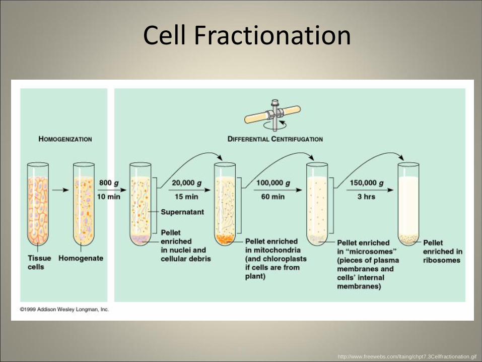
Cell Fractionation
http://www.freewebs.com/ltaing/chpt7.3Cellfractionation.gif
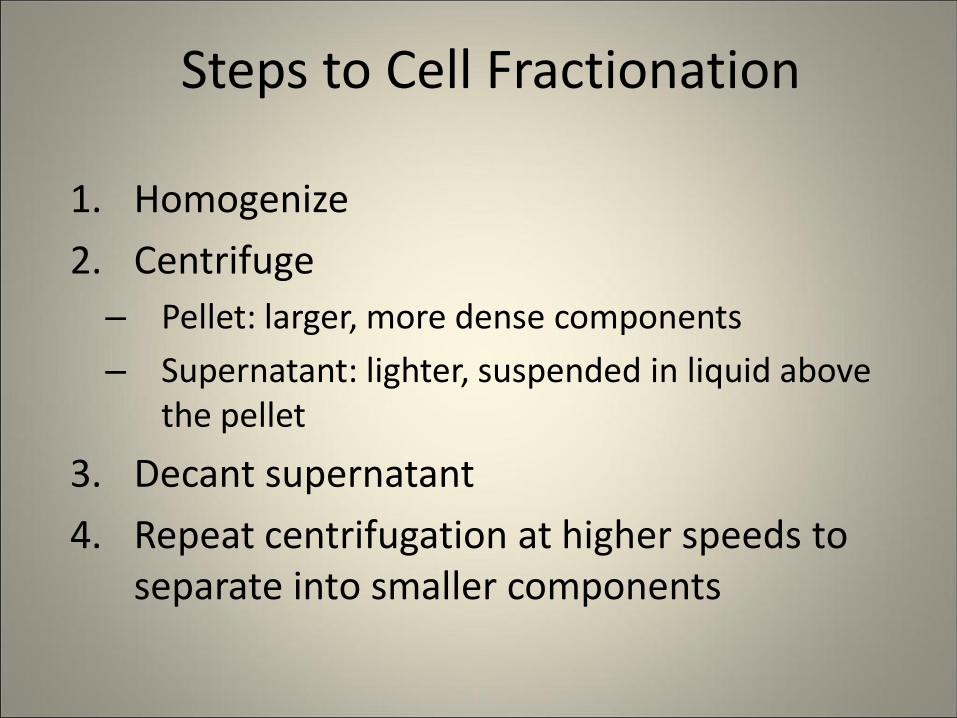
Steps to Cell Fractionation
1. Homogenize
2. Centrifuge
– Pellet: larger, more dense components
– Supernatant: lighter, suspended in liquid above the pellet
3. Decant supernatant
4. Repeat centrifugation at higher speeds to separate into smaller components
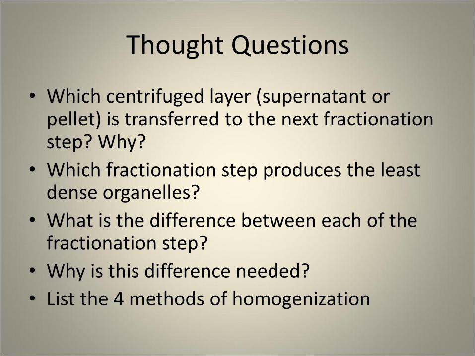
Thought Questions
• Which centrifuged layer (supernatant or pellet) is transferred to the next fractionation step? Why?
• Which fractionation step produces the least dense organelles?
• What is the difference between each of the fractionation step?
• Why is this difference needed?
• List the 4 methods of homogenization
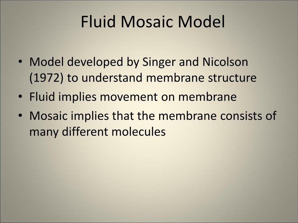
Fluid Mosaic Model
• Model developed by Singer and Nicolson (1972) to understand membrane structure
• Fluid implies movement on membrane
• Mosaic implies that the membrane consists of many different molecules
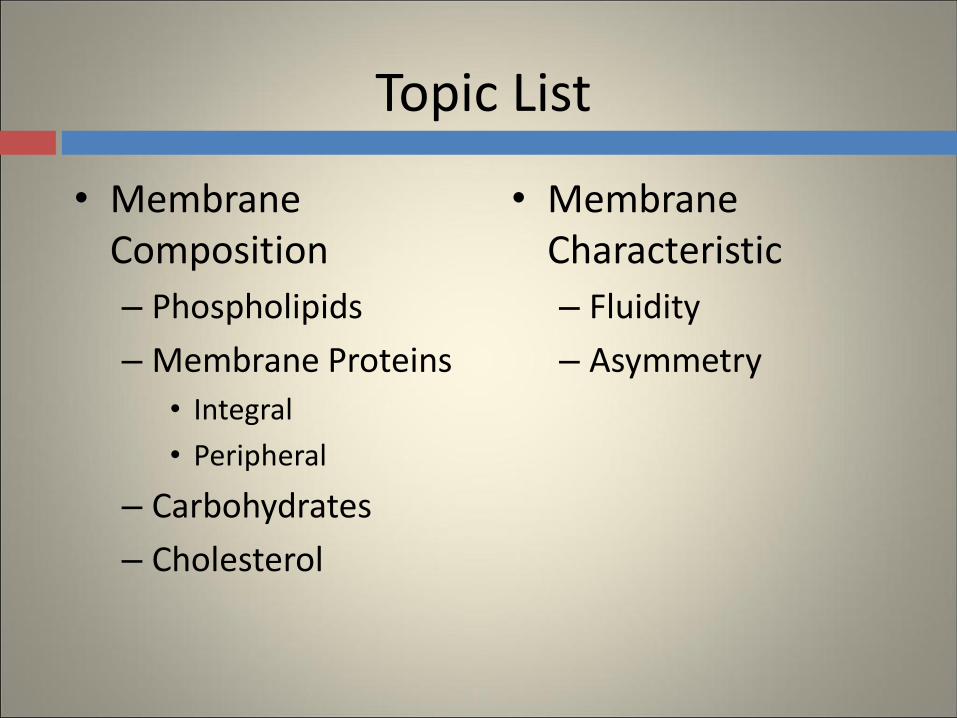
Topic List
• Membrane Composition
– Phospholipids
– Membrane Proteins
• Integral
• Peripheral
– Carbohydrates
– Cholesterol
• Membrane Characteristic
– Fluidity
– Asymmetry
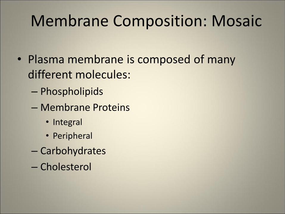
Membrane Composition: Mosaic
• Plasma membrane is composed of many different molecules:
– Phospholipids
– Membrane Proteins
• Integral
• Peripheral
– Carbohydrates
– Cholesterol
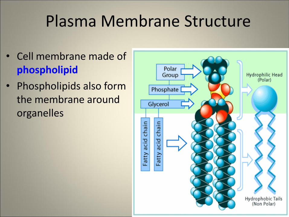
Plasma Membrane Structure
• Cell membrane made of phospholipid
• Phospholipids also form the membrane around organelles
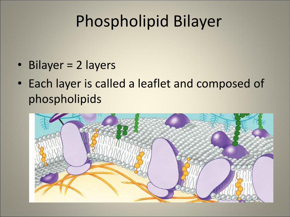
Phospholipid Bilayer
• Bilayer = 2 layers
• Each layer is called a leaflet and composed of phospholipids
Phospholipid Bilayer
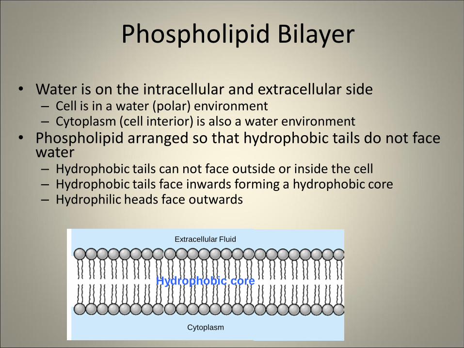
• Water is on the intracellular and extracellular side – Cell is in a water (polar) environment – Cytoplasm (cell interior) is also a water environment
• Phospholipid arranged so that hydrophobic tails do not face water – Hydrophobic tails can not face outside or inside the cell – Hydrophobic tails face inwards forming a hydrophobic core – Hydrophilic heads face outwards
Hydrophobic core
Cytoplasm
Extracellular Fluid
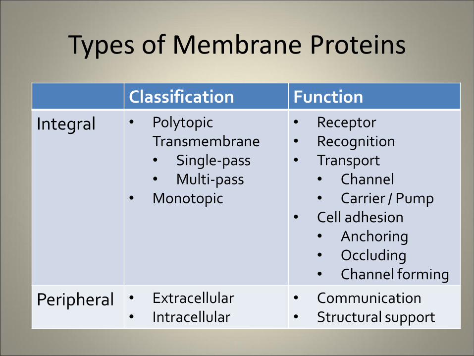
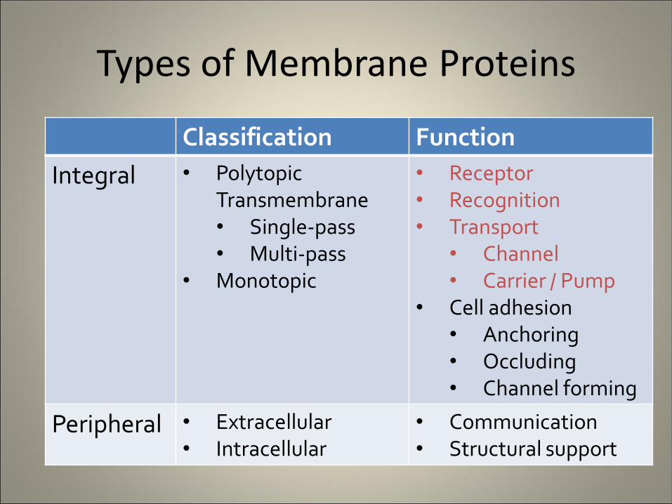
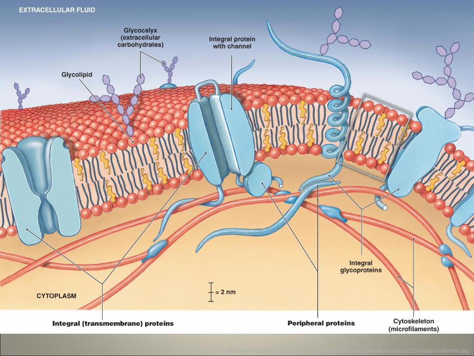
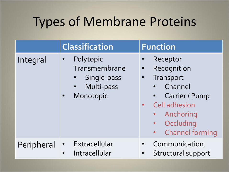
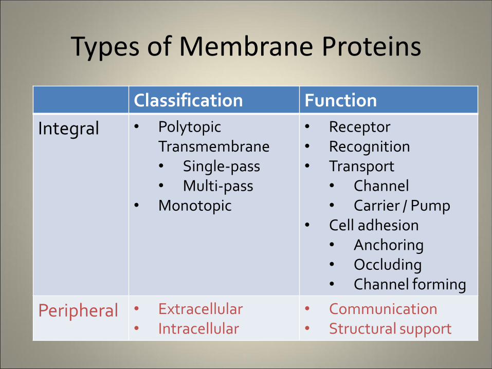
Types of Membrane Proteins
Classification Function
Integral • Polytopic Transmembrane • Single-pass • Multi-pass
• Monotopic
• Receptor • Recognition • Transport
• Channel • Carrier / Pump
• Cell adhesion • Anchoring • Occluding • Channel forming
Peripheral • Extracellular • Intracellular
• Communication • Structural support
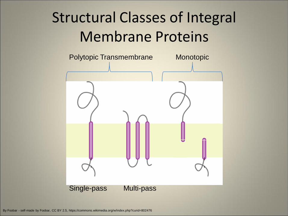
Structural Classes of Integral Membrane Proteins
By Foobar - self-made by Foobar, CC BY 2.5, https://commons.wikimedia.org/w/index.php?curid=802476
Polytopic Transmembrane Monotopic
Single-pass Multi-pass
Structural Classes of Integral Membrane Proteins
• Polytopic: faces both sides of membrane
– Transmembrane: spans entire phospholipid bilayer
– Single-pass: crosses membrane once
– Multi-pass: crosses membrane several times
• Monotopic: associated with membrane on one side (e.g. one leaflet), does not span entire bilayer
Types of Membrane Proteins
Classification Function
Integral • Polytopic Transmembrane • Single-pass • Multi-pass
• Monotopic
• Receptor • Recognition • Transport
• Channel • Carrier / Pump
• Cell adhesion • Anchoring • Occluding • Channel forming
Peripheral • Extracellular • Intracellular
• Communication • Structural support
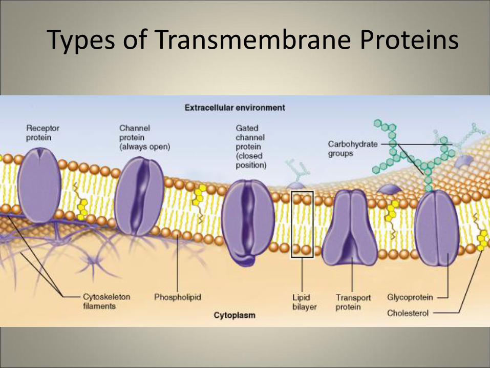
Types of Transmembrane Proteins
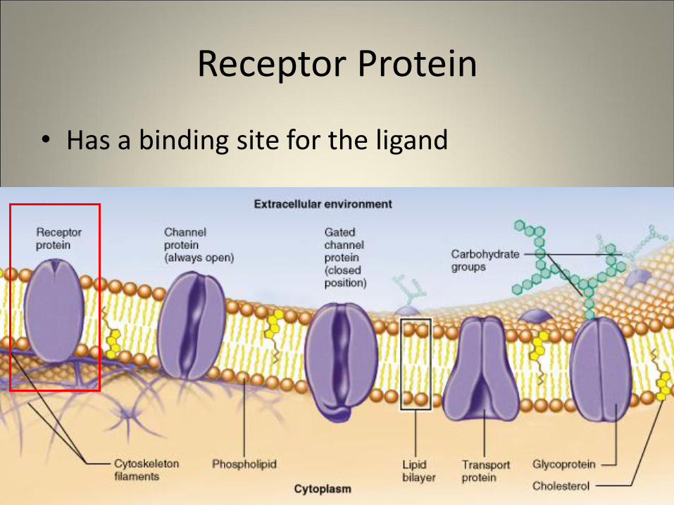
Receptor Protein
• Has a binding site for the ligand
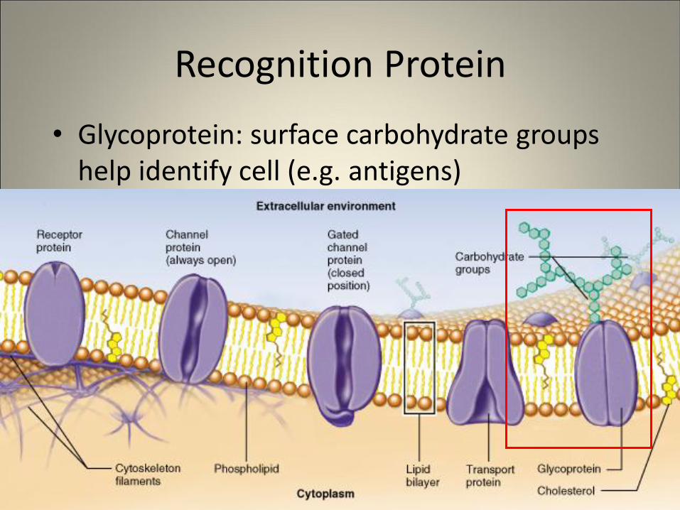
Recognition Protein
• Glycoprotein: surface carbohydrate groups help identify cell (e.g. antigens)
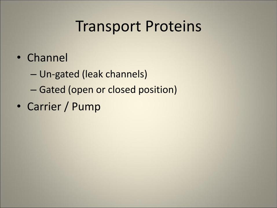
Transport Proteins
• Channel
– Un-gated (leak channels)
– Gated (open or closed position)
• Carrier / Pump

Channel Proteins
• Act like tunnels • Molecules move through protein passively (no energy
involved) • Moves small molecules or charged ions
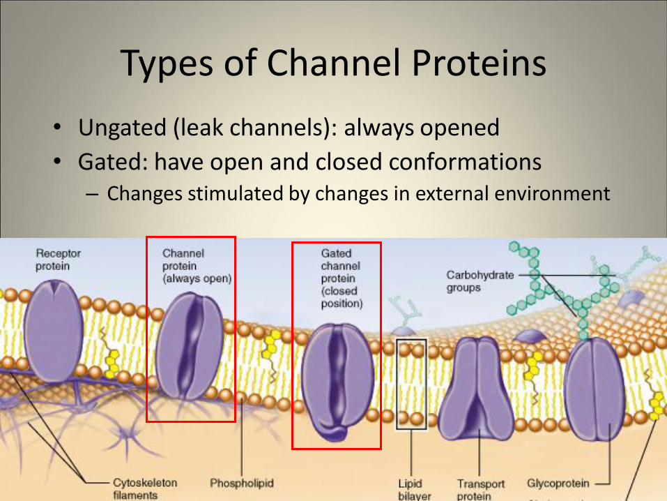
Types of Channel Proteins
• Ungated (leak channels): always opened
• Gated: have open and closed conformations – Changes stimulated by changes in external environment
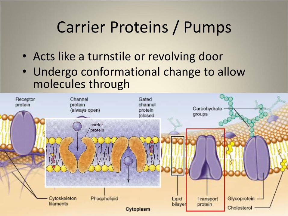
Carrier Proteins / Pumps
• Acts like a turnstile or revolving door • Undergo conformational change to allow
molecules through
http://www.highlands.edu/academics/divisions/scipe/biology/faculty/harnden/2121/images/cellmemb.jpg
Types of Membrane Proteins
Classification Function
Integral • Polytopic Transmembrane • Single-pass • Multi-pass
• Monotopic
• Receptor • Recognition • Transport
• Channel • Carrier / Pump
• Cell adhesion • Anchoring • Occluding • Channel forming
Peripheral • Extracellular • Intracellular
• Communication • Structural support
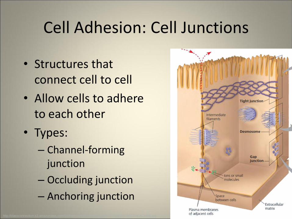
Cell Adhesion: Cell Junctions
• Structures that connect cell to cell
• Allow cells to adhere to each other
• Types:
– Channel-forming junction
– Occluding junction
– Anchoring junction
http://classconnection.s3.amazonaws.com/66/flashcards/2748066/jpg/3434-13ED20C0ECD0C4C340E.jpg
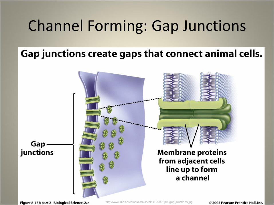
Channel Forming: Gap Junctions
http://www.uic.edu/classes/bios/bios100/f06pm/gap-junctions.jpg
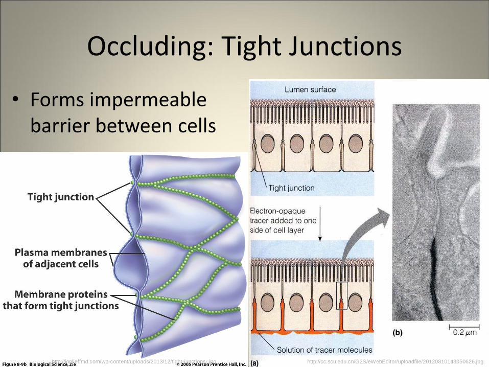
Occluding: Tight Junctions
• Forms impermeable barrier between cells
http://cc.scu.edu.cn/G2S/eWebEditor/uploadfile/20120810143050626.jpg http://jonlieffmd.com/wp-content/uploads/2013/12/tight-junctions-.jpg
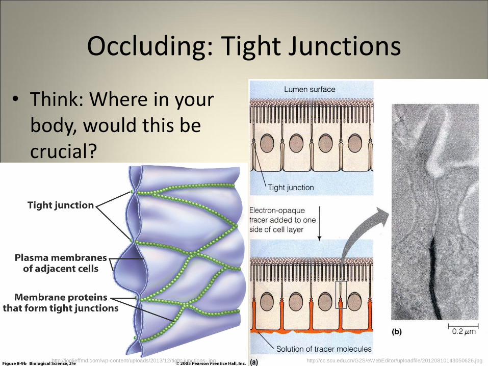
Occluding: Tight Junctions
• Think: Where in your body, would this be crucial?
http://cc.scu.edu.cn/G2S/eWebEditor/uploadfile/20120810143050626.jpg http://jonlieffmd.com/wp-content/uploads/2013/12/tight-junctions-.jpg
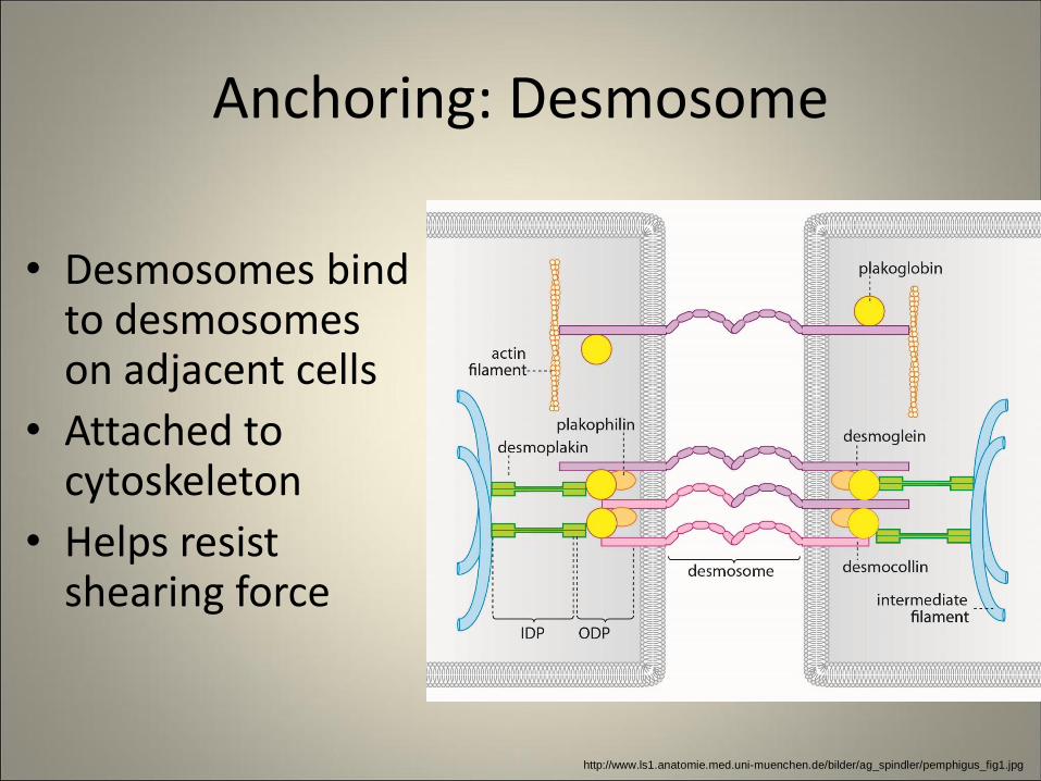
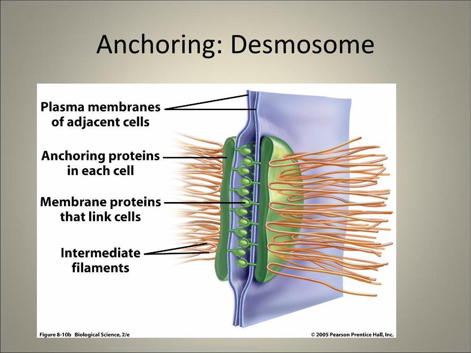
Anchoring: Desmosome
• Desmosomes bind to desmosomes on adjacent cells
• Attached to cytoskeleton
• Helps resist shearing force
http://www.ls1.anatomie.med.uni-muenchen.de/bilder/ag_spindler/pemphigus_fig1.jpg
Anchoring: Desmosome
http://www.uic.edu/classes/bios/bios100/f06pm/desmosomes.jpg
Types of Membrane Proteins
Classification Function
Integral • Polytopic Transmembrane • Single-pass • Multi-pass
• Monotopic
• Receptor • Recognition • Transport
• Channel • Carrier / Pump
• Cell adhesion • Anchoring • Occluding • Channel forming
Peripheral • Extracellular • Intracellular
• Communication • Structural support
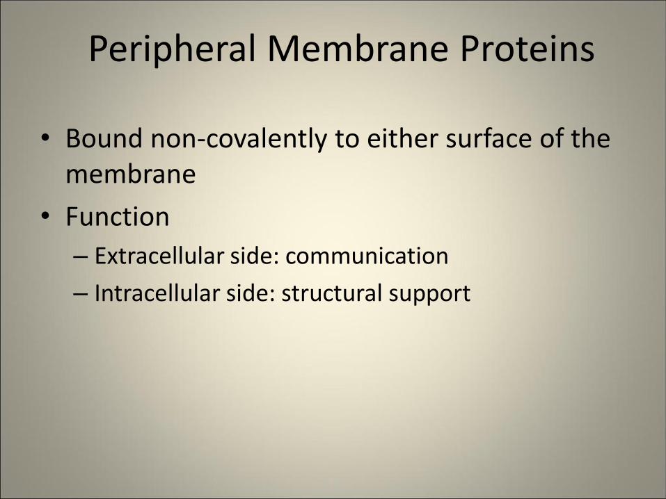
Peripheral Membrane Proteins
• Bound non-covalently to either surface of the membrane
• Function
– Extracellular side: communication
– Intracellular side: structural support
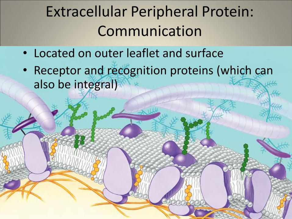
Extracellular Peripheral Protein: Communication
• Located on outer leaflet and surface
• Receptor and recognition proteins (which can also be integral)
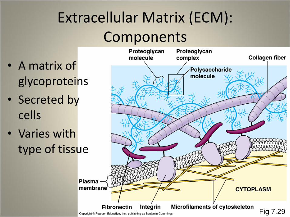
Fibronectin
Extracellular Matrix (ECM): Components
• A matrix of glycoproteins
• Secreted by cells
• Varies with type of tissue
Fig 7.29
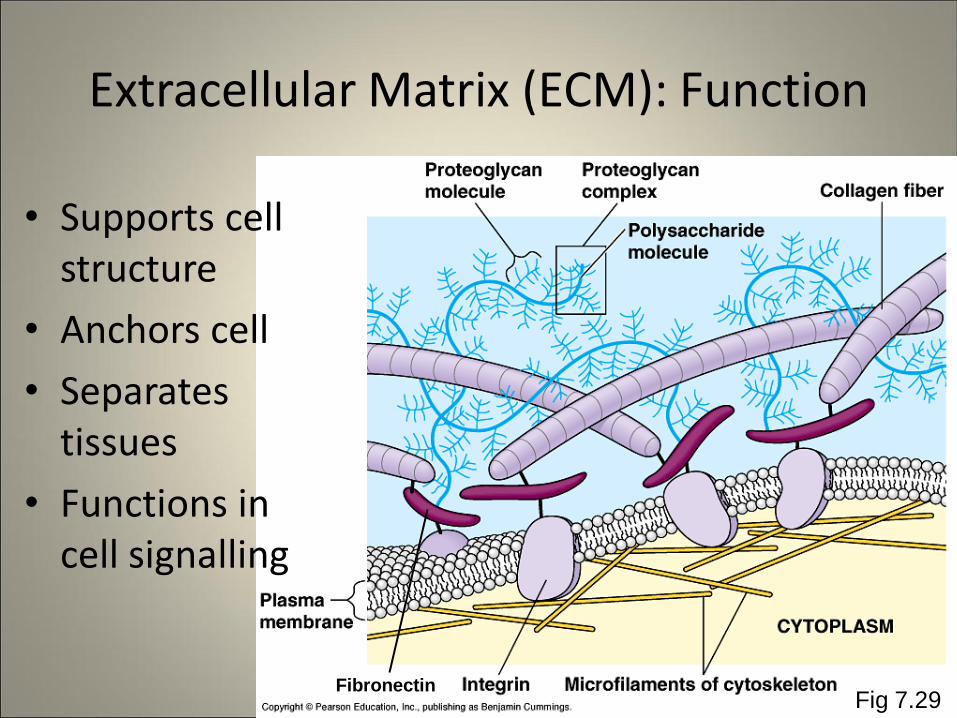
Fibronectin
Extracellular Matrix (ECM): Function
• Supports cell structure
• Anchors cell
• Separates tissues
• Functions in cell signalling
Fig 7.29
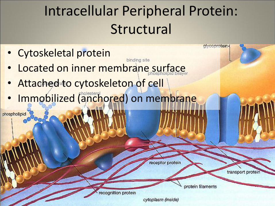
Intracellular Peripheral Protein: Structural
• Cytoskeletal protein
• Located on inner membrane surface
• Attached to cytoskeleton of cell
• Immobilized (anchored) on membrane
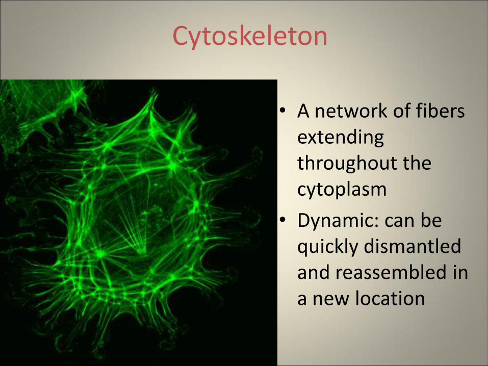
Cytoskeleton
• A network of fibers extending throughout the cytoplasm
• Dynamic: can be quickly dismantled and reassembled in a new location
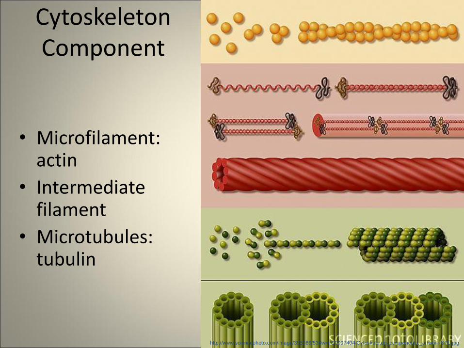
Cytoskeleton Component
• Microfilament: actin
• Intermediate filament
• Microtubules: tubulin
http://www.sciencephoto.com/image/395086/530wm/C0097404-Cytoskeleton_components,_artwork-SPL.jpg
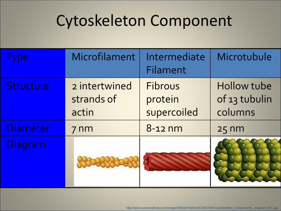
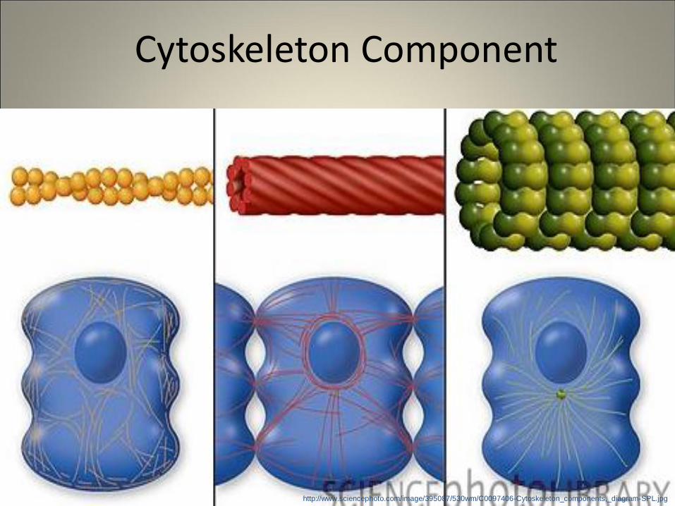
Cytoskeleton Component
Type Microfilament Intermediate Filament
Microtubule
Structure 2 intertwined strands of actin
Fibrous protein supercoiled
Hollow tube of 13 tubulin columns
Diameter 7 nm 8-12 nm 25 nm
Diagram
http://www.sciencephoto.com/image/395087/530wm/C0097406-Cytoskeleton_components,_diagram-SPL.jpg
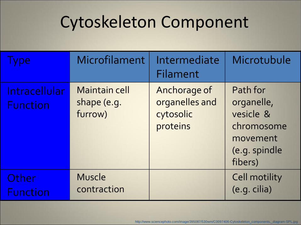
Cytoskeleton Component
Type Microfilament Intermediate Filament
Microtubule
Intracellular Function
Maintain cell shape (e.g. furrow)
Anchorage of organelles and cytosolic proteins
Path for organelle, vesicle & chromosome movement (e.g. spindle fibers)
Other Function
Muscle contraction
Cell motility (e.g. cilia)
http://www.sciencephoto.com/image/395087/530wm/C0097406-Cytoskeleton_components,_diagram-SPL.jpg
Cytoskeleton Component
http://www.sciencephoto.com/image/395087/530wm/C0097406-Cytoskeleton_components,_diagram-SPL.jpg
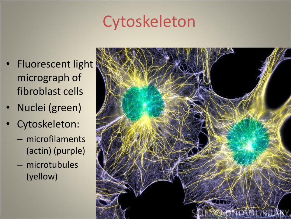
Cytoskeleton
• Fluorescent light micrograph of fibroblast cells
• Nuclei (green)
• Cytoskeleton:
– microfilaments (actin) (purple)
– microtubules (yellow)
http://www.sciencephoto.com/media/316728/enlarge
Carbohydrates
• Glycoprotein = carbohydrate + protein
• Glycolipid = carbohydate + lipid (phospholipid)
• Extracellular side
• Function of cell surface carbohydrates:
– identifies the cell (like a name) helping other cells recognize it
– acts as a signal for communication
Thought Question
• Of the component of the plasma membrane that was just studied, which would have an affect on the fluidity of the membrane?
– Phospholipids
– Proteins
– Carbohydrates
• Explain.
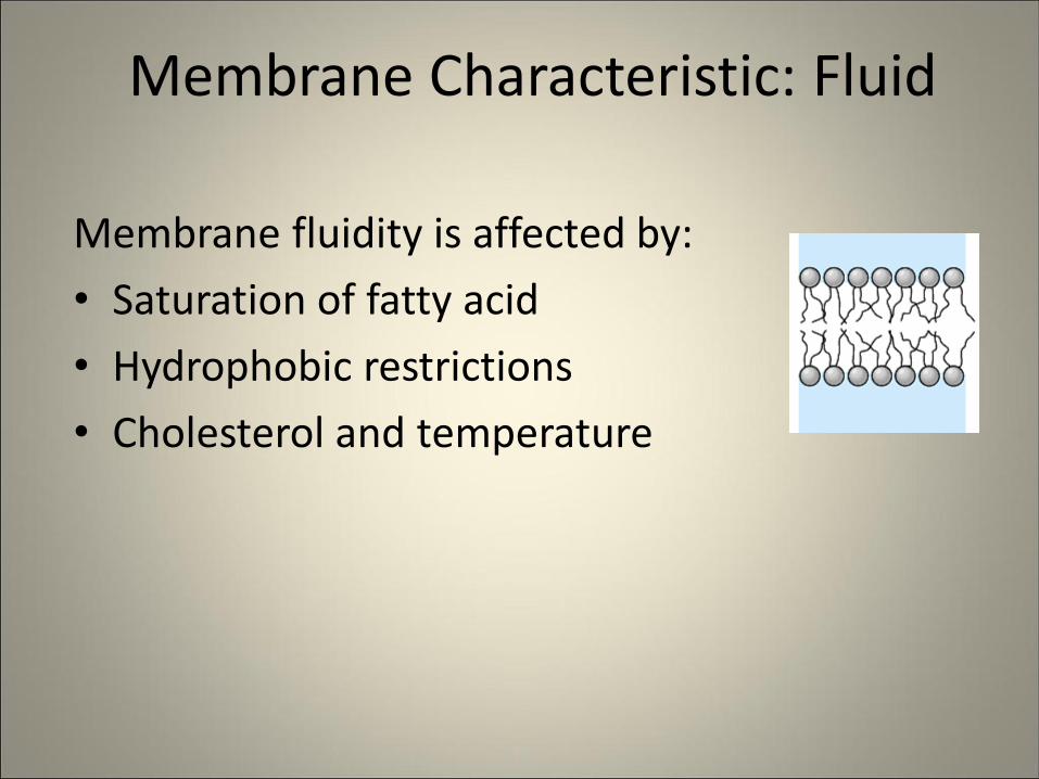
Membrane Characteristic: Fluid
Membrane fluidity is affected by:
• Saturation of fatty acid
• Hydrophobic restrictions
• Cholesterol and temperature
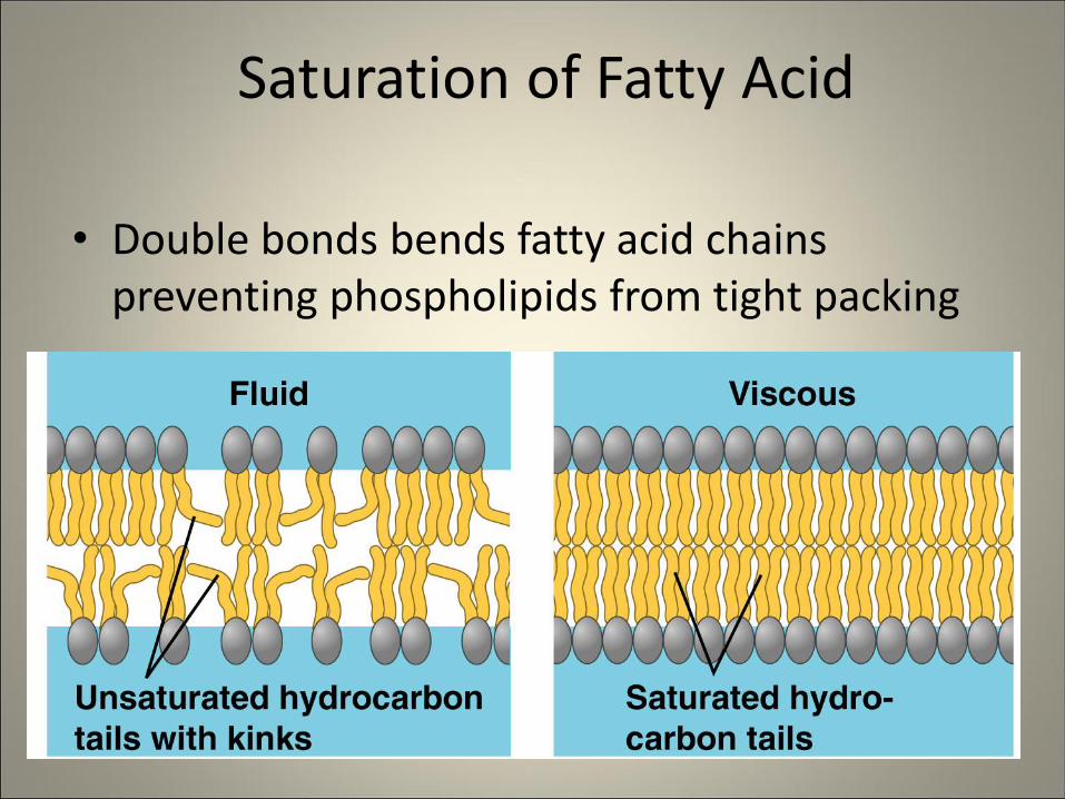
Saturation of Fatty Acid
• Double bonds bends fatty acid chains preventing phospholipids from tight packing
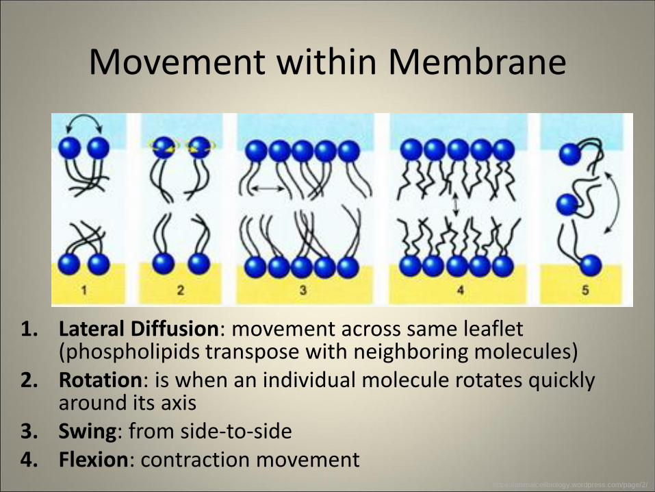
Movement within Membrane
1. Lateral Diffusion: movement across same leaflet (phospholipids transpose with neighboring molecules)
2. Rotation: is when an individual molecule rotates quickly around its axis
3. Swing: from side-to-side 4. Flexion: contraction movement
https://animalcellbiology.wordpress.com/page/2/
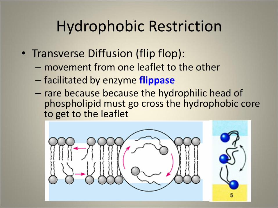
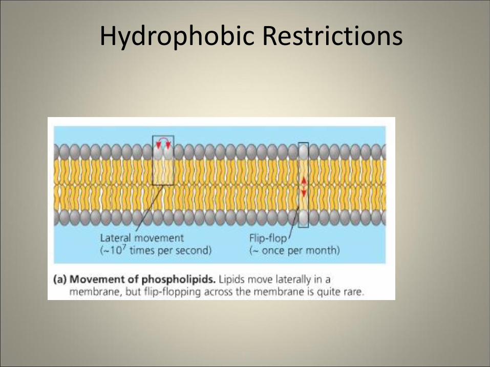
Hydrophobic Restriction
• Transverse Diffusion (flip flop): – movement from one leaflet to the other – facilitated by enzyme flippase – rare because because the hydrophilic head of
phospholipid must go cross the hydrophobic core to get to the leaflet
Hydrophobic Restrictions
Properties of Cholesterol
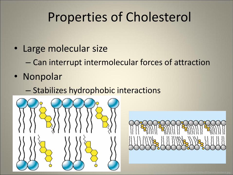
• Large molecular size
– Can interrupt intermolecular forces of attraction
• Nonpolar
– Stabilizes hydrophobic interactions
http://www.uic.edu/classes/bios/bios100/lectf03am/cholesterol.jpg
Properties of Cholesterol
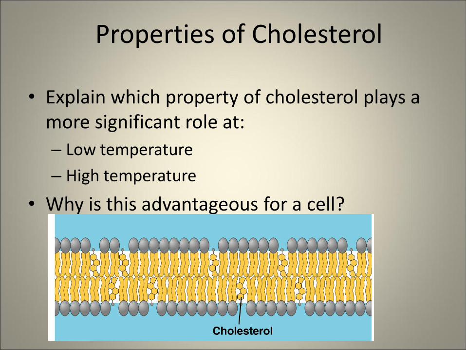
• Explain which property of cholesterol plays a more significant role at:
– Low temperature
– High temperature
• Why is this advantageous for a cell?
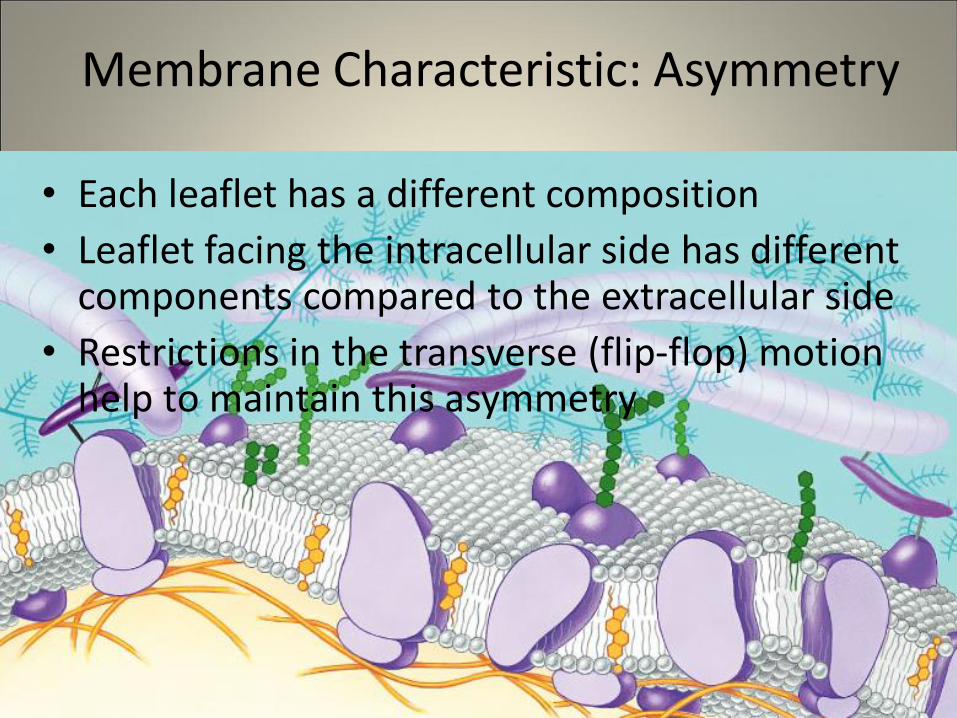
Membrane Characteristic: Asymmetry
• Each leaflet has a different composition
• Leaflet facing the intracellular side has different components compared to the extracellular side
• Restrictions in the transverse (flip-flop) motion help to maintain this asymmetry
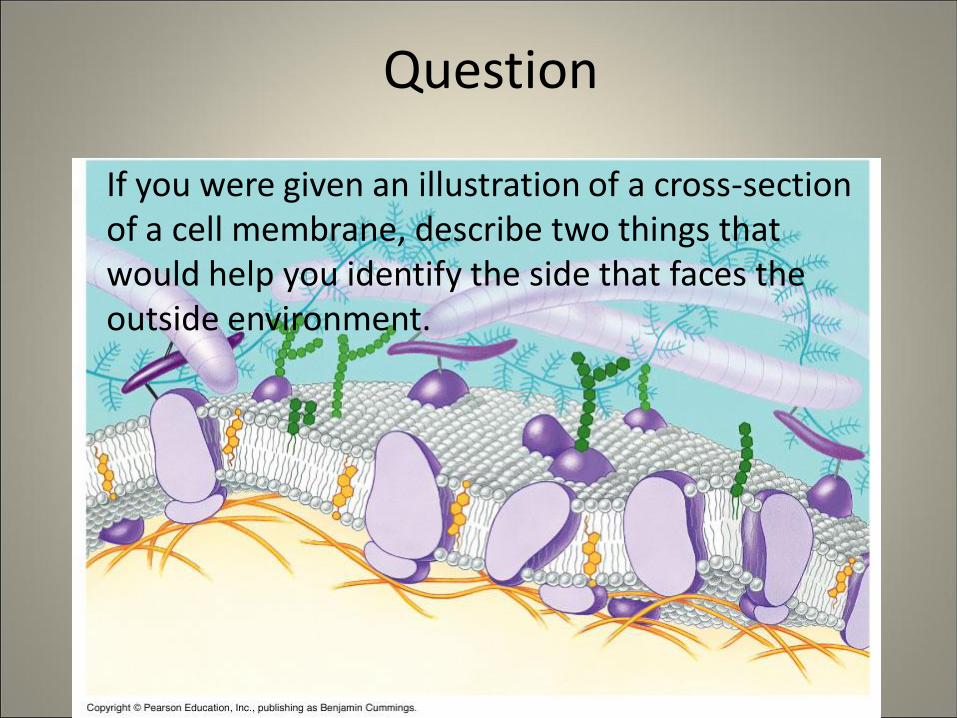
Question
If you were given an illustration of a cross-section of a cell membrane, describe two things that would help you identify the side that faces the outside environment.
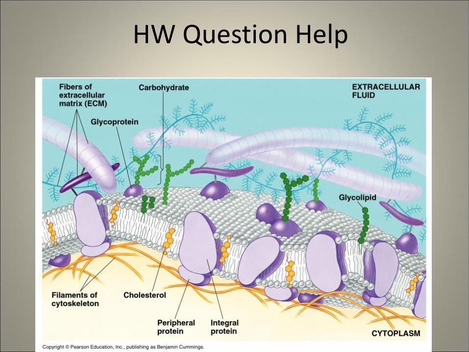
HW Question Help