cell entry of the aphthovirus equine rhinitis a virus is dependent
TRANSCRIPT

JOURNAL OF VIROLOGY, June 2010, p. 6235–6240 Vol. 84, No. 120022-538X/10/$12.00 doi:10.1128/JVI.02375-09Copyright © 2010, American Society for Microbiology. All Rights Reserved.
Cell Entry of the Aphthovirus Equine Rhinitis A Virus IsDependent on Endosome Acidification�
Elisabetta Groppelli, Tobias J. Tuthill,† and David J. Rowlands*Institute of Molecular and Cellular Biology and Astbury Centre for Structural Molecular Biology,
Faculty of Biological Sciences, University of Leeds, Leeds LS2 9JT, United Kingdom
Received 11 November 2009/Accepted 30 March 2010
Equine rhinitis A virus (ERAV) is genetically closely related to foot-and-mouth disease virus (FMDV), andboth are now classified within the genus Aphthovirus of the family Picornaviridae. For disease security reasons,FMDV can be handled only in high-containment facilities, but these constraints do not apply to ERAV, makingit an attractive alternative for the study of aphthovirus biology. Here, we show, using immunofluorescence,pharmacological agents, and dominant negative inhibitors, that ERAV entry occurs (as for FMDV) viaclathrin-mediated endocytosis and acidification of early endosomes. This validates the use of ERAV as a modelsystem to study the mechanism of cell entry by FMDV.
Equine rhinitis A virus (ERAV) belongs to the genus Aph-thovirus of the family Picornaviridae (23) and is closely relatedto foot-and-mouth disease virus (FMDV) as they share phys-icochemical properties (18, 19), nucleotide sequence (15, 28,34), and structural similarities (31). Picornaviruses are smallnonenveloped RNA viruses, comprising a 30-nm-diametercapsid made of 60 copies of each of four capsid proteins, VP1to VP4, which encapsidate a single-stranded RNA genome (7to 8 kb) (24). The family has 12 genera and includes a numberof important pathogens (e.g., poliovirus [PV], human rhinovi-rus [HRV], hepatitis A virus [HAV], etc.).
Despite extensive study of FMDV, there are still many as-pects of aphthovirus biology, such as uncoating and genomedelivery, that are yet to be elucidated. At acidic pHs, theseviruses appear to simply dissociate into subunits during uncoat-ing, and the mechanisms by which they deliver their genomesacross a cellular membrane into the cytoplasm are poorly un-derstood. In contrast, the enterovirus capsid remains intactthroughout the infection process, and models have been pro-posed for the mechanism by which these viruses interact withthe membrane and deliver their genomes into the cytoplasm.However, it has yet to be established how broadly applicablethese models are for all picornaviruses (30).
We have recently shown that acid-induced capsid dissocia-tion of ERAV proceeds via a transient intact empty particle,from which the RNA has been lost (31). This suggests a mech-anism that coordinates genome release and delivery, as in themodel for enteroviruses. To validate these studies in cell cul-ture, we first wished to identify the endocytic route used by thevirus and assess the role of acidification in the entry process.Picornaviruses utilize a variety of endocytic pathways. For ex-ample, FMDV and HRVs enter the cell via clathrin-mediated
endocytosis and are subsequently delivered to the endosome.Here, they encounter an acidic pH, which is an indispensablestep for a productive infection (2, 5, 12, 22). In contrast, echo-virus 1 (enterovirus) uses the caveolin-dependent uptake ofcaveolae and delivery to caveosomes, where no change in pHis observed (16). PV entry is independent of clathrin- andcaveolin-mediated endocytosis (6).
To elucidate the entry route of ERAV, we used a combina-tion of methods that include immunofluorescence (IF) micros-copy, pharmacological inhibitors of specific endocytosis path-ways, and dominant negative proteins.
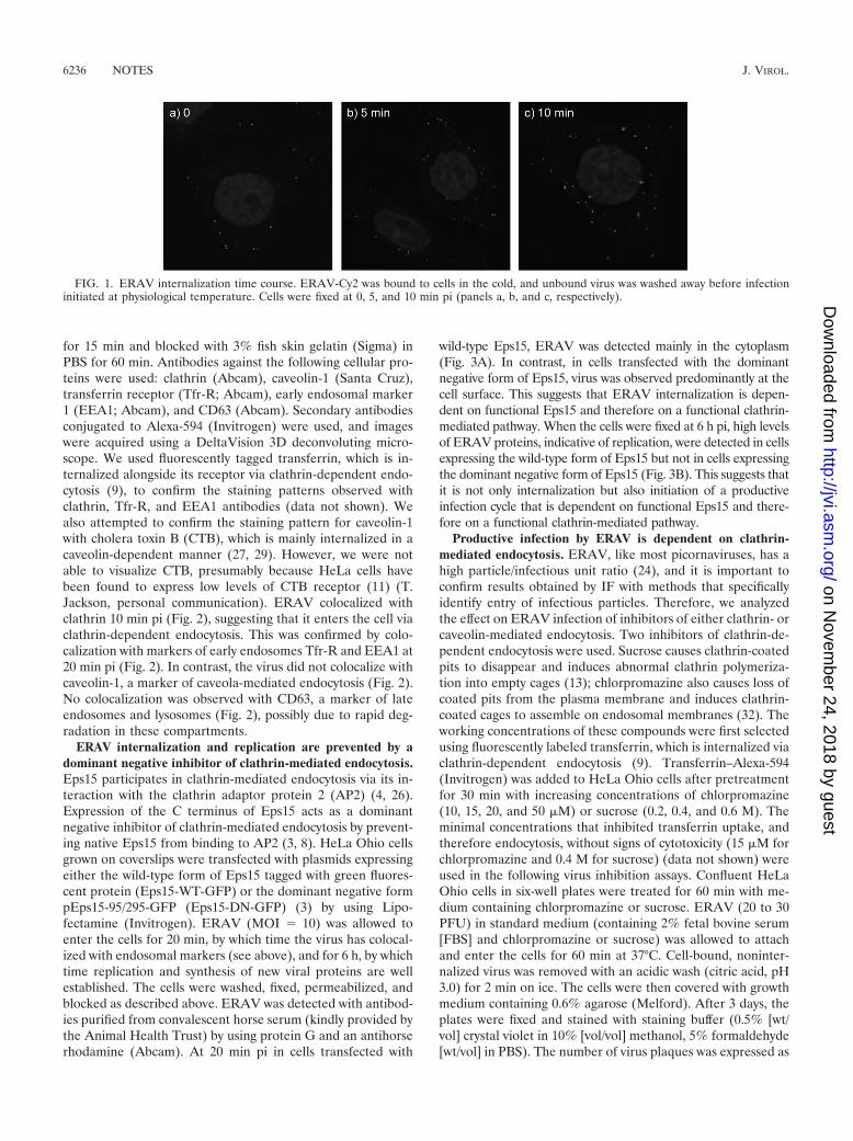
ERAV entry is rapid. Purified ERAV was labeled with thefluorophore Cy2 (GE Healthcare) according to the manufac-turer’s instructions. The conjugation of a fluorophore to theviral capsid can interfere with receptor binding and internal-ization; we therefore titrated the ratio of virus/Cy2 used inlabeling reactions and selected the virus/Cy2 ratio that gave areasonable signal in IF without affecting virus infectivity. HeLaOhio cells were grown in standard medium on glass coverslips(30-mm diameter; Agar Scientific). The cells were cooled at4°C for 30 min before ERAV-Cy2 (multiplicity of infection[MOI] � 10) was adsorbed for 20 min at 4°C. This MOI waschosen to obtain a clear signal without overloading the cellswith virus. Unattached virus was removed, and the cells wereincubated in growth medium at 37°C for 0 to 10 min beforefixing in 4% formaldehyde in phosphate-buffered saline (PBS).Images were acquired using a DeltaVision three-dimensional(3D) deconvoluting microscope. Figure 1 shows progressivemovement of the virus from the cell surface to the interiorwithin 10 min postinfection (pi). This rapid internalization iscompatible with clathrin-dependent entry, as seen with FMDV(5), but not with caveolin-mediated entry.
ERAV colocalizes with markers of clathrin-dependent endo-cytosis. An established method for identification of the routeof viral entry is IF colocalization of virus with markers ofknown endocytosis pathways. HeLa Ohio cells were precooledand infected with ERAV-Cy2 as described above. Cells werethen incubated in growth medium at 37°C for 0 min, 5, 10, 20,or 30 min before being fixed in 4% formaldehyde in PBS. Cellswere permeabilized with 0.1% Triton X-100 (Sigma) in PBS
* Corresponding author. Mailing address: Institute of Molecularand Cellular Biology and Astbury Centre for Structural MolecularBiology, Faculty of Biological Sciences, University of Leeds, Leeds LS29JT, United Kingdom. Phone: 44 0113 34 35641. Fax: 44 0113 3435638. E-mail: [email protected].
† Present address: Institute for Animal Health, Ash Road, Pirbright,Surrey GU24 0NF, United Kingdom.
� Published ahead of print on 7 April 2010.
6235
on Novem
ber 24, 2018 by guesthttp://jvi.asm
.org/D
ownloaded from
for 15 min and blocked with 3% fish skin gelatin (Sigma) inPBS for 60 min. Antibodies against the following cellular pro-teins were used: clathrin (Abcam), caveolin-1 (Santa Cruz),transferrin receptor (Tfr-R; Abcam), early endosomal marker1 (EEA1; Abcam), and CD63 (Abcam). Secondary antibodiesconjugated to Alexa-594 (Invitrogen) were used, and imageswere acquired using a DeltaVision 3D deconvoluting micro-scope. We used fluorescently tagged transferrin, which is in-ternalized alongside its receptor via clathrin-dependent endo-cytosis (9), to confirm the staining patterns observed withclathrin, Tfr-R, and EEA1 antibodies (data not shown). Wealso attempted to confirm the staining pattern for caveolin-1with cholera toxin B (CTB), which is mainly internalized in acaveolin-dependent manner (27, 29). However, we were notable to visualize CTB, presumably because HeLa cells havebeen found to express low levels of CTB receptor (11) (T.Jackson, personal communication). ERAV colocalized withclathrin 10 min pi (Fig. 2), suggesting that it enters the cell viaclathrin-dependent endocytosis. This was confirmed by colo-calization with markers of early endosomes Tfr-R and EEA1 at20 min pi (Fig. 2). In contrast, the virus did not colocalize withcaveolin-1, a marker of caveola-mediated endocytosis (Fig. 2).No colocalization was observed with CD63, a marker of lateendosomes and lysosomes (Fig. 2), possibly due to rapid deg-radation in these compartments.
ERAV internalization and replication are prevented by adominant negative inhibitor of clathrin-mediated endocytosis.Eps15 participates in clathrin-mediated endocytosis via its in-teraction with the clathrin adaptor protein 2 (AP2) (4, 26).Expression of the C terminus of Eps15 acts as a dominantnegative inhibitor of clathrin-mediated endocytosis by prevent-ing native Eps15 from binding to AP2 (3, 8). HeLa Ohio cellsgrown on coverslips were transfected with plasmids expressingeither the wild-type form of Eps15 tagged with green fluores-cent protein (Eps15-WT-GFP) or the dominant negative formpEps15-95/295-GFP (Eps15-DN-GFP) (3) by using Lipo-fectamine (Invitrogen). ERAV (MOI � 10) was allowed toenter the cells for 20 min, by which time the virus has colocal-ized with endosomal markers (see above), and for 6 h, by whichtime replication and synthesis of new viral proteins are wellestablished. The cells were washed, fixed, permeabilized, andblocked as described above. ERAV was detected with antibod-ies purified from convalescent horse serum (kindly provided bythe Animal Health Trust) by using protein G and an antihorserhodamine (Abcam). At 20 min pi in cells transfected with
wild-type Eps15, ERAV was detected mainly in the cytoplasm(Fig. 3A). In contrast, in cells transfected with the dominantnegative form of Eps15, virus was observed predominantly at thecell surface. This suggests that ERAV internalization is depen-dent on functional Eps15 and therefore on a functional clathrin-mediated pathway. When the cells were fixed at 6 h pi, high levelsof ERAV proteins, indicative of replication, were detected in cellsexpressing the wild-type form of Eps15 but not in cells expressingthe dominant negative form of Eps15 (Fig. 3B). This suggests thatit is not only internalization but also initiation of a productiveinfection cycle that is dependent on functional Eps15 and there-fore on a functional clathrin-mediated pathway.
Productive infection by ERAV is dependent on clathrin-mediated endocytosis. ERAV, like most picornaviruses, has ahigh particle/infectious unit ratio (24), and it is important toconfirm results obtained by IF with methods that specificallyidentify entry of infectious particles. Therefore, we analyzedthe effect on ERAV infection of inhibitors of either clathrin- orcaveolin-mediated endocytosis. Two inhibitors of clathrin-de-pendent endocytosis were used. Sucrose causes clathrin-coatedpits to disappear and induces abnormal clathrin polymeriza-tion into empty cages (13); chlorpromazine also causes loss ofcoated pits from the plasma membrane and induces clathrin-coated cages to assemble on endosomal membranes (32). Theworking concentrations of these compounds were first selectedusing fluorescently labeled transferrin, which is internalized viaclathrin-dependent endocytosis (9). Transferrin–Alexa-594(Invitrogen) was added to HeLa Ohio cells after pretreatmentfor 30 min with increasing concentrations of chlorpromazine(10, 15, 20, and 50 �M) or sucrose (0.2, 0.4, and 0.6 M). Theminimal concentrations that inhibited transferrin uptake, andtherefore endocytosis, without signs of cytotoxicity (15 �M forchlorpromazine and 0.4 M for sucrose) (data not shown) wereused in the following virus inhibition assays. Confluent HeLaOhio cells in six-well plates were treated for 60 min with me-dium containing chlorpromazine or sucrose. ERAV (20 to 30PFU) in standard medium (containing 2% fetal bovine serum[FBS] and chlorpromazine or sucrose) was allowed to attachand enter the cells for 60 min at 37°C. Cell-bound, noninter-nalized virus was removed with an acidic wash (citric acid, pH3.0) for 2 min on ice. The cells were then covered with growthmedium containing 0.6% agarose (Melford). After 3 days, theplates were fixed and stained with staining buffer (0.5% [wt/vol] crystal violet in 10% [vol/vol] methanol, 5% formaldehyde[wt/vol] in PBS). The number of virus plaques was expressed as
FIG. 1. ERAV internalization time course. ERAV-Cy2 was bound to cells in the cold, and unbound virus was washed away before infectioninitiated at physiological temperature. Cells were fixed at 0, 5, and 10 min pi (panels a, b, and c, respectively).
6236 NOTES J. VIROL.
on Novem
ber 24, 2018 by guesthttp://jvi.asm
.org/D
ownloaded from

a percentage of the no-drug control. Both clathrin inhibitorycompounds resulted in a reduced number of ERAV plaquescompared to the untreated cells; chlorpromazine or sucrosetreatment resulted in 40% and 85% reductions, respectively
(Fig. 4A). No reduction in the number of plaques was observedif the inhibitors were added 3 h pi (data not shown), confirmingthat they affected ERAV specifically at the entry stage. Toinvestigate alternative routes for ERAV entry, we also used
FIG. 2. ERAV colocalization with endocytosis markers. Cells were infected with Cy2-labeled ERAV for 0 to 30 min before being visualized asdescribed in the text. Antibodies against endocytosis markers were used in combination with secondary antibodies conjugated to Alexa-594(Invitrogen). Cell nuclei were stained with Hoechst. Images were acquired using a DeltaVision deconvoluting microscope. (a) ERAV-Cy2 (green).(b) Clathrin (red) at 10 min pi. (c) Merge of panels a and b, showing colocalization (yellow) of ERAV and clathrin. (e to t) Cells fixed at 20 minpi with ERAV-Cy2 (green) and caveolin-1, Tfr-R, EEA1, or CD63 (red). (g) Overlay of panels e and f, showing ERAV (green) and caveolin-1(red). (k and o) Colocalization (yellow) of ERAV with Tfr-R or EEA1, respectively. Panel k is a merge of panels i and j, and panel o is a mergeof panels m and n. (t) Overlay of panels r and s, showing ERAV (green) and CD63 (red). Panels d, h, l, p, q, and u are enlargements from theMerge panels.
VOL. 84, 2010 NOTES 6237
on Novem
ber 24, 2018 by guesthttp://jvi.asm
.org/D
ownloaded from

two reagents known to inhibit caveola-mediated endocytosis byinterfering with the lipid components of caveolae. Nystatin, acholesterol-complexing agent, and methyl-�-cyclodextrin(M�CD), which stimulates cholesterol efflux from the cellmembrane, were used in plaque assays as described above at 25�M and 7.5 mM, respectively. Neither nystatin nor M�CD
reduced the number of plaques compared to untreated con-trols (Fig. 4B), suggesting that ERAV entry does not requirecaveola-mediated endocytosis.
ERAV infection is dependent upon endosomal acidification.Concanamycin A was used as an inhibitor of endosomal acid-ification in order to assess the role of pH in ERAV infection
FIG. 3. Effects of inhibition of clathrin-mediated endocytosis on ERAV entry. (A) Detection of ERAV (red) 20 min pi (b and f) of cellsexpressing wild-type Eps15 (Eps15-WT-GFP) (green) (a) or dominant negative Eps15 (Eps15-DN-GFP) (green) (e), which inhibits clathrin-mediated endocytosis. ERAV is located predominantly in the cytoplasm in cells expressing wild-type Eps15 (c, overlay of a and b). Panel d is anenlargement from panel c. ERAV is located predominantly at the cell surface in cells expressing dominant negative Eps15 (g, overlay of panelse and f). Panel h is an enlargement from panel g. (B) Panels i to o show cells fixed at 6 h pi. Panel k is a merge of panels i (Eps15-WT-GFP [green])and j (ERAV [red]). The large ERAV signal in the cytoplasm is indicative of replication. Panel o is a merge of panels m (Eps15-DN-GFP [green])and n (ERAV [red]). The lack of ERAV signal in the cytoplasm suggests lack of replication.
6238 NOTES J. VIROL.
on Novem
ber 24, 2018 by guesthttp://jvi.asm
.org/D
ownloaded from

(10). The inhibitory effect of concanamycin A was confirmedby monitoring endosomal pH with the fluorophore acridineorange (AO; Invitrogen). AO is a weak base that becomesconcentrated in acidic endosomes and exhibits a concentra-tion-dependent shift from green (low concentration) to red(high concentration) fluorescence (35). HeLa Ohio cells oncoverslips were incubated for 30 min in the presence or ab-sence of concanamycin A (50, 100, and 200 nM). AO (0.5�g/ml) was added for 15 min before the cells were visualized byfluorescence microscopy and red and green signals overlaid. Inthe absence of concanamycin A, acidic vesicles appeared or-
ange because AO was trapped within the endosomes and itsconcentration increased (Fig. 4C). When concanamycin A wasused at 100 nM (optimal concentration), no red/orange stain-ing was detected, suggesting that endosomal acidification wasinhibited (Fig. 4C). The effect of concanamycin A (100 nM)was then assessed on ERAV infection in virus inhibition as-says, as described above. No plaques were seen in the presenceof concanamycin A, suggesting that acidification is essential forthe entry process (Fig. 4D). To confirm this result, we usedmonensin, a carboxylic ionophore that intercalates in the en-dosomal membrane and exchanges cytoplasmic K� for pro-tons, thereby increasing endosomal pH (17). In the presence ofmonensin (25 �M), ERAV infection was inhibited by 95%(Fig. 4D).
To identify more precisely the pH necessary for productiveinfection, we inhibited the maturation of early (pH 6.5) endo-somes to late (pH 5.5) endosomes. This was achieved by in-ducing depolymerization of microtubules by exposure of cellsto 20 �M nocodazole (1) or by inhibition of phosphoinositide3-kinase signaling with 25 nM wortmannin (33). Neither drugreduced the number of plaques compared to the untreatedcontrols (Fig. 4E) in infectious assays as described above, sug-gesting that maturation from early to late endosome is notnecessary for ERAV infection. It is also worth noting thatmicrotubules are involved in caveolin-dependent endocytosis.Therefore, the lack of an inhibitory effect of nocodazole onERAV suggests that its entry is not dependent on caveolarendocytosis, thus supporting our general conclusions.
Exposure to acidic pH in the endosome is thought to be thetrigger for genome release during FMDV infection (5, 14, 21),and both FMDV and ERAV dissociate under acidic conditionsinto pentameric subunits with release of RNA (7, 31). How-ever, it is difficult to envision how the RNA is protected withinthe endosomal lumen following its release from the dissociatedcapsid and how it is transferred across the endosomal mem-brane. Earlier studies on FMDV described the formation ofempty capsid particles lacking RNA (25), and we have recentlyshown that dissociation of ERAV proceeds via a transientlystable icosahedral empty particle from which the RNA hasbeen ejected (31). We speculate that an uncoating intermedi-ate particle may associate with the endosomal membrane tofacilitate transfer of the RNA into the cytoplasm of the hostcell, perhaps via a pore formed by VP4. This may protect theRNA from damaging exposure to the luminal contents of thevesicle, as would occur if the particle dissociated directly intoits component subunits.
In summary, we have shown that productive infection of cellsby ERAV is dependent on clathrin-mediated endocytosis andendosomal acidification, as is the case for FMDV (5, 14, 20).The requirement for acidification during infection is in agree-ment with recent studies describing the in vitro uncoating ofERAV, and these findings further strengthen the relevance ofERAV as a model for studying FMDV cell entry.
We thank Janet Daley and the Animal Health Trust, Newmarket,United Kingdom, for providing anti-ERAV serum.
This work was funded by the Biotechnology and Biological SciencesResearch Council, United Kingdom, and the Medical Research Coun-cil, United Kingdom.
FIG. 4. Effects of pharmacological inhibitors of clathrin- andcaveolin-dependent endocytosis and endosomal acidification onERAV infectivity. (A) HeLa Ohio cells were treated with inhibitors ofclathrin-dependent endocytosis and then infected with ERAV. Thenumber of virus plaques generated in the presence of the inhibitorswas expressed as a percentage of the no-drug control. ERAV infec-tivity was inhibited 40% by chlorpromazine (Cpz; 15 �M) and 85% bysucrose (Sucr; 0.4 M). (B) Effects of inhibition of caveolin-dependentendocytosis on ERAV. Neither nystatin (Nys) nor M�CD reduced thenumber of plaques compared to untreated controls. (C) Cells weretreated with concanamycin A (ConcA) (b) or dimethyl sulfoxide(DMSO) alone (a) for 30 min at 37°C followed by visualization ofendosomal acidification by AO fluorescence. In mock-treated cells, theacidic vesicles appear orange due to accumulation of AO, while inconcanamycin A-treated cells, no red/orange color is detected, con-firming inhibition of endosomal acidification (see the text). (D) Re-duction of ERAV infectivity by inhibition of endosomal acidificationwith concanamycin A (100 nm) or monensin (Mon; 25 �M). (E) Pro-gression from early to late endosomes is not necessary for ERAVinfectivity since nocodazole (Noc; 20 �M) or wortmannin (Wort; 25nM) did not reduce plaque numbers.
VOL. 84, 2010 NOTES 6239
on Novem
ber 24, 2018 by guesthttp://jvi.asm
.org/D
ownloaded from

REFERENCES
1. Aniento, F., N. Emans, G. Griffiths, and J. Gruenberg. 1993. Cytoplasmicdynein-dependent vesicular transport from early to late endosomes. J. CellBiol. 123:1373–1387.
2. Bayer, N., D. Schober, M. Huttinger, D. Blaas, and R. Fuchs. 2001. Inhibi-tion of clathrin-dependent endocytosis has multiple effects on human rhino-virus serotype 2 cell entry. J. Biol. Chem. 276:3952–3962.
3. Benmerah, A., M. Bayrou, N. Cerf-Bensussan, and A. Dautry-Varsat. 1999.Inhibition of clathrin-coated pit assembly by an Eps15 mutant. J. Cell Sci.112(Pt. 9):1303–1311.
4. Benmerah, A., C. Lamaze, B. Begue, S. L. Schmid, A. Dautry-Varsat, and N.Cerf-Bensussan. 1998. AP-2/Eps15 interaction is required for receptor-me-diated endocytosis. J. Cell Biol. 140:1055–1062.
5. Berryman, S., S. Clark, P. Monaghan, and T. Jackson. 2005. Early events inintegrin �v�6-mediated cell entry of foot-and-mouth disease virus. J. Virol.79:8519–8534.
6. Brandenburg, B., L. Y. Lee, M. Lakadamyali, M. J. Rust, X. Zhuang, andJ. M. Hogle. 2007. Imaging poliovirus entry in live cells. PLoS Biol. 5:e183.
7. Burroughs, J. N., D. J. Rowlands, D. V. Sangar, P. Talbot, and F. Brown.1971. Further evidence for multiple proteins in the foot-and-mouth diseasevirus particle. J. Gen. Virol. 13:73–84.
8. Carbone, R., S. Fre, G. Iannolo, F. Belleudi, P. Mancini, P. G. Pelicci, M. R.Torrisi, and P. P. Di Fiore. 1997. eps15 and eps15R are essential componentsof the endocytic pathway. Cancer Res. 57:5498–5504.
9. Dautry-Varsat, A. 1986. Receptor-mediated endocytosis: the intracellularjourney of transferrin and its receptor. Biochimie 68:375–381.
10. Drose, S., and K. Altendorf. 1997. Bafilomycins and concanamycins as in-hibitors of V-ATPases and P-ATPases. J. Exp. Biol. 200:1–8.
11. Fishman, P. H., R. M. Bradley, J. Moss, and V. C. Manganiello. 1978. Effectof serum on ganglioside uptake and choleragen responsiveness of trans-formed mouse fibroblasts. J. Lipid Res. 19:77–81.
12. Grunert, H. P., K. U. Wolf, K. D. Langner, D. Sawitzky, K. O. Habermehl,and H. Zeichhardt. 1997. Internalization of human rhinovirus 14 into HeLaand ICAM-1-transfected BHK cells. Med. Microbiol. Immunol. 186:1–9.
13. Hansen, S. H., K. Sandvig, and B. van Deurs. 1993. Clathrin and HA2adaptors: effects of potassium depletion, hypertonic medium, and cytosolacidification. J. Cell Biol. 121:61–72.
14. Johns, H. L., S. Berryman, P. Monaghan, G. J. Belsham, and T. Jackson.2009. A dominant negative mutant of rab5 inhibits infection of cells byfoot-and-mouth disease virus: implications for virus entry. J. Virol. 83:6247–6256.
15. Li, F., G. F. Browning, M. J. Studdert, and B. S. Crabb. 1996. Equinerhinovirus 1 is more closely related to foot-and-mouth disease virus than toother picornaviruses. Proc. Natl. Acad. Sci. U. S. A. 93:990–995.
16. Marjomaki, V., V. Pietiainen, H. Matilainen, P. Upla, J. Ivaska, L. Nissinen,H. Reunanen, P. Huttunen, T. Hyypia, and J. Heino. 2002. Internalization ofechovirus 1 in caveolae. J. Virol. 76:1856–1865.
17. Mellman, I., R. Fuchs, and A. Helenius. 1986. Acidification of the endocyticand exocytic pathways. Annu. Rev. Biochem. 55:663–700.
18. Newman, J. F., D. J. Rowlands, and F. Brown. 1973. A physico-chemicalsub-grouping of the mammalian picornaviruses. J. Gen. Virol. 18:171–180.
19. Newman, J. F., D. J. Rowlands, F. Brown, D. Goodridge, R. Burrows, and F.Steck. 1977. Physicochemical characterization of two serologically unrelatedequine rhinoviruses. Intervirology 8:145–154.
20. O’Donnell, V., M. Larocco, and B. Baxt. 2008. Heparan sulfate-bindingfoot-and-mouth disease virus enters cells via caveola-mediated endocytosis.J. Virol. 82:9075–9085.
21. O’Donnell, V., M. LaRocco, H. Duque, and B. Baxt. 2005. Analysis of foot-and-mouth disease virus internalization events in cultured cells. J. Virol.79:8506–8518.
22. Prchla, E., E. Kuechler, D. Blaas, and R. Fuchs. 1994. Uncoating of humanrhinovirus serotype 2 from late endosomes. J. Virol. 68:3713–3723.
23. Pringle, C. R., and M. A. Mayo. 1999. Virus taxonomy at the 11th Interna-tional Congress of Virology, Sydney, Australia, 1999. Arch. Virol. 144:2065–2070.
24. Racaniello, V. R. 2007. Picornaviridae: the viruses and their replication, p.685–722. In Fields virology, vol. 1. Lippincott, Williams, and Wilkins, Phil-adelphia, PA.
25. Rowlands, D. J., D. V. Sangar, and F. Brown. 1975. A comparative chemicaland serological study of the full and empty particles of foot-and-mouthdisease virus. J. Gen. Virol. 26:227–238.
26. Salcini, A. E., H. Chen, G. Iannolo, P. De Camilli, and P. P. Di Fiore. 1999.Epidermal growth factor pathway substrate 15, Eps15. Int. J. Biochem. CellBiol. 31:805–809.
27. Singh, R. D., V. Puri, J. T. Valiyaveettil, D. L. Marks, R. Bittman, and R. E.Pagano. 2003. Selective caveolin-1-dependent endocytosis of glycosphingo-lipids. Mol. Biol. Cell 14:3254–3265.
28. Studdert, M. J., and L. J. Gleeson. 1978. Isolation and characterisation of anequine rhinovirus. Zentralbl. Veterinarmed. B 25:225–237.
29. Torgersen, M. L., G. Skretting, B. van Deurs, and K. Sandvig. 2001. Inter-nalization of cholera toxin by different endocytic mechanisms. J. Cell Sci.114:3737–3747.
30. Tuthill, T. J., E. Groppelli, J. M. Hogle, and D. J. Rowlands. Picornaviruscell entry. Cell entry of non-enveloped viruses. Curr. Top. Microbiol.Immunol., in press.
31. Tuthill, T. J., K. Harlos, T. S. Walter, N. J. Knowles, E. Groppelli, D. J.Rowlands, D. I. Stuart, and E. E. Fry. 2009. Equine rhinitis A virus and itslow pH empty particle: clues towards an aphthovirus entry mechanism?PLoS Pathog. 5:e1000620.
32. Wang, L. H., K. G. Rothberg, and R. G. Anderson. 1993. Mis-assembly ofclathrin lattices on endosomes reveals a regulatory switch for coated pitformation. J. Cell Biol. 123:1107–1117.
33. Wipf, P., and R. J. Halter. 2005. Chemistry and biology of wortmannin. Org.Biomol. Chem. 3:2053–2061.
34. Wutz, G., H. Auer, N. Nowotny, B. Grosse, T. Skern, and E. Kuechler. 1996.Equine rhinovirus serotypes 1 and 2: relationship to each other and toaphthoviruses and cardioviruses. J. Gen. Virol. 77(Pt. 8):1719–1730.
35. Zelenin, A. V. 1993. AO as a probe for molecular and cell biology, p. 83–89.In W. Mason (ed.), Fluorescent and luminescent probes for biological activ-ity. Academic Press, London, United Kingdom.
6240 NOTES J. VIROL.
on Novem
ber 24, 2018 by guesthttp://jvi.asm
.org/D
ownloaded from