cell cycle, signaling to cell cycle, and molecular basis ... · cdk inhibitors – bind and...
TRANSCRIPT

1
Cell cycle, signaling to cell cycle, and molecular basis of oncogenesis
MUDr. Jiří Vachtenheim, CSc.
CELL CYCLE - SUMMARY Basic terminology: Cyclins – conserved proteins with homologous regions; their cellular level profoundly oscilate
during the cell cycle due to transcriptional regulation and different degradation of the protein.Cyclins are catalytic subunits of active cyclin-cdk complexes.CYCLINS A, B, D(1,2,3), ECyclin-dependent kinases (cdks) – kinases which require a catalytic subunit (cyclin) and their
activity is regulated by phosphorylation/dephosphorylation and by cdk-inhibitors.CDK 1,2,3,4,6,7
Substrates of cyclin-cdk complexes – the most important is the retinoblastoma protein (Rb).Rb gene family: Rb, p107, p130. Cdk inhibitors – bind and inactivate cyclin-cdk complexes
E2F transcription factors – heterodimers of E2Fs (1-5) and DPs (1,2) activate transcription of severalgenes important for the S-phase. Transcription by E2F is repressed by Rb protein. Only hypophosphorylated Rb protein is capable of repressing transcription. Upon phosphorylation, Rb protein becomes inactive. E2F targets are promoters of: DNA polymerase α, dihydrofolate-reductase, thimidine kinase,
Cyclin E, cyclin A, c-myc, E2F-1 (positive loop) cdc6

2
Summary of the regulation of cyclin/cdk complexes during cell cycle
Cellcyclephase
Cyclin-cdkcomplex
inhibitor activation Substrate(s)
G1 Cyclin D/cdk4,6
p16 family,p21 family
CAK,Cdc25A
Rb protein
G1/S Cyclin E/cdk 2 p21 family CAK,Cdc25A
Rb protein, NPAT,cdc6
S Cyclin A/cdk 2(CyclinA/cdk 1)
p21 family CAK,Cdc25
Rb protein, pre-RC,E2F
G2/M Cyclin B/cdk 1(Cyclin A/cdk1)
p21 family CAK,Cdc25C
Several substratesrequired formitosis (APC,lamins,condensins,..)
Cell cycle checkpoints - restriction point
a regulatory checkpoint, operates under physiologic conditions (in the absence of DNA damage), regulates S-phase entry. After this point, the cell is committed to enter the S-phase. Other checkpoints enable cells to halt the cell cycle and repair damaged DNA or complete spindleassembly at mitosis:
- DNA replication checkpoint (in G2/M)this checkpoint ensures that mitosis occurs only after DNA has replicated completely and faithfully.
DNA replicates only once during a single cell cycle (exception: endoreduplication)
- spindle assembly checkpointensures proper segregation of chromosomes during mitosis (at the metaphase to anaphase transition)
- DNA damage checkpoint(s)
cell cycle can be arrested in G1, S , or G2

3
Other cell processes that result also in effects on cell cycle
Differentiation.Differentiated cells are in the G1(G0) phase of the cell cycle.Terminal differentiation (nerves, muscles) normally does not allowthe re-entry into the cell cycle. Differentiated cells have specific differentiated phenotype which includesmorphology and expression of cell type-specific markers. Senescence.Replicative senescence results in exit from the cell cycle into G0.Again, normally, senescent cells are unable to re-enter the cell cycle.Senescent cells have also specific morphology and express senescent specific markers. Apoptosis.Apoptosis (programmmed cell death) occurs after the activation of pro-apoptotic genes or inhibition ofanti-apoptotic genes and in most instances it is initiated in G1.
Signaling after DNA damage
(DNA damage checkpoints)
Crucial players:ATM (ataxia-teleangiectasia mutated), ATR
Chk1, Chk2

4

5
Cdk inhibitors INK4 family:p16 (INK4a), p15 (INK4b) p18 (INK4c) p16(INK4d) Inhibit only cyclin D/cdk 4,6 complexes p21 (Cip1) family p21 (Cip1, WAF1), p27 (kip1), p57 (Kip2) Inhibit both cyclin D/cdk 4,6 complexes nad cyclin E/cdk2 and cyclin A/cdk2 complexes p14ARF (p19ARF in mouse)stabilizes the p53 protein

6
APC is mainly required to induce progression and exit from mitosis by inducingproteolysis of different cell cycle regulators including securin and cyclin B. APC has ubiquitin-ligase activity and is a large complex that contains at least 11 subunits.
APC = Anaphase promoting complex
Cdc20 and Cdh1 are adaptors helping APC to couple with substrates .APCCdc20 is activated at the onset of prometaphase (PM) when it initiates the degradation of cyclin A (CycA). During anaphase (A) and telophase (T), APCCdh1 is activated and mediates the destruction of additional substrates. APCCdc20 is inactivated during mitotic exit, whereas APCCdh1 remains active until the onset of the next G1 phase.
Anaphase promoting complex has two different adaptors
APCCdc20 enables activation of the protease separase by mediating the degradation of securin.
Active separase separates sister chromatids from each other by cleaving cohesin complexes.
APCCdc20 also helps to activate separase by initiating the degradation of cyclin B and other mitoticcyclins.
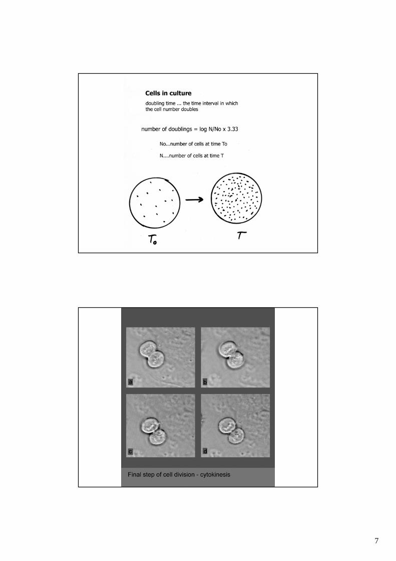
7

8
Ras signalling RAS proteins can be activated constitutively by oncogenic mutations (typically codon 12 in K-ras, codon 13 in K-ras, codon 61 in H-ras), as in cancer cells, or physiologically throughgrowth factors and their receptors (e.g. EGFR = EGF receptor).
Ras proteins activate a checkpoint resulting in G1 cell cycle arrestupon forced ras stimulation, provided all other cell cycle proteins that brake the cycle worknormally (= are not mutated or inactivated). If ras is stimulated in the cell where p16 or p19ARF is inactivated (e.g. by mutation),deregulation of cell cycle occurs, resulting in tumor formation (in mice). Ras is physiologically required for normal cell cycle progression through G1 phase.
The MAPK pathway:encompasses a cascade of phosphorylation events involving three key kinases,namely Raf, MEK and ERK:
RAS signaling (ras: H-ras, K-ras, N-ras – GTP-binding membraneproteins, with GTPase activity), active form: GTP-ras (constitutively active whenmutated, such as in tumors)
RAF = MAP kinase kinase kinase (MAPKKK) MEK = MAP kinase kinase (MAPKK) ERK = MAP kinase (MAPK) protein substrates

9
Activation of growth factor receptor protein tyrosine kinases results inautophosphorylation on tyrosineresidues > PI3K is recruited to the membrane > this leads to allosteric activationof thecatalytic subunit > activation results in production of the second messengerphosphatidylinositol-3,4,5-trisphosphate(PIP3).The lipid product of PI3K, PIP3, recruits a subset of signaling proteins, includingAKT (=PKB). (PTEN, is a PI-3,4,5-P3 phosphatase, which negatively regulates thePI3K/Akt pathway).Once activated, AKT mediates the activation and inhibition of several targets(by phosphorylating them),resulting in cellular survival, growth and proliferation through variousmechanisms.
Activation of AKT kinase by PI 3-kinase
The PI3K–AKT cascade is an anti-apoptotic signalling pathway.
An important downstream effector of PI3Kfunction is the AKT serine/threoninekinase. One of the best-characterizedsubstrates of AKT that is important inpromoting cell survival is the pro-apoptotic BAD protein. BAD interacts withthe anti-apoptotic protein BCL-XL andprevents BCL-XL function, therebyleading to cytochrome c release from themitochondria.

10
Rb- and p53- pathways are deregulated in cancer cells

11
Inserted oncogenes:•Growth factor receptors - One example is epidermal growth factor receptor whichpromotes wound healing by stimulating cell growth. Some factors function astransmembrane protein kinases that are activated by an extracellular signal. An exampleis v-erbB found in the Avian erythroblastosis virus that infects chicken.•Protein kinases - These proteins alter the function of other proteins by phosphorylatingspecific amino acid residues. The v-src from the Rous Sarcoma virus which infectschickens is an example.•G-proteins - These proteins bind the nucleotide GTP, and also exhibit GTPase activity.The v-H-ras oncogene of the Harvey murine sarcoma virus which infects rats is anexample.•Transcription factors - These proteins function by binding to DNA and activatingtranscription. An example is the v-jun oncogene of the Avian sarcoma virus that infectschickens.
RETROVIRAL ONCOGENESIS

12
CANCER GENES - 2004:
•So far, 291 cancer genes have been reported (more than 1% of all the genes inthe human genome).
•90% of cancer genes show somatic mutations in cancer, 20% show germlinemutations and 10% show both.
•The most common mutation class among the known cancer genes is achromosomal translocation that creates a chimeric gene or apposes a gene tothe regulatory elements of another gene.
•Many more cancer genes have been found in leukemias, lymphomas andsarcomas than in other types of cancer, despite the fact that they represent only10% of human cancer. These genes are usually altered by chromosomaltranslocation.
•The most common domain that is encoded by cancer genes is the proteinkinase. Several domains that are involved in DNA binding and transcriptionalregulation are common in proteins that are encoded by cancer genes.
•Futreal et al.: Nature Reviews Cancer 4, 177 -183 (2004)
Oncogenic DNA viruses. Virus: Oncoprotein: Genome size (kb):
SV-40 virus large T Ag 5 Polyomavirus middle T (large T) 5 Adenoviruses (Ad12) E1a (E1b) 35 Papilomaviruses (HPV16) E7, E6 8 ______________________________Hepatitis B virusesHerpesviruses, Epstein-Barr virusPoxviruses

13
Cellular processes disrupted in cancer cells:(Cellular processes that result also in effects on cell cycle) Differentiation:Differentiated cells are usually in the G0 phase of the cell cycle.Terminal differentiation (nerves, muscles) normally does not allowthe re-entry into the cell cycle. Differentiated cells have specific differentiated phenotype which includesmorphology and expression of cell type-specific markers. Differentiation program is often impairedin cancer cells. Senescence:Replicative senescence results in exit from the cell cycle into G0.Again, normally, senescent cells are unable to re-enter the cell cycle.Senescent cells have also specific morphology and express senescent specific markers. Cancer cells areunable to senesce (cancer cells are always immortal). Apoptosis:Apoptosis (programmmed cell death) occurs after the activation of pro-apoptotic genes or inhibition ofanti-apoptotic genes and in most instances it is initiated in G1. The execution of apoptosis is impairedin cancer cells. Cell cycle checkpoints:Cell cycle control, mostly in G1, is deregulated in cancer cells.Also, checkpoints are deregulated in cancer cells.

14
The INK4A locus and proliferation control in human and mouse cells

15
Unphosphorylated Rb blockstranscription
High level of E2F sequestersRb from promoter
Phosphorylation with cdk-cyclincomplexes inactivates Rb
Oncogenic proteins of DNAviruses inactivate Rb
Absence of E2F-1 preventsformation of repressor complex Rb-E2F