cell biology and genetics - nsdl at niscair:...
TRANSCRIPT
1
Cell Biology and Genetics
Cell Division and Genetic Inheritance
Satyawada Rama Rao 1 and Sangeeta Bewal 2
1Department of Bio-Technology & Bio-Informatics
North Eastern Hill University Permanent Campus ,Mawkynroh,
Umshing SHILLONG- 793 022
Meghalaya(India) [email protected]
2 Cytogenetics and Molecular biology Laboratory
Department of Botany Jai Narain Vyas University
JODHPUR-342 005 Rajastahan
2
Introduction
Despite differences between prokaryotes and eukaryotes, there are several common features in their cell division processes like replication of DNA before cell division, distribution/segregation of the genetic material and the cytokinesis which symbolizes the end of the cell division process. However, irrespective of whether the cell is a eukaryotic or prokaryotic in nature, some basic events must precede the cell division. Primarily, there are two kinds of cell divisions taking place in higher organisms. They are:
1. Mitosis / equational division- which is meant for multiplication of cell number.
2. Meiosis / reduction division- which help in reduction of the chromosome number to half and bring in genetic recombinations.
Mitosis is a necessity for maintenance and perpetuation of life as it helps in increasing cell size, surface area as well as the cell volume. It gives rise to two identical daughter cells which are akin to parent cells both qualitatively and quantitatively. In asexual method of reproduction, it is obvious that growth as well as reproduction of organisms includes only mitotic division.
However, during sexual reproduction, the fusion of sex cells (Germ cells: The gametes i.e. sperm and egg cells) occurs. If the gametes have same chromosome number as that of somatic cells (any cell of the body that is not a gamete), the resulting zygote will have double the chromosome number and in subsequent generations, progenies will have multiplied number of chromosomes resulting in raise of ploidy level. To balance the chromosome number, the sex cells undergo a reduction division, in which the chromosome number gets reduced to half (n) from the original somatic chromosome number (2n). Whether the chromosome number is reduced at the time of formation of the sex cells (higher plants and animals) or at any other stage during life cycle (bryophytes and pteridophytes), the resultant condition is referred to as haploid.
Cell Cycle
In higher organisms, growth occurs through mitotic cell division with subsequent enlargement and differentiation of the cells produced. Growth requires an increase in cell mass followed by duplication of genetic material so as to ensure that each daughter cell receives an equal complement of the genetic material leading to perpetuation of the cell line. These steps occur in an orderly progression during the life span of a cell. The sequences of such events are combinedly referred to as of Cell Cycle. In prokaryotes, DNA replication is followed immediately by divisional phase. On contrary, in eukaryotic cells, DNA synthesis and cell division occur in special phases of Cell Cycle. Thus a cell cycle is the sequence of events by which a growing cell duplicates all its components and divides into two daughter cells, each with sufficient machinery and information to repeat the process. The most important components are the cell's chromosomes, which contain linear DNA molecules in association with many proteins. Each DNA molecule must be accurately replicated and the two copies carefully segregated to daughter cells at division.
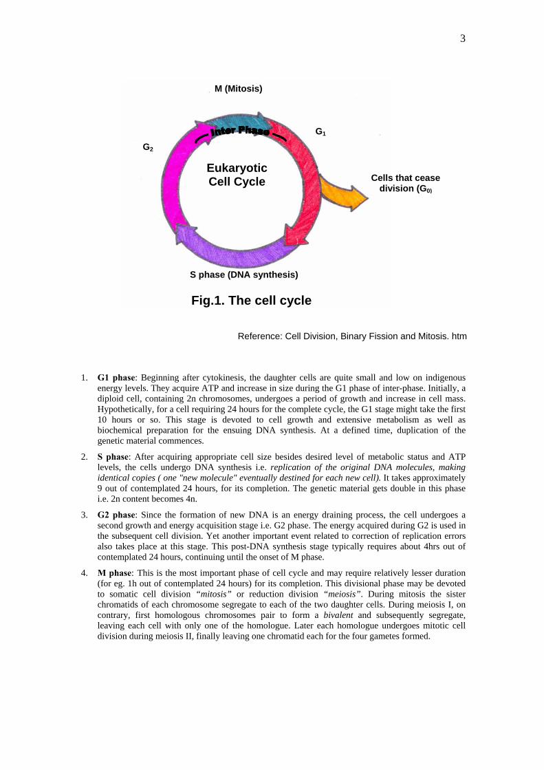
A typical eukaryotic cell cycle includes G1, S, G2 and M phase. The first three phases of cell cycle (G1, S and G2) are collectively known as inter-phase, which is the longest phase of cell cycle. In some cases, an additional phase of cell cycle, known as G0, is refereed to by some workers to denote a condition in which a cell at times might leave the cell cycle, temporarily or permanently. A G0 cell is often called "quiescent", but that is probably more a reflection of the interests of the scientists studying the cell cycle than the cell itself. Many G0 cells are anything but quiescent. They are busy carrying out their normal functions in the organism e.g. secretion, attacking pathogens etc. Often G0 cells are terminally differentiated and will never re-enter the cell cycle. Instead, they will carry out their function in the organism until they die. For other cells, G0 can be followed by reentry into the cell cycle. Most of the lymphocytes in human blood are in G0. However, with proper stimulation, such as encountering the appropriate antigen, they can be stimulated to reenter the cell cycle at G1 and proceed on to new rounds of alternating S phases and mitosis.G0 represents not simply the absence of signals for mitosis but an active repression of the genes needed for mitosis. Cancer cells cannot enter G0 and are destined to repeat the cell cycle infinitely.
3
M (Mitosis)
1. G1 phase: Beginning after cytokinesis, the daughter cells are quite small and low on indigenous energy levels. They acquire ATP and increase in size during the G1 phase of inter-phase. Initially, a diploid cell, containing 2n chromosomes, undergoes a period of growth and increase in cell mass. Hypothetically, for a cell requiring 24 hours for the complete cycle, the G1 stage might take the first 10 hours or so. This stage is devoted to cell growth and extensive metabolism as well as biochemical preparation for the ensuing DNA synthesis. At a defined time, duplication of the genetic material commences.
2. S phase: After acquiring appropriate cell size besides desired level of metabolic status and ATP levels, the cells undergo DNA synthesis i.e. replication of the original DNA molecules, making identical copies ( one "new molecule" eventually destined for each new cell). It takes approximately 9 out of contemplated 24 hours, for its completion. The genetic material gets double in this phase i.e. 2n content becomes 4n.
3. G2 phase: Since the formation of new DNA is an energy draining process, the cell undergoes a second growth and energy acquisition stage i.e. G2 phase. The energy acquired during G2 is used in the subsequent cell division. Yet another important event related to correction of replication errors also takes place at this stage. This post-DNA synthesis stage typically requires about 4hrs out of contemplated 24 hours, continuing until the onset of M phase.
4. M phase: This is the most important phase of cell cycle and may require relatively lesser duration (for eg. 1h out of contemplated 24 hours) for its completion. This divisional phase may be devoted to somatic cell division “mitosis” or reduction division “meiosis”. During mitosis the sister chromatids of each chromosome segregate to each of the two daughter cells. During meiosis I, on contrary, first homologous chromosomes pair to form a bivalent and subsequently segregate, leaving each cell with only one of the homologue. Later each homologue undergoes mitotic cell division during meiosis II, finally leaving one chromatid each for the four gametes formed.
G1
Cells that cease
division (G0)
Eukaryotic Cell Cycle
G2
S phase (DNA synthesis)
Fig.1. The cell cycle
Reference: Cell Division, Binary Fission and Mitosis. htm

4
Molecular Regulation of Cell Cycle
In G1, chromosomes are unreplicated and the cell is non-committed to the replication division process. During the transitional phase from G1 to S phase, often referred to as START (check point), a cell confirms to that internal and external conditions are favorable for a new round of DNA synthesis and division, and commits itself to the process and the decision is irreversible. Once DNA synthesis commences, cell cycle continues uninterrupted till the completion of the cell cycle. During the process of DNA replication, sister chromatids are tethered together by specific proteins, called cohesins. Cell cycle events are controlled by a network of molecular signals, whose central components are cyclins and cyclin-dependent protein kinases (Cdks). At G1 phase, Cdk activity is low since cyclin proteins are rapidly degraded. At START (Fig.2), cyclin degradation is inhibited, and their synthesis is initiated causing a dramatic rise in Cdk activity, which persists throughout S, G2 and M phases. High Cdk activity is essential for DNA replication, chromosome condensation, and spindle assembly. At finish, a group of proteins, making up the anaphase-promoting complex (APC) are activated1. The APC consists of a core complex of about a dozen polypeptides.
Major types of proteins that govern a eukaryotic cell cycle are:
1. Cyclin dependent kinases (Cdks):
a. G1 Cdk (Cdk4)
b. S-phase Cdk ((Cdk2)
c. M-phase Cdk (Cdk1)
The levels of these Cdks in the dividing cell remain fairly stable, but each must bind to the appropriate cyclin (whose levels keep fluctuating) for activation. They add phosphate groups to a variety of protein substrates that control processes in the cell cycle.
2. Cyclins: Some of the important cyclins identified are:
a. G1 cyclin (cyclin D)
b. S-phase cyclins (cyclins E and A)
c. Mitotic cyclins (cyclins B and A)
Their levels in the cell rise and fall with the stages of the cell cycle. These are accumulated at G2 of interphase and are rapidly destroyed at the end of cell division2. Cyclin gene homologous was also reported in various plats viz. carrot, soybean, alfalfa etc.
3. S-phase promoting factor (SPF): This includes Cyclin A bound to Cdk2, which enters the nucleus and prepares the cell to duplicate its DNA and its centrosomes

5
= Cyclin
= Cdk
APC G0G1 cyclin
4. MPF (Maturation promoting factor or M-Phase promoting factor): It is a protein complex comprising p34 kinases and cyclins that regulate meiotic maturation as reported in Xenopus oocytes7. It plays an important role in regulation of cell cycle. MPF initiate the activation of a cascade of protein kinases to act directly on cellular components leading to:
• chromosome condensation • nuclear envelop breakdown • cytoskeletal rearrangement
Activation of MPF is essential for entry into mitosis and progress through metaphase. p34 kinase in S. prombe is coded by cdc2 gene6 and by cdc28 gene in S. cervisiae3. Since then several genes homologous to cdc’s have been isolated from divergent organisms such as Xenopus, Drosophila, chickens, mice, humans besides a number of higher plants. p34 kinases plays a catalytic function in MPF whereas cyclins are regulatory subunits.
5. APC (Anaphase promoting complex or cyclosome): The APC triggers the events leading to destruction of the cohesins thus allowing the sister chromatids to separate and degrades the mitotic cyclin B.
6. p53: It plays an important role in cell cycle control and apoptosis. Defective p53 could allow abnormal cells to proliferate, resulting in cancerous growth. In normal cells, the p53 protein level is low in quantum, but DNA damage and other stress signals may trigger their increase. These proteins are supposedly have three major functions viz. growth arrest, DNA repair and apoptosis.
G1 CdkM G1
M-phase Promoting Factor
Degraded cyclins
START Checkpoint S-phase Promoting Factor
G2 S Degraded cyclins
Fig. 2. Interaction of various cyclins and Cdks during eukaryotic cell cycle
(Reference: The cell cycle. htm)
6
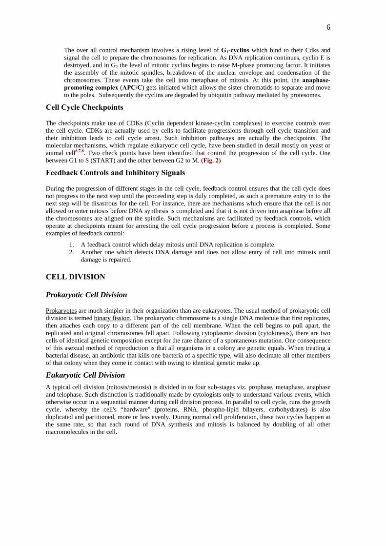
The over all control mechanism involves a rising level of G1-cyclins which bind to their Cdks and signal the cell to prepare the chromosomes for replication. As DNA replication continues, cyclin E is destroyed, and in G2 the level of mitotic cyclins begins to raise M-phase promoting factor. It initiates the assembly of the mitotic spindles, breakdown of the nuclear envelope and condensation of the chromosomes. These events take the cell into metaphase of mitosis. At this point, the anaphase-promoting complex (APC/C) gets initiated which allows the sister chromatids to separate and move to the poles. Subsequently the cyclins are degraded by ubiquitin pathway mediated by protesomes.
Cell Cycle Checkpoints
The checkpoints make use of CDKs (Cyclin dependent kinase-cyclin complexes) to exercise controls over the cell cycle. CDKs are actually used by cells to facilitate progressions through cell cycle transition and their inhibition leads to cell cycle arrest. Such inhibition pathways are actually the checkpoints. The molecular mechanisms, which regulate eukaryotic cell cycle, have been studied in detail mostly on yeast or animal cell6,7,8. Two check points have been identified that control the progression of the cell cycle. One between G1 to S (START) and the other between G2 to M. (Fig. 2)
Feedback Controls and Inhibitory Signals
During the progression of different stages in the cell cycle, feedback control ensures that the cell cycle does not progress to the next step until the proceeding step is duly completed, as such a premature entry in to the next step will be disastrous for the cell. For instance, there are mechanisms which ensure that the cell is not allowed to enter mitosis before DNA synthesis is completed and that it is not driven into anaphase before all the chromosomes are aligned on the spindle. Such mechanisms are facilitated by feedback controls, which operate at checkpoints meant for arresting the cell cycle progression before a process is completed. Some examples of feedback control:
1. A feedback control which delay mitosis until DNA replication is complete. 2. Another one which detects DNA damage and does not allow entry of cell into mitosis until
damage is repaired. CELL DIVISION Prokaryotic Cell Division
Prokaryotes are much simpler in their organization than are eukaryotes. The usual method of prokaryotic cell division is termed binary fission. The prokaryotic chromosome is a single DNA molecule that first replicates, then attaches each copy to a different part of the cell membrane. When the cell begins to pull apart, the replicated and original chromosomes fell apart. Following cytoplasmic division (cytokinesis), there are two cells of identical genetic composition except for the rare chance of a spontaneous mutation. One consequence of this asexual method of reproduction is that all organisms in a colony are genetic equals. When treating a bacterial disease, an antibiotic that kills one bacteria of a specific type, will also decimate all other members of that colony when they come in contact with owing to identical genetic make up.
Eukaryotic Cell Division A typical cell division (mitosis/meiosis) is divided in to four sub-stages viz. prophase, metaphase, anaphase and telophase. Such distinction is traditionally made by cytologists only to understand various events, which otherwise occur in a sequential manner during cell division process. In parallel to cell cycle, runs the growth cycle, whereby the cell's “hardware” (proteins, RNA, phospho-lipid bilayers, carbohydrates) is also duplicated and partitioned, more or less evenly. During normal cell proliferation, these two cycles happen at the same rate, so that each round of DNA synthesis and mitosis is balanced by doubling of all other macromolecules in the cell.
7
Mitosis:
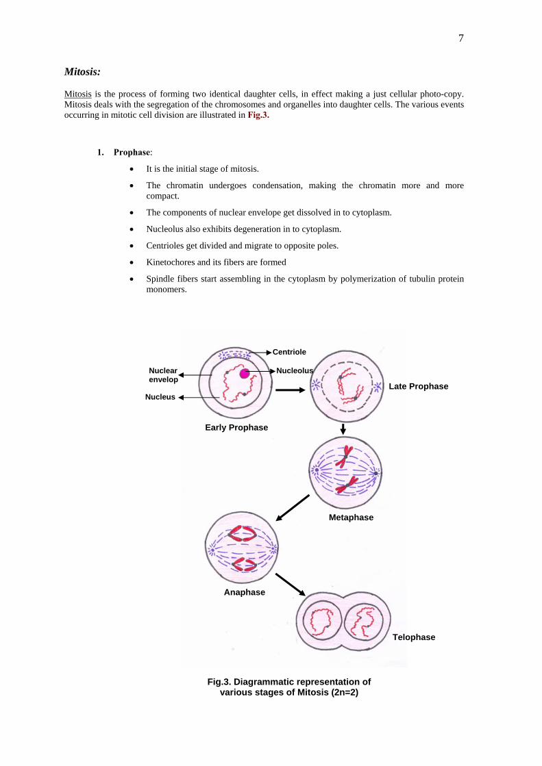
Mitosis is the process of forming two identical daughter cells, in effect making a just cellular photo-copy. Mitosis deals with the segregation of the chromosomes and organelles into daughter cells. The various events occurring in mitotic cell division are illustrated in Fig.3.
1. Prophase:
• It is the initial stage of mitosis.
• The chromatin undergoes condensation, making the chromatin more and more compact.
• The components of nuclear envelope get dissolved in to cytoplasm.
• Nucleolus also exhibits degeneration in to cytoplasm.
• Centrioles get divided and migrate to opposite poles.
• Kinetochores and its fibers are formed
• Spindle fibers start assembling in the cytoplasm by polymerization of tubulin protein monomers.
Centriole
Early Prophase
Late Prophase
Metaphase
Anaphase
Telophase
Nuclear envelop
Nucleolus
Nucleus
Fig.3. Diagrammatic representation ofvarious stages of Mitosis (2n=2)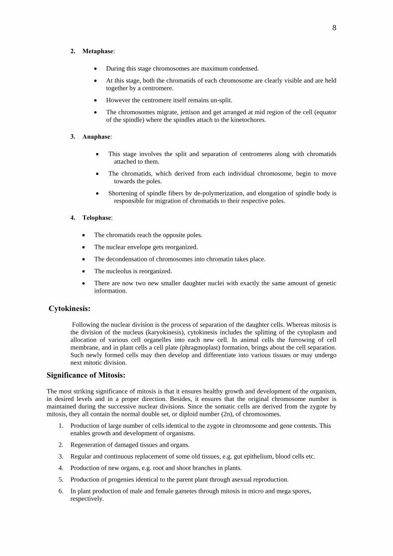
8
2. Metaphase:
ng this stage chromosomes are maximum condensed.
• clearly visible and are held
ged at mid region of the cell (equator netochores.
3. Anapha
is stage involves the split and separation of centromeres along with chromatids attached to them.
ration of chromatids to their respective poles.
4. Telophase:
matids reach the opposite poles.
•
• hromatin takes place.
er daughter nuclei with exactly the same amount of genetic
Cytokinesis:
g the nuclear division is the process of separation of the daughter cells. Whereas mitosis is of the nucleus (karyokinesis), cytokinesis includes the splitting of the cytoplasm and
Signifi
itosis is that it ensures healthy growth and development of the organism, oper direction. Besides, it ensures that the original chromosome number is
3. d tissues, e.g. gut epithelium, blood cells etc.
ores,
• Duri
At this stage, both the chromatids of each chromosome are together by a centromere.
• However the centromere itself remains un-split.
• The chromosomes migrate, jettison and get arranof the spindle) where the spindles attach to the ki
se:
• Th
• The chromatids, which derived from each individual chromosome, begin to move towards the poles.
• Shortening of spindle fibers by de-polymerization, and elongation of spindle body is responsible for mig
• The chro
The nuclear envelope gets reorganized.
The decondensation of chromosomes into c
• The nucleolus is reorganized.
• There are now two new smallinformation.
Followinthe divisionallocation of various cell organelles into each new cell. In animal cells the furrowing of cell membrane, and in plant cells a cell plate (phragmoplast) formation, brings about the cell separation. Such newly formed cells may then develop and differentiate into various tissues or may undergo next mitotic division.
cance of Mitosis:
The most striking significance of min desired levels and in a prmaintained during the successive nuclear divisions. Since the somatic cells are derived from the zygote by mitosis, they all contain the normal double set, or diploid number (2n), of chromosomes.
1. Production of large number of cells identical to the zygote in chromosome and gene contents. This enables growth and development of organisms.
2. Regeneration of damaged tissues and organs.
Regular and continuous replacement of some ol
4. Production of new organs, e.g. root and shoot branches in plants.
5. Production of progenies identical to the parent plant through asexual reproduction.
6. In plant production of male and female gametes through mitosis in micro and mega sprespectively.

9
Meiosi
exual reproduction is the characteristic feature of the eukaryotes. During the formation of gametes, the chromosomes is reduced by half, and returned to the normal diploid number when the two
gametes fuse during fertilization. Thus, meiosis is a special type of cell division found in organisms in which
o mely meiosis I (reduction division) and meiosis II (an equational division). The
sequence of events taking place in meiosis is as follows:
anaphase I and telophase I (Figs.4 & 5).
vents like synapsis of homologues; genetic recombination through crossing over takes place here. It is divided into five sub stages viz. leptotene, zygotene, pachytene, diplotene, and diakinesis for understanding
stranded. The chromosomes show chromomeres, which are darkly
(b)
of a special structure called synaptonemal complex. The
(c)
al complex is maintained through pachytene and is also responsible for
(d)
s
Snumber of
there is sexual reproduction. Meiosis is the mechanism that prevents the doubling of chromosomal set in successive generations. By a combination of two divisions, the number of chromosomes is reduced by half, giving rise to four haploid cells. Another essential feature of meiosis is genetic recombination by way of exchange of chromosome segments (crossing over) and random assortment of homologous chromosomes.
Stages of Meiosis
Meiosis is a prolonged process in comparison to mitosis, which is rather brief. Meiosis is divided into twconsecutive phases na
Meiosis I:
This division is characterized by prophase I, metaphase I,
1. Prophase I: This is the most significant stage of meiotic process since many important e
various events more clearly.
(a) Leptotene: It is a phase in which the metabolically active cell and nucleus with increased size and chromosomes become more clearly visible. In this stage chromosomes look single rather than double stained chromatin bodies.
Zygotene: The homologous chromosomes become aligned and undergo pairing in a process called synapsis of the chromosome forming ‘bivalents’. The pairing process involves the formation synaptonemal complex is considered the structural basis for pairing and synapsis of homolougues.
Pachytene: The chromosomes in a bivalent get longitudinally split and sister chromatids of each homolougue are clearly visible. Thus each unit of bivalent appears in a tetrad form. The synaptonemconsequent crossing over.( The term crossing over refers to the cytological phenomenon involving exchange of corresponding segments of nonsister chromatids of a pair of homologous chromosomes by breakage and reunion of following synapsis). The recombinational nodules present on the synaptenemal complex which contain the required enzyme machinery for exchange of chromosomal segments, are responsible for genetic crossing over. Diplotene: At diplotene, the nuclear envelope and the nucleolus start degenerating into the cytoplasm. The intimately paired chromosomes repel each other and begin to fall apart. However, this separation is not complete because the homologous chromosomes remain united by their points of interchange called “chiasmata”. Chiasmata are generally regarded as the sites where the phenomenon of crossing over or recombination has taken place. The chiasma may be intercalary or terminal in position over bivalents.

10
Nucleolus
Leptotene Zygotene
Synaptonemal complex
Pachytene Diplotene
Diakinesis
Fig.4. Diagrammatic representation of
Prophase I of Meiosis I (2n=4)
(e) Diakinesis: The nuclear envelope and the nucleolus completely degenerate and are no more visible. The chiasma starts sliding towards the terminal region of bivalents and the process is called “terminalization of chiasma”. At diakinesis the bivalents are more or less evenly distributed. Even at this stage the homologues are held together by the terminalized chiasma.
11
Metaphase I Anaphase I
Telophase I
Meiosis I
Meiosis II
Metaphase II
12
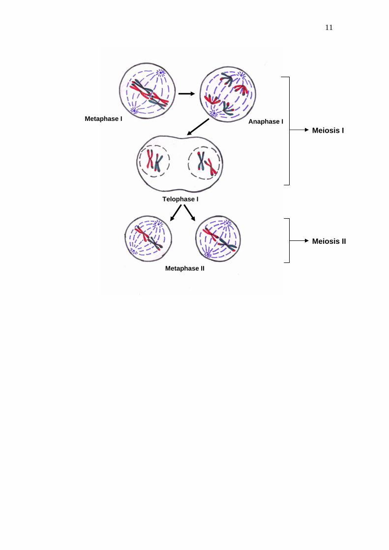
Fig.5. Metaphase I, anaphase I, Telophase I and meiosis II
Anaphase II
Meiosis II
Telophase II
Four haploid daughter cells
2. Metaphase I: This stage is characterized the maximum condensation of bivalents and their orderly arrangement at metaphase plate or at mid region of the cell (i.e. equator of the spindle). The spindle fibers help in this arrangement through their attachment to the kinetochores of the partner chromosomes in bivalents. The homologues are still attached by the terminal chiasma while the kinetochores are being pulled towards the poles (Fig. 5)
3. Anaphase I: The homologues of each bivalent, move towards opposite poles of the cell, leaving one chromosome (with un-split centromere), of each pair at the poles, there by reducing the somatic chromosome number to half (Fig. 5).
4. Telophase I: When the anaphase groups arrive at their respective poles, a short telophase I follows. In fact, at telophase I chromosomes may remain in a condensed state for some time. Generally cytokinesis does not take place after telophase I. However these two daughter nuclei directly proceed to second meiotic division. (Fig. 5).
Meiosis II:
Meiosis II is similar to the mitotic division including prophase II, metaphase II, anaphase II and telophase II (Fig. 5).
5. Prophase II: It is very short phase during which no chromosomal duplication occurs. Formation of spindle marks the end of this phase and the beginning of metaphase II .
6. Metaphase II. Individual chromosomes get arranged at mid region of the cell (equator of the spindle) with the help of spindle fibers and the sister chromatids get ready for separation.
7. Anaphase II: The sister chromatids of each chromosome separate and move towards the opposite poles with the help of spindle apparatus. The main difference between anaphase I and anaphase II is
13
that during anaphase I bivalents get separated and centromeres remain intact whereas in anaphase II, the centromeres split and the sister chromatids are segregated into opposite sides of the cells
8. Telophase II: It is identical to the telophase of mitosis. The nuclear envelope gets reorganized, and the de-condensation of chromosomes into chromatin takes place. Meanwhile the nucleolus also gets reorganized. There are now four new smaller daughter nuclei per cell. Each of the four nuclei formed at telophase II has a haploid number of chromosomes.
9. Cytokinesis : Following the meiotic karyokinesis, the four haploid nuclei that are newly formed will now develop in to gametes through separation of cytoplasm and cell membrane and/or cell wall formation as discussed earlier in the context of mitotic cell division.
Significance of Meiosis
Meiosis not only results in halving the chromosome number but also segregates each member of the homologous pair/ bivalent into four different nuclei. The process of crossing over results in the exchange of genetic material by the homologous partners. The segregation of the homologues and recombination has important genetic consequences. From this point of view, meiosis is a mechanism for distributing the genes between the gametes which permits their recombination and random segregation. As a result of the crossing over, each chromosome does not consist solely of maternal or paternal material but of alternating segments of each. The random assortment of the chromatids results in the formation of four gametes having different genetic constitution which ultimately results in genetic variation among population of the organism. Meiosis is the mechanism by which genetic variation is brought about.
A comparison between Mitosis and Meiosis
Mitosis and meiosis are two different patterns of chromosomal division within the cells in life cycle of an organism, however both the divisions are different in many aspects. Mitosis occurs in all somatic or vegetative cells of an individual, but meiosis is limited to the germinal cells. Mitosis includes one cell division resulting in two daughter cells with diploid number of chromosomes, whereas meiosis includes the two successive divisions resulting in four daughter cells with haploid number of chromosomes from the original diploid set. In mitosis DNA synthesis occurs in the S phase followed by extended G2 phase before the onset of division, however the G2 phase is short or almost nonexistent in meiosis. This is because pre-meiotic DNA synthesis is relatively longer than that of mitosis and followed immediately by meiosis. Another aspect which significantly differentiates mitosis and meiosis is behavior of chromosomes, in mitosis every chromosome behaves independently whereas in meiosis the homologous chromosomes become mechanically related during the first meiotic division i.e. chromosomal pairing during prophase I. A fundamental difference between the two types of cell divisions is that in mitosis the genetic material remains constant with only rare mutations or chromosomal aberrations, while genetic variability is one of the main consequences of meiosis.
References:
1. Bayliss, M. W (1985). The cell division cycle in plants. Bryant, J.A. & Francis (ed.), pp157-187. Cambridge University Press, New York.
2. Evans, T., Rosenthal, E., Youngbloom, J.,Distel, D. and Hunt, J. (1983), Cell 33:389-396. 3. Hartwell, L.M. and Wienert, T.A. (1989). Science 246 : 629-634 4. Jacobs, T. (1992). Dev.Biol. 153: 1-5. 5. Masui, Y and Markert, C.L. (1971). Exp.Zool.177: 129-146. 6. Murray, A.W. (1992). Nature 359: 388-393. 7. Nurse, P. and Bisset, Y. (1981). Nature 292 : 558-560. 8. Reed, S.I., Hadwiger, J.A. and Lorinez, A.T.(1985). Proc. Natl. Acad.Sci.,(USA) 82:4055-4057.
14
Genetic Inheritance
Genetics is the study of heredity and variation. The term genetics was used for the first time by W. Bateson in 1905. Heredity includes those traits or characteristics which are transmitted from one generation to generation with or with out changes. Variations are mainly of two types namely hereditary and non-hereditary. A hereditary variation refers to differences in inherited traits and such variation is found not only in progenies of different parents but also among progeny from the same parents. Identical twins, however, are an exception. Non-hereditary variations are those which are mainly due to environment.
Genetic inheritance has been of immense interest to human civilization and efforts to understand how characters are transmitted from one generation to the other were made by several early biologists (Pre- Mendelian Genetics). Some of them are described in brief here:
1. W. Harvey (1578-1657) speculated that all animals arise from eggs and that semen plays only a vitalizing role.
2. R. de Graaf (1641-1673) observed that the progeny would have characteristics of father as well as mother and therefore suggested that both the parents should be contributing to heredity. He also studied the development of embryo to some extent.
3. A. V. Leeuwenhoek observed sperms of several animals in 1677 and also suggested their association with eggs for reproduction.
4. Reproductive parts of plants were described for the first time by N. Grew in 1682.
5. R. Camerarius in 1694 described sexual reproduction in plants for the first time. He is also known to be the first to produce a hybrid between two different plant species.
6. It was, however, in 1717 that T. Fairchild produced a hybrid having characters of both parents. This hybrid was called “Fairchild Sweet William” or “Fairchild’s mule”.
Pre-formation and Epigenesis
1. In 1679, J. Swammerdam studied insects and suggested that development of an organism is a simple enlargement of a minute but preformed individuals that were called Homunculus.
2. K. W. Wolff (1738-1794) proposed that neither egg nor sperm had a structure like homunculus but the gametes contained undifferentiated living substance capable of forming the organized body after fertilization. Such an idea was called the theory of Epigenesis.
Pangenesis and Acquired characters
1. J. B. Lamarck (1744-1829) postulated that characters which are acquired during the lifetime of an individual are inherited. This concept is known as “Lamarckism” or “Theory of Inheritance of Acquired Characters”.
2. Charles Darwin (1809-1882) tried to suggest that the physical basis of heredity and suggested that every part of body produced very small invisible bodies called gemmules or pangenes, which are transported through the blood stream to the sex organs and are assemble there into gametes. During fertilization gemmules from both parents are brought together for redistribution to different organs during development, thus determining different characters.
Germplasm Theory
A. Weismann (1834-1914) demonstrated that pangenesis could not be practically verified. He proved it by cutting the tail of mice for 22 generations but complete tail structure was still inherited in next generation. According to germplasm theory, to account for heredity, the body of an individual can be divided
15
into two types of tissues, germplasm and somatoplasm. Changes in somatoplasm does not inherit while those occur in germplasm does transmit from one generation to the other. Mendel’s Laws of Inheritance
Gregor Johann Mendel (1822-1884) is popularly known as the “Father of Genetics”. With the help of his experiments on garden pea, he was able to formulate laws, which explain the pattern of inheritance of characters. The transmission of characters is known to be governed by the structure and behavior of the chromosomes during meiosis. However, in 1865 when Gregor Mendel discovered the fundamental laws of inheritance, nothing was known about the chromosomes and/or meiosis. His discovery was based on exceptional abstract thinking supported by precise quantitative analysis of experimental crosses and their progeny.
Mendel selected pea (Pisum sativum; Papilionaceae) as his experimental plant. This plant completes its life cycle in short duration and is amenable for both self or cross pollinations. Most important feature of this plant is that it exhibits several pairs of contrasting characters for study. Mendel crossed pea plants with pairs of differential or contrasting characteristics. By crossing plants of parental generation (P1&P2), he observed the resulting hybrids in the first filial generation (F1). Then he crossed the hybrids (F1s) among themselves and studied their progeny in the second filial generation (F2).
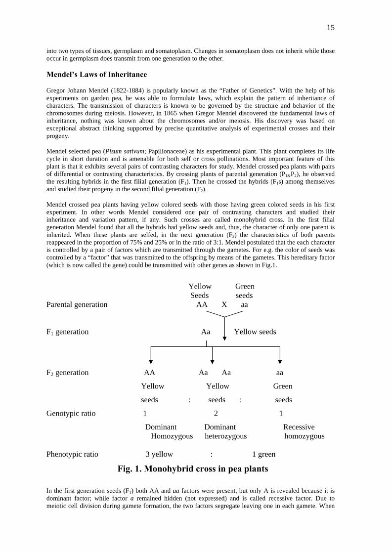
Mendel crossed pea plants having yellow colored seeds with those having green colored seeds in his first experiment. In other words Mendel considered one pair of contrasting characters and studied their inheritance and variation pattern, if any. Such crosses are called monohybrid cross. In the first filial generation Mendel found that all the hybrids had yellow seeds and, thus, the character of only one parent is inherited. When these plants are selfed, in the next generation (F2) the characteristics of both parents reappeared in the proportion of 75% and 25% or in the ratio of 3:1. Mendel postulated that the each character is controlled by a pair of factors which are transmitted through the gametes. For e.g. the color of seeds was controlled by a “factor” that was transmitted to the offspring by means of the gametes. This hereditary factor (which is now called the gene) could be transmitted with other genes as shown in Fig.1.
Yellow Green Seeds seeds Parental generation AA X aa
F1 generation Aa Yellow seeds
F2 generation AA Aa Aa aa
Yellow Yellow Green
seeds : seeds : seeds
Genotypic ratio 1 2 1
Dominant Dominant Recessive Homozygous heterozygous homozygous
Phenotypic ratio 3 yellow : 1 green
Fig. 1. Monohybrid cross in pea plants
In the first generation seeds (F1) both AA and aa factors were present, but only A is revealed because it is dominant factor; while factor a remained hidden (not expressed) and is called recessive factor. Due to meiotic cell division during gamete formation, the two factors segregate leaving one in each gamete. When
16
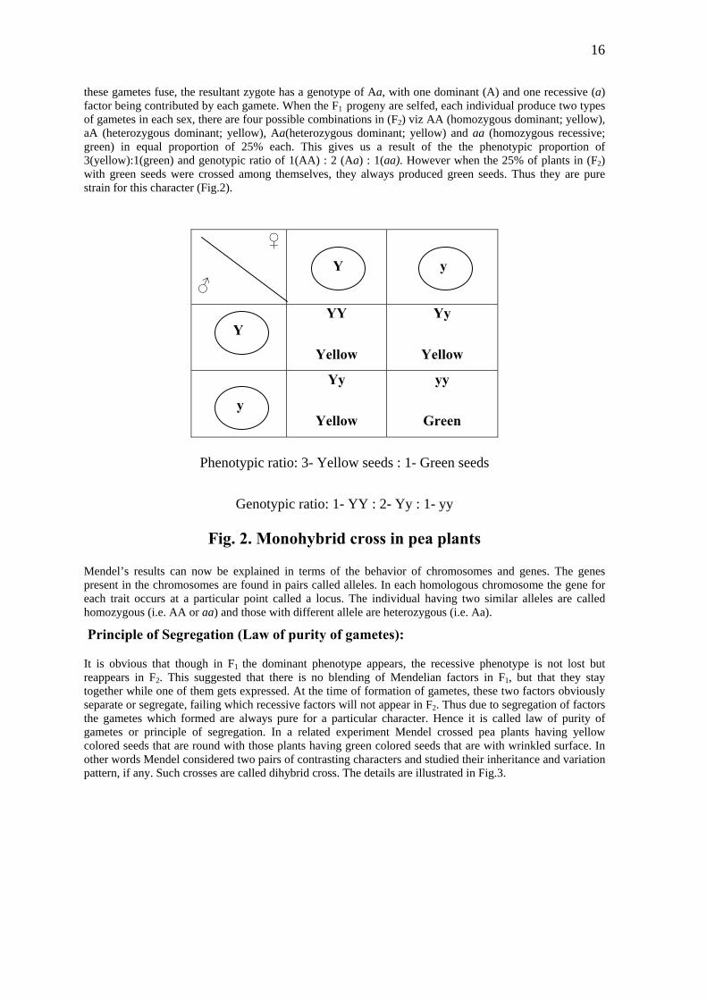
these gametes fuse, the resultant zygote has a genotype of Aa, with one dominant (A) and one recessive (a) factor being contributed by each gamete. When the F1 progeny are selfed, each individual produce two types of gametes in each sex, there are four possible combinations in (F2) viz AA (homozygous dominant; yellow), aA (heterozygous dominant; yellow), Aa(heterozygous dominant; yellow) and aa (homozygous recessive; green) in equal proportion of 25% each. This gives us a result of the the phenotypic proportion of 3(yellow):1(green) and genotypic ratio of 1(AA) : 2 (Aa) : 1(aa). However when the 25% of plants in (F2) with green seeds were crossed among themselves, they always produced green seeds. Thus they are pure strain for this character (Fig.2).
♀
♂
YY
Yellow
Yy
Yellow
Yy
Yellow
yy
Green
Y y
Y
y
Phenotypic ratio: 3- Yellow seeds : 1- Green seeds
Genotypic ratio: 1- YY : 2- Yy : 1- yy
Fig. 2. Monohybrid cross in pea plants
Mendel’s results can now be explained in terms of the behavior of chromosomes and genes. The genes present in the chromosomes are found in pairs called alleles. In each homologous chromosome the gene for each trait occurs at a particular point called a locus. The individual having two similar alleles are called homozygous (i.e. AA or aa) and those with different allele are heterozygous (i.e. Aa).
Principle of Segregation (Law of purity of gametes):
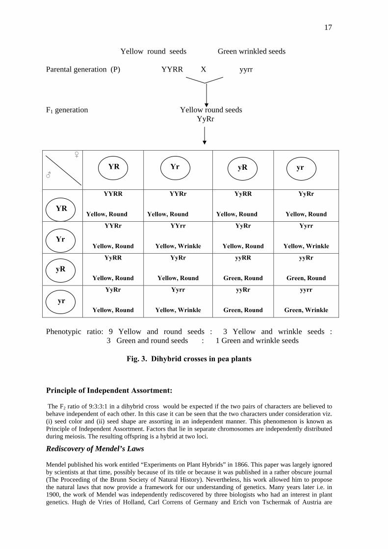
It is obvious that though in F1 the dominant phenotype appears, the recessive phenotype is not lost but reappears in F2. This suggested that there is no blending of Mendelian factors in F1, but that they stay together while one of them gets expressed. At the time of formation of gametes, these two factors obviously separate or segregate, failing which recessive factors will not appear in F2. Thus due to segregation of factors the gametes which formed are always pure for a particular character. Hence it is called law of purity of gametes or principle of segregation. In a related experiment Mendel crossed pea plants having yellow colored seeds that are round with those plants having green colored seeds that are with wrinkled surface. In other words Mendel considered two pairs of contrasting characters and studied their inheritance and variation pattern, if any. Such crosses are called dihybrid cross. The details are illustrated in Fig.3.
17
Yellow round seeds Green wrinkled seeds Parental generation (P) YYRR X yyrr
F1 generation Yellow round seeds YyRr
♀
♂
YYRR
Yellow, Round
YYRr
Yellow, Round
YyRR
Yellow, Round
YyRr
Yellow, Round
YYRr
Yellow, Round
YYrr
Yellow, Wrinkle
YyRr
Yellow, Round
Yyrr
Yellow, Wrinkle
YyRR
Yellow, Round
YyRr
Yellow, Round
yyRR
Green, Round
yyRr
Green, Round
YyRr
Yellow, Round
Yyrr
Yellow, Wrinkle
yyRr
Green, Round
yyrr
Green, Wrinkle
yR yr YR Yr
YR
Yr
yR
yr
Phenotypic ratio: 9 Yellow and round seeds : 3 Yellow and wrinkle seeds : 3 Green and round seeds : 1 Green and wrinkle seeds
Fig. 3. Dihybrid crosses in pea plants
Principle of Independent Assortment:
The F2 ratio of 9:3:3:1 in a dihybrid cross would be expected if the two pairs of characters are believed to behave independent of each other. In this case it can be seen that the two characters under consideration viz. (i) seed color and (ii) seed shape are assorting in an independent manner. This phenomenon is known as Principle of Independent Assortment. Factors that lie in separate chromosomes are independently distributed during meiosis. The resulting offspring is a hybrid at two loci.
Rediscovery of Mendel’s Laws
Mendel published his work entitled “Experiments on Plant Hybrids” in 1866. This paper was largely ignored by scientists at that time, possibly because of its title or because it was published in a rather obscure journal (The Proceeding of the Brunn Society of Natural History). Nevertheless, his work allowed him to propose the natural laws that now provide a framework for our understanding of genetics. Many years later i.e. in 1900, the work of Mendel was independently rediscovered by three biologists who had an interest in plant genetics. Hugh de Vries of Holland, Carl Correns of Germany and Erich von Tschermak of Austria are

18
credited with the rediscovery Mendelism. Within a few years, the impact of Mendel’s studies, experiments and their significance in the field of genetics was felt around the world.
Deviations to Mendel’s laws During meiosis there is a random distribution of the chromosomes which leads to the segregation of the
Usually a single gene controls one character and the alleles of a pair are related as dominant and cessive
(A) Interallelic or Intra-allelic gene interaction
Incomplete dominance (1:2:1): F1 hybrids were not related to either of the parents but exhibited a blending
generation Pink
F2 generation: rr
e
Phenotypic ratio
1. Co-dominance (1:2:1): In co-dominance, both the factors of an allelomorphic pair express
genes in the gametes. When this type of study was carried out in the fruit fly Drosophila melanogaster by Morgan and collaborators (1910-1915), however, it become evident that the law of independent assortment was not universally applicable and that in certain crosses where two or more allelic pairs control a character, free segregation was limited. After studying a considerable number of different crosses in Drosophila, Morgan reached the conclusion that all genes of this fly were clustered into four linkage groups corresponding to the four pairs of chromosomes. re . But various exceptions have been noticed to these facts. In some cases more than one pair of genes may influence the same character. These may interact in different ways by adding, subtracting or modifying the characters, or may inhibit or reverse the effect of another pair of genes. This concept was introduced by Bateson and is called as “Bateson factor Hypothesis”.
of characters. It means that two factors in a allelomorphic pair are not conventionally related as dominant and recessive factors. Rather the dominant gene in heterozygous condition has reduced expression, so that each of them expresses itself partially. This is called incomplete dominance or blending inheritance e.g. Mirabilis jalapa (Pink colored flowers). This phenomenon was discovered during the study of inheritance of flower colour in Mirabilis jalapa (the four –o- clock plant). This plant has two varieties one red flower and the other white flowers. A cross between two varieties produces a progeny (F1) with pink flowered plants. The selfing among F1 – pink flowered plant F2 progeny having three phenotypes viz. red flowered, white flowered and pink flowered plants. On counting, the phenotypic ratio between red, pink and white flowered plants. On counting the phenotypic ratio between red, pink and white flowered plants is found to be 1:2:1. A comparison of this with Mendelian 3:1 ratio due to dominance, reveals that the third phenotype is due to heterozygous nature of 50% of the F2 individuals. Since the dominant gene (R) is not able to mask its alleles ‘r’ for white coloured flowers completely, the phenomenon is called partial or incomplete dominance.
Pa rents: Red X White Parental generation (P) RR X rr
F1 Rr
RR Rr
Red Pink Whit
1 2 1
themselves equally in F1 hybrids. It means a heterozygous for codominant genes exhibits both the characters side by side. These follow the law of segregation and F2 progeny exhibits 1:2:1 ratio both in genotype and phenotypes.

19
(B) Non-allelic or inter-genic interaction entary genes are two pairs of non-allelic dominant
W. Bateson and R. C. Punnett crossed two white flowered varieties of Sweet pea (Lathyrus
Summary:
Red flowered plants with both complementary genes C and R=9, white flowered plants having either ‘C’ or
From the checker board it is clear that the red colouration of the flower is due to interaction of two
X White flowered Sweet pea
RRcc
F1 generation Red flowered plants
Gametes of F1 generation: CR,Cr,cR,cr
2. Complementary factors (9:7): The complemgenes (i.e. present on separate gene loci), which interact to produce only one phenotypic trait but neither of them if present alone can not produces the phenotypic trait. It means that for the development of the dominant character in question, both the complementary genes must be represented at least by their one dominant allele. Absence of even one of the two genes produces recessive phenotype.
odoratus). In the experiment all the plants of F1 generation developed red flowers instead of expected white flowers. These F1 plants, on self pollination, produced red and white flowered plants in the ratio 9:7 in F2 generation. . Bateson explained his experiment in the following way:
‘R’ =7.
complementary factors C and R. The parents developed white flowers because they lacked either gene ‘C’ or gene ‘R’ in their genotypic expressions CCcc and RRrr. F1 hybrids received both the complementary genes C and R, hence all the flowers in that generation were red. The ratio 9:7 obtained in F2 generation is actually a modification of normal dihybrid ratio (9:3:3:1).
Pa rents: White flowered Sweet pea Parental generation (P) CCrr X
CcRr
F2 generation:
♀
♂
CCRR
Red
CC
Red
CcRR
Red
CcRr
Red
Rr
C
Red White Red White
CRr CCrr CcRr Ccrr
C
Red Red White White
cRR CcRr ccRR ccRr
CcRr
Red White White White
Ccrr ccRr ccrr
CR Cr cR cr
Cr
cR
c
CR
r
20
Red flowered plants with both complementary genes C and R = 9, White flowered plants having either ‘C’ or ‘R’ = 7. 3. Epistasis: Epistasis is the interaction between non-allelic factors in which one gene masks, inhibits
or suppresses the expression of other gene. The gene that suppresses the other gene is known as inhibiting or epistatic factor and one which is prevented from exhibiting itself is known as hypostatic factor.
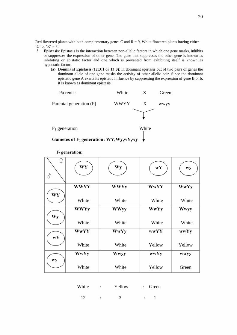
(a) Dominant Epistasis (12:3:1 or 13:3): In dominant epistasis out of two pairs of genes the dominant allele of one gene masks the activity of other allelic pair. Since the dominant epistatic gene A exerts its epistatic influence by suppressing the expression of gene B or b, it is known as dominant epistasis.
Pa rents: White X Green Parental generation (P) WWYY X wwyy
F1 generation White Gametes of F1 generation: WY,Wy,wY,wy
F2 generation:
♀
♂
WWYY
White
WWYy
White
WwYY
White
WwYy
White
WWYy
White
WWyy
White
WwYy
White
Wwyy
White
WwYY
White
WwYy
White
wwYY
Yellow
wwYy
Yellow
WwYy
White
Wwyy
White
wwYy
Yellow
wwyy
Green
White : Yellow : Green
12 : 3 : 1
WY Wy wY wy
WY
Wy
wY
wy
21
In this modification or interaction, one dominant factor inhibits or suppresses the phenotypic expression of another dominant gene found on different chromosome. A similar effect is seen in the inheritance of plume colors in fowl. White Wyandotte and white leghorn are two breeds of fowl with white plumes. The white plumage of the white wyandottes is recessive to the colored plumage. The white leghorns possess a colour factor which is prevented from being expressed by an inhibiting gene. If the inhibiting factor is symbolized by ‘I’ and colour factor by ‘C’, the genotype of white leghorn can be expressed by CCII and that of white Wyandotte by ccii. When white Leghorns (CCII) and white Wyandotte (ccii) are crossed, the F1 hybrids are all white (Cc Ii). When the F1 hybrids are hybrids are inbred, the offsprings in F2 appear in the ratio 13 white: 3 colored.
(b) Recessive Epistasis (9:3:4): Epistasis due to recessive gene is known as recessive epistasis, i.e. out of the two pairs of genes, the recessive epistatic gene masks the activity of the dominant gene of the other gene locus. The dominant A gene expresses itself only when the epistasis locus B also has the dominant gene, if the epistatic locus has recessive B, gene A fails to express.
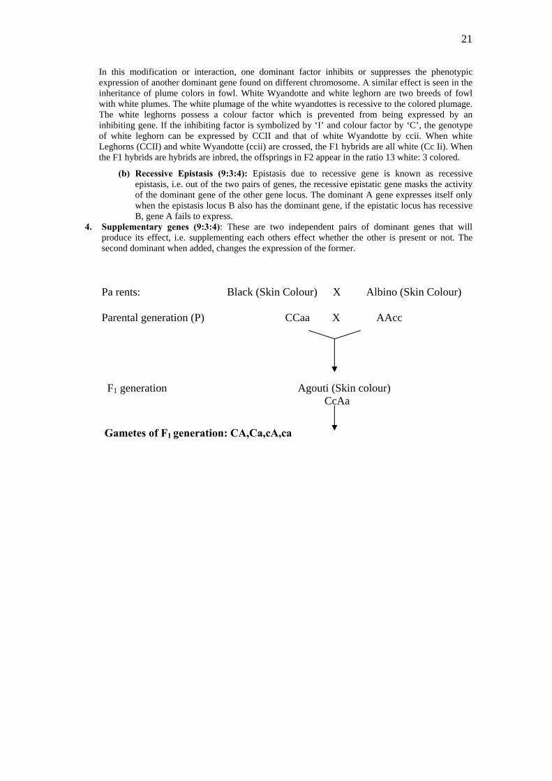
4. Supplementary genes (9:3:4): These are two independent pairs of dominant genes that will produce its effect, i.e. supplementing each others effect whether the other is present or not. The second dominant when added, changes the expression of the former.
Pa rents: Black (Skin Colour) X Albino (Skin Colour)
Parental generation (P) CCaa X AAcc
F1 generation Agouti (Skin colour) CcAa
Gametes of F1 generation: CA,Ca,cA,ca
22
F2 generation: ♀
♂
cA ca CA Ca
CCAA
Agouti
CCAa
Agouti
CcAA
Agouti
CcAa
Agouti
CCAa
Agouti
CCaa
Black
CcAa
Agouti
Ccaa
Black
CcAA
Agouti
CcAa
Agouti
ccAA
Albino
ccAa
Albino
CcAa
Agouti
Ccaa
Black
ccAa
Albino
ccaa
Albino
CA
Ca
cA
ca
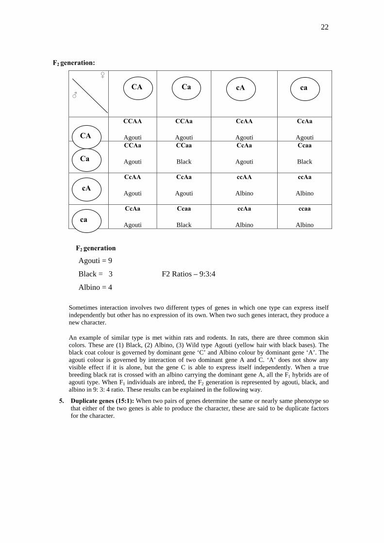
F2 generation
Agouti = 9
Black = 3 F2 Ratios – 9:3:4
Albino = 4
Sometimes interaction involves two different types of genes in which one type can express itself independently but other has no expression of its own. When two such genes interact, they produce a new character.
An example of similar type is met within rats and rodents. In rats, there are three common skin colors. These are (1) Black, (2) Albino, (3) Wild type Agouti (yellow hair with black bases). The black coat colour is governed by dominant gene ‘C’ and Albino colour by dominant gene ‘A’. The agouti colour is governed by interaction of two dominant gene A and C. ‘A’ does not show any visible effect if it is alone, but the gene C is able to express itself independently. When a true breeding black rat is crossed with an albino carrying the dominant gene A, all the F1 hybrids are of agouti type. When F1 individuals are inbred, the F2 generation is represented by agouti, black, and albino in 9: 3: 4 ratio. These results can be explained in the following way.
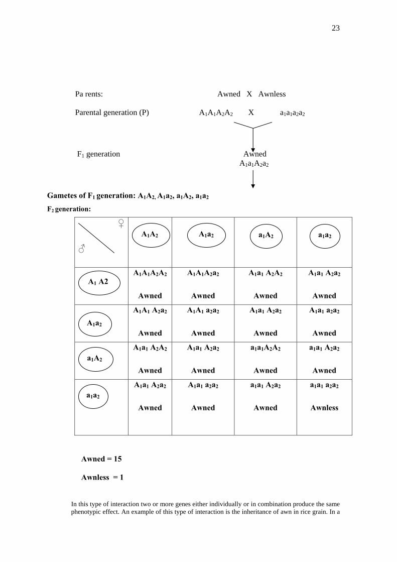
5. Duplicate genes (15:1): When two pairs of genes determine the same or nearly same phenotype so that either of the two genes is able to produce the character, these are said to be duplicate factors for the character.
23
Pa rents: Awned X Awnless Parental generation (P) A1A1A2A2 X a1a1a2a2
F1 generation Awned A1a1A2a2
Gametes of F1 generation: A1A2, A1a2, a1A2, a1a2
F2 generation:
♀
♂
a1A2 a1a2A1A2 A1a2
A1A1A2A2
Awned
A1A1A2a2
Awned
A1a1 A2A2
Awned
A1a1 A2a2
Awned
A1A1 A2a2
Awned
A1A1 a2a2
Awned
A1a1 A2a2
Awned
A1a1 a2a2
Awned
A1a1 A2A2
Awned
A1a1 A2a2
Awned
a1a1A2A2
Awned
a1a1 A2a2
Awned
A1a1 A2a2
Awned
A1a1 a2a2
Awned
a1a1 A2a2
Awned
a1a1 a2a2
Awnless
A1 A2
A1a2
a1A2
a1a2
Awned = 15
Awnless = 1
In this type of interaction two or more genes either individually or in combination produce the same phenotypic effect. An example of this type of interaction is the inheritance of awn in rice grain. In a
24
cross between ‘awned’ and ‘awnless’ types, F1 rice grains were all awned. Awned character is thus dominant over awnless. The F1 plants on self-pollination give awned and awnless types in 15:1 ratio in F2 generation. This result can be explained if it is supposed that awned character is governed by two dominant genes, say A1 and A2. The presence of one or both dominant genes results awned grains and in absence of both these dominant genes awnless grains develop. From the above cross it is clear that out of sixteen offsprings in the F2 generation, fifteen have one or both the dominant genes, so they are all awned type and only one is such which is double recessive, hence awnless.
6. Collaborator genes: In collaboration two gene pairs which are present on separate loci but influence the same trait, interact to produce some totally new trait or phenotype that neither of the genes independently produce.
(C) Pleio-tropic effect of genes 7. Lethal genes: are known to control the manifestation of some phenotypic trait as well as affect the
viability of the organism. While some other genes may exclusively affect the viability alone. These genes are known as lethals or semi-lethals depending on their influence. Complete lethal genes in homozygous condition kill all or nearly all the homozygous individuals, while in case of semi-lethal genes some homozygous individuals are able to survive. The lethal genes are always recessive for their lethality and express the lethal effect only in homozygous recessive condition.
(a) Dominant lethals: The dominant lethal genes are lethal in homozygous condition and produce some defective or abnormal phenotype in heterozygous condition. Their most serious effect in heterozygotes may also cause death.
(b) Recessive lethals: The recessive genes produce lethal effect only in homozygous condition. Their heterozygotes are normal. Therefore, recessive lethals remain unnoticed in the population (floating lethals) but are generally get established in the population.
(c) Conditional lethals: The genes which may be normal to the individual in a particular environment may prove to be lethal when the environment is changed.
8. Atavism: Characters may and often remain hidden generation after generation through the effect of
inhibiting or epistatic factor or some other gene interaction. Occasionally, some wild character which was present in the ancestral forms reappears in the off-springs. This was first discovered by Charles Darwin and is called “atavism”.
9. Multiple genes: In certain cases, the final expression of a single character involves a number of
steps. Each step controlled by a specific enzyme controlled by a specific gene. In such cases each biochemical reaction involves stepwise conversion of precursor into another compound which acts as a precursor for the next step till the formation of final product is obtained. All these steps of a biochemical reaction constitute biosynthetic pathway.
Further Reading:
1. Gupta, P.K. 1995 Cytogenetics. Rastogi and company, Merrut, India.
2. John, B. and Lewis, K.R.1965 The meiotic system. Protoplamatologia VI. Springer verlag, Wien, Newyork
3. Singh, R.J.1993 Plant Cytogenetics.CRC press, Inc. Boca Raton, Florida, USA.
4. Sybenga, J. 1972 General Cytogenetics. North-Holland Publisher Co., Amsterdam
Web Links consulted:
1. Mitosis: http://www.emc.maricopa.edu/faculty/farabee/biobk/BioBookmito.html
2. 2.Meiosis: http://www.emc.maricopa.edu/faculty/farabee/biobk/BioBookmeiosis.html
3. The Cell Cycle: users.rcn.com/.../BiologyPages/C/CellCycle.html