c/ebp transcription factors in human squamous cell carcinoma: selective changes in expression of...
DESCRIPTION
The CCAAT/Enhancer Binding Proteins (C/EBPs) are a family of leucine-zipper transcription factors that regulate physiological processes such as energy metabolism, inflammation, cell cycle, and the development and differentiation of several tissues including skin. Recently, a role for C/EBPs in tumor cell proliferation and differentiation has been proposed, but the incomplete characterization in the literature of multiple translational isoforms of these proteins has made interpretation of these roles difficult. Therefore, we have carefully reexamined C/EBP isoform expression in human nonmelanoma skin cancers. C/EBPa, C/EBPb, and C/EBPd were analyzed histologically in squamous cell carcinomas (SCC). The individual isoforms of C/EBPa and C/EBPb were examined by immunofluorescent digital imaging, western blotting and DNA binding activity (electrophoretic mobility shift analysis). Expression of all C/EBP family proteins was decreased in SCC tumors. Suppression was greatest for C/EBPa, less for C/EBPb, and least for C/EBPd. Western analyses confirmed that C/EBPa p42 and p30 isoforms were decreased. For C/EBPb, only the abundant full-length isoform (C/EBPb21, LAP*, 55 kD) was reduced, whereas the smaller isoforms, C/EBPb22 (LAP, 48 kD) and C/EBPb23 (LIP, 20 kD), which are predominantly nuclear, were significantly increased in well- and moderately-differentiated SCC (up to 14-fold for C/EBPb23). These elevations correlated with increases in PCNA, a marker of proliferation. Although C/EBPb displayed increased posttranslational modifications in SCC, phosphorylation of C/EBPb21 (Thr 235) was not altered. C/EBP-specific DNA binding activity in nuclear and whole-cell extracts of cultured cells and tumors was predominantly attributable to C/EBPb. In summary, two short C/EBPb isoforms, C/EBPb22 and C/EBPb23, represent strong candidate markers for epithelial skin malignancy, due to their preferential expression in carcinoma versus normal skin, and their strong correlation with tumor proliferation.TRANSCRIPT

C/EBP Transcription Factors in Human Squamous CellCarcinoma: Selective Changes in Expression of IsoformsCorrelate with the Neoplastic StateSanjay Anand1,2, John Ebner1, Christine B. Warren1, Manu S. Raam2, Melissa Piliang1,3,
Steven D. Billings1,3, Edward V. Maytin1,2,4*
1Department of Dermatology, Cleveland Clinic, Cleveland, Ohio, United States of America, 2Department of Biomedical Engineering, Cleveland Clinic, Cleveland, Ohio,
United States of America, 3Department of Anatomic Pathology, Cleveland Clinic, Cleveland, Ohio, United States of America, 4Wellman Center for Photomedicine,
Massachusetts General Hospital, Harvard Medical School, Boston, Massachusetts, United States of America
Abstract
The CCAAT/Enhancer Binding Proteins (C/EBPs) are a family of leucine-zipper transcription factors that regulatephysiological processes such as energy metabolism, inflammation, cell cycle, and the development and differentiation ofseveral tissues including skin. Recently, a role for C/EBPs in tumor cell proliferation and differentiation has been proposed,but the incomplete characterization in the literature of multiple translational isoforms of these proteins has madeinterpretation of these roles difficult. Therefore, we have carefully reexamined C/EBP isoform expression in human non-melanoma skin cancers. C/EBPa, C/EBPb, and C/EBPd were analyzed histologically in squamous cell carcinomas (SCC). Theindividual isoforms of C/EBPa and C/EBPb were examined by immunofluorescent digital imaging, western blotting and DNAbinding activity (electrophoretic mobility shift analysis). Expression of all C/EBP family proteins was decreased in SCCtumors. Suppression was greatest for C/EBPa, less for C/EBPb, and least for C/EBPd. Western analyses confirmed that C/EBPap42 and p30 isoforms were decreased. For C/EBPb, only the abundant full-length isoform (C/EBPb21, LAP*, 55 kD) wasreduced, whereas the smaller isoforms, C/EBPb22 (LAP, 48 kD) and C/EBPb23 (LIP, 20 kD), which are predominantlynuclear, were significantly increased in well- and moderately-differentiated SCC (up to 14-fold for C/EBPb23). Theseelevations correlated with increases in PCNA, a marker of proliferation. Although C/EBPb displayed increased post-translational modifications in SCC, phosphorylation of C/EBPb21 (Thr 235) was not altered. C/EBP-specific DNA bindingactivity in nuclear and whole-cell extracts of cultured cells and tumors was predominantly attributable to C/EBPb. Insummary, two short C/EBPb isoforms, C/EBPb22 and C/EBPb23, represent strong candidate markers for epithelial skinmalignancy, due to their preferential expression in carcinoma versus normal skin, and their strong correlation with tumorproliferation.
Citation: Anand S, Ebner J, Warren CB, Raam MS, Piliang M, et al. (2014) C/EBP Transcription Factors in Human Squamous Cell Carcinoma: Selective Changes inExpression of Isoforms Correlate with the Neoplastic State. PLoS ONE 9(11): e112073. doi:10.1371/journal.pone.0112073
Editor: Roberto Mantovani, Universita degli Studi di Milano, Italy
Received June 6, 2014; Accepted October 13, 2014; Published November 17, 2014
Copyright: � 2014 Anand et al. This is an open-access article distributed under the terms of the Creative Commons Attribution License, which permitsunrestricted use, distribution, and reproduction in any medium, provided the original author and source are credited.
Data Availability: The authors confirm that all data underlying the findings are fully available without restriction. All data are within the paper andsupplementary figures.
Funding: This study was funded by the National Cancer Institute, of the National Institute of Health, grant number PO1-CA84203. The funders had no role instudy design, data collection and analysis, decision to publish, or preparation of the manuscript.
Competing Interests: The authors have declared that no competing interests exist.
* Email: [email protected]
Introduction
C/EBP transcription factors (C/EBPs), a family of six gene
members in which C/EBPa, C/EBPb, and C/EBPd have been
the most intensively studied, were originally identified as regulators
of growth and differentiation in normal tissues [1–3]. More
recently, C/EBPs have received considerable attention as potential
molecular markers that define prognostic risk in cancer; reviewed
in [4–6]. C/EBPa is known to be a tumor suppressor in acute
myelogenous leukemia [reviewed in [7,8]], and is reportedly
down-regulated in human epithelial cancers of the breast [9], lung
[10], liver [11], head and neck [12], endometrium [13], and
squamous cell carcinoma (SCC) of the skin [14]. C/EBPaexpression levels in actinic keratoses and keratoacanthoma
(precancerous skin conditions that can progress to SCC) were
reduced relative to normal epidermis, and fully invasive SCCs
expressed no detectable levels of C/EBPa, indicating a direct
correlation between expression levels of C/EBPa and the severity
of neoplasia [14]. C/EBPb expression, in contrast to C/EBPa,was reportedly increased in advanced cases of breast, ovarian,
colorectal, renal, and gastric carcinoma [15–21]. However,
interpretation of those reports is complicated by the existence of
three different isoforms of C/EBPb, a major topic of the current
study (see below). C/EBPd gene has been reported to be
methylated in acute myelomoncytic leukemia (AML), cervical,
breast and hepatocellular carcinoma and reduced expression
associated with progression of breast tumors [22]. Other C/EBP
family genes have received relatively less attention, but potential
involvement of dysregulated C/EBPe [23], and C/EBPf/gadd153[24] in leukemia and melanoma, respectively, has been suggested.
PLOS ONE | www.plosone.org 1 November 2014 | Volume 9 | Issue 11 | e112073

The question of whether absolute protein levels of the various
C/EBPs reflect biological functions in human cancers remains
unresolved, but it appears increasingly clear that changes in the
ratio of C/EBP isoforms could have prognostic significance. C/
EBPa and C/EBPb are intronless genes, each producing a single
mRNA transcript. Different protein isoforms can be produced
from a single C/EBPa or C/EBb transcript via a mechanism of
alternative translation that uses different ATG start codons within
the ribonucleotide sequence [25]. The human C/EBPa transcript
produces two proteins, ,42 kDa and ,30 kDa, whereas the C/
EBPb transcript produces three proteins named LAP*, LAP, and
LIP in mice, or C/EBPb-1, C/EBPb-2, and C/EBPb-3 in
humans, in order of decreasing size [6]. The shortest form can act
as a dominant-negative inhibitor since it contains a DNA-binding
region, yet lacks N-terminal amino acids necessary to transactivate
gene transcription. For this reason, the relative expression levels of
a long and short form, the ‘‘LAP/LIP ratio,’’ was proposed to
participate in regulation of proliferation and differentiation in
normal cells [26,27], and in cancer [28]. This idea now deserves a
revisit as more detailed information has emerged. Linda Sealy
et al. have established that the largest form of C/EBPb observed
in transformed breast cancer cell lines is C/EBPb-2 (LAP),
whereas the full-length C/EBPb-1 (LAP*) is only expressed in
whole tissues in vivo or in primary epithelial cells in vitro [29,30].Also, elevated expression of C/EBPb-2 in MCF10A normal
human mammary epithelial cells results in transformation, an
epithelial to mesenchymal transition (EMT) and acquisition of an
invasive phenotype, directly linking C/EBPb-2 to severity of
neoplasia [31]. The implications here are two-fold: (i), current
information about C/EBPb-1 is not clear, given many older
reports in which the analyses were performed immunohistologi-
cally and distinction between C/EBPb-1 and C/EBPb-2 was not
recognized; (ii), previous interpretations regarding proteins
extracted from tissues in vivo and detected by western blotting
may need to be reconsidered, in light of newer information.
Murine models of cutaneous carcinogenesis may offer particular
insight because recent reports have indicated important functional
roles for C/EBPa and C/EBPb during tumorigenesis in murine
skin. Mice lacking C/EBPa in the epidermis show normal
proliferation and differentiation but were highly susceptible to
skin tumorigenesis in response to carcinogens. These mice
displayed decreased tumor latency, increased tumor incidence,
multiplicity, growth rate and malignant progression [14,32].
Chemically induced squamous cell carcinomas and also primary
cell lines established from these SCC, showed negligible expression
of C/EBPa as compared with normal epidermis [33]. Also, the
expression levels of C/EBPa or C/EBPb were a direct indicator of
the state of neoplasia, since benign papillomas in mice showed an
intermediate expression level, when compared to normal epider-
mis (highest level) and SCCs (lowest level) in the same study [34].
C/EBPb knockout mice are completely refractory to skin tumor
development in response to chemical carcinogens [35] or UVB
exposure [Anand et al., unpublished results]. A robust increase in
apoptosis accompanied with elevated p53 levels in epidermis has
been suggested as the causative mechanism for this resistance to
tumor formation in C/EBPb null mice [36]. In a similar study, C/
EBPd KO mice showed no difference in the tumor phenotype as
compared to wild type, in response to chemical carcinogen [37].
However, in murine model of mammary tumerigenesis, loss of C/
EBPd resulted in increased mammary tumor multiplicity and
reduced lung metastasis involving the regulation of HIF-1a,mTOR and FBXW7 [22,38]. The role of C/EBPd as an
inflammatory response gene and also a candidate tumor suppres-
sor gene has been supported by the sensitivity of C/EBPd null
mice to ionizing radiation-induced hematopoietic and intestinal
injury [39].
In this paper, we have re-examined the question of change in
histological expression of the three most abundant C/EBPs (a, band d) in human SCC, by looking comprehensively in vivo at
expression of individual protein isoforms and their DNA-binding
ability. Our data confirm an across-the-board downregulation of
C/EBPa but more interestingly, the data show a strong
upregulation of C/EBPb-2 and C/EBPb-3 that correlates with
cellular proliferation in moderately- and well-differentiated SCC
of the skin.
Materials and Methods
Culture of primary keratinocytes and carcinoma cellsNormal human epidermal keratinocytes (NHEKs; Cascade
Biologics, Portland, OR); HEK1 cells (HEK001; from ATCC,
Manassas, VA); and SCC13 cells (gift from Jonathan Garlick,
Tufts University, Boston, MA, [40,41]) were cultured at 37uC in a
humidified CO2 incubator as previously described [42]. Human
prostate carcinoma cells (LNCAP; ATCC) were cultured as
described [43]. cos-7 cells (ATCC) were cultured at 37uC in a 5%
humidified CO2 incubator in DMEM (4 mM L-glutamine, 1.5 g/l
sodium bicarbonate, 4.5 g/l glucose, with10% FBS, 100 units/ml
Penicillin and 100 mg/ml streptomycin). Cells were maintained at
subconfluence (,80–90%) levels, and seeded at a 1:6 to 1:8
dilutions during passaging.
Collection of human tumor specimensDiscarded skin tissues, taken during Moh’s surgery for removal
of SCC tumors diagnosed previously by biopsy, were obtained in a
de-identified manner. This study was approved by the Institutional
Review Board of the Cleveland Clinic. Frozen tissue blocks in
OCT compound (Tissue Tek, Torrance, CA) were screened
histologically, and those that contained usable tumor were stored
at 280uC until further study.
Expression vectors, cell transfection and preparation ofwhole-cell and nuclear extractsExpression vectors (pcDNA3.1) encoding rat C/EBPa and
mouse C/EBPd were kind gifts of David Ron (NYU Medical Ctr,
New York, NY) and James Dewille (Ohio State University,
Columbus, OH), respectively. An expression vector (pCMV6)
encoding human C/EBPb was purchased from Origene (Rock-
ville, MD). An expression vector for human C/EBPb1(pcDNA3.1hisA) that exclusively expressed the longest form of
C/EBPb due to modification of the ATG for C/EBPb2 was kindlyprovided by Linda Sealy (Vanderbilt University, Nashville, TN).
Nuclear extracts from cos-7 cells overexpressing various C/EBPs
(a, b and d) were prepared exactly as described by Schreiber et al.
[44] following transfection of cells in 100 mm dishes using 3 mg ofexpression vector and GenePORTER reagent (Genlantis, San
Diego, CA) according to the manufacturer’s protocol. Whole cell
extracts from cells overexpressing the C/EBPs were prepared by
lysis in urea buffer (7 M urea, 2% IGEPAL, 5% b-mercaptoeth-
anol and protease inhibitor cocktail), followed by 3–5 pulses (4–
6 sec each) of sonication to disrupt membranes, and a 5 min high-
speed microfuge spin to clear the lysates. All procedures were
carried out at 4uC [45].
C/EBP Protein Isoforms in Human Skin Carcinoma
PLOS ONE | www.plosone.org 2 November 2014 | Volume 9 | Issue 11 | e112073

Histological and immunofluorescent analysis of C/EBPexpression in human tumorsCryosections (5 mm) of frozen tumor samples were fixed 5 min
in ice-cold methanol and stained with hematoxylin and eosin by
standard methods. For immunohistochemical detection of C/
EBPs, methanol-fixed cryosections were washed in PBS, and
permeabilized in 0.1% Triton X-100 (Sigma, St Louis, MO) in
PBS for 10 min on ice. Sections were serially incubated in the
following solutions: 3% normal donkey serum, 30 min, room
temperature (RT); primary antisera, overnight, 4uC; PBS rinses,
5 min x 3; Cy3-conjugated donkey anti-rabbit IgG (Jackson
ImmunoResearch, West Grove, PA, 1:1500, for 4 h at RT), PBS
rinse x1; mounting in Vectashield (Vector Lab, Burlingame, CA)
under coverslips. Antisera for C/EBPa, C/EBPb, C/EBPd, E-cad(all from Santa Cruz Biotechnology, Santa Cruz, CA) and Ki67
(Thermo Fisher, Waltham, MA) were used at 1:50 dilution in PBS.
Semiquantitative analysis of fluorescence intensity inimmunostained sections of human skin tumorsImaging of fluorescence intensity from immunostained skin
specimens was standardized as follows. Seven different exposures
of a given tumor specimen, and of a normal skin specimen from
the same patient, was digitally captured on an Olympus BX50
fluorescent microscope (Olympus America, Center Valley, PA)
equipped with a OolSNAP-Pro color CCD camera (Media
Cybernetics, Bethesda, MD). Exposure times for each successive
image were lengthened so as to double the amount of light
collected. Pairs of digital images from tumor and normal hair
follicle were compared side-by-side on a computer monitor, and
the two images whose intensities were visually most closely
matched were noted. From the ratio of exposure times of the best-
matched image pair, the relative difference (fold) in C/EBP
protein between tumor and normal was determined. For this
analysis it was assumed that the fluorescent signal is directly
proportional to the amount of C/EBP protein bound by primary
antibody, since no amplification step was employed during
immunostaining. Some examples of images analyzed in this way
are illustrated in Fig. S2.
Western blot analysesCells were lysed and for human tumor samples, 30 mm
cryosections (cut perpendicular to the skin surface) were scraped
from the glass slide and pooled, then homogenized, lysed and
sonicated in urea lysis buffer as described [42]. Protein content was
determined by Bradford assay kit (Bio-Rad, Hercules, CA), and
equal amounts were analyzed on western blots using 4–12% Bis-
Tris acrylamide mini gels (Invitrogen, Carlsbad, CA) or 10% Tris-
Glycine gels, as described [42]. The source and dilution of antisera
used here were as follows: C/EBPa, C/EBPb, C/EBPd, Actin and
GAPDH (Santa Cruz,1:5000); PCNA and E-Cadherin (Santa
Cruz, 1:2000); a-Tubulin (Sigma,1:10000), Phospho-C/EBPb(Cell Signaling, Danvers, MA, 1:1000); and peroxidase-conjugated
goat anti-rabbit IgG (Jackson ImmunoResearch, 1:20,000).
Western blot signals were quantitated using IPLab software
(Scanalytics Inc., Fairfax, VA).
Electrophoretic Mobility Shift Assay (EMSA)Complementary oligonucleotides (oligos; 29-mer) that spanned
either a well-established C/EBP motif, or a mutant C/EBP motif,
were synthesized (Integrated DNA Technologies, San Diego, CA)
and used for EMSA. The sense and antisense sequences for each
of the oligos are as follows:
C/EBP consensus; (Sense 59 CTAGCATCTGCAGATTGCG-CAATCTGCAC 39; Antisense 59 TCGAGTGCAGATTGCG-CAATCTGCAGATG 39).
Mutant C/EBP consensus; (Sense 59 CTAGCATCTGCA-
GAGGTATACCTCTGCAC 39; Antisense 59 TCGAGTGCA-
GAGGTATACCTCTGCAGATG 39). The C/EBP consensus
sequence is shown in bold, and mutant sequences are underlined
[46]. Two pM of duplex oligos, heat denatured and annealed,
were labeled with [a-32P] dCTP (3,000 Ci/mM, ICN Pharma-
ceuticals) using Klenow polymerase (New England Biolabs,
Ipswich, MA) and purified using Probequant spin columns (GE
Healthcare, Piscataway, NJ). Nuclear extracts (2–3 mg) from cos-7cells, keratinocytes, or tumor cells were prepared as described [44]
and incubated with 50 fM of labeled oligos in DNA binding buffer
(Hepes pH 7.9, 20 mM, glycerol 10%, KCl 40 mM, NP-40 0.1%,
EDTA 0.5 mM, PMSF 1 mM, DTT 0.5 mM) along with 1 mg of
non-specific DNA competitor [poly(dG-dC). poly (dG-dC)] and
1 ml of filtered FBS for 30 min at room temperature. DNA-protein
complexes were resolved on 4% non-denaturing polyacrylamide
gels in Tris-Glycine-EDTA buffer (Tris 25 mM, Glycine 200 mM
and EDTA 2.25 mM), dried and exposed to X-ray film between
intensifying screens. For supershift experiments, 1 ml of antibodywas added to the mixture and incubated for 15 min prior to
addition of labeled oligos [45].
Statistical analysesRelative levels of C/EBPs (compared to normal skin) were
quantitated using immunohistological images (three images per
sample) from 10–13 SCC samples and data presented as
mean6SD in Table 1 and Fig. 1B. Protein expression levels
by western blot were quantitated using blots from two independent
experiments and the data presented as average6range in Figs. 2–4.
Results
Expression of C/EBPa, C/EBPb, and C/EBPd is reduced inhuman cutaneous SCCTo obtain qualitative information about expression levels and
intracellular localization of C/EBP transcription factors in skin
cancers in vivo, we used primary antibodies to C/EBPa, C/
EBPb, and C/EBPd to perform immunostaining on frozen SCC
specimens and adjacent normal skin (Fig. 1). Staining patterns forC/EBPa and C/EBPb in normal epidermis, hair follicles, and
sebaceous glands were consistent with previous reports [47,48] i.e.
C/EBPa and C/EBPb staining was primarily cytoplasmic in the
lower epidermal layers and hair follicle epithelium (Fig. 1A,‘‘ep’’), but became nuclear in the upper epidermal layers. The
difference between C/EBPa and C/EBPb was that greater
amount of cytoplasmic C/EBPb was also seen in the normal
follicles (Fig. 1A). For C/EBPd, expression was strongly nuclear
throughout all layers of the epidermis (not shown) and in the
follicles (Fig. 1A).
Within SCC tumor nests, the signal intensity for each C/EBP
protein was markedly lower in tumor cells than in normal cells
(Fig. 1A), with some exceptions. To obtain more objective
estimates of changes in C/EBP protein expression in the biopsy
specimens, we devised an ‘‘f-stop’’ technique to quantify changes
in the immunofluorescent signal in digitally-recorded micrographs;
see Methods and Fig. S2. The graphs in Fig. 1B summarize the
results of this enhanced analysis for multiple SCC tumors; data for
the individual tumors are in Table 1. For SCC, C/EBPa was
suppressed in 7/8 specimens, C/EBPb in 6/11 specimens, and C/
EBPd in 5/11 specimens. Thus, in terms of both frequency and
C/EBP Protein Isoforms in Human Skin Carcinoma
PLOS ONE | www.plosone.org 3 November 2014 | Volume 9 | Issue 11 | e112073
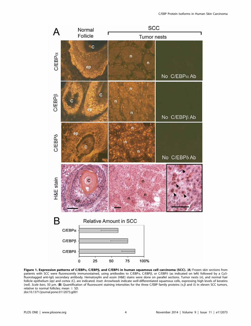
Figure 1. Expression patterns of C/EBPa, C/EBPb, and C/EBPd in human squamous cell carcinoma (SCC). (A) Frozen skin sections frompatients with SCC were fluorescently immunostained, using antibodies to C/EBPa, C/EBPb, or C/EBPd (as indicated on left) followed by a Cy3-fluorotagged anti-IgG secondary antibody. Hematoxylin and eosin (H&E) stains were done on parallel sections. Tumor nests (n), and normal hairfollicle epithelium (ep) and cortex (C), are indicated. Inset: Arrowheads indicate well-differentiated squamous cells, expressing high levels of keratins(red). Scale bars, 50 mm. (B) Quantification of fluorescent staining intensities for the three C/EBP family proteins (a,b and d) in eleven SCC tumors,relative to normal follicles; mean 6 SD.doi:10.1371/journal.pone.0112073.g001
C/EBP Protein Isoforms in Human Skin Carcinoma
PLOS ONE | www.plosone.org 4 November 2014 | Volume 9 | Issue 11 | e112073
magnitude of change in expression, C/EBPa was suppressed the
most, C/EBPb less so, and C/EBPd the least.
Both C/EBPa isoforms and CEBPb-1 isoform aredecreased, whereas CEBPb-2 and CEBPb-3 are increasedin malignant SCC cell linesBecause standard immunohistological technique can not
differentiate between isoforms, we used western analysis to further
refine our initial observations of C/EBP protein expression in
SCC, focusing on C/EBPa and C/EBPb. For a preliminary look,
and to confirm the specificity of antibody reagents, we tested
several cell lines that lie along a spectrum of increasingly
malignant behavior. In the order of benign to malignant, the
lines were: (i), NHEK, a normal human epidermal keratinocytes,
(ii), HEK1, a virally transformed human keratinocyte line, (iii),
SCC13, a spontaneously-tranformed squamous cell carcinoma
line, and (iv), LNCAP, a prostate carcinoma line. In these cell
lines, expression profiles of markers of growth-arrest and
differentiation had already been established [see Fig. S1 in ref.
[42]]. Here, western analyses revealed a decrease in both C/EBPaisoforms (42 kD and 30 kD) in parallel with the hierarchy of
malignant progression (Fig. 2A, B). This finding was consistent
with clinical reports in breast carcinoma [9], human SCC [14] and
experimental cutaneous carcinoma in mice [33,49]. For C/EBPb,the situation was more complicated. Because previous literature
did not always distinguish between the two large isoforms of C/
EBPb, we took care to confirm the identity of C/EBPb-1 and C/
EBPb-2 on gels by comparing their location on the western blots
with the location of recombinant proteins translated in cos-7 cells.
For calibration purposes, we used: (i), a normal full-length human
C/EBPb sequence that expresses C/EBPb-2 and 23 but not C/
EBPb-1 in the cos-7 cells (Fig. 2C, lanes 2 and 5), and (ii), a
cDNA engineered to express only C/EBPb-1 and not the other
isoforms (lane 4). In the cell lines, the abundant C/EBPb-1 isoformappeared to decrease with malignant progression (Fig. 2C, lanes6–8), whereas the two smaller isoforms C/EBPb-2 and C/EBPb-3were increased 2- to 5-fold (Fig. 2D). These results agree with
others who reported that C/EBPb-1 is expressed in normal
mammary cells and tissues, but not expressed in immortalized cell
lines. These data are also consistent with reports that high C/
EBPb-2 expression can results in transformation, EMT and
acquisition of an invasive phenotype in normal human mammary
epithelial cells [29,31]. We also tested for C/EBPd, but signals onWestern blots were too weak to assess. Low expression levels of C/
EBPd in these cell lines were later confirmed by functional DNA
binding assays (see below).
In human SCC cancers, overall expression of C/EBPa andC/EBPb is reduced, but expression of two small C/EBPbisoforms is increasedWe next examined human SCC specimens by western blotting.
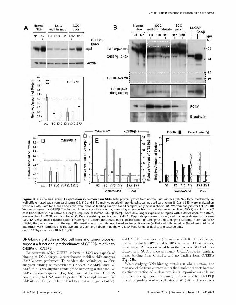
Obtaining sufficient tumor tissue from skin biopsies was only
possible by pooling multiple thick frozen sections from the largest
tumors, which comprised five invasive SCC. Those specimens
were evaluated by two certified dermatopathologists to determine
the histological tumor grade (Fig. S1). Protein lysates from these
tumors were loaded onto western blots in the order of worsening
neoplasia, using duplicate blots to provide an estimate of
variability (Fig. 3). Three proteins (GAPDH, tubulin and actin)
were used as housekeeping loading controls, instead of only one,
because expression of ‘‘invariant’’ control genes often differs
between skin samples as demonstrated by Minner and Poumay
[50]. Fig. 3A illustrates a typical Western blot for C/EBPa in
normal and carcinoma tissues (only the actin control is shown). C/
EBPa (p42) expression decreased by 60–75% in most of the SCCs,
although less so in the most poorly differentiated tumor (S13). The
C/EBPa p30 isoform was not detected.
For C/EBPb, each isoform behaved differently. The largest
protein, C/EBPb-1, appeared as a thick multi-banded complex
(55–70 kD), indicating a high level of post-translational modifica-
tion (Fig. 3B), as described by Zahnow [6]. The abundance of C/
EBPb-1 was moderately reduced in all SCC (Fig. 3D), although a
more remarkable loss of the highest MW (most post-translationally
modified) forms of C/EBPb-1 was absent in the well-to-
moderately differentiated SCC tumors, S9–S11 (Fig. 3B, dottedoval). In contrast, the shorter isoforms, C/EBPb-2 and b-3, wereboth strongly induced in tumors S9–S11 (Fig. 3B). Although
expressed in low amounts, C/EBPb-2 and C/EBPb-3 differed
Table 1. Relative levels of C/EBPa, C/EBPb and C/EBPd in histological sections of human squamous cell carcinoma.
Protein Expression (% of normal)
Tumor ID# C/EBPa C/EBPb C/EBPd
S1 ND 100 83
S2 53 80 100
S3 ND 43 100
S4 61 100 53
S5 43 55 66
S6 100 66 100
S9 45 100 111
S10 31 100 100
S11 33 42 66
S12 71 100 100
S13 ND 40 64
Mean 6 SD 60626 75626 86620
The relative intensity of immunofluorescent staining (compared to normal skin) for each C/EBP family member was determined semi-quantitatively by making pairwisevisual comparisons between tumor nests and normal hair follicles within the same patient, using the f-stop technique (see Methods and Figure S2). ND, not done.doi:10.1371/journal.pone.0112073.t001
C/EBP Protein Isoforms in Human Skin Carcinoma
PLOS ONE | www.plosone.org 5 November 2014 | Volume 9 | Issue 11 | e112073

qualitatively from C/EBPb-1 in that both were undetectable in
normal skin. By densitometry, C/EBPb-2 and C/EBPb-3 were
increased in the well- and moderately-differentiated SCC, but not
in the poorly-differentiated SCC (Fig. 3E). C/EBPb-3 appeared
to offer the greatest signal-to-noise ratio, rising 8- to14-fold in the
moderately-differentiated tumors (Fig. 3E).
The expression patterns of C/EBPb-2 and C/EBPb-3 incutaneous SCC correlate with changes in markers ofcellular proliferation and differentiationTo ask whether changes in the short C/EBP isoforms were
correlated with any physiologic markers within tumors, markers of
proliferation (PCNA) and differentiation (E-cadherin) were ana-
lyzed in the SCC by western blot (Fig. 3B, bottom two panels).
PCNA expression levels tracked very closely with expression of the
smallest isoform, C/EBPb-3, such that PCNA and C/EBPb-3were elevated in the same tumor subset (Fig. 3E and 3F). E-cadherin did not bear any clear relationship with C/EBPbexpression. However, within SCC tumors as a group, E-cadherin
expression rose as PCNA expression dropped (Fig. 3F), consistentwith a permissive relationship between growth arrest and the onset
of terminal differentiation often seen in squamous epithelia.
Immunohistological analyses of proliferation (Ki67) and differen-
tiation (E-cad) in SCC revealed a similar pattern as described
above (Fig. S3).
The C/EBPb-1 isoform is phosphorylated in SCC, butphosphorylation does not appear to be differentiallyregulatedTo begin to examine post-translational modifications of C/
EBPb, we evaluated the phosphorylation state of C/EBPb by
probing blots with a selective antibody against C/EBPbphosphorylated at threonine-235 (Fig. 4A). Phosphorylation of
the threonine 235 residue in human C/EBPb by Ras-MAPK-
ERK kinase (MEK) signaling pathway results in transcriptional
activation of C/EBPb [6]. Here only one band corresponding to
phosphorylated C/EBPb-1/LAP*, was detected. However, up to
four bands corresponding to various other forms of C/EBPb-1were apparent in the tumor specimens (Fig. 4B). In normal skin,
only one major form of C/EBPb-1 was seen (Fig. 4B, lanes 1 and
2) and it corresponded to phosphorylated C/EBPb-1. Slower-
migrating forms of C/EBPb were observed in the SCC tumors
and may correspond to acetylation, methylation, or sumoylation,
as reported by others and summarized by Zahnow [6]. The ratio
of phosphorylated C/EBPb-1 to total C/EBPb-1 (defined as those
isoforms located above the dotted line in Fig. 4B) did not change
significantly in SCC relative to normal skin.
Figure 2. C/EBPa and C/EBPb isoforms are differentially expressed in SCC cell lines as compared to normal human keratinocytes.Western analyses of normal primary keratinocytes (NHEK), and two squamous cell carcinoma cell lines, HEK1 and SCC13. (A) Western blot for C/EBPa,in the skin cell lines and also in prostate carcinoma cells (LNCAP). GAPDH is a loading control. (B) Densitometric quantification for C/EBPa. (C) Westernblots for C/EBPb. Lanes 2 and 5 (cos b2) contain recombinant human cDNA for C/EBPb overexpressed in cos-7 cells, which translate only LAP and LIP.Lane 4 (cos b1) contains recombinant human T7 his-tagged C/EBPb-1 plasmid expressed in cos-7 cells. Lanes 1 and 3, extracts from cos-7 cell thatwere untransfected or transfected with the empty vector (pVector), respectively. Box, longer exposure of C/EBPb-3. GAPDH, loading control. (D)Densitometric quantification of each isoform of C/EBPb. Graphs represent the average of two independent Western analyses, with protein levelsnormalized to GAPDH and expressed relative to NHEK (dotted line).doi:10.1371/journal.pone.0112073.g002
C/EBP Protein Isoforms in Human Skin Carcinoma
PLOS ONE | www.plosone.org 6 November 2014 | Volume 9 | Issue 11 | e112073
DNA-binding studies in SCC cell lines and tumor biopsiessuggest a functional predominance of C/EBPb, relative toC/EBPa or C/EBPdTo determine which C/EBP isoforms in SCC are capable of
binding to DNA targets, electrophoretic mobility shift analyses
(EMSA) were performed. To validate the techniques, we first
analyzed binding of recombinant C/EBPa, C/EBPb, and C/
EBPd to a DNA oligonucleotide probe harboring a standard C/
EBP consensus sequence (Fig. 5A). Each of the three C/EBPs
bound avidly to DNA, and the protein-DNA complexes were C/
EBP site-specific (i.e., failed to bind to a mutant oligonucleotide),
and C/EBP protein-specific (i.e., were supershifted by preincuba-
tion with anti-C/EBPa, anti-C/EBPb, or anti-C/EBPd antisera,
respectively). Proteins extracted from the nuclei of SCC cell lines
HEK-1 and SCC13 showed mainly C/EBPb-specific binding,
minor binding from C/EBPd, and no binding from C/EBPa(Fig. 5B).
When studying DNA-binding proteins in whole tumors, one
must use whole-tissue extracts rather than nuclear extracts because
selective extraction of nuclear proteins is impossible (as cells are
disrupted during frozen sectioning). To ask whether C/EBPbexpression profiles in whole cell extracts (WC) vs. nuclear extracts
Figure 3. C/EBPa and C/EBPb expression in human skin SCC. Total protein lysates from normal skin samples (N1, N2), three moderately- orwell-differentiated squamous carcinomas (S9, S10 and S11), and two poorly-differentiated squamous cell carcinomas (S12 and S13) were analyzed onwestern blots. Blots for tubulin and actin were done as loading controls for all samples; only actin is shown. (A) Western analyses for C/EBPa. (B)Western analyses for C/EBPb. The last two lanes are positive controls, consisting of lysates from a prostate cancer cell line (LNCAP) and from cos-7cells transfected with a native full-length sequence of human C/EBPb (cos/b). Solid box, longer exposure of region within dotted lines. At bottom,western blots for PCNA and E-cadherin. (C) Densitometric quantification of C/EBPa. Duplicate gels were scanned, and the range shown by the errorbars. (D) Densitometric quantification of C/EBPb21 isoform. (E) Densitometric quantification of C/EBPb22 and C/EBPb23 isoforms. Note that for C/EBPb-3, the y-axis scale is on the right. (F) Densitometric quantitation of markers for proliferation (PCNA) and differentiation (E-cadherin). All bandintensities were normalized to the average of actin and tubulin (not shown). Error bars, range of duplicate measurements.doi:10.1371/journal.pone.0112073.g003
C/EBP Protein Isoforms in Human Skin Carcinoma
PLOS ONE | www.plosone.org 7 November 2014 | Volume 9 | Issue 11 | e112073

(NucX) would be similar or different, we compared WC to NucX
prepared from normal keratinocytes or from HEK1 carcinoma
cells (Fig. 5C). NucX contained C/EBPb-2 and C/EBPb-3almost exclusively, whereas WC contained mostly C/EBPb-1(Fig. 5C). When these two lysates were compared by EMSA
(Fig. 5D), the abundance of the protein-DNA complexes that
formed [see bracket with a double asterisk] tended to reflect the
size distribution of C/EBPb isoforms in the extracts, i.e. larger
complexes in WC than in NucX. However, the predominant C/
EBP family member expressed was always C/EBPb, regardless ofwhether WC or NucX was examined. We next did DNA binding
studies on total tissue lysates from normal skin and tumor
specimens (Fig. 5E). C/EBPb binding was easily detectable in
all specimens. C/EBPd was only weakly positive, and C/EBPawas absent. In summary, C/EBPb appears to be the major C/
EBP species present and capable of binding conventional C/EBP
sites on DNA, both in normal skin and in SSC tumors.
Discussion
Nonmelanoma skin cancers (NMSC), comprising basal cell
carcinomas (BCC) and squamous cell carcinomas (SCC), consti-
tute the most common of all human cancers [51]. SCCs, while
representing only 10% of NMSC incidence, are very important
because SCC can readily invade and metastasize. Thus an
important research goal is to recognize features within tissue
biopsies that might identify SCC cancers with the most aggressive
biological behavior. In this manuscript, we have examined the
expression of three members of the C/EBP transcription factor
family that have gained attention as potential prognostic indicators
in cancer. Our finding of decreased expression of C/EBPa in SCC
concurs with previous reports implicating C/EBPa as a tumor
suppressor in myeloid leukemia [8] and cutaneous SCC [14], is
consistent with suggestions that low C/EBPa expression in tumors
contributes to failure of cell cycle arrest [7,9,10,12,33]. For C/
EBPb our data agree with some findings, yet contradict other
aspects of previous clinicopathological studies on C/EBPbexpression in malignancies of various origins [15,18,20,21]. In
agreement, we noted an overall increase in C/EBPb expression in
carcinoma cell lines and in SCC tumors in vivo relative to normal
keratinocytes. However, these increases were entirely due to
elevated amounts of the short isoforms, C/EBPb22 and C/
EBPb23. The full-length isoform C/EBPb21 was actually
slightly decreased. Because most previous studies were not clear
about the exact identity of the large C/EBPb isoforms being
observed, it is not possible to know whether apparent discrepancies
reflect differences between skin and other tissues, or instead reflect
differences in methodologies for protein separation and detection.
In the current study, two additional techniques were employed to
validate our histological findings; namely, western blotting was
calibrated by the use of recombinant C/EBPb isoform standards,
and EMSA was used to determine functional DNA binding
capacity.
Changes in C/EBP expression observed on our western blots
cannot be ascribed to differences in cell populations within
different tissue specimens, because protein lysates were prepared
from frozen biopsy sections with very similar cellular components
by histological examination. Thus, in both the normal skin
specimens and the SCC tumor sections, .90% of the cells were
epithelial (either hair follicles and epidermis, or squamous tumor
cells; see (Fig. S1). Given these equal proportions of normal and
neoplastic epithelial cells, relative changes in C/EBPb isoforms we
observed most likely reflect changes in intracellular C/EBPbexpression, probably at the level of alternative mRNA translation
(see Introduction). However, we cannot rule out a role for
increased transcription of the C/EBPb gene, since studies with
cultured cells show that ER stress (from glucose deprivation or
amino acid deprivation) can induce the expression of human C/
EBPb through unfolded protein response elements in the 39-UTR
of the gene [52,53]. We also cannot rule out the possibility that
post-translational modification of C/EBPb (via upstream signaling
pathways) might affect protein stability and accumulation.
Although changes in phosphorylation appeared to be relatively
minor, other modifications to these proteins were apparent (see
next paragraph).
Changes in differential expression of the C/EBPb isoforms were
among the most interesting findings. C/EBPb-1 (or LAP*)
constituted the vast majority of C/EBPb expressed in the skin,
both normal and SCC, a fact not previously appreciated.
Phosphorylation of this isoform at threonine-235 [54] was not
significantly altered in SCC specimens (Fig. 4A), suggesting that
other modifications such as C/EBPb acetylation, methylation, and
sumoylation as reported in other systems [6] might account for
shifted C/EBPb-1 bands that were apparent in SCC (Fig. 4B). Arecent report by Atwood et al. reported sumoylation of C/EBPb-1in breast cancer cells as a possible mechanism to circumvent
oncogene induced senescence (OIS) in tumors [55]. Overall, the
changes in C/EBPb-1 were not very impressive, with only a
,50% decrease observed in the SCC along with modest decreases
in post-translational modification (phosphorylation) in well and
Figure 4. C/EBPb-1 is phosphorylated at threonine-235 inhuman SCC. Western blots from the same lysates used in Fig. 3were run and developed with antibodies to either: (A) phospho-C/EBPb(Thr235), or (B) total C/EBPb. The last lane contains lysates from cos-7cells transfected with full-length human C/EBPb vector. Dashed linesindicate the approximate location of recombinant C/EBPb-2 to facilitatecomparisons between the tumor lysates. MW markers in kD areindicated. Bottom, ratio of phosphorylated-to-total C/EBPb (mean 6range) from densitometry of two western blot analyses.doi:10.1371/journal.pone.0112073.g004
C/EBP Protein Isoforms in Human Skin Carcinoma
PLOS ONE | www.plosone.org 8 November 2014 | Volume 9 | Issue 11 | e112073

Figure 5. C/EBPb, and to a lesser extent C/EBPd, are the predominant C/EBP proteins that bind DNA in human SCC tumors. Proteinlysates from cultured cells (A–D) or from tumors (E) analyzed by EMSA (panels A, B, D and E) or by western blot (panel C). Lysates were incubated witha 32P-labeled double-stranded DNA probe containing an authentic C/EBP consensus binding sequence (*), or a mutated sequence (m), and theprotein-DNA complexes were run on an acrylamide gel and detected by autoradiography. In some lanes, a supershifting antibody to C/EBPa, C/EBPb,or C/EBPd, was added to the mixture prior to electrophoresis, as indicated. Lysates for the experiments were: (A) Nuclear extracts from cos-7 cellstransfected with plasmids encoding full-length C/EBPa (cos/a), C/EBPb (cos/b), or C/EBPd (cos/d); (B) Nuclear extracts from normal keratinocytes(NHEK) or from HEK1 or SCC13 cells; (C) Nuclear extracts (NucX) or whole-cell (WC) extracts from NHEK or HEK1; this panel is an analysis by western
C/EBP Protein Isoforms in Human Skin Carcinoma
PLOS ONE | www.plosone.org 9 November 2014 | Volume 9 | Issue 11 | e112073

moderately differentiated SSC. In contrast, C/EBPb-2 and C/
EBPb-3 (especially C/EBPb-3) were highly induced in SSC
relative to normal skin. Isoform levels correlated directly with
levels of PCNA and therefore may reflect cell division within the
tumors, either directly or indirectly. Overall, these findings suggest
that C/EBPb-2 and C/EBPb-3 represent potential biomarkers of
proliferative potential in cutaneous SCC.
From the literature, C/EBPb appears to be involved in tumor
cell proliferation through regulation of cyclin D1 and its target
genes. Cyclin D1 is often overexpressed in cancers, driving the cell
cycle inappropriately and preventing normal G1 arrest. C/EBPbmay be co-regulatory with cyclin D1, both directly and indirectly
[29,56]. In transient transfection studies, C/EBPb-2 was capable
of binding and activating the cyclin D1 gene promoter; which
would tend to drive the cell cycle [29]. Another mechanism may
involve coregulation of a common set of target genes by C/EBPband cyclin D1, as shown by an elegant study in which more than
500 human tumor specimens were examined by gene expression
profiling; the C/EBPb gene was consistently coexpressed with the
same set of genes activated by cyclin D1 [56]. The promoters of
seven of the cyclin D1-responsive genes that were examined in
more detail contained classical C/EBPb binding sites. However,
those promoters were atypical; they were suppressed by wildtype
C/EBPb and activated by a dominant-negative mutant of C/
EBPb (functionally similar to LIP). Thus, C/EBPb21 and/or C/
EBPb22 normally appear to repress cyclin D1 target genes, and
cyclin D1 acts by antagonizing this repressor function. In tumors,
high levels of dominant-negative C/EBPb23 could antagonize the
repression of cyclin D1 target genes by displacing the long C/
EBPb isoforms, activating cell cycle progression.
Our in vivo data in tumors tend to support previous cell culture
and animal experiments demonstrating that C/EBPb23 exerts
preferential effects upon gene transcription that may favor cancer
progression. Zahnow et al. created transgenic mice in which LIP
(C/EBPb-3) was targeted to the mammary gland, leading to
hyperplasia and tumorigenesis [57]. Human breast cancer cells
lose their ability to undergo growth-arrest in response to TGFb;Gomis et al. showed that forced overexpresion of LIP exacerbated
this loss of TGFb cytostatic response, whereas C/EBPb-2 (LAP)
overexpression restored the response [17]. Positive correlations
between elevated levels of LIP and neoplastic transformation have
been reported in murine mammary epithelial tumors [16] and in
human breast cancers [29]. Such results argue that a high
LIP:LAP ratio is pro-oncogenic whereas a low LIP:LAP ratio
favors normal differentiation. The mechanism of action most often
quoted involves LIP as a dominant negative inhibitor. Because LIP
binds DNA yet lacks a transactivation domain, LIP can displace
other activating isoforms (such as LAP) from sites on DNA and
block transcription of at least some target genes [26,57]. However,
such effects are gene-dependent since LIP actually activates certain
genes in different cellular contexts, as reviewed by Zahnow [6].
Complexities and contradictions abound. For example, C/EBPb-2(LAP) when overexpressed at high levels using a retroviral vector,
caused neoplastic transformation in human mammary epithelial
cells [30,31], and C/EBPb-3 (LIP) did not cause transformation
and in fact blocked proliferation in that system [31].
C/EBPb-3 is a relatively minor component when compared to
C/EBPb-1, which presents a puzzle when thinking about how C/
EBPb-3 manages to exert such profound effects during malignant
transformation. However, our data suggest that C/EBPb-3 and
C/EBPb-2 may reside in a different geographic and functional
compartment than C/EBPb-1. In whole-cell lysates of HEK1 cells
and NHEK cells, C/EBPb-1 is the major constituent (Fig. 5C).Yet, C/EBPb-1 is nearly absent in classical nuclear extracts
(nuclear proteins extracted using high salt), where C/EBPb-2 and
C/EBPb-3 are abundant (Fig. 5C). Eaton et al. reported
substantial C/EBPb-1 as well as C/EBPb-2 and C/EBPb-3 in
crude nuclear lysates of normal mammary epithelial cells [29]. In
that case, however, the C/EBPb-1 may have been tightly bound to
nuclear/perinuclear membranes or to chromatin, which were
spun down in the nuclear pellet prior to collection [29]. The fact
that immunostained C/EBPb is consistently observed in cytoplas-
mic/perinuclear locations within basal keratinocytes of normal
epidermis [47], which contain little or no C/EBPb-2 nor C/
EBPb-3, suggests that the abundant C/EBPb-1 isoform resides
preferentially in cytoplasmic/perinuclear membranes in those
locations. In addition, our western data show that C/EBPb-2 and
C/EBPb-3 exist in a more loosely bound state than C/EBPb-1,being preferentially extractable in high salt buffer. C/EBPb-2 and
C/EBPb-3 may even comprise a majority of the active, DNA-
binding C/EBPs detected within SSC tumor lysates, since in the
EMSA experiments there is a correlation between (i), expression
levels of the individual proteins C/EBPb-2 and C/EBPb-3(Fig. 3B) and (ii), intensity of C/EBPb-containing DNA/protein
complexes in the EMSA experiments (Fig. 5E); these two
parameters are both high for SCC9 and SCC10, and low for
SCC12 and SCC13.
The idea that C/EBPb-1 and C/EBPb-2 may have different
functional roles is consistent with previous studies. C/EBPb-1 and
-2 were each capable of binding to the cyclin D1 promoter, yet
only C/EBPb-2 could activate a cyclin D1 promoter-reporter
construct in human mammary epithelial cells [29]. Many other
studies showed that C/EBPb-2 is a stronger transactivator than
C/EBPb-1; reviewed in [6]. Distinct functional and binding
properties of C/EBPb-1 may be attributable to the unique N-
terminal region (21 amino acids of C/EBPb-1), that can
specifically bind the SWI-SNF nucleosome remodeling complex
[58], and possibly other proteins as well [6].
The potential clinical utility of C/EBPb-2 and -3 isoforms, as
biomarkers for cancer prognosis, will be difficult to evaluate until a
more sensitive assay for C/EBPb isoform detection in routine skin
biopsy specimens is developed. Our data, however, suggest that
such a developmental effort could be worthwhile. The high
correlation between C/EBPb-3 expression and tumor prolifera-
tion in SCC offers promise. On the other hand, very anaplastic
tumors may lie so far along a pathway to neoplastic degeneration
that normal mechanisms of squamous differentiation no longer
apply. Thus, the poorly-differentiated tumors S12 and S13 showed
no C/EBPb-3 elevation, and showed very low proliferation levels.
In summary, we have demonstrated that levels of C/EBPa, C/EBPb, and to a lesser extent C/EBPd are decreased in human
SSC. Observed losses of C/EBPa are in accord with the widely
acknowledged tumor suppressor function of C/EBPa, now well-
established in myeloid leukemia and in some solid tumors
including SCCs [4,7,59]. More novel is our finding that C/
EBPb-1 (the most abundant C/EBPb isoform) is also reduced in
skin carcinomas, a fact not previously recognized. Most interest-
ing, however, is our demonstration of a robust and qualitative
induction of C/EBPb-3 which correlates with proliferative activity
and could contribute to gene dysregulation in SSC tumors. Our
blot for C/EBPb; (D) Nuclear extracts or whole-cell extracts from HEK1, analyzed by EMSA; (E) Whole-cell extracts of frozen tissue from SCC specimenspreviously analyzed in Fig. 3. The unbound 32P-probe is shown only in panel D.doi:10.1371/journal.pone.0112073.g005
C/EBP Protein Isoforms in Human Skin Carcinoma
PLOS ONE | www.plosone.org 10 November 2014 | Volume 9 | Issue 11 | e112073
analysis of DNA-binding activity suggests that C/EBPb isoforms
constitute most of the functional C/EBP family proteins in SCC.
Combined with experimental evidence from other systems, these
data further strengthen the possibility that C/EBPb-3 (and maybe
also C/EBPb-2) are important players in aberrant gene regulation
in carcinomas, and should be investigated as potentially useful
markers of neoplastic progression in SCC. In other words, the
short C/EBPb proteins should be on the short list of future
biomarker studies.
Supporting Information
Figure S1 Histological grading of the human SCCtumors analyzed by Western blot and EMSA in Figs 3–5. Post-fixed frozen skin sections from samples of normal skin, and
five squamous cell carcinomas (SCC9 to SCC13), were stained
with hematoxylin and eosin (H&E). These were the specimens
amongst the specimens in Table 1 that were large enough to
provide sufficient material for Western blot and DNA-binding
analyses. The fields shown here illustrate the following: in normal
skin, the appearance of normal hair follicles (hf) and normal
keratinocytes within a follicular root sheath (arrowheads); in
squamous tumors SCC9 to SCC11, the presence of cells with an
epithelial morphology and eosinophilic keratinizing islands (ker); inSCC12, unusual clear-cell changes; and in SCC13, nuclear
pleomorphism and a marked stromal response. Scale bar,100 mm. A histological grade for each SCC specimen, S9 to
S13, was determined by two board-certified dermatopathologists
(Drs. MP and SB). Tumors were scored as well-differentiated
(well), moderately-differentiated (mod), or poorly-differentiated
(poor) using histopathologic criteria such as the presence/absence
of keratinization, nuclear pleomorphism, stromal reaction, etc.
The relative concordance of the two pathologists’ scores is
indicated on each image. S9 was an extensive tumor on the back,
untreated for years. S13 was a metastatic tumor on the face.
(TIF)
Figure S2 Semiquantitative analysis of relative differ-ences in immunofluorescence, using micrographs ofnormal skin and tumor tissue, captured as a continuousseries of timed exposures. Imaging of fluorescence intensity
was standardized as follows. Seven different exposures of a given
tumor specimen, and a normal skin specimen from the same
patient were digitally captured on an Olympus BX50 fluorescent
microscope (Olympus America, Center Valley, PA) equipped with
a CoolSNAP-Pro color CCD camera (Media Cybernetics,
Bethesda, MD). Each exposure time in the series doubled the
amount of collected light. Pairs of digital images from tumor and
normal hair follicles were compared side-by-side on a computer
monitor, and the two images with the most closely matched
intensities were noted. The relative difference in C/EBP protein
between tumor and normal was determined from these exposure
times. This analysis assumes that fluorescent signal is directly
proportional to C/EBP protein bound by primary antibody, since
no amplification step was employed during immunostaining. Two
examples of this analytic approach are shown below. For C/EBPa:The best match (equal intensities) is for the normal follicle at 2.0 s
and tumor at 6.4 s. From the ratio of exposure times (2.0/6.4), the
tumor is,0.3 the intensity of normal tissue, or,3-fold less intense
than the normal hair follicle. For C/EBPb: The best match is the
normal follicle at 2.0 s and the tumor at 3.2 s. (Higher exposure
times were in the saturation range, and therefore less reliable).
From the ratio of exposure times (2.0/3.2), the tumor intensity is
,0.63 or 63% of the normal tissue intensity, a reduction in
intensity of approximately one-third. (Scale bar: 100 mm.)
(TIF)
Figure S3 Immunohistological analyses of differentia-tion and proliferation in normal skin and SCC tumors.Methanol fixed frozen sections from normal skin and SCC tumors
(two tumors each) were stained with E-cadherin (E-cad) and Ki67,
markers of differentiation and proliferation, respectively. (A)Membranous expression of E-cad in cells in epidermis (normal
skin; left panel), in tumor nests from well to moderate
differentiated (middle panel) and poorly differentiated SCCs (right
panel). Note the increase in E-cad expression associated with well
to moderate differentiated state of the SCC. (B) Nuclear
expression of Ki67 in cells of basal layer in epidermis (normal
skin; left panel), in tumor nests from well to moderate
differentiated (middle panel) and poorly differentiated SCCs (right
panel). Note the increase in Ki67 expressing cells in SCCs, a
marker routinely used by dermatopathologists. Scale bar, 50 mM.
(TIF)
Acknowledgments
We thank Dr. Jonathan Garlick, Dr. David Ron, Dr. James Dewille and
Dr. Linda Sealy for generous gifts of cells and plasmid vectors as specified
in the manuscript. We thank Dr. Tayyaba Hasan (Massachusetts General
Hospital) for her leadership and support. We thank Mr. Kishore Rollakanti
for his help with experiments.
Author Contributions
Conceived and designed the experiments: SA EVM. Performed the
experiments: SA JAE CBW MSR. Analyzed the data: SA MSR MP SDB
EVM. Contributed reagents/materials/analysis tools: SA JAE CBW MSR
EVM. Contributed to the writing of the manuscript: SA EVM.
References
1. Birkenmeier EH, Gwynn B, Howard S, Jerry J, Gordon JI, et al. (1989) Tissue-
specific expression, developmental regulation, and genetic mapping of the gene
encoding CCAAT/enhancer binding protein. Genes Dev 3: 1146–1156.
2. Johnson PF, Landschulz WH, Graves BJ, McKnight SL (1987) Identification of
a rat liver nuclear protein that binds to the enhancer core element of three
animal viruses. Genes Dev 1: 133–146.
3. Cao Z, Umek RM, McKnight SL (1991) Regulated expression of three C/EBP
isoforms during adipose conversion of 3T3-L1 cells. Genes Dev 5: 1538–1552.
4. Johnson PF (2005) Molecular stop signs: regulation of cell-cycle arrest by C/EBP
transcription factors. J Cell Sci 118: 2545–2555.
5. Nerlov C (2007) The C/EBP family of transcription factors: a paradigm for
interaction between gene expression and proliferation control. Trends Cell Biol
17: 318–324.
6. Zahnow CA (2009) CCAAT/enhancer-binding protein beta: its role in breast
cancer and associations with receptor tyrosine kinases. Expert Rev Mol Med 11:
e12.
7. Koschmieder S, Halmos B, Levantini E, Tenen DG (2009) Dysregulation of the
C/EBPalpha differentiation pathway in human cancer. J Clin Oncol 27: 619–
628.
8. Pabst T, Mueller BU (2009) Complexity of CEBPA dysregulation in human
acute myeloid leukemia. Clin Cancer Res 15: 5303–5307.
9. Gery S, Tanosaki S, Bose S, Bose N, Vadgama J, et al. (2005) Down-regulation
and growth inhibitory role of C/EBPalpha in breast cancer. Clin Cancer Res 11:
3184–3190.
10. Halmos B, Huettner CS, Kocher O, Ferenczi K, Karp DD, et al. (2002) Down-
regulation and antiproliferative role of C/EBPalpha in lung cancer. Cancer Res
62: 528–534.
11. Xu L, Hui L, Wang S, Gong J, Jin Y, et al. (2001) Expression profiling suggested
a regulatory role of liver-enriched transcription factors in human hepatocellular
carcinoma. Cancer Res 61: 3176–3181.
12. Bennett KL, Hackanson B, Smith LT, Morrison CD, Lang JC, et al. (2007)
Tumor suppressor activity of CCAAT/enhancer binding protein alpha is
C/EBP Protein Isoforms in Human Skin Carcinoma
PLOS ONE | www.plosone.org 11 November 2014 | Volume 9 | Issue 11 | e112073

epigenetically down-regulated in head and neck squamous cell carcinoma.
Cancer Res 67: 4657–4664.13. Takai N, Kawamata N, Walsh CS, Gery S, Desmond JC, et al. (2005) Discovery
of epigenetically masked tumor suppressor genes in endometrial cancer. Mol
Cancer Res 3: 261–269.14. Thompson EA, Zhu S, Hall JR, House JS, Ranjan R, et al. (2011) C/EBPalpha
expression is downregulated in human nonmelanoma skin cancers andinactivation of C/EBPalpha confers susceptibility to UVB-induced skin
squamous cell carcinomas. J Invest Dermatol 131: 1339–1346.
15. Milde-Langosch K, Loning T, Bamberger AM (2003) Expression of theCCAAT/enhancer-binding proteins C/EBPalpha, C/EBPbeta and C/EBP-
delta in breast cancer: correlations with clinicopathologic parameters and cell-cycle regulatory proteins. Breast Cancer Res Treat 79: 175–185.
16. Raught B, Gingras AC, James A, Medina D, Sonenberg N, et al. (1996)Expression of a translationally regulated, dominant-negative CCAAT/enhanc-
er-binding protein beta isoform and up-regulation of the eukaryotic translation
initiation factor 2alpha are correlated with neoplastic transformation ofmammary epithelial cells. Cancer Res 56: 4382–4386.
17. Gomis RR, Alarcon C, Nadal C, Van Poznak C, Massague J (2006) C/EBPbetaat the core of the TGFbeta cytostatic response and its evasion in metastatic
breast cancer cells. Cancer Cell 10: 203–214.
18. Sundfeldt K, Ivarsson K, Carlsson M, Enerback S, Janson PO, et al. (1999) Theexpression of CCAAT/enhancer binding protein (C/EBP) in the human ovary
in vivo: specific increase in C/EBPbeta during epithelial tumour progression.Br J Cancer 79: 1240–1248.
19. Oya M, Horiguchi A, Mizuno R, Marumo K, Murai M (2003) Increasedactivation of CCAAT/enhancer binding protein-beta correlates with the
invasiveness of renal cell carcinoma. Clin Cancer Res 9: 1021–1027.
20. Rask K, Thorn M, Ponten F, Kraaz W, Sundfeldt K, et al. (2000) Increasedexpression of the transcription factors CCAAT-enhancer binding protein-beta
(C/EBBeta) and C/EBzeta (CHOP) correlate with invasiveness of humancolorectal cancer. Int J Cancer 86: 337–343.
21. Sankpal NV, Moskaluk CA, Hampton GM, Powell SM (2006) Overexpression
of CEBPbeta correlates with decreased TFF1 in gastric cancer. Oncogene 25:643–649.
22. Balamurugan K, Sterneck E (2013) The many faces of C/EBPdelta and theirrelevance for inflammation and cancer. Int J Biol Sci 9: 917–933.
23. Verbeek W, Wachter M, Lekstrom-Himes J, Koeffler HP (2001) C/EBPepsilon2/2 mice: increased rate of myeloid proliferation and apoptosis. Leukemia 15:
103–111.
24. Korabiowska M, Cordon-Cardo C, Betke H, Schlott T, Kotthaus M, et al.(2002) GADD153 is an independent prognostic factor in melanoma:
immunohistochemical and molecular genetic analysis. Histol Histopathol 17:805–811.
25. Calkhoven CF, Muller C, Leutz A (2000) Translational control of C/EBPalpha
and C/EBPbeta isoform expression. Genes Dev 14: 1920–1932.26. Descombes P, Schibler U (1991) A liver-enriched transcriptional activator
protein, LAP, and a transcriptional inhibitory protein, LIP, are translated fromthe same mRNA. Cell 67: 569–579.
27. Dearth LR, Hutt J, Sattler A, Gigliotti A, DeWille J (2001) Expression andfunction of CCAAT/enhancer binding proteinbeta (C/EBPbeta) LAP and LIP
isoforms in mouse mammary gland, tumors and cultured mammary epithelial
cells. J Cell Biochem 82: 357–370.28. Zahnow CA, Younes P, Laucirica R, Rosen JM (1997) Overexpression of C/
EBPbeta-LIP, a naturally occurring, dominant-negative transcription factor, inhuman breast cancer. J Natl Cancer Inst 89: 1887–1891.
29. Eaton EM, Hanlon M, Bundy L, Sealy L (2001) Characterization of C/EBPbeta
isoforms in normal versus neoplastic mammary epithelial cells. J Cell Physiol189: 91–105.
30. Bundy LM, Sealy L (2003) CCAAT/enhancer binding protein beta (C/EBPbeta)-2 transforms normal mammary epithelial cells and induces epithelial
to mesenchymal transition in culture. Oncogene 22: 869–883.
31. Bundy L, Wells S, Sealy L (2005) C/EBPbeta-2 confers EGF-independentgrowth and disrupts the normal acinar architecture of human mammary
epithelial cells. Mol Cancer 4: 43.32. Loomis KD, Zhu S, Yoon K, Johnson PF, Smart RC (2007) Genetic ablation of
CCAAT/enhancer binding protein alpha in epidermis reveals its role insuppression of epithelial tumorigenesis. Cancer Res 67: 6768–6776.
33. Shim M, Powers KL, Ewing SJ, Zhu S, Smart RC (2005) Diminished expression
of C/EBPalpha in skin carcinomas is linked to oncogenic Ras and reexpressionof C/EBPalpha in carcinoma cells inhibits proliferation. Cancer Res 65: 861–
867.34. Oh HS, Smart RC (1998) Expression of CCAAT/enhancer binding proteins
(C/EBP) is associated with squamous differentiation in epidermis and isolated
primary keratinocytes and is altered in skin neoplasms. J Invest Dermatol 110:939–945.
35. Zhu S, Yoon K, Sterneck E, Johnson PF, Smart RC (2002) CCAAT/enhancer
binding protein-beta is a mediator of keratinocyte survival and skintumorigenesis involving oncogenic Ras signaling. Proc Natl Acad Sci U S A
99: 207–212.
36. Yoon K, Zhu S, Ewing SJ, Smart RC (2007) Decreased survival of C/EBP beta-deficient keratinocytes is due to aberrant regulation of p53 levels and function.
Oncogene 26: 360–367.37. Sterneck E, Zhu S, Ramirez A, Jorcano JL, Smart RC (2006) Conditional
ablation of C/EBP beta demonstrates its keratinocyte-specific requirement for
cell survival and mouse skin tumorigenesis. Oncogene 25: 1272–1276.38. Balamurugan K, Wang JM, Tsai HH, Sharan S, Anver M, et al. (2010) The
tumour suppressor C/EBPdelta inhibits FBXW7 expression and promotesmammary tumour metastasis. EMBO J 29: 4106–4117.
39. Pawar SA, Shao L, Chang J, Wang W, Pathak R, et al. (2014) C/EBPdeltadeficiency sensitizes mice to ionizing radiation-induced hematopoietic and
intestinal injury. PLoS One 9: e94967.
40. Rheinwald JG, Beckett MA (1981) Tumorigenic keratinocyte lines requiringanchorage and fibroblast support cultured from human squamous cell
carcinomas. Cancer Res 41: 1657–1663.41. Zhang W, Vaccariello MA, Wang Y, Alt-Holland A, Fusenig NE, et al. (2005)
Escape from microenvironmental control and progression of intraepithelial
neoplasia. Int J Cancer 116: 885–893.42. Anand S, Honari G, Hasan T, Elson P, Maytin EV (2009) Low-dose
methotrexate enhances aminolevulinate-based photodynamic therapy in skincarcinoma cells in vitro and in vivo. Clin Cancer Res 15: 3333–3343.
43. Sinha AK, Anand S, Ortel BJ, Chang Y, Mai Z, et al. (2006) Methotrexate usedin combination with aminolaevulinic acid for photodynamic killing of prostate
cancer cells. Br J Cancer 95: 485–495.
44. Schreiber E, Matthias P, Muller MM, Schaffner W (1989) Rapid detection ofoctamer binding proteins with ‘mini-extracts’, prepared from a small number of
cells. Nucleic Acids Res 17: 6419.45. Anand S, Hasan T, Maytin EV (2013) Mechanism of differentiation-enhanced
photodynamic therapy for cancer: upregulation of coproporphyrinogen oxidase
by C/EBP transcription factors. Mol Cancer Ther 12: 1638–1650.46. Taniguchi M, Hashimoto M, Hori N, Sato K (2005) CCAAT/enhancer binding
protein-beta (C/EBP-beta), a pivotal regulator of the TATA-less promoter in therat catalase gene. FEBS Lett 579: 5785–5790.
47. Maytin EV, Habener JF (1998) Transcription factors C/EBP alpha, C/EBPbeta, and CHOP (Gadd153) expressed during the differentiation program of
keratinocytes in vitro and in vivo. J Invest Dermatol 110: 238–246.
48. Bull JJ, Muller-Rover S, Chronnell CM, Paus R, Philpott MP, et al. (2002)Contrasting expression patterns of CCAAT/enhancer-binding protein tran-
scription factors in the hair follicle and at different stages of the hair growthcycle. J Invest Dermatol 118: 17–24.
49. Yoon K, Smart RC (2004) C/EBPalpha is a DNA damage-inducible p53-
regulated mediator of the G1 checkpoint in keratinocytes. Mol Cell Biol 24:10650–10660.
50. Minner F, Poumay Y (2009) Candidate housekeeping genes require evaluationbefore their selection for studies of human epidermal keratinocytes. J Invest
Dermatol 129: 770–773.51. Ridky TW (2007) Nonmelanoma skin cancer. J Am Acad Dermatol 57: 484–
501.
52. Chen C, Dudenhausen EE, Pan YX, Zhong C, Kilberg MS (2004) HumanCCAAT/enhancer-binding protein beta gene expression is activated by
endoplasmic reticulum stress through an unfolded protein response elementdownstream of the protein coding sequence. J Biol Chem 279: 27948–27956.
53. Thiaville MM, Dudenhausen EE, Zhong C, Pan YX, Kilberg MS (2008)
Deprivation of protein or amino acid induces C/EBPbeta synthesis and bindingto amino acid response elements, but its action is not an absolute requirement for
enhanced transcription. Biochem J 410: 473–484.54. Nakajima T, Kinoshita S, Sasagawa T, Sasaki K, Naruto M, et al. (1993)
Phosphorylation at threonine-235 by a ras-dependent mitogen-activated protein
kinase cascade is essential for transcription factor NF-IL6. Proc Natl AcadSci U S A 90: 2207–2211.
55. Atwood AA, Jerrell R, Sealy L (2011) Negative regulation of C/EBPbeta1 bysumoylation in breast cancer cells. PLoS One 6: e25205.
56. Lamb J, Ramaswamy S, Ford HL, Contreras B, Martinez RV, et al. (2003) Amechanism of cyclin D1 action encoded in the patterns of gene expression in
human cancer. Cell 114: 323–334.
57. Zahnow CA, Cardiff RD, Laucirica R, Medina D, Rosen JM (2001) A role forCCAAT/enhancer binding protein beta-liver-enriched inhibitory protein in
mammary epithelial cell proliferation. Cancer Res 61: 261–269.58. Kowenz-Leutz E, Leutz A (1999) A C/EBP beta isoform recruits the SWI/SNF
complex to activate myeloid genes. Mol Cell 4: 735–743.
59. Schuster MB, Porse BT (2006) C/EBPalpha: a tumour suppressor in multipletissues? Biochim Biophys Acta 1766: 88–103.
C/EBP Protein Isoforms in Human Skin Carcinoma
PLOS ONE | www.plosone.org 12 November 2014 | Volume 9 | Issue 11 | e112073