cd109 is a novel marker for squamous cell/adenosquamous ... · letter to the editor open access...
TRANSCRIPT

Dong et al. Diagnostic Pathology (2015) 10:137 DOI 10.1186/s13000-015-0375-0
LETTER TO THE EDITOR Open Access
CD109 is a novel marker for squamous cell/adenosquamous carcinomas of the gallbladderFengyun Dong†, Chuanfeng Lu†, Xiaocui Chen, Yuan Guo and Ju Liu*
Abstract
Gallbladder cancer is the most common biliary tract malignancy with the worst overall prognosis. CD109 is aco-receptor of TGF-β1 and suppresses TGF-β signaling. In this study, CD109 protein expression in three subtypes ofgallbladder cancer was examined by immunohistochemistry on human tissue samples and tissue microarrays. Wefound that CD109 is specifically expressed in malignant squamous cells in squamous cell carcinomas (86.7 %) andadenosquamous carcinomas (91.7 %), but not in adenocarcinomas or normal gallbladder tissues. Thus, CD109 may bea potential pathology marker for gallbladder squamous cell/adenosquamous carcinomas.
FindingsGallbladder cancer (GBC), the most common malig-nancy of the biliary tract, is a highly lethal diseasewith an overall 5 year survival of less than 5 % andmean survival of 6 months [1, 2]. GBC belongs to theepithelium originated tumor of the digestive systemwith a highly variable presentation [2]. More than 80 % ofGBCs are adenocarcinomas (AC) arising from the gall-bladder mucosa. Squamous cell carcinoma (SCC), adenos-quamous carcinoma (ASC), undifferentiated or anaplasticcarcinoma represent less common types of GBCs [3]. Upto date, no biological markers for effectively identifyingGBC subtypes have been reported. CD109, a glycosyl-phosphatidylinositol anchored protein, is a member of thea2-macroglobulin/complement family. CD109 bindstransforming growth factor (TGF)-β1 with high affinity,and suppresses TGF-β signaling [4]. In mammals, CD109is highly expressed in squamous cells of skin, uterus andesophagus [5–7], but has never been examined in GBCtissues. In this study, we determined CD109 expression in3 subtypes of GBC by immunohistochemistry.This study was performed in accordance with the
“Code for Proper Secondary Use of Human Tissue”. Thespecimens were obtained from Pathology Center atShandong Provincial Qianfoshan Hospital (Jinan, China)and the tissue microarrays (TMAs) from US Biomax Inc(GA801, Rockville, MD). In total, we collected the
* Correspondence: [email protected]†Equal contributorsMedical Research Center, Shandong Provincial Qianfoshan Hospital,Shandong University, 16766 Jingshi Road, Jinan, Shandong 250014, China
© 2015 Dong et al. Open Access This articleInternational License (http://creativecommoreproduction in any medium, provided youlink to the Creative Commons license, andDedication waiver (http://creativecommonsarticle, unless otherwise stated.
sections of 21 gallbladder ACs, 16 SCCs, 12 ASCsand 10 normal gallbladder tissues. Immunohistochem-ical staining was performed using a rabbit polyclonalanti-CD109 antibody (HPA009292; Sigma-Aldrich, StLouis, MO) with appropriate controls. The experi-ments were repeated to confirm reproducibility. Afterstaining, the tissue sections and TMAs were photo-graphed using an OLYMPUS FSX100 imaging system(Olympus Corporation, Tokyo, Japan). The data wereanalyzed by Pearson's chi-squared test using SPSSsoftware (SPSS Inc, Chicago, IL).As shown on Table 1, CD109 staining was negative in
all normal gallbladder tissues and AC tissues. CD109 posi-tive cells were found in 86.7 % of SCCs and 91.7 % ofASCs. On the sections of SCCs and ASCs, CD109 stainingis restricted in the membrane and cytosol of malignantsquamous cells, which are stratified, disorganized and in-vaded into the submucosa layer (Fig. 1, C,D). For tumorgrade, the percentage of CD109 positive cases are similarin well- (100 %) and moderately- (83.3 %) differentiatedSCCs (p = 0.26). In addition, the level of CD109 expressionshowed no significant difference in well- and moderately-differentiated SCCs (percentage of expression: 24.85 ±3.48 vs. 18.64 ± 4.62, p = 0.14;intensity of cytoplasmicstaining: 0.36 ± 0.02 vs. 0.37 ± 0.04, p = 0.42). CD 109 isnot expressed in other cell types, including column epithe-lial cells, fibroblast, endothelial cells, and smooth musclecells in all the normal and tumor tissues (Fig. 1).SCCs and ASCs account for an estimated 1.4–10.6 %
of all incidences of gallbladder carcinoma [8]. The clini-copathological features of SCC/ASCs and the differences
is distributed under the terms of the Creative Commons Attribution 4.0ns.org/licenses/by/4.0/), which permits unrestricted use, distribution, andgive appropriate credit to the original author(s) and the source, provide aindicate if changes were made. The Creative Commons Public Domain.org/publicdomain/zero/1.0/) applies to the data made available in this
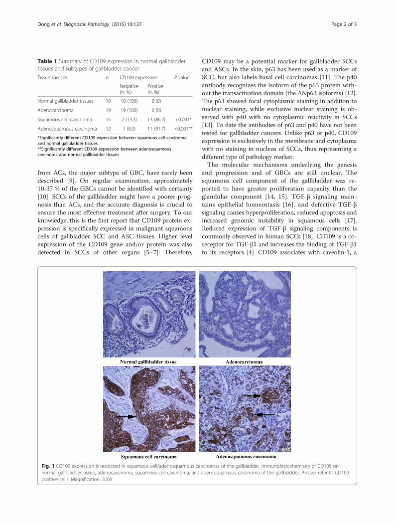
Table 1 Summary of CD109 expression in normal gallbladdertissues and subtypes of gallbladder cancer
Tissue sample n CD109 expression P value
Negative(n, %)
Positive(n, %)
Normal gallbladder tissues 10 10 (100) 0 (0)
Adenocarcinoma 19 19 (100) 0 (0)
Squamous cell carcinoma 15 2 (13.3) 13 (86.7) <0.001*
Adenosquamous carcinoma 12 1 (8.3) 11 (91.7) <0.001**
*Significantly different CD109 expression between squamous cell carcinomaand normal gallbladder tissues**Significantly different CD109 expression between adenosquamouscarcinoma and normal gallbladder tissues
Dong et al. Diagnostic Pathology (2015) 10:137 Page 2 of 3
from ACs, the major subtype of GBC, have rarely beendescribed [9]. On regular examination, approximately10-37 % of the GBCs cannot be identified with certainty[10]. SCCs of the gallbladder might have a poorer prog-nosis than ACs, and the accurate diagnosis is crucial toensure the most effective treatment after surgery. To ourknowledge, this is the first report that CD109 protein ex-pression is specifically expressed in malignant squamouscells of gallbladder SCC and ASC tissues. Higher levelexpression of the CD109 gene and/or protein was alsodetected in SCCs of other organs [5–7]. Therefore,
Fig. 1 CD109 expression is restricted in squamous cell/adenosquamous canormal gallbladder tissue, adenocarcinoma, squamous cell carcinoma, andpositive cells. Magnification: 200X
CD109 may be a potential marker for gallbladder SCCsand ASCs. In the skin, p63 has been used as a marker ofSCC, but also labels basal cell carcinomas [11]. The p40antibody recognizes the isoform of the p63 protein with-out the transactivation domain (the ΔNp63 isoforms) [12].The p63 showed focal cytoplasmic staining in addition tonuclear staining, while exclusive nuclear staining is ob-served with p40 with no cytoplasmic reactivity in SCCs[13]. To date the antibodies of p63 and p40 have not beentested for gallbladder cancers. Unlike p63 or p40, CD109expression is exclusively in the membrane and cytoplasmawith no staining in nucleus of SCCs, thus representing adifferent type of pathology marker.The molecular mechanisms underlying the genesis
and progression and of GBCs are still unclear. Thesquamous cell component of the gallbladder was re-ported to have greater proliferation capacity than theglandular component [14, 15]. TGF-β signaling main-tains epithelial homeostasis [16], and defective TGF-βsignaling causes hyperproliferation, reduced apoptosis andincreased genomic instability in squamous cells [17].Reduced expression of TGF-β signaling components iscommonly observed in human SCCs [18]. CD109 is a co-receptor for TGF-β1 and increases the binding of TGF-β1to its receptors [4]. CD109 associates with caveolin-1, a
rcinomas of the gallbladder. Immunohistochemistry of CD109 onadenosquamous carcinoma of the gallbladder. Arrows refer to CD109

Dong et al. Diagnostic Pathology (2015) 10:137 Page 3 of 3
major component of the caveolae, and facilitateslocalization of the TGF-β receptors into the caveolarcompartment where they degrade [19]. Thus, CD109enhances TGF-β receptor endocytosis and negatively reg-ulates TGF-β signaling. Elevated expression of CD109 ingallbladder squamous cells may inhibit TGF-β signalingand subsequently promote the development of SCC.Taken together, CD109 may involve in the pathogenesis ofgallbladder SCCs and present as a novel target for thera-peutic intervention.
AbbreviationsGBC: Gallbladder cancer; AC: adenocarcinomas; SCC: Squamous cell carcinoma;ASC: adenosquamous carcinoma; TMA: tissue microarrays; TGF-β: Transforminggrowth factor –β.
Competing interestsThe authors declared that they have no competing interests.
Authors’ contributionsFD, CL, XC, YG performed the experiments and analyzed data. JL designedthe experiment, analyzed data and wrote the manuscript. All authors readand approved the final manuscript.
Author informationJL, MD., Ph.D., Professor of Medicine, Associate Chair of Medical ResearchCenter, Shandong Provincial Qianfoshan Hospital, Shandong UniversityJL earned a M.D. degree at Shandong University in China, and a Ph.D.degree at University of Toronto in Canada. Later he performed a researchfellowship at Beth Israel Deaconess Medical Center, and joined the faculty atYale University School of Medicine. In 2012, JL moved to China andestablished a new laboratory at Shandong University. JL’s research focuseson carcinogenesis and angiogenesis. JL has authored a number of papers inscientific journals including Blood, PNAS USA, and Oncogene.
AcknowledgementWe are grateful for the support by Shandong Taishan Scholarship (Ju Liu).
Received: 22 June 2015 Accepted: 29 July 2015
References1. Lai CH, Lau WY. Gallbladder cancer–a comprehensive review. Surgeon.
2008;6(2):101–10.2. Wernberg JA, Lucarelli DD. Gallbladder cancer. Surg Clin North Am.
2014;94(2):343–60. doi:10.1016/j.suc.2014.01.009.3. Gourgiotis S, Kocher HM, Solaini L, Yarollahi A, Tsiambas E, Salemis NS.
Gallbladder cancer. Am J Surg. 2008;196(2):252–64. doi:10.1016/j.amjsurg.2007.11.011.
4. Finnson KW, Tam BY, Liu K, Marcoux A, Lepage P, Roy S, et al. Identificationof CD109 as part of the TGF-beta receptor system in human keratinocytes.FASEB J. 2006;20(9):1525–7. doi:10.1096/fj.05-5229fje.
5. Dong F, Wang Y, Li L, Wang Y, Liu X, Liu J. CD109 expression is increased incutaneous squamous cell carcinoma. J Dermatol. 2014;41(10):947–9.doi:10.1111/1346-8138.12620.
6. Zhang JM, Hashimoto M, Kawai K, Murakumo Y, Sato T, Ichihara M, et al.CD109 expression in squamous cell carcinoma of the uterine cervix. PatholInt. 2005;55(4):165–9. doi:10.1111/j.1440-1827.2005.01807.x.
7. Dong F, Liu F, Yan S, Liu X, Jiang Z, Liu J. Elevated Expression of CD109 inEsophageal Squamous Cell Carcinoma. Pathology oncology research : POR.2015. doi:10.1007/s12253-014-9894-3.
8. Chan KM, Yu MC, Lee WC, Jan YY, Chen MF. Adenosquamous/squamouscell carcinoma of the gallbladder. J Surg Oncol. 2007;95(2):129–34.doi:10.1002/jso.20576.
9. Kim WS, Jang KT, Choi DW, Choi SH, Heo JS, You DD, et al.Clinicopathologic analysis of adenosquamous/squamous cell carcinoma ofthe gallbladder. J Surg Oncol. 2011;103(3):239–42. doi:10.1002/jso.21813.
10. Roa I, Araya JC, Villaseca M, Roa J, de Aretxabala X, Ibacache G. Gallbladdercancer in a high risk area: morphological features and spread patterns.Hepatogastroenterology. 1999;46(27):1540–6.
11. Reis-Filho JS, Torio B, Albergaria A, Schmitt FC. p63 expression in normalskin and usual cutaneous carcinomas. J Cutan Pathol. 2002;29(9):517–23.
12. Henderson SA, Torres-Cabala CA, Curry JL, Bassett RL, Ivan D, Prieto VG,et al. p40 is more specific than p63 for the distinction of atypicalfibroxanthoma from other cutaneous spindle cell malignancies. Am J SurgPathol. 2014;38(8):1102–10. doi:10.1097/PAS.0000000000000245.
13. Alomari AK, Glusac EJ, McNiff JM. p40 is a more specific marker than p63 forcutaneous poorly differentiated squamous cell carcinoma. J Cutan Pathol.2014;41(11):839–45. doi:10.1111/cup.12388.
14. Charbit A, Malaise EP, Tubiana M. Relation between the pathological natureand the growth rate of human tumors. Eur J Cancer. 1971;7(4):307–15.
15. Nishihara K, Nagai E, Izumi Y, Yamaguchi K, Tsuneyoshi M. Adenosquamouscarcinoma of the gallbladder: a clinicopathological, immunohistochemicaland flow-cytometric study of twenty cases. Japanese journal of cancerresearch : Gann. 1994;85(4):389–99.
16. Taylor MA, Lee YH, Schiemann WP. Role of TGF-beta and the tumormicroenvironment during mammary tumorigenesis. Gene Expr.2011;15(3):117–32.
17. White RA, Malkoski SP, Wang XJ. TGFbeta signaling in head and neck squamouscell carcinoma. Oncogene. 2010;29(40):5437–46. doi:10.1038/onc.2010.306.
18. Han G, Wang XJ. Roles of TGFbeta signaling Smads in squamous cellcarcinoma. Cell & bioscience. 2011;1:41. doi:10.1186/2045-3701-1-41.
19. Bizet AA, Liu K, Tran-Khanh N, Saksena A, Vorstenbosch J, Finnson KW, et al.The TGF-beta co-receptor, CD109, promotes internalization and degradationof TGF-beta receptors. Biochim Biophys Acta. 2011;1813(5):742–53.doi:10.1016/j.bbamcr.2011.01.028.
Submit your next manuscript to BioMed Centraland take full advantage of:
• Convenient online submission
• Thorough peer review
• No space constraints or color figure charges
• Immediate publication on acceptance
• Inclusion in PubMed, CAS, Scopus and Google Scholar
• Research which is freely available for redistribution
Submit your manuscript at www.biomedcentral.com/submit