castro_personality traits in rats predict vulnerability - infoscience
TRANSCRIPT

Personality traits in rats predict vulnerability andresilience to developing stress-induced depression-like behaviors, HPA axis hyper-reactivity and brainchanges in pERK1/2 activity
Jorge E. Castro, Shanaz Diessler, Emilio Varea, Cristina Marquez,Marianne H. Larsen, M. Isabel Cordero, Carmen Sandi *
Laboratory of Behavioral Genetics, Brain Mind Institute, School of Life Sciences, Ecole Polytechnique Federale de Lausanne (EPFL),Lausanne, Switzerland
Received 30 August 2011; received in revised form 14 December 2011; accepted 14 December 2011
Psychoneuroendocrinology (2012) 37, 1209—1223
KEYWORDSStress;Personality;Depression;Anxiety;Exploration;Corticosterone;ERK1/2
Summary Emerging evidence indicates that certain behavioral traits, such as anxiety, areassociated with the development of depression-like behaviors after exposure to chronic stress.However, single traits do not explain the wide variability in vulnerability to stress observed inoutbred populations. We hypothesized that a combination of behavioral traits might provide abetter characterization of an individual’s vulnerability to prolonged stress. Here, we sought todetermine whether the characterization of relevant behavioral traits in rats could aid inidentifying individuals with different vulnerabilities to developing stress-induced depression-like behavioral alterations. We also investigated whether behavioral traits would be related tothe development of alterations in the hypothalamic-pituitary-adrenal axis and in brain activity —as measured through phosphorylation of extracellular signal-regulated kinase 1/2 (ERK1/2) — inresponse to an acute stressor following either sub-chronic (2 weeks) or chronic (4 weeks)unpredictable stress (CUS). Sprague-Dawley rats were characterized using a battery of behavioraltasks, and three principal traits were identified: anxiety, exploration and activity. Whencombined, the first two traits were found to explain the variability in the stress responses.Our findings confirm the increased risk of animals with high anxiety developing certain depres-sion-like behaviors (e.g., increased floating time in the forced swim test) when progressivelyexposed to stress. In contrast, the behavioral profile based on combined low anxiety and lowexploration was resistant to alterations related to social behaviors, while the high anxiety and lowexploration profile displayed a particularly vulnerable pattern of physiological and neurobiologi-cal responses after sub-chronic stress exposure. Our findings indicate important differences in
* Corresponding author at: Laboratory of Behavioral Genetics, Brain Mind Institute, Ecole Polytechnique Federale de Lausanne (EPFL), Station19, CH-1015 Lausanne, Switzerland. Tel.: +41 021 693 1762; fax: +41 021 693 9636.
E-mail address: [email protected] (C. Sandi).
Available online at www.sciencedirect.com
j our na l h omepa g e: www.e l se v ie r.c om/l oca te/ psyne ue n
0306-4530/$ — see front matter # 2011 Elsevier Ltd. All rights reserved.
doi:10.1016/j.psyneuen.2011.12.014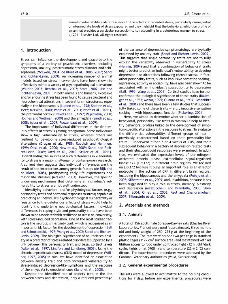
animals’ vulnerability and/or resilience to the effects of repeated stress, particularly during initialor intermediate levels of stress exposure, and they highlight that the behavioral inhibition profile ofan animal provides a particular susceptibility to responding in a deleterious manner to stress.# 2011 Elsevier Ltd. All rights reserved.
1210 J.E. Castro et al.
1. Introduction
Stress can influence the development and exacerbate thesymptoms of a variety of psychiatric disorders, includingdepression, anxiety, posttraumatic stress disorder and schi-zophrenia (McEwen, 2004; de Kloet et al., 2005, 2007; Sandiand Richter-Levin, 2009). An increasing number of animalmodels based on stress interventions have been shown toeffectively mimic a variety of psychopathological alterations(Willner, 2005; Renthal et al., 2007; Stam, 2007; Ilin andRichter-Levin, 2009). In both animals and humans, excessiveand/or enduring stress has been found to cause structural andneurochemical alterations in several brain structures, espe-cially in the hippocampus (Lupien et al., 1998; Sheline et al.,1999; McEwen, 2000; Pham et al., 2003; Bisaz et al., 2011),the prefrontal cortex (Drevets et al., 1997; Rajkowska, 2000;Holmes and Wellman, 2009) and the amygdala (Sandi et al.,2008; Mitra et al., 2009; Roozendaal et al., 2009).
The importance of individual differences in the deleter-ious effects of stress is gaining recognition. Some individualsshow a high vulnerability to stress, whereas others areresilient to developing stress-induced psychopathologicalalterations (Drugan et al., 1989; Rudolph and Hammen,1999; Oitzl et al., 2000; New et al., 2009; Sandi and Rich-ter-Levin, 2009; Oitzl et al., 2010; Stiller et al., 2011).Understanding the sources of such differences in vulnerabil-ity to stress is a major challenge for contemporary research.A current view suggests that individual differences are theresult of the interactions among genetic factors (de Rijk andde Kloet, 2005), predisposing early life experiences andmajor life stressors (McEwen, 2003). However, the specificunderlying mechanisms that determine an individual’s vul-nerability to stress are not well understood.
Identifying behavioral and/or physiological factors (e.g.,personality traits and hormonal responses to stress) capable ofpredicting an individual’s psychopathological vulnerability orresistance to the deleterious effects of stress would help toidentify the underlying neurobiological factors. Individualdifferences in coping style and personality traits have beenshown to be associated with resilience to stress or, conversely,with stress-induced depression. One of the most studied fac-tors is the neuroticism-anxiety trait, which is recognized as animportant risk factor for the development of depression (Balland Schottenfeld, 1997; Wang et al., 2002; Sandi and Richter-Levin, 2009). The biological significance of neuroticism-anxi-ety as a predictor of stress-related disorders is supported by alink between this personality trait and basal cortisol levels(Adler et al., 1997; Lindfors and Lundberg, 2002). Using thechronic unpredictable stress (CUS) model of depression (Will-ner, 1997, 2005) in rats, we have identified an associationbetween anxiety trait and both increased vulnerability tostress-induced depression-like symptoms and the responseof the amygdala to emotional cues (Sandi et al., 2008).
Despite the identified role of anxiety trait in the linkbetween stress and depression, only a reduced percentage
of the variance of depressive symptomatology are typicallyexplained by anxiety trait (Sandi and Richter-Levin, 2009).This suggests that single personality traits are not to fullyexplain the variability observed in vulnerability to stress(Hennig, 2004) and that a combination of behavioral traitsmight better predict an individual’s vulnerability to developdepression-like alterations following chronic stress. In fact,other personality traits, such as impulsive sensation seeking,aggression, activity or sociability, have also been shown to beassociated with an individual’s susceptibility to depression(Ball, 1995; Wang et al., 2004). Cortisol studies have furtherconfirmed the biological significance of these traits (Ballen-ger et al., 1983; Mazur, 1995; Gunnar et al., 1997; Rosenblittet al., 2001) and there have been a few studies that success-fully linked some of these traits — e.g., impulsive sensationseeking — with hippocampal function (Pickering, 2004).
Here, we aimed to determine whether a combination ofbehavioral, personality-like traits in rats would help to iden-tify behavioral profiles linked to the development of symp-tom-specific alterations in the response to stress. To evaluatethe differential vulnerability, different groups of rats —previously characterized based on their personality-liketraits — underwent either 2 or 4 weeks of CUS, and theirsubsequent behavior in a battery of depression-related testsand their glucocorticoid responses were examined. In addi-tion, we evaluated the expression levels of the mitogen-activated protein kinase extracellular signal-regulatedkinase 1/2 (ERK1/2) in different brain regions. We focusedon ERK1/2 because it plays an important role as an effectormolecule in the actions of CRF in different brain regions,including the hippocampus and the amygdala (Refojo et al.,2005; Silberstein et al., 2009) and, in addition, because it hasbeen suggested to play a role in stress, memory, plasticityand depression (Mazzucchelli and Brambilla, 2000; Shenet al., 2004; Qi et al., 2006; Reul and Chandramohan,2007; Silberstein et al., 2009).
2. Materials and methods
2.1. Animals
A total of 156 adult male Sprague-Dawley rats (Charles RiverLaboratories, France) were used (approximately three monthsold and body weight of 250—275 g at the beginning of theexperiment). The rats were housed two per cage in standardplastic cages (1177 cm2 surface area) and maintained with adlibitum access to food under controlled light (12 h light/darkcycle; lights on at 0700 h) and temperature (22 � 2 8C) con-ditions. The experimental procedures were approved by theCantonal Veterinary Authorities (Vaud, Switzerland).
2.2. General experimental procedures
The rats were allowed to acclimatize to the housing condi-tions for 7 days before any experimental procedures were
EPM OF/NO LD C C
EPM OF/NO LD C C CUS 4w
CUS 2w
Blood samples
Perfusio n
Perfusio n
Behavioral characterization Behavioral outcomesStress
H
H
Day 1 3 5 7 10 24 38 39 40 41 +15min
SC SP FST
SC SP FST
Figure 1 Schematic plan of the experimental procedures. After handling (H), the rats were characterized using the elevated plusmaze (EPM), the open field and novel object test (OF/NO), the light and dark box (LD) and the circular corridor (CC). After three days ofrest, the rats were submitted to the chronic unpredictable stress protocol (CUS) for either 2 or 4 weeks. Depression-like behaviors wereevaluated using the saccharin consumption test (SC), the social preference test (SP), and the forced swim test (FST). Blood sampleswere taken 15 min after the end of the FST, and the animals were subsequently euthanized by transcardiac perfusion to process thebrains for the immunohistochemical essays.
Anxiety traits and vulnerability to depression 1211
initiated. Before the start of the CUS protocol, the animalswere tested in a battery of behavioral tasks, including theelevated plus maze (EPM), the open field/novel object test(OF/NO), the light and dark box (LD) and the circular corridor(CC), to determine their individual personality traits. Tostudy the effect of individual differences on the impact ofthe stress protocol, the animals were matched according totheir behavioral traits and body weights to ensure that thesame personality traits and body weight distributions wereobtained in each group. Subsequently, the matched rats wererandomly assigned to one of the following experimentalgroups: (1) control (daily handling), (2) chronic unpredictablestress for 14 days, and (3) chronic unpredictable stress for28 days.
Behavioral indexes of the impact of CUS were evaluatedusing a saccharin consumption test, a social preference testand a forced swim test performed on days zero, three andfive, respectively, after exposure to CUS (Fig. 1). The bodyweight was measured the day before starting the stress-induction protocol and then every 3rd day throughout thestress period.
2.3. Characterization of behavioral traits
2.3.1. Elevated plus mazeAnxiety levels were evaluated using the EPM test (Herreroet al., 2006). Briefly, the test consists of two opposing openarms (45 cm � 10 cm) perpendicular to two enclosed arms(45 cm � 10 cm � 50 cm) that extend from a central plat-form (10 cm � 10 cm), elevated 65 cm above the floor. Therats were placed individually on the central platform andallowed to explore the maze for 5 min. Their behavior wasmonitored using a video camera and analyzed with a com-puterized tracking system (EthoVision 3.1.16, Noldus IT, TheNetherlands). The time spent in the open and closed arms,distance moved and transitions between the different armswere recorded.
2.3.2. Open field and novel object reactivityAnxiety, exploration and activity-related behaviors weretested in the open field (OF) and novel object (NO) tests.The open field test consisted of a black circular arena (1 m indiameter and 40 cm high). The floor of the arena was dividedinto three zones: the outer zone, with a diameter of 1 m; the
inner zone, with a diameter of 75 cm; and the center zone,with a diameter of 25 cm. The light was adjusted to a level of8—10 lx in the center of the pool. The animals were placed inthe center of the arena, and the open field activity wastested over a 10 min period. Subsequently, a novel object wasintroduced into the center of the arena, and the animal’sbehavior was observed over the following 5 min. The activityand behavior during the whole session was recorded with avideo camera and automatically registered and analyzedwith the EthoVision computerized tracking system (Color-Pro 3.0.15, Noldus Information Technology, The Nether-lands). The time spent in each of the virtually created zones(central zone, 25 cm diameter in the center of the apparatus;inner zone, 75 cm diameter in the center of the apparatus;and outer zone, 12.5 cm in the periphery of the apparatusnext to the walls) was measured. Behavioral scoring of thetime spent freezing and the time spent touching the novelobject was done manually (Larsen et al., 2010).
2.3.3. Light and dark boxAnxiety levels were also evaluated using a modified light anddark box (Crawley and Goodwin, 1980) consisting of twoequally sized chambers (24 cm high � 25 cm wide � 33 cmlong) that were connected by an 8 cm � 8 cm opening. Onechamber was black and covered by a lid (i.e., the dark box).The alternate chamber was white and remained uncoveredduring the test (i.e., the light box). A light positioned 165 cmabove the light box provided illumination at a level of 500 lx.There was no appreciable illumination (i.e., <2 lx) in thedark box. The rats were placed in the center of the light boxand allowed to freely explore the apparatus for 5 min. Theapparatus was cleaned with a 5% alcohol solution beforetesting each rat. Behaviors recorded during the testincluded: (1) the latency to enter the dark box, (2) thelatency to return to the light box after entering the darkchamber, (3) the percent of time spent in the light box, (4)the number of chamber transitions, and (5) rearing in thelight box.
2.3.4. Circular corridorExploration and activity-related behaviors were evaluatedusing a modified circular corridor (Rotllant et al., 2010;Piazza et al., 1989) consisting of a black circular arena(80 cm in diameter � 34 cm high) with a cylinder placed in

1212 J.E. Castro et al.
the center (50 cm in diameter), forming a circular corridor14 cm wide. The test was performed under infrared light, andthe behavior during the entire session was recorded with avideo camera that detected infrared light and was automa-tically registered and analyzed with the EthoVision compu-terized tracking system (Color-Pro 3.0.15, NoldusInformation Technology, The Netherlands). During the30 min test, the rat was placed inside the corridor and wasallowed to move around the circular track between the walls.The following measures were taken in intervals of 5 min: (1)the total distance traveled, (2) the time spent in movement,and (3) the number of defecations.
2.4. Chronic unpredictable stress
As in our previous studies (Sandi et al., 2008; Larsen et al.,2010), the rats submitted to the CUS protocol were subjectedeach day to one of nine stressors in an unpredictable orderand at an unpredictable time of the day for a period of either2 or 4 weeks. The stressors included acoustic stimulation(78—115 dB 20—40 ms noise bursts; intertrial intervals ran-ged from 4 to 22 s and averaged 13 s: total duration ofsession: 15 min), inverse light and dark cycle (over a 48 hperiod), exposure to overcrowding under a bright light (sixrats in a standard home cage, 1000 lx, 2 h), inescapable footshock (three foot shocks of 1 mA, 1 s, 1 min intershockinterval), an elevated platform (rats were placed on a plat-form (20 cm � 20 cm) elevated 1 m above the ground for2 h), predator odor (1 h exposure to 2,5-dihydro-2,4,5-tri-methylthiazoline, a synthetic compound originally isolatedfrom fox feces; Fendt and Endres, 2008), bright light andwater deprivation (1000 lx, 30 min), food deprivation (24 h),and forced swim stress (15 min). The stressed pairs of ratswere housed separated by transparent plastic wall, whichallowed visual and olfactory contact between them. Non-stressed control rats were housed 2 per cage without theseparator and were briefly handled every day during thestress period.
2.5. Behavioral testing
2.5.1. Saccharin consumptionA saccharin consumption test was performed on days 0, 7, 14,21 and 28 of the stress period. The rats were given a freechoice between two bottles, one containing normal drinkingwater and the other containing a 0.02% saccharin solution.The bottles were available for 12 h. The saccharin intake wascalculated as the amount consumed in grams per 100 g bodyweight. The saccharin preference was calculated as sac-charin intake/total fluid intake (water + saccharin) (Larsenet al., 2010).
2.5.2. Social preferenceOn the third day after CUS, the social preference test wasconducted in a rectangular, three chambered box (a centercompartment of 20 cm � 35 cm � 35 cm with a left and aright compartment of 30 cm � 35 cm � 35 cm) fabricatedfrom opaque gray polycarbonate. The dividing walls hadretractable doorways allowing access to each chamber.The test rat was placed in the middle chamber and allowedto explore the entire apparatus for 10 min. Each of the two
side chambers contained an empty wire cage. The wire cageswere 10 cm in height, with a bottom diameter of 9 cm andbars spaced 1 mm apart. A weighted plastic cone was placedon the top of each cage to prevent climbing by the test rats.Four sets of wire cages were used during the experiment, andall of the cages were washed with water and dried properlybetween each use.
For habituation to the wire cage, each novel pre-pubertalmale rat used in the social interaction test had been pre-viously placed in the wire cage in the apparatus without thetest rat for 5 min on 3 consecutive days preceding the socialtest. On the day after the last habituation session, a test ratwas placed in the center compartment and allowed toexplore the entire apparatus for 10 min. An unfamiliar ratwas placed in one of the wire cages located on either side ofthe social test box during the 10 min session. A rectangularcolored object was placed in the other wire cage on the otherside of the box. The location of the stranger and the object inthe left and right sides of the chamber was counterbalancedfor different animals. Placing the strange rat in a wire cageprevented direct physical contact between the rats andensured that the social approach was only initiated by thetest rat. The time spent sniffing each wire cage was video-recorded and manually scored to evaluate the level of pre-ference for the unfamiliar rat as compared to the object. Theentire apparatus was cleaned with water and dried thor-oughly between each tested rat.
2.5.3. Forced-swim testOn day 5 after the CUS, the animals were subjected to aforced swim test (Porsolt et al., 1977) to evaluate potentialdifferences in depression-like behavior between thestressed and control animals (Castro et al., 2010; Larsenet al., 2010). Briefly, the animals were individually placed ina plastic beaker (25 cm in diameter, 46 cm deep) containing30 cm of water (25 � 1 8C) for 5 min. Their behavior wasrecorded with a video camera, and the time spent immobile(making only those movements necessary to keep the snoutabove the water), swimming, climbing and diving was quan-tified using a computer program (The Observer 5.0.25,Noldus IT, 2003). Although the standard use of the forcedswim test when applied for psychopharmacology purposestypically includes a first 15-min induction session followedby a 5-min testing session 24 h later, our study — not focusingon drug effects but on the assessment of the impact of theprior stress experience — only included a single 15-min testsession. The reason for this procedure was two-fold. First, asjust indicated, to serve as a readout of the impact ofexposure to different lengths of repeated stress in differentanimal groups. Second, to act as a stress challenge for theimmediate evaluation of ERK activation without the con-founding of potential memory factors that could be mani-fested on a second, repetition test.
2.6. Histological procedures
Fifteen minutes after the end of the forced-swim test, theanimals were anesthetized with isoflurane and transcardiallyperfused using a 0.9% saline solution followed by a fixativesolution of paraformaldehyde 4% in phosphate buffered sal-ine (PBS, pH = 7.5). After perfusion-fixation, the brains wereremoved and post-fixed in the same solution for 4 h. Next,

Anxiety traits and vulnerability to depression 1213
50 mm thick coronal sections were cut on a vibratome (VT1000S; Leica, Glattbrugg, Switzerland) and stored at 4 8Cin PBS. Brain ERK1/2 activity was assayed by staining forpERK1/2.
An alternate series of 1 in 10 sections was processed‘‘free-floating’’ for immunohistochemical visualization ofpERK1/2. Briefly, the sections were incubated with 10%H2O2 in PBS for 10 min to block the endogenous peroxidaseactivity. After washing in PBS (3� 10 min), the sections weretreated for 1 h with 10% normal donkey serum (NDS, JacksonImmunoResearch Laboratories, West Grove, PA) in PBS with0.2% Triton-X100 (Sigma—Aldrich, St. Louis, MO). After wash-ing in PBS, the sections were incubated overnight at 4 8C withrabbit polyclonal anti pERK1/2 IgG, which stains the acti-vated form of ERK1/2 (1:500; Cell Signaling Technology). PBScontaining 0.2% Triton-X100 and 5% NDS (PBST) was used todilute the primary and secondary antibodies. After washing inPBS, the sections were incubated for 120 min with goat anti-rabbit IgG (1:2000; Jackson ImmunoResearch Laboratories).After washing in PBS, the sections were incubated for 1 h withthe avidin—biotin—peroxidase complex (ABC; A 1:200, B1:200; Vector Laboratories, Peterborough, UK), which wasprepared 30 min prior to incubation in PBS. After washing inPBS, color development was achieved by incubating thesections in 3,30-diaminobenzidine tetrahydrochloride (DAB;0.5 mg/ml, Sigma—Aldrich) for 15 min.
2.7. Image analysis
The levels of brain ERK1/2 activity (phosphorylated ERK1/2;pERK1/2) were estimated by densitometric analyses of thestaining in the basolateral, central, medial and basomedialnuclei of the amygdala, the CA1, the CA3 and the dentategyrus of the hippocampus, and the corpus callosum. Digitalpictures were taken from coronal sections using a NorthernLight transilluminator (model R95) and a Photometrics Cool-snap camera (Roper scientific) system. Protein expressionlevels were semi-quantitatively determined by measuringthe optical densities and the number of pixels in the definedareas with Analysis Pro 5.0. The mean pERK1/2 proteinexpression levels per area are expressed in arbitrary units(number of pixels per optic density). All of the comparedsamples were processed in the same immunohistochemicalassay to avoid inter-assay variability. The optical density datafor all of the brain regions were normalized to the opticaldensity obtained for the corpus callosum.
2.8. Corticosterone analyses
Corticosterone responses to stress were evaluated in plasmasamples obtained 15 min after testing in the forced swimtest. Plasma was obtained from blood collected cardiacpuncture immediately after anesthesia and just before per-fusion. Three hundred microliters of blood was taken using acapillary tube containing heparin (Sarted, Germany). Theblood samples were centrifuged, and the plasma was storedat �20 8C until the assay. The plasma corticosterone levelswere quantified using the Correlate-EIA CorticosteroneEnzyme Immunoassay Kit (Assay Design, MI, USA) accordingto the manufacturer’s instructions. The intra-assay coeffi-cient of variance was 8%.
2.9. Statistical analyses
The SPSS 13.0 (SPSS, Chicago, IL) statistical package was usedfor the statistical analyses. The normality and homogeneityof variance of the data were tested, and the adjustedstatistics were used as required.
2.9.1. Factorial analysesTwo-fold exploratory factor analyses were used to obtain acontinuous interval scale score by using principal componentsas an extraction method and the varimax rotation with theKaiser normalization rotation (Doremus et al., 2006). Indivi-dual factor scores from the parameters of the EPM, OF/NO,LD and CC were calculated for each subject based on therelative weight and orientation (eigenvalues) of the loadingof the parameters for each obtained factor. The scores weregenerated using a Z distribution, where a value of 0 corre-sponds to the mean, and the values were expressed in termsof their standard deviations. The animals were matchedbased on their scores for the different factors and classifiedinto different experimental groups to yield groups withsimilar personality-like traits. In addition, the data fromthe factorial analyses were used to investigate the modula-tory effect of the factor score differences on the stresseffects investigated. The animals were classified into groupssuch that the group scores were either above or below theoverall mean for each of the factors. Using the extreme lowand high values of the continuous scores obtained from thefactor analyses, categorical dichotomous scores wereobtained by classifying the subjects into groups of low andhigh scores for each factor; the subjects with scores within�0.25 standard deviations from the mean were excludedfrom this classification.
2.9.2. Parametric statisticsThe general effects of chronic stress on body weight gain,behavioral indexes, corticosterone response, and ERK1/2activity were analyzed with one-way ANOVAs followed bypost hoc comparisons. Sequential three-way ANOVAs fol-lowed by simple effects analyses were used to assess theinteraction effect of chronic stress conditions and the levelsof anxiety exploration traits (3 � 2 � 2, whenever analysesincluding the three personality-like traits were not signifi-cant; analyses of different combinations of two traits fol-lowed 2 � 2 � 2) on body weight gain, behavioral indices,corticosterone response, and ERK1/2.
3. Results
3.1. Characterization of personality traits
To characterize the animals according to their personalitytraits, factorial analyses were applied separately to a rangeof extracted parameters from the EPM, OF/NO, LD and CCtests, which were conducted before the animals’ exposure tothe CUS (Table S1). Then, an overall factor analysis wasperformed on the extracted factors, and this analysisrevealed three factors, which were termed activity, anxietyand exploration according to the parameters that definedthem (Table S1). The data obtained from the animals’ scoresfor each of these factors were used to classify the animals

1214 J.E. Castro et al.
into dichotomized variables for each behavioral trait (i.e.,activity, anxiety and exploration). Thus, the animals wereclassified as follows according to whether their score wasabove or below the mean for each factor: low (LL) or high(HL) locomotion, low (LA) or high (HA) anxiety, and low (LE)or high (HE) exploration. The orthogonality of the personalitytraits was confirmed by comparing the mean scores for eachvariable (see Fig. S1 for an example of no differences in theexploration trait values between the animals classified aseither LA or HA). Throughout the study, factorial ANOVAs withthe three personality traits revealed a lack of significantinteraction (n.s.). Further factorial ANOVAs that were per-formed on combinations of two behavioral traits as thefactors did not yield statistical significance, except when‘anxiety’ and ‘exploration’ traits were combined. These twobehavioral traits were also previously identified to explainvariance in stressful learning (Salehi et al., 2010). Therefore,this factorial ANOVA will subsequently be referred to inthe text.
3.2. Effects of CUS and personality traits on bodyweight
One-way ANOVAs were performed to assess the impact of twoand four weeks of CUS on body weight. The ANOVA showed asignificant effect of chronic stress on body weight gain duringthe CUS period [F(2,123) = 34.13; p = 0.001]. Post hoc ana-lyses showed a progressive decrease after two ( p = 0.001)and four weeks ( p = 0.001) of CUS compared to the controlgroup. Moreover, the group receiving four weeks of CUSdiffered from the group receiving two weeks of CUS( p = 0.05) (Fig. 2A). Three-way ANOVAs were performed toassess the impact of CUS, and anxiety and exploration traitson body weight. ANOVA showed a significant interactioneffect on body weight gain [F(2,114) = 3.60; p = 0.03]. Afurther simple effect analysis indicated a decrease in bodyweight in the groups receiving two weeks of CUS compared tothe control group in all of the personality profiles except the
Stress 4 weeks
Stress 2 weeks
Control
Rel
ativ
e b
od
y w
eig
ht
gai
n (
% c
on
tro
l)
115
110
105
100
95
90
85
80Anxiety Lo w
Exploration L
Rel
ativ
e b
od
y w
eig
ht
gai
n (
% c
on
tro
l)
115
110
105
100
95
90
85
80
*
**
****
**
A B
Figure 2 A. The effects of chronic unpredictable stress on body weiCUS: n = 28. B. Interaction effect of chronic unpredictable stress ann = 10, LA/HE n = 14, HA/LE n = 16, HA/HE n = 16; 2 weeks of CUS: LACUS: LA/LE n = 7, LA/HE n = 6, HA/LE n = 5, HA/HE n = 10. *p < 0.0
‘low anxiety and low exploration’ profile (LA/HE p = 0.02;HA/LE p = 0.001; HA/HE p = 0.001). Additionally, only the‘low anxiety and low exploration’ ( p = 0.03) and ‘high anxi-ety and high exploration’ ( p = 0.001) personality profilesshowed significant decreases in body weight in the groupreceiving four weeks of CUS compared to the group receivingtwo weeks of CUS (Fig. 2B).
3.3. Effects of CUS and personality traits ondepression-like behaviors
One-way ANOVAs were performed to assess the impact of twoand four weeks of CUS on depression-like behaviors, includinganhedonia measured by saccharin intake in a preferencetask, social avoidance measured by the interaction with aconspecific juvenile animal versus an object in a preferencetask and immobility in the forced swim test. Three-wayANOVAs were performed to assess the impact of CUS, anxietyand exploration traits on these depression-like behaviors.
3.3.1. Effects of CUS and personality traits onsaccharin consumptionAn ANOVA showed a significant effect of chronic stress onanhedonia measured by saccharin intake in a preference task[F(2,139) = 8.16; p = 0.001]. Post hoc analyses showed adecrease in saccharin intake both in animals receiving two( p = 0.001) or four ( p = 0.02) weeks of CUS compared to thecontrol group. This result was observed both when analyzed asaverage intake (Fig. 3A) and as consumption across weeklytesting (Fig. 3B). A factorial ANOVA showed non-significantinteractions of the personality traits with anhedonia measuredby saccharin intake in a preference task (data not shown).
3.3.2. Effects of CUS and personality traits on socialpreferenceAn ANOVA showed a significant effect of chronic stress onsocial avoidance measured by the interaction with an animalversus an object in the preference task [F(2,128) = 33.38;
Anxiety High Exploration High
Anxiety High Exploration Low
Anxiety Low Exploration Highow
****
****
****
ght gain. Control group: n = 56, 2 weeks of CUS: n = 42, 4 weeks ofd personality traits on body weight gain. Control group: LA/LE/LE n = 10, LA/HE n = 12, HA/LE n = 6, HA/HE n = 14; 4 weeks of5, **p < 0.01.
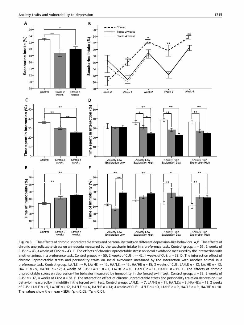
Figure 3 The effects of chronic unpredictable stress and personality traits on different depression-like behaviors. A,B. The effects ofchronic unpredictable stress on anhedonia measured by the saccharin intake in a preference task. Control group: n = 56, 2 weeks ofCUS: n = 43, 4 weeks of CUS: n = 43. C. The effects of chronic unpredictable stress on social avoidance measured by the interaction withanother animal in a preference task. Control group: n = 50, 2 weeks of CUS: n = 42, 4 weeks of CUS: n = 39. D. The interaction effect ofchronic unpredictable stress and personality traits on social avoidance measured by the interaction with another animal in apreference task. Control group: LA/LE n = 9, LA/HE n = 13, HA/LE n = 13, HA/HE n = 15; 2 weeks of CUS: LA/LE n = 12, LA/HE n = 13,HA/LE n = 5, HA/HE n = 12; 4 weeks of CUS: LA/LE n = 7, LA/HE n = 10, HA/LE n = 11, HA/HE n = 11. E. The effects of chronicunpredictable stress on depression-like behavior measured by immobility in the forced swim test. Control group: n = 39, 2 weeks ofCUS: n = 37, 4 weeks of CUS: n = 38. F. The interaction effect of chronic unpredictable stress and personality traits on depression-likebehavior measured by immobility in the forced swim test. Control group: LA/LE n = 7, LA/HE n = 11, HA/LE n = 8, HA/HE n = 13; 2 weeksof CUS: LA/LE n = 5, LA/HE n = 12, HA/LE n = 6, HA/HE n = 14; 4 weeks of CUS: LA/LE n = 10, LA/HE n = 9, HA/LE n = 9, HA/HE n = 10.The values show the mean + SEM; *p < 0.05, **p < 0.01.
Anxiety traits and vulnerability to depression 1215

1216 J.E. Castro et al.
p = 0.001], and post hoc analyses showed a progressivedecrease in the interaction with the conspecific juvenileafter two ( p = 0.001) and four ( p = 0.001) weeks of CUS(Fig. 3B). A factorial ANOVA showed a significant interactioneffect on social avoidance measured by the interaction withanother animal in a preference task [F(2,119) = 3.61;p = 0.03], and a simple effect analysis demonstrated adecrease in the interaction with another animal in animalsreceiving two (LA/HE p = 0.04; HA/LE p = 0.001; HA/HEp = 0.001) or four (LA/HE p = 0.001; HA/LE p = 0.001; HA/HE p = 0.001) weeks of CUS compared to the control group forall of the personality profiles except the ‘low anxiety and lowexploration’ profile (Fig. 3C).
3.3.3. Effects of CUS and personality traits on theforced swim testAn ANOVA showed a significant effect of chronic stress ondepression-like behavior measured by immobility in theforced swim test [F(2,111) = 16.03; p = 0.001], and posthoc analyses showed an increase in the time spent immobilein animals receiving either two ( p = 0.001) or four ( p = 0.001)weeks of CUS compared to the control group (Fig. 3C). Afactorial ANOVA showed a significant interaction effect ondepression-like behavior measured by immobility in theforced swim test [F(2,102) = 3.48; p = 0.03], and simpleeffect analyses showed an increase in the time spent immo-bile in the animals receiving two weeks of CUS compared tothe control group only in the ‘high anxiety and low explora-tion’ ( p = 0.001) and ‘high anxiety and high exploration’( p = 0.001) personality profiles (Fig. 3E).
3.4. Effects of CUS and personality traits on thecorticosterone response
One-way ANOVAs were performed to assess the impact of twoand four weeks of CUS on the corticosterone response toacute stress, which was analyzed 15 min after the forced
Stress 4 weeks
Stress 2 weeks
Control
Co
rtic
ost
ero
ne
(ng
/ml)
450
400
350
300
250
200
150
100
50
0Anxiety Lo
Exploration
Co
rtic
ost
ero
ne
(ng
/ml)
450
400
350
300
250
200
150
100
50
0
**
**
A B
Figure 4 A. The effects of chronic unpredictable stress on corticon = 22, 2 weeks of CUS: n = 39, 4 weeks of CUS: n = 14. B. The interacticorticosterone levels after 15 min of forced swimming. Control groupCUS: LA/LE n = 10, LA/HE n = 13, HA/LE n = 6, HA/HE n = 11; 4 weeksdata show the mean + SEM; *p < 0.05, **p < 0.01.
swim test. An ANOVA showed a significant effect of chronicstress on the corticosterone response [F(2,72) = 9.09;p = 0.001], and post hoc analyses showed a gradual increasein the corticosterone response both in animals receiving twoweeks ( p = 0.02) or four weeks ( p = 0.001) of CUS comparedto the control group (Fig. 4A). Factorial ANOVAs were per-formed to assess the impact of CUS and anxiety and explora-tion traits on the corticosterone response. An ANOVA showeda marginal interaction effect on the corticosterone responseto the acute stress induced by the forced swim test[F(2,67) = 2.58; p = 0.08], and simple effect analyses indi-cated an increase in the corticosterone response in theanimals receiving two weeks of CUS compared to the controlgroup only in animals with the ‘high anxiety and low explora-tion’ ( p = 0.001) personality profile. Additionally, an increasein the corticosterone response in animals receiving fourweeks of CUS compared to animals receiving two weeks ofCUS was only found in the ‘low anxiety and low exploration’( p = 0.04) and ‘high anxiety and high exploration’ ( p = 0.04)personality profiles (Fig. 4B).
3.5. Effects of CUS and personality traits onbrain ERK1/2 activity
One-way ANOVAs were performed to assess the impact of twoand four weeks of CUS on brain ERK activity, and factorialANOVAs were performed to assess the impact of CUS andanxiety and exploration traits on brain ERK activity. AnANOVA showed no significant effect of chronic stress on pERKlevels in the corpus callosum, which was used as a negativecontrol for pERK immunohistochemical signal (n.s.; data notshown).
3.5.1. Effects of CUS and personality traits on ERK1/2activity in the amygdalaANOVAs showed a significant effect of chronic stress on pERKlevels in the basolateral [F(2,91) = 4.80; p = 0.01] and
Anxiety High Exploration High
Anxiety High Exploration Low
Anxiety Low Exploration High
w Low
***
**
sterone levels after 15 min of forced swimming. Control group:on effect of chronic unpredictable stress and personality traits on: LA/LE n = 6, LA/HE n = 4, HA/LE n = 7, HA/HE n = 6; 2 weeks of
of CUS: LA/LE n = 4, LA/HE n = 4, HA/LE n = 4, HA/HE n = 4. The

Anxiety High Exploration High
Anxiety High Exploration Low
Anxiety Low Exploration High
Anxiety Low Exploration Low
pE
RK
OD
Am
ygd
ala
120
90
60
30
0
Stress 4 weeks
Stress 2 weeks
Control
pE
RK
OD
BL
A120
90
60
30
0Stress 4 weeks
Stress 2 weeks
Control
pE
RK
OD
CeA
120
90
60
30
0
**
**
A B
C D
Stress 4 weeks
Stress 2 weeks
Control
pE
RK
OD
BM
A
120
90
60
30
0
** **
*** *
***
Figure 5 A. The effects of chronic unpredictable stress on ERK activity in the basolateral amygdala. Control group: n = 30, 2 weeks ofCUS: n = 46, 4 weeks of CUS: n = 18. B. The effects of chronic unpredictable stress on ERK activity in the central amygdala. Controlgroup: n = 30, 2 weeks of CUS: n = 46, 4 weeks of CUS: n = 18. C. The effects of chronic unpredictable stress on ERK activity in thebasomedial amygdala. Control group: n = 29, 2 weeks of CUS: n = 45, 4 weeks of CUS: n = 18. D. The interaction effect of chronicunpredictable stress and personality traits on ERK activity in the amygdala. Control group: LA/LE n = 6, LA/HE n = 6, HA/LE n = 9, HA/HE n = 9; 2 weeks of CUS: LA/LE n = 14, LA/HE n = 13, HA/LE n = 6, HA/HE n = 13; 4 weeks of CUS: LA/LE n = 5, LA/HE n = 4, HA/LE n = 5,HA/HE n = 4. The data show the mean + SEM; *p < 0.05, **p < 0.01.
Anxiety traits and vulnerability to depression 1217
basomedial [F(2,89) = 7.38; p = 0.001] amygdala, and posthoc analyses showed a ‘‘U-shaped’’ effect of stress in both ofthese regions. Thus, after two weeks of CUS, there was asignificant decrease of pERK levels compared to controls( p = 0.05 and p = 0.04, respectively) and animals receivingfour weeks of CUS ( p = 0.02 and p = 0.001, respectively)(Fig. 5A and C). An ANOVA also showed a significant effectof chronic stress on pERK levels in the central amygdala[F(2,91) = 4.96; p = 0.01], and post hoc analyses specifiedan increase in pERK in the central amygdala after four weeksof CUS compared to two weeks of CUS ( p = 0.01) (Fig. 5B).
A factorial ANOVA indicated a significant interactioneffect on pERK levels in the amygdala (a compound measurefor this nucleus was used because the data for individualnuclei depicted similar results) (F(2,82) = 3.51; p = 0.03).Inthe ‘high anxiety and low exploration’, the groups receivingtwo ( p = 0.05) or four ( p = 0.001) weeks of CUS showed anincrease in pERK levels compared to the control group. This isat odds with the other three behavioral profiles, that in allcases showed a decrease in pERK1/2 levels after two weeks ofCUS as compared to controls (LA/LE p = 0.05; LA/HE p = 0.04;
HA/HE p = 0.001) while pERK1/2 levels after four weeks ofCUS did not differ from control (n.s.) (Fig. 5D).
3.5.2. Effects of CUS and personality traits on ERK1/2activity in the hippocampusAn ANOVA showed no significant effect of chronic stress onpERK levels in the CA1 area of the hippocampus (Fig. 6A).However, an ANOVA showed a significant effect of chronicstress on pERK levels in the CA3 area of the hippocampus[F(2,93) = 3.66; p = 0.03], and post hoc analyses indicated anincrease in pERK levels after four weeks of CUS compared totwo weeks of CUS ( p = 0.03) (Fig. 6B). Additionally, an ANOVAshowed a significant effect of chronic stress on pERK levels inthe hippocampal dentate gyrus [F(2,93) = 3.98; p = 0.02],and post hoc analyses showed a decrease in pERK levels inthe group receiving two weeks of CUS compared to thecontrol group ( p = 0.05) (Fig. 6C).
An ANOVA showed a significant interaction effect of theanxiety and exploration traits on pERK levels in the hippo-campus (a compound measure for this area was used becausethe data for the different hippocampal regions depicted

C D
Stress 4 weeks
Stress 2 weeks
Control
pE
RK
OD
DG
120
90
60
30
0Anxiety High
Exploration HighAnxiety High
Exploration LowAnxiety Low
Exploration HighAnxiety Low
Exploration Low
pE
RK
OD
Hip
po
cam
pu
s
120
90
60
30
0
**
* ****
Stress 4 weeks
Stress 2 weeks
Control
pE
RK
OD
CA
1120
90
60
30
0Stress 4 weeks
Stress 2 weeks
Control
pE
RK
OD
CA
3
120
90
60
30
0
*
A B
Figure 6 A. The effects of chronic unpredictable stress on ERK activity in the hippocampal CA1 area. Control group: n = 30, 2 weeks ofCUS: n = 48, 4 weeks of CUS: n = 18. B. The effects of chronic unpredictable stress on ERK activity in the hippocampal CA3 area. Controlgroup: n = 30, 2 weeks of CUS: n = 48, 4 weeks of CUS: n = 18. C. The effects of chronic unpredictable stress on ERK activity in thehippocampal dentate gyrus. Control group: n = 30, 2 weeks of CUS: n = 48, 4 weeks of CUS: n = 18. D. The interaction effect of chronicunpredictable stress and personality traits on ERK activity in the hippocampus. Control group: LA/LE n = 6, LA/HE n = 6, HA/LE n = 9,HA/HE n = 9; 2 weeks of CUS: LA/LE n = 14, LA/HE n = 13, HA/LE n = 7, HA/HE n = 14; 4 weeks of CUS: LA/LE n = 5, LA/HE n = 4, HA/LEn = 5, HA/HE n = 4. The data show the mean + SEM; *p < 0.05, **p < 0.01.
1218 J.E. Castro et al.
similar results) [F(2,84) = 6.41; p = 0.001]. Further simpleeffect analyses showed a decrease in pERK levels in the‘low anxiety and low exploration’ and ‘high anxiety and highexploration’ personality profiles in the group receiving twoweeks of CUS compared to the control group (LA/LE p = 0.01;HA/HE p = 0.001). Simple effect analyses showed an increasein pERK levels in the ‘high anxiety and low exploration’personality profile in the group receiving two weeks of CUScompared to the control group ( p = 0.01). In addition, incontrol animals, the ‘high anxiety and low exploration per-sonality’ profile displayed lower pERK levels than the ‘lowanxiety and low exploration’ ( p = 0.001) personality profile(Fig. 6D).
4. Discussion
Chronic stress is a known risk factor for the development ofdepression in the general population (McEwen, 2004; de Kloetet al., 2005; Sandi and Richter-Levin, 2009). However, allindividuals are not equally susceptible to the adverse effectsof stress; some individuals are resistant to stress, whereas
others show a high vulnerability to stress (Southwick et al.,2005; Feder et al., 2009). Both genetic factors and factorsrelated to the individual’s life history have been implicated inthis susceptibility variation (Southwick et al., 2005; Federet al., 2009; Sandi and Richter-Levin, 2009). These factorsdetermine, among other things, an individual’s personalitytraits, coping styles and other behavioral and physiologicalcharacteristics that have been hypothesized to be involved inthe link between stress and psychopathology. Emerging evi-dence indicates that anxiety (Sandi and Richter-Levin, 2009)and exploration (Lara and Akiskal, 2006) traits are linked tostress-induced depression (Sandi et al., 2008; Revest et al.,2009; Fuss et al., 2010) and high glucocorticoid reactivity(Tyrka et al., 2008). A key challenge in this field is to determinethe moderating effects of personality factors on an individual’svulnerability to depression and the neurobiological pathwaysunderlying this differential vulnerability to stress. The aim ofthis study was to identify the behavioral traits and/or profilesthat are associated with vulnerability and resilience to stress-induced depression-like behaviors and to evaluate HPA axisactivity and pERK1/2 reactivity. To evaluate the differentialvulnerability linked to specific personality profiles that may
Anxiety traits and vulnerability to depression 1219
emerge with different levels of stress exposure, rats wereexposed to either two or four weeks of CUS.
We first focused our analyses on the impact of progressiveexposure to stress in the entire population. Thus, we eval-uated the data from all of the animals submitted to CUSregardless of their personality profiles. All of the physiolo-gical (i.e., body weight gain, plasma corticosterone to acutestress), behavioral (i.e., tests for different depression-likebehaviors) and neurobiological (i.e., pERK activity in theamygdala) parameters evaluated showed significant altera-tions after exposure to 2 weeks of stress. Except for pERKactivity in the amygdala, which returned to control levelsafter a longer stress exposure, physiological and behavioralalterations became more pronounced with 4 weeks of stressexposure. However, pERK activation in the hippocampus(except for the dentate gyrus, which showed a decreaseafter two weeks of CUS) was not affected after 2 weeks ofchronic stress. This apparent resistance of the hippocampusto the accumulated effects of stress exposure is consistentwith morphological studies showing that changes in hippo-campal dendritic morphology observed after chronic stressrequire several weeks to develop, with pyramidal cell den-dritic atrophy observed after 21 but not after 14 days ofrepeated stress exposure (Magarinos and McEwen, 1995).More generally, these results are consistent with a growingbody of data indicating that the amygdala is more vulnerablethan the hippocampus to the effects of stress. This differenceis reflected by a faster reactivity of the structural features ofthe amygdala to the effects of stress exposure; 10 days ofimmobilization stress was shown to be sufficient to inducedendritic hypertrophy and spine formation in the basolateralamygdala (Mitra et al., 2005). Additionally, the difference isreflected by a slower ability of the amygdala to recover fromchronic stress; even after 21 days of stress-free recovery froma previous chronic immobilization stress, a persistentincrease in dendritic arborization in the basolateral amyg-dala spiny neurons was observed, whereas hippocampal CA3atrophy had completely recovered by this time point (Vyaset al., 2004). Only a 10-day stress-free period was sufficientto reverse hippocampal CA3 atrophy (Conrad et al., 1999).
In addition to a robust (in terms of statistical significance)response in pERK activation in the amygdala as compared tothe hippocampus after two weeks of stress, our resultsindicate a highly similar time-dependent, U-shaped patternof ERK1/2 activity in these two structures. Interestingly, inthe basolateral amygdala, the ERK1/2 signaling pathway hasbeen shown to be critical for mediating stress effects onhippocampal synaptic plasticity (Yang et al., 2008). Thissimilarity in the pattern of ERK1/2 activation in these twostructures might appear surprising given the contrastingpatterns of structural remodeling frequently reported forchronic stress effects in the hippocampus (dendritic atrophyin the CA3 area) and the amygdala (dendritic hypertrophy inthe BLA) (Vyas et al., 2002). However, these opposing mor-phological effects were observed when immobilization stresswas used, whereas a CUS model that was similar to the oneused in our study was found to induce similar dendriticatrophy-like effects in the hippocampus and BLA (Vyaset al., 2002), which is consistent with the similar patternof pERK activation observed in these two structures in ourstudy. In contrast, the U-shaped pattern of activationobserved in our study might suggest a habituation to the
repeated exposure of stress, which has been speculated inthe case of the BLA (Vyas et al., 2006), because no effects ofstress were observed following 21 days of restraint stress inthe levels of polysialylated neural cell adhesion molecule(PSA-NCAM), a key mediator of structural plasticity (Corderoet al., 2005). Our results on pERK levels, particularly in theamygdala, support the habituation hypothesis, particularlywhen taking into account the differences in the pattern ofactivation related to the personality profiles and their linkwith the behavioral and physiological results (see below).The habituation of behavioral or plasticity markers torepeated stress exposure might depend on many factors,such as individual differences, as suggested by our results,or the intensity of the stressor used (Vyas et al., 2006).
However, the data suggest an alternative explanationwhen individual vulnerability or resistance to stress areanalyzed as a result of the combination of the anxiety andexploration behavioral traits. While all of the groups showedsimilar reductions in body weight with repeated exposure tostress, at the behavioral level, the animals with the ‘lowanxiety and low exploration’ profile were less vulnerable toalterations in sociability measures following different lengthsof stress exposure. In contrast, the other three personalityprofiles showed a progressive reduction in social motivation,which was already apparent after two weeks of stress. The‘low anxiety and low exploration’ group, in addition to theother low anxiety group (the ‘low anxiety and high explora-tion’ profile), also showed higher resilience to increases infloating time in the forced swim test. While the two highanxiety groups showed enhanced floating after only 2 weeksof stress, changes in this parameter were only significant forthe animals with the low anxiety profiles after 4 weeks ofstress. Thus, at the behavioral level, the high anxiety traitseems to increase the animal’s likelihood to develop depres-sion-like behaviors, which is consistent with the literaturefrom both animals and humans (Kendler et al., 2006; seeSection 1). Additionally, the low anxiety trait in combinationwith the low exploration trait provided resilience againstdeveloping stress-induced depression-like behaviors. Impor-tantly, behavioral responses in tests for anxiety (Stiller et al.,2011), exploration (Minor et al., 1994; Walker et al., 2008)and stress coping (Drugan et al., 1989) in rats had beenpreviously shown to predict the development of certainstress-induced depressive-like and/or conditioned fear beha-viors. In addition, individual differences in novelty reactivity— generally termed ‘‘novelty-seeking’’ trait — in rats werefound to be associated with the emergence of anxiety- anddepression-like behaviors following repeated exposure tosocial defeat (Duclot et al., 2011). Specifically, animals thatdisplay a low reactivity to novelty were found to have ahigher vulnerability to develop those psychopathologicalbehaviors than rats displaying a high locomotor reactivity.In this connection, individual differences in novelty-seekingbehavior in rats were also shown to predict differentialresponses to the antidepressant desipramine in the forcedswim test (Jama et al., 2008).
The impact of stress on these behavioral profiles wasfurther examined at the physiological and neurobiologicallevels. Interestingly, the ‘high anxiety and low exploration’profile (one of the two highly vulnerable groups at thebehavioral level) was the only profile that showed enhancedplasma corticosterone levels and pERK1/2 expression in

1220 J.E. Castro et al.
amygdala and hippocampus (in virtually all of the othercases, these levels were reduced) following acute stressexposure. It should be noted that our corticosterone mea-surement 15 min after acute stress exposure is a readout ofthe maximum capacity of the adrenal gland to release thishormone after exposure to CUS, which is normally increasedby chronic stress (Marquez et al., 2004). This is consistentwith the fact that the higher corticosterone response wasfound in the most vulnerable group from a behavioral per-spective. Our findings, therefore, seem to be in line withrecent work suggesting that pERK activity in the amygdala isan inverse regulator of the animal’s habituation to stress(Grissom and Bhatnagar, 2011). Following 2 weeks of expo-sure to restraint stress [a higher intensity stress protocol thanCUS in terms of circuitry impact (Vyas et al., 2002)], stress-induced depression-like behaviors were correlated with anincrease in pERK1/2 in the hippocampus (Bravo et al., 2009).The highest levels of pERK activation in the amygdala werefound when the animals were submitted to uncontrollablestress, whereas it was reduced when they learn to control thestressor and were less anxious (Ilin and Richter-Levin, 2009).Pharmacological inhibition of pERK activation was shown toreverse depression-like behaviors in a CRF-2-deficient mousemodel of depression (Todorovic et al., 2009). Further evi-dence links sustained increases in pERK1/2 with increasedanxiety, as shown by intra-amygdala infusions of d-cycloser-ine (Wu et al., 2008). Additionally, it is interesting to notethat individual differences in coping with predator stress — asmanifested by post-stress anxiety responses in the elevatedplus maze — were previously found in the literature tocorrelate with differences in the structure of the dendritictrees in the basolateral amygdala; thus, maladapted (extre-mely anxious) rats showed larger dendrites than well-adapted (less anxious) animals (Mitra et al., 2009). Corticos-terone treatment was found to mimic dendritic hypertrophyin the BLA and the enhanced anxiety observed after chronicstress (Mitra and Sapolsky, 2008). Differences in corticoster-one responses and in neural activity in the amygdala werealso previously found to be associated with coping responsesto stress, with active coping leading to lower activation onthose parameters (Walker et al., 2009).
In summary, our findings indicate that the high behavioralvulnerability of the ‘high anxiety and low exploration’ groupwas accompanied by this group’s vulnerability to physiologi-cal and neurobiological indexes of anxiety and maladaptationto repeated stress. These characteristics resemble a beha-vioral inhibition profile that has been linked to significantdifferences in stress physiology and is associated with acuteand basal glucocorticoid overproduction (Cavigelli et al.,2007). These findings suggest that the interaction betweenhigh anxiety and low exploration traits represent a higher riskto the effects of stress in congruence with the human traits ofneuroticism and extraversion, which have been suggested tobe predictors of an individual’s vulnerability or resilience todevelop depression (Lara and Akiskal, 2006).
In contrast, the other three experimental groups (bothlow anxiety groups and the ‘high anxiety and high explora-tion’ group) showed, overall, a mild reduction of pERK1/2responses after 2 weeks of stress that was followed by asubsequent recovery to control levels with further stressexposure. This finding is consistent with experimental evi-dence that suggests that a reduction in pERK1/2 activity
might indicate adaptation to repeated stress. Whereas acuteexposure to stressors has been shown to lead to enhancedpERK activation (Ilin and Richter-Levin, 2009; Gutierrez-Mecinas et al., 2011), repeated exposure to stress for 5consecutive days reduced pERK expression in the BLA, whichwas prevented by a b-adrenergic receptor blockade (Grissomand Bhatnagar, 2011). In animals that are well-trained in acued avoidance response, exposure to the cue was found toreduce pERK levels in the amygdala (Botreau and Gisquet-Verrier, 2006). Importantly, the inhibition of pERK in the BLAmimicked the neuroendocrine and behavioral habituationinduced by repeated exposure to stress (Grissom and Bhat-nagar, 2011).
Altogether, our findings confirm the increased vulnerabil-ity of animals with high anxiety to certain behavioral altera-tions associated with depression (e.g., learned helplessnessin the forced swim test) when progressively exposed tostress. Additionally, our findings suggest that the behavioralprofile that combines ‘low anxiety and low exploration’might be resistant to the development of behavioral altera-tions related to social behaviors. Among the highly anxiousanimals, the animals that showed a behavioral profile char-acterized by behavioral inhibition (i.e., the ‘high anxiety andlow exploration’ group) displayed a particularly vulnerablepattern of physiological (higher corticosterone levels thanthe other groups when exposed to acute stress following 2weeks of CUS) and neurobiological responses (increasedpERK1/2 activation in the amygdala and hippocampus withprogressive exposure to stress, as opposed to the othergroups that showed an initial reaction followed by a returnto control levels by the 4th week of CUS). Overall, thedifferential vulnerability observed among the different beha-vioral profiles was particularly evident after the sub-chronicexposure to stress (i.e., 2 weeks), whereas the animals’responses after further stress exposure (i.e., 4 weeks) tendedto be similarly affected. Our findings suggest that individualvulnerability and/or resilience to the effects of stress mightbe particularly apparent during the initial or intermediatelevels of stress exposure, and they highlight that the beha-vioral inhibition profile of an animal indicates whether it islikely to be susceptible to responding in a deleterious mannerto stress. Thus, we provide a relevant model to furtherinvestigate key neurobiological mechanisms that underlievulnerability and resilience to stress.
Role of the funding sources
The founding sources have contributed to the development ofthe research but do not have any interest in the results. Theyhave not participated in the specific design of experimentsand are not involved in the exploitation of the obtained data.
Conflict of interest
The authors declare not to have any conflict of interest.
Acknowledgments
We thank Coralie Siegmund, Mark Fajans, Laura Leon andLorena Moreno for their excellent technical assistance. Thiswork was supported by grants from the Swiss National Science

Anxiety traits and vulnerability to depression 1221
Foundation (310000-120791 and 31003AB-135710, SinergiaCRSIK3-122691 and CRSIK0-122697, and the NCCR ‘‘Thesynaptic basis of mental diseases’’), the FP7 Health program(MemStick) and intramural funding from the EPFL.
Appendix A. Supplementary data
Supplementary data associated with this article can befound, in the online version, at doi:10.1016/j.psyneuen.2011.12.014.
References
Adler, L., Wedekind, D., Pilz, J., Weniger, G., Huether, G., 1997.Endocrine correlates of personality traits: a comparison betweenemotionally stable and emotionally labile healthy young men.Neuropsychobiology 35, 205—210.
Ball, S.A., 1995. The validity of an alternative five-factor measure ofpersonality in cocaine abusers. Psychol. Assess. 7, 148—154.
Ball, S.A., Schottenfeld, R.S., 1997. A five-factor model of personal-ity and addiction, psychiatric, and AIDS risk severity in pregnantand postpartum cocaine misusers. Subst. Use Misuse 32, 25—41.
Ballenger, J.C., Post, R.M., Jimerson, D.C., Lake, C.R., Murphy, D.L.,Zuckerman, M., Cronin, C., 1983. Biochemical correlates ofpersonality traits in normals: an exploratory study. Pers. Indiv.Differ. 4, 615—625.
Bisaz, R., Schachner, M., Sandi, C., 2011. Causal evidence for theinvolvement of the neural cell adhesion molecule, NCAM, inchronic stress-induced cognitive impairments. Hippocampus 21(1), 56—71.
Botreau, F., Gisquet-Verrier, P., 2006. Memory reactivation, dissoci-ated from behavioural expression, decreases ERK phosphorylationin the rat prefrontal cortex and amygdala. Behav. Brain Res. 169(1), 176—180.
Bravo, J.A., Dıaz-Veliz, G., Mora, S., Ulloa, J.L., Berthoud, V.M.,Morales, P., Arancibia, S., Fiedler, J.L., 2009. Desipramine pre-vents stress-induced changes in depressive-like behavior andhippocampal markers of neuroprotection. Behav. Pharmacol. 20(3), 273—285.
Castro, J.E., Varea, E., Marquez, C., Cordero, M.I., Poirier, G., Sandi,C., 2010. Role of the amygdala in antidepressant effects onhippocampal cell proliferation and survival and on depression-like behavior in the rat. PLoS One 5 (1), e8618.
Cavigelli, S.A., Stine, M.M., Kovacsics, C., Jefferson, A., Diep, M.N.,Barrett, C.E., 2007. Behavioral inhibition and glucocorticoiddynamics in a rodent model. Physiol. Behav. 92 (5), 897—905.
Conrad, C.D., LeDoux, J.E., Magarinos, A.M., McEwen, B.S., 1999.Repeated restraint stress facilitates fear conditioning indepen-dently of causing hippocampal CA3 dendritic atrophy. Behav.Neurosci. 113 (5), 902—913.
Cordero, M.I., Rodrıguez, J.J., Davies, H.A., Peddie, C.J., Sandi, C.,Stewart, M.G., 2005. Chronic restraint stress down-regulatesamygdaloid expression of polysialylated neural cell adhesionmolecule. Neuroscience 133 (4), 903—910.
Crawley, J., Goodwin, F.K., 1980. Preliminary report of a simpleanimal behavior model for the anxiolytic effects of benzodiaze-pines. Pharmacol. Biochem. Behav. 13 (2), 167—170.
de Kloet, C.S., Vermetten, E., Heijnen, C.J., Geuze, E., Lentjes,E.G., Westenberg, H.G., 2007. Enhanced cortisol suppression inresponse to dexamethasone administration in traumatized veter-ans with and without post-traumatic stress disorder. Psychoneur-oendocrinology 32 (3), 215—226.
de Kloet, E.R., Joels, M., Holsboer, F., 2005. Stress and the brain:from adaptation to disease. Nat. Rev. Neurosci. 6, 463—475.
de Rijk, R., de Kloet, E.R., 2005. Corticosteroid receptor geneticpolymorphisms and stress responsivity. Endocrine 28 (3), 263—270.
Doremus, T.L., Varlinskaya, E.I., Spear, L.P., 2006. Factor analysis ofelevated plus-maze behavior in adolescent and adult rats. Phar-macol. Biochem. Behav. 83, 570—577.
Drevets, W.C., Price, J.L., Simpson, J.R., Todd, R.D., Reich, T.,Vannier, M., 1997. Subgenual prefrontal cortex abnormalities inmood disorders. Nature 386 (6627), 824—827.
Drugan, R.C., Skolnick, P., Paul, S.M., Crawley, J.N., 1989. A pretestprocedure reliably predicts performance in two animal models ofinescapable stress. Pharmacol. Biochem. Behav. 33 (3), 649—654.
Duclot, F., Hollis, F., Darcy, M.J., Kabbaj, M., 2011. Individualdifferences in novelty-seeking behavior in rats as a model forpsychosocial stress-related mood disorders. Physiol. Behav. 104(2), 296—305.
Feder, A., Nestler, E.J., Charney, D.S., 2009. Psychobiology andmolecular genetics of resilience. Nat. Rev. Neurosci. 10 (6),446—457.
Fendt, M., Endres, T., 2008. 2,3,5-Trimethyl-3-thiazoline (TMT), acomponent of fox odor — just repugnant or really fear-inducing?Neurosci. Biobehav. Rev. 32 (7), 1259—1266.
Fuss, J., Ben Abdallah, N.M., Vogt, M.A., Touma, C., Pacifici, P.G.,Palme, R., 2010. Voluntary exercise induces anxiety-like behaviorin adult C57BL/6J mice correlating with hippocampal neurogen-esis. Hippocampus 20 (3), 364—376.
Grissom, N.M., Bhatnagar, S., 2011. The basolateral amygdala reg-ulates adaptation to stress via b-adrenergic receptor-mediatedreductions in phosphorylated extracellular signal-regulated ki-nase. Neuroscience 31 (178), 108—122.
Gunnar, M.R., Tout, K., De Haan, M., Pierce, S., Stansbury, K., 1997.Temperament, social competence, and adrenocortical activity inpreschoolers. Dev. Psychobiol. 31, 65—85.
Gutierrez-Mecinas, M., Trollope, A.F., Collins, A., Morfett, H., Hes-keth, S.A., Kersante, F., Reul, J.M., 2011. Long-lasting behavioralresponses to stress involve a direct interaction of glucocorticoidreceptors with ERK1/2-MSK1-Elk-1 signaling. Proc. Natl. Acad.Sci. U. S. A. 108 (33), 13806—13811.
Hennig, J., 2004. Personality, serotonin, and noradrenaline. In:Stelmack, M. (Ed.), On the Psychobiology of Personality, vol.20. Elsevier B.V., Amsterdam, pp. 379—408.
Herrero, A.I., Sandi, C., Venero, C., 2006. Individual differences inanxiety trait are related to spatial learning abilities and hippo-campal expression of mineralocorticoid receptors. Neurobiol.Learn. Mem. 86 (2), 150—159.
Holmes, A., Wellman, C.L., 2009. Stress-induced prefrontal reorga-nization and executive dysfunction in rodents. Neurosci. Biobe-hav. Rev. 33 (6), 773—783.
Ilin, Y., Richter-Levin, G., 2009. Enriched environment experienceovercomes learning deficits and depressive-like behavior inducedby juvenile stress. PLoS One 4 (1), e4329.
Jama, A., Cecchi, M., Calvo, N., Watson, S.J., Akil, H., 2008. Inter-individual differences in novelty-seeking behavior in rats predictdifferential responses to desipramine in the forced swim test.Psychopharmacology (Berl) 198 (3), 333—340.
Kendler, K.S., Gatz, M., Gardner, C.O., Pedersen, N.L., 2006. Per-sonality and major depression: a Swedish longitudinal, popula-tion-based twin study. Arch. Gen. Psychiatry 63 (10), 1113—1120.
Lara, D.R., Akiskal, H.S., 2006. Toward an integrative model of thespectrum of mood, behavioral and personality disorders based onfear and anger traits: II. Implications for neurobiology, geneticsand psychopharmacological treatment. J. Affect. Disord. 94 (1—3), 89—103.
Larsen, M.H., Mikkelsen, J.D., Hay-Schmidt, A., Sandi, C., 2010.Regulation of brain-derived neurotrophic factor (BDNF) in thechronic unpredictable stress rat model and the effects of chronicantidepressant treatment. J. Psychiatr. Res. 44 (13), 808—816.
Lindfors, P., Lundberg, U., 2002. Is low cortisol release an indicator ofpositive health. Stress Health 18, 153—160.
Lupien, S.J., de Leon, M., de Santi, S., Convit, A., Tarshish, C., Nair,N.P., et al., 1998. Cortisol levels during human aging predict

1222 J.E. Castro et al.
hippocampal atrophy and memory deficits. Nat. Neurosci. 1 (1),69—73.
Magarinos, A.M., McEwen, B.S., 1995. Stress-induced atrophy ofapical dendrites of hippocampal CA3c neurons: comparison ofstressors. Neuroscience 69 (1), 83—88.
Marquez, C., Nadal, R., Armario, A., 2004. The hypothalamic-pitui-tary-adrenal and glucose responses to daily repeated immobilisa-tion stress in rats: individual differences. Neuroscience 123 (3),601—612.
Mazur, A., 1995. Biosocial models of deviant behavior among malearmy veterans. Biol. Psychol. 41, 271—293.
Mazzucchelli, C., Brambilla, R., 2000. Ras-related and MAPK signal-ling in neuronal plasticity and memory formation. Cell. Mol. LifeSci. 57 (4), 604—611.
McEwen, B.S., 2000. Effects of adverse experiences for brain struc-ture and function. Biol. Psychiatry 48 (8), 721—731.
McEwen, B.S., 2003. Early life influences on life-long patterns ofbehavior and health. Ment. Retard. Dev. Disabil. Res. Rev. 9 (3),149—154.
McEwen, B.S., 2004. Protection and damage from acute and chronicstress: allostasis and allostatic overload and relevance to thepathophysiology of psychiatric disorders. Ann. N. Y. Acad. Sci.1032, 1—7.
Minor, T.R., Dess, N.K., Ben-David, E., Chang, W.-C., 1994. Individualdifferences in vulnerability to inescapable shock in rats. J. Exp.Psychol. Anim. Behav. Process. 20 (4), 402—412.
Mitra, R., Ferguson, D., Sapolsky, R.M., 2009. SK2 potassium channeloverexpression in basolateral amygdala reduces anxiety, stress-induced corticosterone secretion and dendritic arborization. Mol.Psychiatry 14 (9), 847—855 827.
Mitra, R., Jadhav, S., McEwen, B.S., Vyas, A., Chattarji, S., 2005.Stress duration modulates the spatiotemporal patterns of spineformation in the basolateral amygdala. Proc. Natl. Acad. Sci. U. S.A. 102 (26), 9371—9376.
Mitra, R., Sapolsky, R.M., 2008. Acute corticosterone treatment issufficient to induce anxiety and amygdaloid dendritic hypertro-phy. Proc. Natl. Acad. Sci. U. S. A. 105 (14), 5573—5578.
New, A.S., Fan, J., Murrough, J.W., Liu, X., Liebman, R.E., Guise,K.G., Tang, C.Y., Charney, D.S., 2009. A functional magneticresonance imaging study of deliberate emotion regulation inresilience and posttraumatic stress disorder. Biol. Psychiatry 66(7), 656—664.
Oitzl, M.S., Champagne, D.L., van der Veen, R., de Kloet, E.R., 2010.Brain development under stress: hypotheses of glucocorticoidactions revisited. Neurosci. Biobehav. Rev. 34 (6), 853—856.
Oitzl, M.S., Workel, J.O., Fluttert, M., Frosch, F., de Kloet, E.R.,2000. Maternal deprivation affects behaviour from youth tosenescence: amplification of individual differences in spatiallearning and memory in senescent Brown Norway rats. Eur. J.Neurosci. 12 (10), 3771—3780.
Pham, K., Nacher, J., Hof, P.R., McEwen, B.S., 2003. Repeatedrestraint stress suppresses neurogenesis and induces biphasicPSA-NCAM expression in the adult rat dentate gyrus. Eur. J.Neurosci. 17 (4), 879—886.
Piazza, P.V., Deminiere, J.M., Le Moal, M., Simon, H., 1989. Factorsthat predict individual vulnerability to amphetamine self-admin-istration. Science 245 (4925), 1511—1513.
Pickering, A.D., 2004. The neuropsychology of impulsive antisocialsensation seeking personality traits: from dopamine to hippocam-pal function? In: Stelmack, M. (Ed.), On the Psychobiology ofPersonality, vol. 23. Elsevier B.V., Amsterdam, pp. 459—476.
Porsolt, R.D., Le Pichon, M., Jalfre, M., 1977. Depression: a newanimal model sensitive to antidepressant treatments. Nature 266(5604), 730—732.
Qi, X., Lin, W., Li, J., Pan, Y., Wang, W., 2006. The depressive-likebehaviors are correlated with decreased phosphorylation of mi-togen-activated protein kinases in rat brain following chronicforced swim stress. Behav. Brain Res. 175, 233—240.
Rajkowska, G., 2000. Histopathology of the prefrontal cortex inmajor depression: what does it tell us about dysfunctional mono-aminergic circuits? Prog. Brain Res. 126, 397—412.
Refojo, D., Echenique, C., Muller, M.B., Reul, J.M., Deussing, J.M.,Wurst, W., Sillaber, I., Paez-Pereda, M., Holsboer, F., Arzt, E.,2005. Corticotropin-releasing hormone activates ERK1/2 MAPK inspecific brain areas. Proc. Natl. Acad. Sci. U. S. A. 102, 6183—6188.
Renthal, W., Maze, I., Krishnan, V., Covington 3rd, H.E., Xiao, G.,Kumar, A., et al., 2007. Histone deacetylase 5 epigeneticallycontrols behavioral adaptations to chronic emotional stimuli.Neuron 56 (3), 517—529.
Reul, J.M., Chandramohan, Y., 2007. Epigenetic mechanisms instress-related memory formation. Psychoneuroendocrinology 32(Suppl. 1), S21—S25.
Revest, J.M., Dupret, D., Koehl, M., Funk-Reiter, C., Grosjean, N.,Piazza, P.V., et al., 2009. Adult hippocampal neurogenesis isinvolved in anxiety-related behaviors. Mol. Psychiatry 14, 959—967.
Roozendaal, B., McEwen, B.S., Chattarji, S., 2009. Stress, memoryand the amygdala. Nat. Rev. Neurosci. 10, 423—433.
Rosenblitt, J.C., Soler, H., Johnson, S.E., Quadagno, D.M., 2001.Sensation seeking and hormones in men and women: exploringthe link. Horm. Behav. 40, 396—402.
Rotllant, D., Marquez, C., Nadal, R., Armario, A., 2010. The brainpattern of c-fos induction by two doses of amphetamine suggestsdifferent brain processing pathways and minor contribution ofbehavioural traits. Neuroscience 168 (3), 691—705.
Rudolph, K.D., Hammen, C., 1999. Age and gender as determinantsof stress exposure, generation, and reactions in youngsters: atransactional perspective. Child Dev. 70 (3), 660—677.
Salehi, B., Cordero, M.I., Sandi, C., 2010. Learning under stress: theinverted-U-shape function revisited. Learn. Mem. 17 (10), 522—530.
Sandi, C., Cordero, M.I., Ugolini, A., Varea, E., Caberlotto, L., et al.,2008. Chronic stress-induced alterations in amygdala responsive-ness and behavior–—modulation by trait anxiety and corticotropin-releasing factor systems. Eur. J. Neurosci. 28, 1836—1848.
Sandi, C., Richter-Levin, G., 2009. From high anxiety trait to de-pression: a neurocognitive hypothesis. Trends Neurosci. 32, 312—320.
Sheline, Y.I., Sanghavi, M., Mintun, M.A., Gado, M.H., 1999. Depres-sion duration but not age predicts hippocampal volume loss inmedically healthy women with recurrent major depression. J.Neurosci. 19 (12), 5034—5043.
Shen, C.P., Tsimberg, Y., Salvadore, C., Meller, E., 2004. Activation ofErk and JNK MAPK pathways by acute swim stress in rat brainregions. BMC Neurosci. 5, 36.
Silberstein, S., Vogl, A.M., Refojo, D., Senin, S.A., Wurst, W.,Holsboer, F., Deussing, J.M., Arzt, E., 2009. AmygdaloidpERK1/2 in corticotropin-releasing hormone overexpressing miceunder basal and acute stress conditions. Neuroscience 159, 610—617.
Southwick, S.M., Vythilingam, M., Charney, D.S., 2005. The psycho-biology of depression and resilience to stress: implications forprevention and treatment. Annu. Rev. Clin. Psychol. 1, 255—291.
Stam, R., 2007. PTSD and stress sensitisation: a tale of brain and bodyPart 2: animal models. Neurosci. Biobehav. Rev. 31 (4), 558—584.
Stiller, A.L., Drugan, R.C., Hazi, A., Kent, S.P., 2011. Stress resilienceand vulnerability: the association with rearing conditions, endo-crine function, immunology, and anxious behavior. Psychoneur-oendocrinology 36 (9), 1383—1395.
Todorovic, C., Sherrin, T., Pitts, M., Hippel, C., Rayner, M., Spiess, J.,2009. Suppression of the MEK/ERK signaling pathway reversesdepression-like behaviors of CRF2-deficient mice. Neuropsycho-pharmacology 34 (6), 1416—1426.
Tyrka, A.R., Wier, L.M., Price, L.H., Rikhye, K., Ross, N.S., Anderson,G.M., Wilkinson, C.W., Carpenter, L.L., 2008. Cortisol and ACTH

Anxiety traits and vulnerability to depression 1223
responses to the Dex/CRH test: influence of temperament. Horm.Behav. 53 (4), 518—525.
Vyas, A., Jadhav, S., Chattarji, S., 2006. Prolonged behavioral stressenhances synaptic connectivity in the basolateral amygdala.Neuroscience 143 (2), 387—393.
Vyas, A., Mitra, R., ShankaranarayanaRao, B.S., Chattarji, S., 2002.Chronic stress induces contrasting patterns of dendritic remodel-ing in hippocampal and amygdaloid neurons. J. Neurosci. 22 (15),6810—6818.
Vyas, A., Pillai, A.G., Chattarji, S., 2004. Recovery after chronicstress fails to reverse amygdaloid neuronal hypertrophy andenhanced anxiety-like behavior. Neuroscience 128 (4),667—673.
Walker, F.R., Masters, L.M., Dielenberg, R.A., Day, T.A., 2009. Copingwith defeat: acute glucocorticoid and forebrain responses tosocial defeat vary with defeat episode behaviour. Neuroscience162 (2), 244—253.
Walker, F.R., Hinwood, M., Masters, L., Deilenberg, R.A., Day, T.A.,2008. Individual differences predict susceptibility to conditionedfear arising from psychosocial trauma. J. Psychiatr. Res. 42 (5),371—383.
Wang, W., Du, W., Lieu, P., Lieu, J., Wang, Y., 2002. Five-factormeasures in Chinese university students: effects of one-childpolicy? Psychiatry Res. 109, 37—44.
Wang, W., Du, W., Wang, Y., Livesley, W.J., Jang, K.L., 2004. Therelationship between the Zuckerman-Kuhlman Personality Ques-tionnaire and traits delineating personality pathology. Pers. Indiv.Differ. 36, 155—162.
Willner, P., 2005. Chronic mild stress (CMS) revisited: consistency andbehavioural-neurobiological concordance in the effects of CMS.Neuropsychobiology 52 (2), 90—110.
Willner, P., 1997. Validity, reliability and utility of the chronic mildstress model of depression: a 10-year review and evaluation.Psychopharmacology (Berl) 134, 319—329.
Wu, S.L., Hsu, L.S., Tu, W.T., Wang, W.F., Huang, Y.T., Pawlak, C.R.,Ho, Y.J., 2008. Effects of D-cycloserine on the behavior and ERKactivity in the amygdala: role of individual anxiety levels. Behav.Brain Res. 187, 246—253.
Yang, R.J., Mozhui, K., Karlsson, R.M., Cameron, H.A., Williams,R.W., et al., 2008. Variation in mouse basolateral amygdalavolume is associated with differences in stress reactivity and fearlearning. Neuropsychopharmacology 33, 2595—2604.