case report unusual transalveolar and transmuco-gingival...
TRANSCRIPT
Case ReportUnusual Transalveolar and Transmuco-GingivalRoot Avulsion of a Fractured Primary Central Incisor:A Case with an 8-Year Follow-Up
E. Ferrés-Amat,1,2 C. Díaz-Martínez,3 S. Herrera-Martínez,3
I. Maura-Solivellas,3 and E. Ferrés-Padró4
1Pediatric Dentistry Service and Oral and Maxillofacial Surgery Service, Fundacio Hospital de Nens de Barcelona,Consell de Cent 437, 08009 Barcelona, Spain2Department of Oral and Maxillofacial Surgery, Faculty of Dentistry, Universitat Internacional de Catalunya, Barcelona, Spain3Service of Pediatric Dentistry, Fundacio Hospital de Nens de Barcelona, Barcelona, Spain4Service of Oral and Maxillofacial Surgery, Fundacio Hospital de Nens de Barcelona, Barcelona, Spain
Correspondence should be addressed to E. Ferres-Amat; [email protected]
Received 5 January 2015; Accepted 23 January 2015
Academic Editor: Hamdi Cem Gungor
Copyright © 2015 E. Ferres-Amat et al.This is an open access article distributed under the Creative Commons Attribution License,which permits unrestricted use, distribution, and reproduction in any medium, provided the original work is properly cited.
The purpose of this unique case report is to describe a very unusual dentoalveolar fracture associated with avulsion of the near-complete root. A 3-year-oldmale patient came for consultation after a dentoalveolar traumawith a “fragment that looks like canine”found in his mouth by his mother. This boy suffered root fracture of the upper primary central right incisor, accompanied bytransalveolar and transmuco-gingival avulsion of the tooth root fragment, leaving the crown in its position in the dental arch.Clinical and radiological examinations were performed in order to follow up the case: 15 days, one month, and three months aftertrauma, the crown had a slight mobility without other clinical or radiological signs. After six months, the upper primary centralright incisor’s crown was exfoliated. Open bite due to the persistence of the pacifier habit favored the crown retention in the mouth.This case emphasizes the importance of primary diagnosis and follow-up of trauma cases. To the best of our knowledge, this kind ofdental injury has not been previously described in the literature nor in the current Dental Trauma guidelines for the managementof traumatic dental injuries in the primary dentition.
1. Introduction
Dental traumas are the second cause of pediatric dental care,after dental caries, although its incidence is increasing. Inthe primary dentition, many accidents usually occur duringthe first three years of life, because it is during this periodwhen the child moves from a state of total dependence inhis or her movements to a relative stable situation, as he orshe learns to bend down, crawl, stand, and walk [1–4]. Themost common reason of dental trauma in preschool childrenwas reported to be falls [3–15]. Most accidents involvingthe primary dentition take place in a domestic environment[6, 14].
There is a similar prevalence in both sexes [3–6]. Manystudies report that the gender differences are statisticallynonsignificant in primary dentition [11–14].
The maxillary arch is more affected than the mandibulararch [16]. The upper front teeth are the most commonlyinvolved because of their exposed position in the dentalarch [6–16]. The maxillary incisors and, more specifically thecentral incisors, are the most commonly injured teeth [1–7].
The literature reports luxations and avulsions as the mostfrequent types of trauma on the deciduous dentition ratherthan hard tissue injuries in primary dentition. This is due tohigher elasticity of the bone and relatively short roots of smallchildren that may favour luxation injuries [12–18].
A root fracture is a fracture confined to the root ofthe tooth involving cementum, dentin, and the pulp. Theperiodontium is also damaged and the coronal fragment isdisplaced or avulsed. The diagnosis is usually clinical andradiological. Sometimes radiological diagnosis of the fracturecan be difficult [16–19].
Hindawi Publishing CorporationCase Reports in DentistryVolume 2015, Article ID 914846, 5 pageshttp://dx.doi.org/10.1155/2015/914846
2 Case Reports in Dentistry
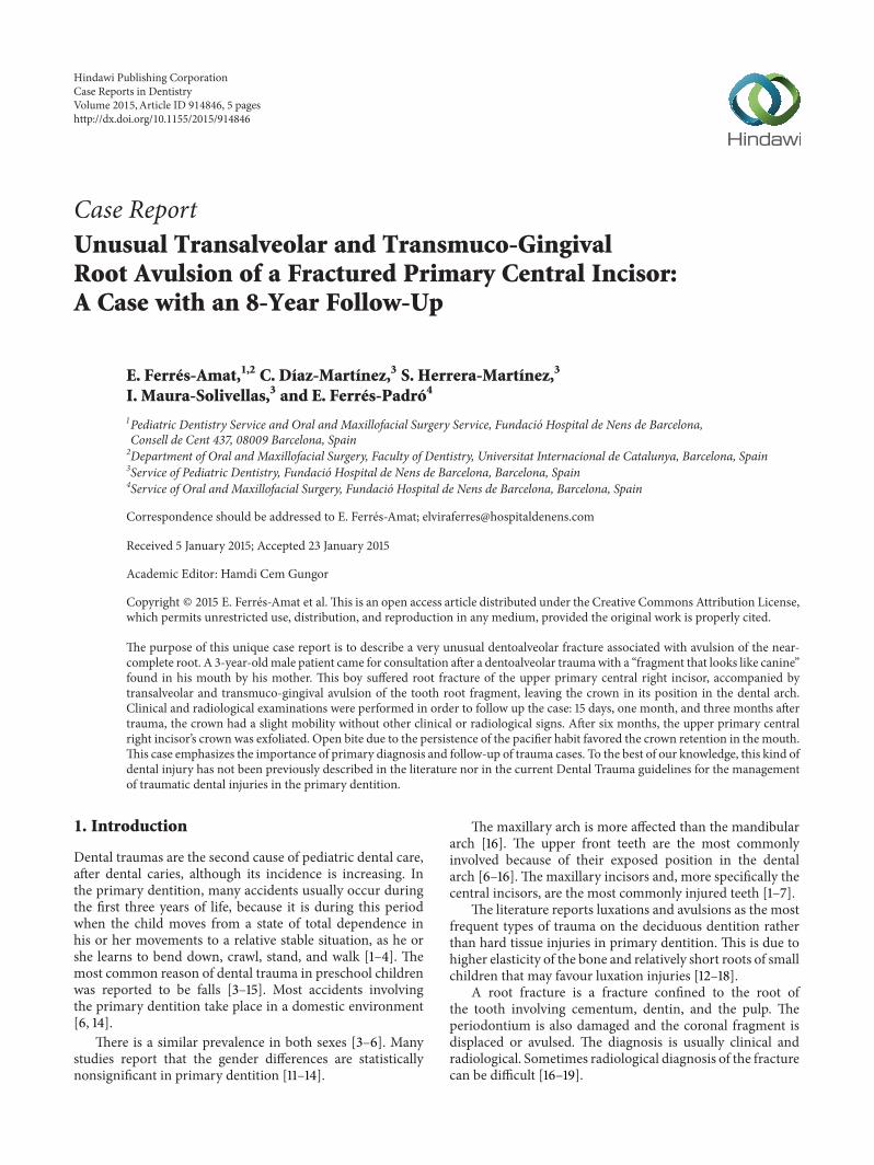
Figure 1: Intraoral view two hours after trauma. Periapical radiography. Root of the primary maxillary right central incisor root.
An avulsion is the complete displacement of the tooth outof its socket. It often affects central incisors in both dentitions,and, in most cases, only a single tooth is involved [19–21].
This case report is about a transmuco-gingival andtransalveolar fractured root avulsion, which means that it is apartial avulsion.
The aim of this first case report is to present a unique den-tal injury, which has not been previously described in theliterature or in the current Dental Trauma guidelines forthe management of traumatic dental injuries in the primarydentition.
2. Case Presentation
This is a case of a 3-year-old male patient, with no medicalor dental history of interest, suffered a dental trauma after adomestic accident and went as an emergency to the PediatricDentistry Service of the Hospital.
Chief complaint: his mother went to the emergency roomwith the fragment of her child’s tooth root in her hand,explaining that she had found this “loose canine in the child’smouth” after a dental trauma. In the physical examination,the following was found: a bruise, abrasion, and lacerationof the maxillary buccal mucosa and gingiva of the primarymaxillary central incisors zone. There was mobility in theprimary maxillary right central incisor and light mobilitywithout displacement in the primary maxillary left centralincisor.The clinical and radiological exploration of the lateralincisors was normal, without any pathology. The patientpresented with anterior open bite because of pacifier habitpersistence.
Periapical radiographic examination revealed the nearcomplete absence of the primary maxillary right centralincisor root. This exploration provided evidence that thepatient had suffered a root fracture of the primary maxillaryright central incisor (fracture located to cervical part of theroot) accompanied by transalveolar and transmuco-gingivalavulsion of the tooth root fragment (Figure 1). The dentalcrypts of themaxillary alveolar process of the developing per-manent teeth were found without any radiological alteration(the dental follicle of the permanent maxillary right centralincisor was in 5 Nolla stage).
Root avulsion of the primary maxillary right centralincisor, subluxation of the primary maxillary left centralincisor, and fracture of the alveolar process were diagnosed.
An antibiotic regimen of amoxicillin-clavulanic acid wasstarted as a treatment for one week. Soft diet was recom-mended and oral hygiene instructions were explained (brushwith a soft brush after every meal). It was also recommendedto apply chlorhexidine 0.1% topically to the affected area withcotton swabs twice a day for one week and to restrict theuse of a pacifier, as well as to avoid putting pressure on thetraumatized area.
Several posttrauma follow-up appointments were per-formed, exactly 15 days, one month, and three months afterthe trauma.
During the first examination, 15 days after the trauma,the clinical intraoral examination showed that the buccalmucosa of the primary maxillary right central incisor washealing, and the periapical radiography showed absence ofthe primary maxillary right central incisor root, but withoutany alteration or pathological image. The crown of theprimary right central incisor did not present mobility (it wasin its initial position in the dental arch) and there was not anyrisk of aspiration. In addition to this, the child had alreadygiven up the pacifier habit, as it was recommended in theposttrauma instructions.
Another examination was performed onemonth after thetrauma. The clinical intraoral examination showed normalbuccal mucosa and the primary maxillary right centralincisor crown did not show mobility. A periapical radiogra-phy was also performed.
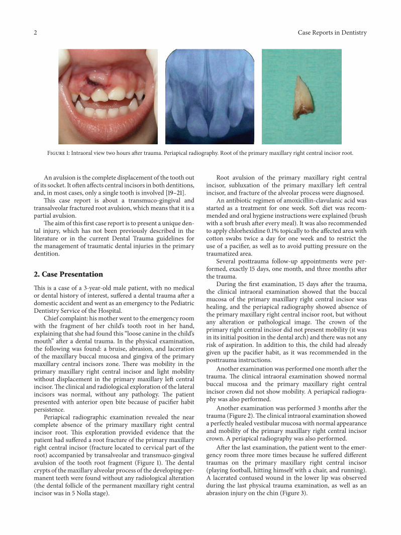
Another examination was performed 3 months after thetrauma (Figure 2).The clinical intraoral examination showeda perfectly healed vestibular mucosa with normal appearanceand mobility of the primary maxillary right central incisorcrown. A periapical radiography was also performed.
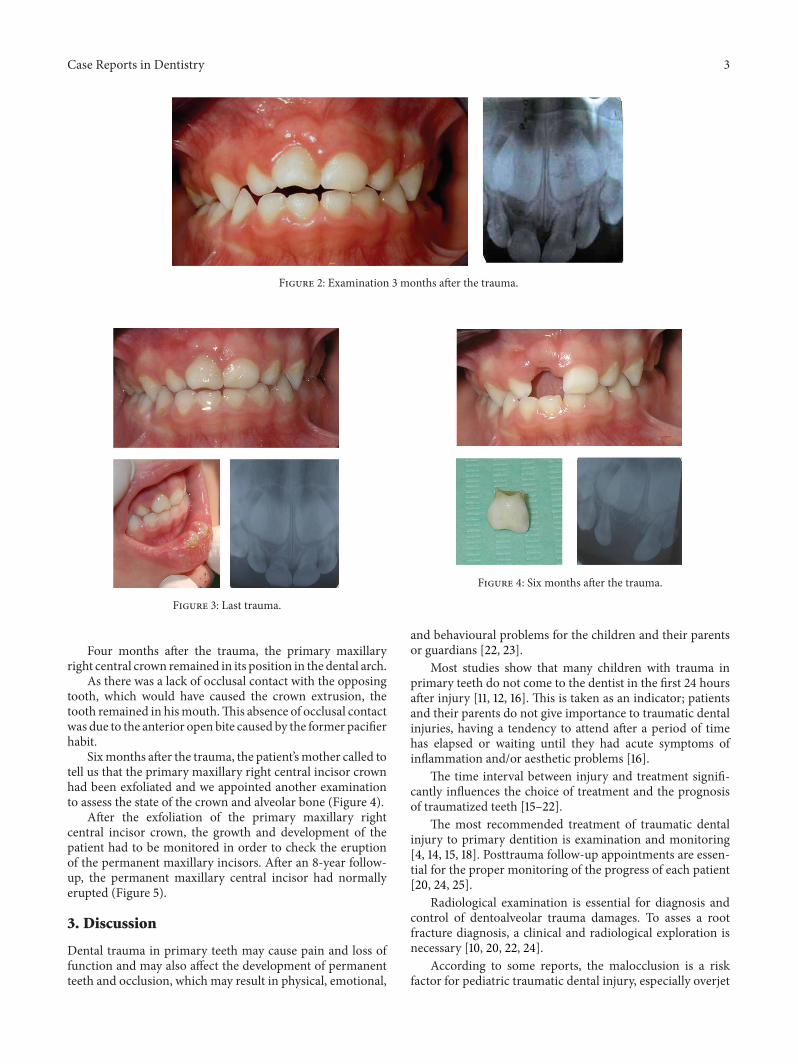
After the last examination, the patient went to the emer-gency room three more times because he suffered differenttraumas on the primary maxillary right central incisor(playing football, hitting himself with a chair, and running).A lacerated contused wound in the lower lip was observedduring the last physical trauma examination, as well as anabrasion injury on the chin (Figure 3).
Case Reports in Dentistry 3
Figure 2: Examination 3 months after the trauma.
Figure 3: Last trauma.
Four months after the trauma, the primary maxillaryright central crown remained in its position in the dental arch.
As there was a lack of occlusal contact with the opposingtooth, which would have caused the crown extrusion, thetooth remained in hismouth.This absence of occlusal contactwas due to the anterior open bite caused by the former pacifierhabit.
Sixmonths after the trauma, the patient’smother called totell us that the primary maxillary right central incisor crownhad been exfoliated and we appointed another examinationto assess the state of the crown and alveolar bone (Figure 4).
After the exfoliation of the primary maxillary rightcentral incisor crown, the growth and development of thepatient had to be monitored in order to check the eruptionof the permanent maxillary incisors. After an 8-year follow-up, the permanent maxillary central incisor had normallyerupted (Figure 5).
3. Discussion
Dental trauma in primary teeth may cause pain and loss offunction and may also affect the development of permanentteeth and occlusion, which may result in physical, emotional,
Figure 4: Six months after the trauma.
and behavioural problems for the children and their parentsor guardians [22, 23].
Most studies show that many children with trauma inprimary teeth do not come to the dentist in the first 24 hoursafter injury [11, 12, 16]. This is taken as an indicator; patientsand their parents do not give importance to traumatic dentalinjuries, having a tendency to attend after a period of timehas elapsed or waiting until they had acute symptoms ofinflammation and/or aesthetic problems [16].
The time interval between injury and treatment signifi-cantly influences the choice of treatment and the prognosisof traumatized teeth [15–22].
The most recommended treatment of traumatic dentalinjury to primary dentition is examination and monitoring[4, 14, 15, 18]. Posttrauma follow-up appointments are essen-tial for the proper monitoring of the progress of each patient[20, 24, 25].
Radiological examination is essential for diagnosis andcontrol of dentoalveolar trauma damages. To asses a rootfracture diagnosis, a clinical and radiological exploration isnecessary [10, 20, 22, 24].
According to some reports, the malocclusion is a riskfactor for pediatric traumatic dental injury, especially overjet

4 Case Reports in Dentistry
Figure 5: Permanent maxillary incisors normally erupted after an 8-year follow-up.
in the anterior incisor region, open bite, and cross-bite [23,26, 27].
In this particular case the open bite of the patient haspromoted the retention of the crown in the mouth, due tothe absence of occlusal contact with the opposing tooth. Theauthors decided to leave a rootless tooth in the mouth due toits unusual clinical situation, which was similar to the rest oftemporary tooth, since exfoliation is a spontaneous processin nature.
The clinical reasons why the authors did not perform thecrown extraction are the following ones: the absence of thecrown mobility and the lack of aspiration risk.
Additionally, the root avulsion through the buccal peri-odontium (alveolar bone process, mucosa, and gingiva)without injury of the dental follicle inside the dental cryptallowed the normal odontogenesis and the permanent centralincisor eruption.
No similar cases to the one that has been described havebeen found in the literature research. Moreover, this type ofunusual fracture is not included in the Dental TraumatologyGuidelines classifications.
Conflict of Interests
The authors declare that there is no conflict of interestsregarding the publication of this paper.
References
[1] T. C. L. Coutinho andM.R. RodriguesCajazeira, “Retrospectivestudy on the occurrence of primary incisor trauma in preschoolchildren of a low-income area in Brazil,” European Journal ofPaediatric Dentistry, vol. 12, no. 3, pp. 159–162, 2011.
[2] M. T. Flores, “Traumatic injuries in the primary dentition,”Dental Traumatology, vol. 18, no. 6, pp. 287–298, 2002.
[3] M. Kovacs, M. Pacurar, B. Petcu, and C. Bukhari, “Prevalence oftraumatic dental injuries in children who attended two dentalclinics in TarguMures between 2003 and 2011,”Oral Health andDental Management, vol. 11, no. 3, pp. 116–124, 2012.
[4] A. Avsar and B. Topaloglu, “Traumatic tooth injuries to primaryteeth of children aged 0–3 years,” Dental Traumatology, vol. 25,no. 3, pp. 323–327, 2009.
[5] M. L. Goettems, M. S. Azevedo, M. B. Correa et al., “Dentaltrauma occurrence and occlusal characteristics in Brazilianpreschool children,” Pediatric Dentistry, vol. 34, no. 2, pp. 104–107, 2012.
[6] D. Atabek, A. Alacam, I. Aydintug, and G. Konakoglu, “A retro-spective study of traumatic dental injuries,” Dental Traumatol-ogy, vol. 30, no. 2, pp. 154–161, 2014.
[7] K. Bucher, C. Neumann, R. Hickel, and J. Kuhnisch, “Traumaticdental injuries at a German University Clinic 2004–2008,”Dental Traumatology, vol. 29, no. 2, pp. 127–133, 2013.
[8] H. C. Gungor, “Management of crown-related fractures in chil-dren: an update review,”Dental Traumatology, vol. 30, no. 2, pp.88–99, 2014.
[9] C. O. Dummett Jr., “Dental management of traumatic injuriesto the primary dentition,” Journal of the California Dental Asso-ciation, vol. 28, no. 11, pp. 838–845, 2000.
[10] E. C. Marchiori, S. E. Santos, L. Asprino, M. de Moraes, and R.W. F. Moreira, “Occurrence of dental avulsion and associatedinjuries in patientswith facial trauma over a 9-year period,”Oraland Maxillofacial Surgery, vol. 17, no. 2, pp. 119–126, 2013.
[11] L. Ekanayake and M. Perera, “Pattern of traumatic dentalinjuries in children attending theUniversityDentalHospital, SriLanka,” Dental Traumatology, vol. 24, no. 4, pp. 471–474, 2008.
[12] M. Govindarajan, V. N. Reddy, K. Ramalingam, K. S. Durai, P.A. Rao, and A. Prabhu, “Prevalence of traumatic dental injuriesto the anterior teeth among three to thirteen-year-old schoolchildren of Tamilnadu,” Contemporary Clinical Dentistry, vol. 3,no. 2, pp. 164–167, 2012.
[13] L.H. Straffon andT. C. Pink, “Trauma to the primary and youngpermanent dentitions,” The Journal of the Michigan DentalAssociation, vol. 82, no. 1, pp. 40–45, 2000.
[14] J. O. Andreasen, E. Lauridsen, and S. S. A. Christensen, “Devel-opment of an interactive dental trauma guide,” Pediatric Den-tistry, vol. 3, pp. 133–136, 2009.
[15] A. A. Hasan, M. A. Qudeimat, and L. Andersson, “Prevalenceof traumatic dental injuries in preschool children in Kuwait-ascreening study,” Dental Traumatology, vol. 26, no. 4, pp. 346–350, 2010.
[16] F. Garcıa-Godoy and F. Pulver, “Treatment of trauma to theprimary and young permanent dentitions,” Dental Clinics ofNorth America, vol. 44, no. 3, pp. 597–632, 2000.
[17] Z. Kirzioglu, H. Karayilmaz, M. S. O. Erturk, and T. K.Sentut, “Epidemiology of traumatised primary teeth in thewest-Mediterranean region of Turkey,” International Dental Journal,vol. 55, no. 5, pp. 329–333, 2005.
[18] E. Canoglu, C. A. Akcan, E. Baharoglu, H. Cem Gungor, and Z.C. Cehreli, “Unusual ectopic eruption of a permanent centralincisor following an intrusion injury to the primary tooth,”Journal of the Canadian Dental Association, vol. 74, no. 8, pp.723–726, 2008.
[19] M. Unal, F. Oznurhan, A. Kapdan, S. Aksoy, and A. Durer,“Traumatic dental injuries in children. Experience of a hospital

Case Reports in Dentistry 5
in the central Anatolia region of Turkey,” European Journal ofPaediatric Dentistry, vol. 15, no. 1, pp. 17–22, 2014.
[20] H. Haueisen, K. Gartner, L. Kaiser, D. Trohorsch, and D. Heide-mann, “Vertical root fracture: prevalence, etiology, and diagno-sis,”Quintessence International, vol. 44, no. 7, pp. 467–474, 2013.
[21] G. T. Kim,M. Sohn, H. J. Ahn, D.W. Lee, and S. C. Choi, “Intra-alveolar root fracture in primary teeth,” Pediatric Dentistry, vol.34, no. 7, pp. e215–e218, 2012.
[22] B. Malmgren, J. O. Andreasen, M. T. Flores et al., “InternationalAssociation of Dental Traumatology guidelines for the man-agement of traumatic dental injuries: 3. Injuries in the primarydentition,”Dental Traumatology, vol. 28, no. 3, pp. 174–182, 2012.
[23] C. Altun, Z. C. Cehreli, G. Guven, and C. Acikel, “Traumaticintrusion of primary teeth and its effects on the permanent suc-cessors: a clinical follow-up study,”Oral Surgery, Oral Medicine,Oral Pathology, Oral Radiology and Endodontology, vol. 107, no.4, pp. 493–498, 2009.
[24] Y. Zhang, Y. Zhu, W. Su, Z. Zhou, Y. Jin, and X. Wang, “Aretrospective study of pediatric traumatic dental injuries inXi’an, China,” Dental Traumatology, vol. 30, no. 3, pp. 211–215,2014.
[25] B. Celikten, C. F. Uzuntas, R. Safaralizadeh, G. Demirel, and S.Sevimay, “Multidisciplinary approach for the treatment of hor-izontal root-fractured maxillary anterior teeth,” Case Reports inDentistry, vol. 2014, Article ID 472759, 7 pages, 2014.
[26] G.-T. Kim,M. Sohn,H. J. Ahn, D.-W. Lee, and S. C. Choi, “Intra-alveolar root fracture in primary teeth,” Pediatric Dentistry, vol.34, no. 7, pp. e215–e218, 2012.
[27] G. C. Bonini, M. Bonecker, M. M. Braga, and F. M. Mendes,“Combined effect of anterior malocclusion and inadequate lipcoverage on dental trauma in primary teeth,” Dental Trauma-tology, vol. 28, no. 6, pp. 437–440, 2012.

Submit your manuscripts athttp://www.hindawi.com
Hindawi Publishing Corporationhttp://www.hindawi.com Volume 2014
Oral OncologyJournal of
DentistryInternational Journal of
Hindawi Publishing Corporationhttp://www.hindawi.com Volume 2014
Hindawi Publishing Corporationhttp://www.hindawi.com Volume 2014
International Journal of
Biomaterials
Hindawi Publishing Corporationhttp://www.hindawi.com Volume 2014
BioMed Research International
Hindawi Publishing Corporationhttp://www.hindawi.com Volume 2014
Case Reports in Dentistry
Hindawi Publishing Corporationhttp://www.hindawi.com Volume 2014
Oral ImplantsJournal of
Hindawi Publishing Corporationhttp://www.hindawi.com Volume 2014
Anesthesiology Research and Practice
Hindawi Publishing Corporationhttp://www.hindawi.com Volume 2014
Radiology Research and Practice
Environmental and Public Health
Journal of
Hindawi Publishing Corporationhttp://www.hindawi.com Volume 2014
The Scientific World JournalHindawi Publishing Corporation http://www.hindawi.com Volume 2014
Hindawi Publishing Corporationhttp://www.hindawi.com Volume 2014
Dental SurgeryJournal of
Drug DeliveryJournal of
Hindawi Publishing Corporationhttp://www.hindawi.com Volume 2014
Hindawi Publishing Corporationhttp://www.hindawi.com Volume 2014
Oral DiseasesJournal of
Hindawi Publishing Corporationhttp://www.hindawi.com Volume 2014
Computational and Mathematical Methods in Medicine
ScientificaHindawi Publishing Corporationhttp://www.hindawi.com Volume 2014
PainResearch and TreatmentHindawi Publishing Corporationhttp://www.hindawi.com Volume 2014
Preventive MedicineAdvances in
Hindawi Publishing Corporationhttp://www.hindawi.com Volume 2014
EndocrinologyInternational Journal of
Hindawi Publishing Corporationhttp://www.hindawi.com Volume 2014
Hindawi Publishing Corporationhttp://www.hindawi.com Volume 2014
OrthopedicsAdvances in