case report synchronous orbital and gastrointestinal metastases...
TRANSCRIPT
Case ReportSynchronous Orbital and Gastrointestinal Metastases fromBreast Cancer: A Case Report and Review of Literature
Ramawad Soobrah,1 Fiona Tsang,2 Veronica Grassi,3 Hassan Hirji,4
Sreelakshmi Mallappa,2 and Robert Reichert4
1Victoria Hospital, Ministry of Health & Quality of Life, Mauritius2Ealing Hospital, Uxbridge Road, Southall, Middlesex UB1 3HW, UK3King’s College Hospital, Denmark Hill, London SE5 9RS, UK4Northwick Park Hospital, Watford Road, Harrow HA13UJ, UK
Correspondence should be addressed to Ramawad Soobrah; [email protected]
Received 18 March 2015; Accepted 4 May 2015
Academic Editor: Jose I. Mayordomo
Copyright © 2015 Ramawad Soobrah et al. This is an open access article distributed under the Creative Commons AttributionLicense, which permits unrestricted use, distribution, and reproduction in any medium, provided the original work is properlycited.
Breast cancer is themost commonmalignancy amongwomen and is a significant cause ofmorbidity andmortality worldwide.Withthe advent of improved imaging techniques and screening programmes, only a small proportion of women present with metastaticdisease. Metastases involving the gastrointestinal (GI) tract and orbit are rare occurrences. We describe the case of a woman withsimultaneous GI and orbital metastases from breast cancer who initially presented with abdominal pain and blurred vision andalso summarise a review of the literature.
1. Introduction
With 5.2 million cases diagnosed in 2008, breast cancerremains the most prevalent neoplasm in women after non-melanoma skin cancer [1] and is the second leading causeof cancer deaths in women [2]. Histologically, there are twomain subtypes of breast cancer: invasive ductal carcinoma(IDC) and invasive lobular carcinoma (ILC). Invasive lobularcarcinoma comprises approximately 10% to 15% of these casesand it is reported that its incidence has been increasingover the last several decades, especially in postmenopausalwomen [3–5]. ILCs have a distinctive growth pattern whichcan sometimes lead to vague findings on clinical breastexamination making it difficult to diagnose; they fail to formpalpable discrete nodules like IDC tumours [6]. Lobularcarcinomas often do not have characteristic mammographicappearances and present with subtle abnormalities such asfocal asymmetry or architectural distortion [7–10]. About16% of cases are mammographically occult and the reportedsensitivity of mammography to detect ILCs varies between57% and 81% [11]. This makes early clinical and radiologicaldetection of these tumours very challenging, particularly in
women with dense breast tissue. Due to such limitations,other modalities such as ultrasonography and magnetic res-onance imaging (MRI) are recommended to further evaluateknown cases of ILC [12]. About 10% of breast cancers havealready metastasized at the time of presentation and themetastatic patterns of IDC and ILC vary considerably [13].Various studies have reported that ductal carcinomas tendto spread to the lungs, bones, and liver, whereas lobularcarcinomas have a tendency to involve the GI tract, gynaeco-logical organs, and the peritoneum/retroperitoneum [13–15].Gastrointestinal metastases from ILC are rare andmany largeseries have reported an incidence of less than 1% [13, 16, 17].
The first case of orbital metastasis from lung cancer wasdescribed by Horner in 1864 [18]. Around 2.5% to 13% ofall orbital tumours are metastatic in nature [19]. Orbitaltumours from metastatic breast cancer (MBC) are relativelyuncommon. In a large observational case series involving1264 patients, only 4% were found to be metastatic lesionsfrom breast cancer [20]. However, this figure is likely to bean underestimate because smaller asymptomatic lesions areoften undiagnosed [21]. Although orbital and GI metastasesfrom lobular carcinomas are unusual, it is important to
Hindawi Publishing CorporationCase Reports in Oncological MedicineVolume 2015, Article ID 282790, 6 pageshttp://dx.doi.org/10.1155/2015/282790
2 Case Reports in Oncological Medicine
(a) (b)
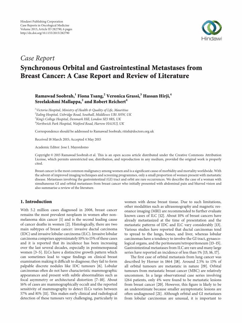
Figure 1: (a) Axial CT with intravenous contrast.The appendix is seen anterior to the right psoas muscle belly and is with a little surroundingfree fluid (solid black arrow). Increased attenuating soft tissue in the right paracolic gutter suggests free fluid or secondary peritonealmetastases (broken arrow). (b) Coronal section of the same patient demonstrating the enhancing appendix medial to the caecum. Muralenhancement is commonly seen in appendicitis. However, in this case, there is little surrounding fat stranding and no fluid filled lumen isseen; these features are more atypical. Enlarged lymph nodes are seen in the mesentery (solid white arrow).
recognise these entities to enable physicians to make promptand accurate diagnosis and provide appropriate treatment. Toour knowledge this is the only reported case of breast cancerwith synchronous metastasis to the orbit and appendix.
2. Case Presentation
A 42-year-old lady presented to the emergency departmentwith a three-day history of nausea, vomiting, fever, abdomi-nal pain, and disturbed vision. She also complained of a three-week history of bilateral breast swelling and right-sided breastpain. On examination, she was apyrexial and had a pulserate of 76 beats per minute. Both breasts were dense with nodiscrete palpable lumps; however, she was also noted to haveenlarged left axillary lymph nodes. Her abdomen was verytender in the right lower quadrant. White cell count and C-reactive protein were within normal limits.
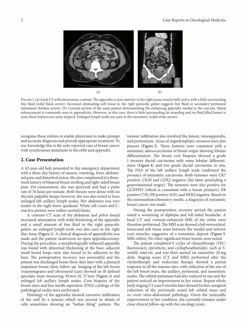
A contrast CT scan of the abdomen and pelvis foundincreased attenuation with mild thickening of the appendixand a small amount of free fluid in the right paracolicgutter; an enlarged lymph node was also seen in the rightiliac fossa (Figure 1). A clinical diagnosis of appendicitis wasmade and the patient underwent an open appendicectomy.During the procedure, a morphologically inflamed appendixwas found with abnormal thickening at the base; adjacentsmall bowel loops were also found to be adherent to thebase. The postoperative recovery was uneventful and thepatient was discharged home three days later with a plannedoutpatient breast clinic follow-up. Imaging of the left breast(mammograms and ultrasound scan) showed an ill-definedspiculate mass measuring 30mm by 27mm (Figure 2) andenlarged left axillary lymph nodes. Core biopsies of thebreast mass and fine needle aspiration (FNA) cytology of thepathological nodes were performed.
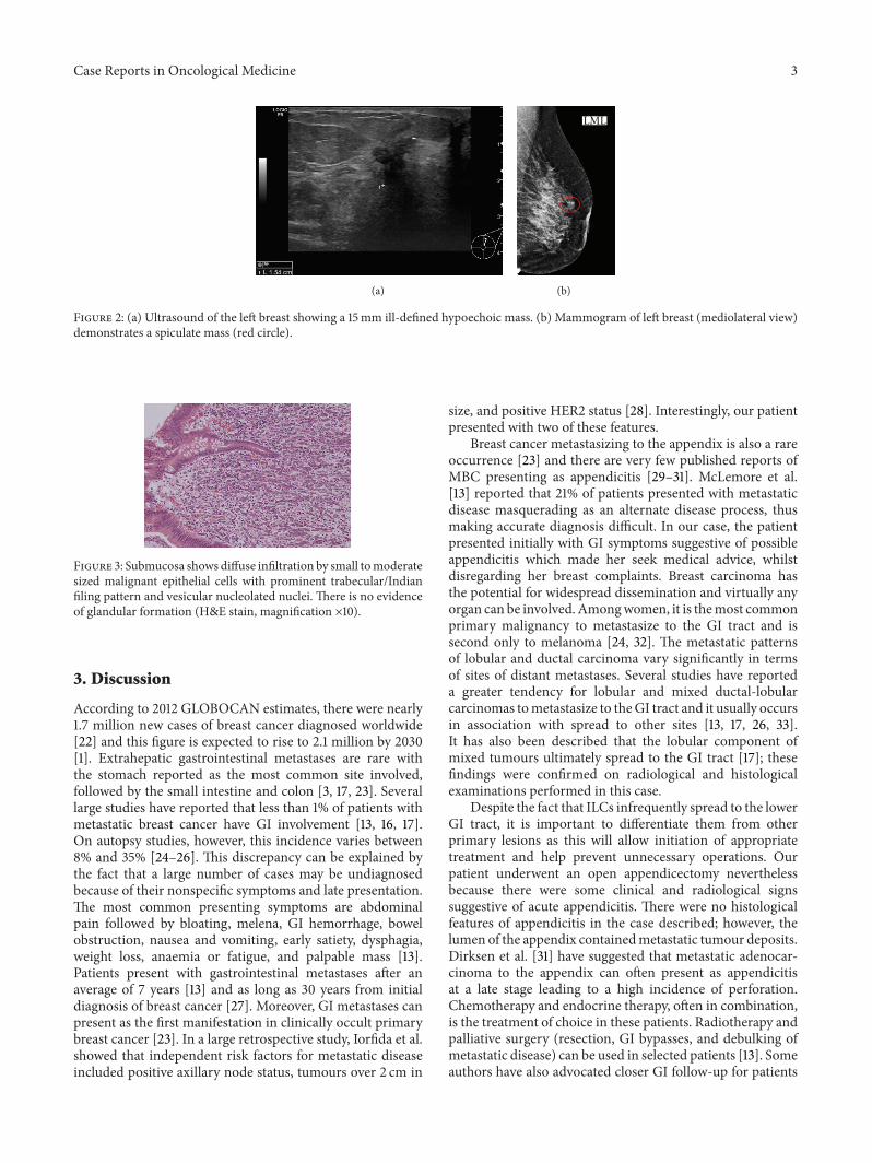
Histology of the appendix showed extensive infiltrationof the wall by a tumour which was present in sheets ofcells sometimes showing an “Indian filing” pattern. The
tumour infiltration also involved the lumen, mesoappendix,and peritoneum. Areas of angiolymphatic invasion were alsopresent (Figure 3). These features were consistent with ametastatic adenocarcinoma of breast origin showing lobulardifferentiation. The breast core biopsies showed a grade1 invasive ductal carcinoma with some lobular differenti-ation (Figure 4) and low grade ductal carcinoma in situ.The FNA of the left axillary lymph node confirmed thepresence of metastatic carcinoma. Both tumours were CK7positive, CK20 and CDX2 negative (the latter precluding agastrointestinal origin). The tumours were also positive forGCDFP15 (which is consistent with a breast primary), ERpositive (7/8), PR positive (8/8), andHER2negative. Based onthe immunohistochemistry results, a diagnosis of metastaticbreast cancer was made.
During the postoperative recovery period the patientnoted a worsening of diplopia and left-sided headache. Ahead CT and contrast-enhanced MRI of the orbits weretherefore performed.TheMRI scan showed a left retroorbital,intraconal soft tissue mass between the medial and inferiorrecti muscles, suggestive of a metastatic deposit (Figure 5:MRI orbits). No other significant brain lesions were noted.
The patient completed 6 cycles of chemotherapy (FEC:fluorouracil, epirubicin, and cyclophosphamide), each at 3-weekly interval, and was then started on tamoxifen 20mgdaily. Staging scans (CT and MRI) performed after thechemotherapy and endocrine therapy showed a partialresponse in all the tumour sites, with reduction of the size ofthe left breast mass, the axillary, peritoneal, and mesentericnodes.The orbital metastasis had also reduced in size and thepatient noticed an improvement in her vision. Repeat wholebody stagingCT scan 9months later showed furthermarginalreduction of the previously noted left orbital mass andno overt intra-abdominal pathology. Given the noticeableimprovement in her condition, she currently remains underclose clinical follow-up with the oncology team.
Case Reports in Oncological Medicine 3
(a)
LML
(b)
Figure 2: (a) Ultrasound of the left breast showing a 15mm ill-defined hypoechoic mass. (b) Mammogram of left breast (mediolateral view)demonstrates a spiculate mass (red circle).
Figure 3: Submucosa shows diffuse infiltration by small tomoderatesized malignant epithelial cells with prominent trabecular/Indianfiling pattern and vesicular nucleolated nuclei. There is no evidenceof glandular formation (H&E stain, magnification ×10).
3. Discussion
According to 2012 GLOBOCAN estimates, there were nearly1.7 million new cases of breast cancer diagnosed worldwide[22] and this figure is expected to rise to 2.1 million by 2030[1]. Extrahepatic gastrointestinal metastases are rare withthe stomach reported as the most common site involved,followed by the small intestine and colon [3, 17, 23]. Severallarge studies have reported that less than 1% of patients withmetastatic breast cancer have GI involvement [13, 16, 17].On autopsy studies, however, this incidence varies between8% and 35% [24–26]. This discrepancy can be explained bythe fact that a large number of cases may be undiagnosedbecause of their nonspecific symptoms and late presentation.The most common presenting symptoms are abdominalpain followed by bloating, melena, GI hemorrhage, bowelobstruction, nausea and vomiting, early satiety, dysphagia,weight loss, anaemia or fatigue, and palpable mass [13].Patients present with gastrointestinal metastases after anaverage of 7 years [13] and as long as 30 years from initialdiagnosis of breast cancer [27]. Moreover, GI metastases canpresent as the first manifestation in clinically occult primarybreast cancer [23]. In a large retrospective study, Iorfida et al.showed that independent risk factors for metastatic diseaseincluded positive axillary node status, tumours over 2 cm in
size, and positive HER2 status [28]. Interestingly, our patientpresented with two of these features.
Breast cancer metastasizing to the appendix is also a rareoccurrence [23] and there are very few published reports ofMBC presenting as appendicitis [29–31]. McLemore et al.[13] reported that 21% of patients presented with metastaticdisease masquerading as an alternate disease process, thusmaking accurate diagnosis difficult. In our case, the patientpresented initially with GI symptoms suggestive of possibleappendicitis which made her seek medical advice, whilstdisregarding her breast complaints. Breast carcinoma hasthe potential for widespread dissemination and virtually anyorgan can be involved. Amongwomen, it is themost commonprimary malignancy to metastasize to the GI tract and issecond only to melanoma [24, 32]. The metastatic patternsof lobular and ductal carcinoma vary significantly in termsof sites of distant metastases. Several studies have reporteda greater tendency for lobular and mixed ductal-lobularcarcinomas tometastasize to theGI tract and it usually occursin association with spread to other sites [13, 17, 26, 33].It has also been described that the lobular component ofmixed tumours ultimately spread to the GI tract [17]; thesefindings were confirmed on radiological and histologicalexaminations performed in this case.
Despite the fact that ILCs infrequently spread to the lowerGI tract, it is important to differentiate them from otherprimary lesions as this will allow initiation of appropriatetreatment and help prevent unnecessary operations. Ourpatient underwent an open appendicectomy neverthelessbecause there were some clinical and radiological signssuggestive of acute appendicitis. There were no histologicalfeatures of appendicitis in the case described; however, thelumen of the appendix containedmetastatic tumour deposits.Dirksen et al. [31] have suggested that metastatic adenocar-cinoma to the appendix can often present as appendicitisat a late stage leading to a high incidence of perforation.Chemotherapy and endocrine therapy, often in combination,is the treatment of choice in these patients. Radiotherapy andpalliative surgery (resection, GI bypasses, and debulking ofmetastatic disease) can be used in selected patients [13]. Someauthors have also advocated closer GI follow-up for patients
4 Case Reports in Oncological Medicine
(a) (b)
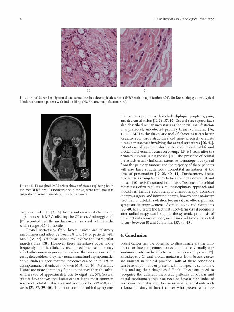
Figure 4: (a) Several malignant ductal structures in a desmoplastic stroma (H&E stain, magnification ×20). (b) Breast biopsy shows typicallobular carcinoma pattern with Indian filing (H&E stain, magnification ×40).
Figure 5: T1 weighted MRI orbits show soft tissue replacing fat inthe medial left orbit is isointense with the adjacent recti and it issuggestive of a soft tissue deposit (white arrows).
diagnosed with ILC [3, 34]. In a recent review article lookingat patients with MBC affecting the GI tract, Ambroggi et al.[17] reported that the median overall survival is 16 monthswith a range of 5–41 months.
Orbital metastases from breast cancer are relativelyuncommon and affect between 2% and 6% of patients withMBC [35–37]. Of those, about 5% involve the extraocularmuscles only [38]. However, these metastases occur morefrequently than is clinically recognised because they mayaffect other major organ systems where the consequences areeasily detectable or theymay remain small and asymptomatic.Some studies suggest that the incidence can be up to 30% inasymptomatic patients with known MBC [21, 36]. Metastaticlesions are more commonly found in the uvea than the orbit,with a ratio of approximately one to eight [21, 37]. Severalstudies have shown that breast cancer is the most commonsource of orbital metastases and accounts for 29%–50% ofcases [21, 37, 39, 40]. The most common orbital symptoms
that patients present with include diplopia, proptosis, pain,and decreased vision [19, 36, 37, 40]. Several case reports havealso described ocular metastasis as the initial manifestationof a previously undetected primary breast carcinoma [36,41, 42]. MRI is the diagnostic tool of choice as it can bettervisualize soft tissue structures and more precisely evaluatetumour metastases involving the orbital structures [20, 43].Patients usually present during the sixth decade of life andorbital involvement occurs on average 4.5–6.5 years after theprimary tumour is diagnosed [21]. The presence of orbitalmetastasis usually indicates extensive haematogenous spreadfrom the primary tumour and the majority of these patientswill also have simultaneous nonorbital metastases at thetime of presentation [19, 21, 40, 44]. Furthermore, breastcancer has a strong tendency to localize in the orbital fat andmuscle [40], as is illustrated in our case. Treatment for orbitalmetastases often requires a multidisciplinary approach andmodalities include radiotherapy, chemotherapy, hormonetherapy, surgery, and immunotherapy; however, themainstaytreatment is orbital irradiation because it can offer significantsymptomatic improvement of orbital signs and symptoms[20, 40, 43]. Despite the fact that short-term visual prognosisafter radiotherapy can be good, the systemic prognosis ofthese patients remains poor; mean survival time is reportedto vary between 10 and 20 months [37, 44, 45].
4. Conclusion
Breast cancer has the potential to disseminate via the lym-phatic or haematogenous routes and hence virtually anyanatomical site can be affected with metastatic deposits [19].Extrahepatic GI and orbital metastases from breast cancerare unusual in clinical practice. Both of these conditionscan be asymptomatic or present with nonspecific symptoms,thus making their diagnosis difficult. Physicians need torecognise the different metastatic patterns of lobular andductal carcinomas; they also need to have a high index ofsuspicion for metastatic disease especially in patients witha known history of breast cancer who present with new
Case Reports in Oncological Medicine 5
gastrointestinal complaints or orbital symptoms. For lesionsin the gastrointestinal tract, it is important to distinguish aprimary carcinoma from a metastatic one in order to initiateappropriate treatment in such patients and help preventunnecessary surgical procedures. Similarly, early recognitionand treatment of patients affected by ocular metastases canhelp preserve their vision and maximize their quality of life.
Conflict of Interests
The authors declare that there is no conflict of interestsregarding the publication of this paper.
Acknowledgments
The authors would like to thank Dr. Nayef Aqel (consultanthistopathologist) for providing the histopathology imagesandDr. SanjayMugon (consultant pathologist) for describingthose images.
References
[1] F. Bray, J.-S. Ren, E. Masuyer, and J. Ferlay, “Global estimates ofcancer prevalence for 27 sites in the adult population in 2008,”International Journal of Cancer, vol. 132, no. 5, pp. 1133–1145,2013.
[2] R. Siegel, D.Naishadham, andA. Jemal, “Cancer statistics, 2012,”CA: A Cancer Journal for Clinicians, vol. 62, no. 1, pp. 10–29,2012.
[3] P. Carcoforo, M. T. Raiji, R. C. Langan et al., “Infiltratinglobular carcinoma of the breast presenting as gastrointestinalobstruction: a mini review,” Journal of Cancer, vol. 3, no. 1, pp.328–332, 2012.
[4] C. I. Li, B. O. Anderson, P. Porter, S. K. Holt, J. R. Daling, andR. E. Moe, “Changing incidence rate of invasive lobular breastcarcinoma among older women,” Cancer, vol. 88, no. 11, pp.2561–2569, 2000.
[5] J. V. Alvarez, D. Perez, and L. A. Chodosh, “mILC-ing themousemammary gland: a model for invasive lobular carcinoma,”Cancer Cell, vol. 10, no. 5, pp. 347–349, 2006.
[6] K. J. Dedes and D. Fink, “Clinical presentation and surgicalmanagement of invasive lobular carcinoma of the breast,” BreastDiseases, vol. 30, pp. 31–37, 2008-2009.
[7] W. P. Evans, L. J. W. Burhenne, L. Laurie, K. F. O’Shaughnessy,and R. A. Castellino, “Invasive lobular carcinoma of the breast:mammographic characteristics and computer-aided detection,”Radiology, vol. 225, no. 1, pp. 182–189, 2002.
[8] M. A. Helvie, C. Paramagul, H. A. Oberman, and D. D. Adler,“Invasive lobular carcinoma: imaging features and clinicaldetection,” Investigative Radiology, vol. 28, no. 3, pp. 202–207,1993.
[9] K.N. Krecke and J. J. Gisvold, “Invasive lobular carcinoma of thebreast:mammographic findings and extent of disease at diagno-sis in 184 patients,”The American Journal of Roentgenology, vol.161, no. 5, pp. 957–960, 1993.
[10] E. A. Sickles, “The subtle and atypical mammographic featuresof invasive lobular carcinoma,” Radiology, vol. 178, no. 1, pp. 25–26, 1991.
[11] J. K. Lopez and L. W. Bassett, “Invasive lobular carcinoma ofthe breast: spectrum of mammographic, US, and MR imagingfindings,” Radiographics, vol. 29, no. 1, pp. 165–176, 2009.
[12] W. A. Berg, L. Gutierrez, M. S. NessAiver et al., “Diagnosticaccuracy of mammography, clinical examination, US, and MRimaging in preoperative assessment of breast cancer,”Radiology,vol. 233, no. 3, pp. 830–849, 2004.
[13] E. C.McLemore, B. A. Pockaj, C. Reynolds et al., “Breast cancer:presentation and intervention in women with gastrointestinalmetastasis and carcinomatosis,” Annals of Surgical Oncology,vol. 12, no. 11, pp. 886–894, 2005.
[14] S. Ferlicot, A. Vincent-Salomon, J. Medioni et al., “Widemetastatic spreading in infiltrating lobular carcinoma of thebreast,” European Journal of Cancer, vol. 40, no. 3, pp. 336–341,2004.
[15] M. Harris, A. Howell, M. Chrissohou, R. I. Swindell, M.Hudson, and R. A. Sellwood, “A comparison of the metastaticpattern of infiltrating lobular carcinoma and infiltrating ductcarcinoma of the breast,” British Journal of Cancer, vol. 50, no. 1,pp. 23–30, 1984.
[16] M. J. Borst and J. A. Ingold, “Metastatic patterns of invasivelobular versus invasive ductal carcinoma of the breast,” Surgery,vol. 114, no. 4, pp. 637–642, 1993.
[17] M. Ambroggi, E. M. Stroppa, P. Mordenti et al., “Metastaticbreast cancer to the gastrointestinal tract: report of five casesand review of the literature,” International Journal of BreastCancer, vol. 2012, Article ID 439023, 8 pages, 2012.
[18] F. Horner, “Carcinoma der dura mater exophthalmus,” KlinMonatsbl Augenheilkd, vol. 2, pp. 186–190, 1864.
[19] K. K. Toller, J. W. Gigantelli, and M. J. Spalding, “Bilateralorbital metastases from breast carcinoma. A case of falsepseudotumor,” Ophthalmology, vol. 105, no. 10, pp. 1897–1901,1998.
[20] J. A. Shields, C. L. Shields, and R. Scartozzi, “Survey of 1264patients with orbital tumors and simulating lesions: the 2002Montgomery Lecture, part 1,”Ophthalmology, vol. 111, no. 5, pp.997–1008, 2004.
[21] S. Fenton, E. G. Kemp, and A. N. Harnett, “Screening for Oph-thalmic involvement in asymptomatic patients with metastaticbreast carcinoma,” Eye, vol. 18, no. 1, pp. 38–40, 2004.
[22] International Agency for Cancer Research.World Health Orga-nization, GLOBOCAN 2012: Estimated Cancer Incidence, Mor-tality and Prevalence Worldwide in 2012, 2012, http://globocan.iarc.fr/Pages/fact sheets cancer.aspx.
[23] C. B. Winston, O. Hadar, J. B. Teitcher et al., “Metastaticlobular carcinoma of the breast: patterns of spread in the chest,abdomen, and pelvis on CT,” American Journal of Roentgenol-ogy, vol. 175, no. 3, pp. 795–800, 2000.
[24] K. Washington and D. McDonagh, “Secondary tumors of thegastrointestinal tract: surgical pathologic findings and compar-ison with autopsy survey,” Modern Pathology, vol. 8, no. 4, pp.427–433, 1995.
[25] E. Caramella, J. N. Bruneton, P. Roux, D. Aubanel, and P.Lecomte, “Metastases of the digestive tract. Report of 77 casesand review of the literature,” European Journal of Radiology, vol.3, no. 4, pp. 331–338, 1983.
[26] G. Cervi, N. Vettoretto, A. Vinco et al., “Rectal localization ofmetastatic lobular breast cancer: report of a case,” Diseases ofthe Colon & Rectum, vol. 44, no. 3, pp. 453–455, 2001.
6 Case Reports in Oncological Medicine
[27] A. Benfiguig, M.-L. Anciaux, C. Eugene, G. Benkemoun, and J.-C. Etienne, “Gastric metastasis of breast cancer occurring aftera cancer-free interval of 30 years,” Annales de Gastroenterologieet d’Hepatologie, vol. 28, no. 4, pp. 175–177, 1992.
[28] M. Iorfida, E. Maiorano, E. Orvieto et al., “Invasive lobularbreast cancer: subtypes and outcome,” Breast Cancer Researchand Treatment, vol. 133, no. 2, pp. 713–723, 2012.
[29] K. S. Latchis and J. W. Canter, “Acute appendicitis secondary tometastatic carcinoma,”TheAmerican Journal of Surgery, vol. 111,no. 2, pp. 220–223, 1966.
[30] R. E. Burney, N. Koss, and I. S. Goldenberg, “Acute appendicitissecondary to metastatic carcinoma of the breast: a report andreview of two cases,”Archives of Surgery, vol. 108, no. 6, pp. 872–875, 1974.
[31] J. L. Dirksen, M. G. Souder, and A. J. Burick, “Metastatic breastcarcinoma presenting as perforated appendicitis,” Breast Care,vol. 5, no. 6, pp. 409–410, 2010.
[32] A. Ciulla, G. Castronovo, G. Tomasello et al., “Gastric metas-tases originating from occult breast lobular carcinoma: diag-nostic and therapeutic problems,” World Journal of SurgicalOncology, vol. 6, article 78, 2008.
[33] S. Dhar, M. N. Kulaylat, K. Gordon, P. Lall, and R. J. Doerr,“Solitary papillary breast carcinoma metastasis to the largebowel presenting as primary colon carcinoma: case report andreview of the literature,” The American Surgeon, vol. 69, no. 9,pp. 799–803, 2003.
[34] J. Nazareno, D. Taves, and H. G. Preiksaitis, “Metastatic breastcancer to the gastrointestinal tract: a case series and review ofthe literature,”World Journal of Gastroenterology, vol. 12, no. 38,pp. 6219–6224, 2006.
[35] J. A. Shields, C. L. Shields, H. K. Brotman, C. Carvalho, N.Perez, and R. C. Eagle Jr., “Cancer metastatic to the orbit:the 2000 Robert M. Curts Lecture,” Ophthalmic Plastic andReconstructive Surgery, vol. 17, no. 5, pp. 346–354, 2001.
[36] S. Wickremasinghe, K. K. Dansingani, P. Tranos, S. Liyanage,A. Jones, and C. Davey, “Ocular presentations of breast cancer,”Acta Ophthalmologica Scandinavica, vol. 85, no. 2, pp. 133–142,2007.
[37] D.H.Char, T.Miller, and S. Kroll, “Orbitalmetastases: diagnosisand course,” British Journal of Ophthalmology, vol. 81, no. 5, pp.386–390, 1997.
[38] R. W. Arnold, B. A. Adams, J. K. Camoriano, and J. A. Dyer,“Acquired divergent strabismus: presumed metastatic gastriccarcinoma to the medial rectus muscle,” Journal of PediatricOphthalmology and Strabismus, vol. 26, no. 1, pp. 50–51, 1989.
[39] C. F. Merrill, D. I. Kaufman, and N. V. Dimitrov, “Breast cancermetastatic to the eye is a common entity,” Cancer, vol. 68, no. 3,pp. 623–627, 1991.
[40] A. A. Valenzuela, C. W. Archibald, B. Fleming et al., “Orbitalmetastasis: clinical features, management and outcome,” Orbit,vol. 28, no. 2-3, pp. 153–159, 2009.
[41] P. J. Vlachostergios, I. A. Voutsadakis, and C. N. Papandreou,“Orbital metastasis of breast carcinoma,” Breast Cancer: Basicand Clinical Research, vol. 3, no. 1, pp. 91–97, 2009.
[42] M. Kadivar, A. Joulaee, M. B. Kashkouli, H. H. Kharazi, M.Kalantari, and P. V. Kumar, “Orbital metastasis as the firstpresentation of nonpalpable invasive lobular carcinoma of thebreast,”The Breast Journal, vol. 12, no. 1, pp. 75–76, 2006.
[43] S. M. Ahmad and B. Esmaeli, “Metastatic tumors of the orbitand ocular adnexa,” Current Opinion in Ophthalmology, vol. 18,no. 5, pp. 405–413, 2007.
[44] E. Ng and P. F. Ilsen, “Orbital metastases,” Optometry, vol. 81,no. 12, pp. 647–657, 2010.
[45] D. Holland, S. Maune, G. Kovacs, and S. Behrendt, “Metastatictumors of the orbit: a retrospective study,” Orbit, vol. 22, no. 1,pp. 15–24, 2003.

Submit your manuscripts athttp://www.hindawi.com
Stem CellsInternational
Hindawi Publishing Corporationhttp://www.hindawi.com Volume 2014
Hindawi Publishing Corporationhttp://www.hindawi.com Volume 2014
MEDIATORSINFLAMMATION
of
Hindawi Publishing Corporationhttp://www.hindawi.com Volume 2014
Behavioural Neurology
EndocrinologyInternational Journal of
Hindawi Publishing Corporationhttp://www.hindawi.com Volume 2014
Hindawi Publishing Corporationhttp://www.hindawi.com Volume 2014
Disease Markers
Hindawi Publishing Corporationhttp://www.hindawi.com Volume 2014
BioMed Research International
OncologyJournal of
Hindawi Publishing Corporationhttp://www.hindawi.com Volume 2014
Hindawi Publishing Corporationhttp://www.hindawi.com Volume 2014
Oxidative Medicine and Cellular Longevity
Hindawi Publishing Corporationhttp://www.hindawi.com Volume 2014
PPAR Research
The Scientific World JournalHindawi Publishing Corporation http://www.hindawi.com Volume 2014
Immunology ResearchHindawi Publishing Corporationhttp://www.hindawi.com Volume 2014
Journal of
ObesityJournal of
Hindawi Publishing Corporationhttp://www.hindawi.com Volume 2014
Hindawi Publishing Corporationhttp://www.hindawi.com Volume 2014
Computational and Mathematical Methods in Medicine
OphthalmologyJournal of
Hindawi Publishing Corporationhttp://www.hindawi.com Volume 2014
Diabetes ResearchJournal of
Hindawi Publishing Corporationhttp://www.hindawi.com Volume 2014
Hindawi Publishing Corporationhttp://www.hindawi.com Volume 2014
Research and TreatmentAIDS
Hindawi Publishing Corporationhttp://www.hindawi.com Volume 2014
Gastroenterology Research and Practice
Hindawi Publishing Corporationhttp://www.hindawi.com Volume 2014
Parkinson’s Disease
Evidence-Based Complementary and Alternative Medicine
Volume 2014Hindawi Publishing Corporationhttp://www.hindawi.com