case report stump cholecystitis: laparoscopic...
TRANSCRIPT

Case ReportStump Cholecystitis: Laparoscopic Completion Cholecystectomywith Basic Laparoscopic Equipment in a Resource Poor Setting
Shamir O. Cawich,1 Carlos Wilson,2 Lindberg K. Simpson,3 and Akil J. Baker4
1 Department of Clinical Surgical Sciences, University of the West Indies, St. Augustine, Trinidad and Tobago2Department of Surgery, Percy Junor Hospital, Spalding, Jamaica3 Department of Surgery, Kingston Public Hospital, Kingston, Jamaica4Department of Surgery, Mandeville Public Hospital, Manchester, Jamaica
Correspondence should be addressed to Shamir O. Cawich; [email protected]
Received 18 July 2014; Accepted 19 August 2014; Published 21 August 2014
Academic Editor: William B. Silverman
Copyright © 2014 Shamir O. Cawich et al. This is an open access article distributed under the Creative Commons AttributionLicense, which permits unrestricted use, distribution, and reproduction in any medium, provided the original work is properlycited.
Introduction. Stump cholecystitis is a recognised condition in which a large gallbladder remnant becomes inflamed after subtotalcholecystectomy. When this occurs, a completion cholecystectomy is indicated. Traditionally, these patients were subjected toopen surgery because the laparoscopic approach was anticipated to be technically difficult. We present a case of completioncholecystectomy using basic laparoscopic equipment in a resource poor setting to demonstrate that the laparoscopic approachis feasible. Case Description. A 57-year-old woman presented with right upper quadrant pain and vomiting. She had an electiveopen cholecystectomy seven years before but reported remarkably similar symptoms. Abdominal ultrasound suggested calculousacute cholecystitis. MRCP confirmed the presence of a large gallbladder remnant with stones. Gastroduodenoscopy excludedother differentials. She had an uneventful laparoscopic completion cholecystectomy performed. Discussion. Although traditionaldogma suggested that a completion cholecystectomy should be performed through the open approach, several small studies havedemonstrated that laparoscopic completion cholecystectomy is feasible and safe. This report adds to the existing data in support ofthe laparoscopic approach.
1. Introduction
After a partial or subtotal cholecystectomy, symptoms mayrecur from pathology in the gallbladder remnant. When thisoccurs, a completion cholecystectomy is required to preventa recurrence [1]. Many of these patients were traditionallysubjected to open cholecystectomy because the laparoscopicapproach was anticipated to be technically challenging. Wepresent a case of a patient with stump cholecystitis ina gallbladder remnant to demonstrate that a laparoscopicapproach to completion cholecystectomy is feasible usingbasic equipment in a resource poor setting.
2. Case Description
A 57-year-old woman with diabetes and hypertension pre-sented to our hospital complaining of severe right upper
quadrant pain and vomiting. She reported having an electiveopen cholecystectomy done seven years before and notedthat the symptoms she now experienced were similar. Shehad been hospitalized six times in the preceding two yearsfor similar episodes. Although abdominal ultrasounds weredone on these occasions, the emergency room physicians didnot entertain the possibility of a stump cholecystitis becausethe patient insisted that her gallbladder had already beenremoved. Operative notes were not available but she reportedthat the operationwas uncomplicated and shewas discharged72 hours after open surgery.
Upper gastrointestinal studies excluded the presence ofupper gastrointestinal pathology. Liver function panel andserum amylase levels were normal. Abdominal ultrasoundsuggested that stones were present in a thick walled gallblad-der.
Hindawi Publishing CorporationCase Reports in MedicineVolume 2014, Article ID 787631, 3 pageshttp://dx.doi.org/10.1155/2014/787631

2 Case Reports in Medicine
GB
CBD
RHD LHD
Figure 1: MRCP showing a normal extrahepatic biliary tree and alarge gallbladder remnant (GB). CBD: common bile duct; LHD: lefthepatic duct; RHD: right hepatic duct.
Inferior liver surface(segment 4b)
Figure 2: Dense adhesions occupy the gallbladder bed precludingclear visualization of anatomy.
The patient’s history was revisited on this presentation.She insisted that open cholecystectomy was performed sevenyears before and there was a surgical scar at the right upperquadrant, but no operative records or histology reportswere available to corroborate the surgical history. Once thesymptomatology resolved, the patient was discharged witha request for magnetic resonance cholangiography (MRCP)from another institution. This confirmed the presence of agallbladder remnant with normal biliary anatomy. There wasno evidence of choledocholithiasis (Figure 1).
She was prepared for general anaesthesia and taken tothe operating theatre for laparoscopic completion cholecys-tectomy. Laparoscopic shears were used to perform adhesi-olysis in order to visualize the structures in the right upperquadrant. There were dense adhesions at gallbladder bedprecluding clear visualization of relevant anatomy (Figure 2).Careful and patient dissection with the cautery hook even-tually presented the gallbladder remnant (Figure 3). Theremnant was followed in an anterograde fashion down to the
Figure 3: Careful dissection has presented the gallbladder remnant.
Figure 4: Anterograde dissection down to infundibulum presentsstructures in Calot’s triangle and allows demonstration of Strasberg’scritical view of safety.
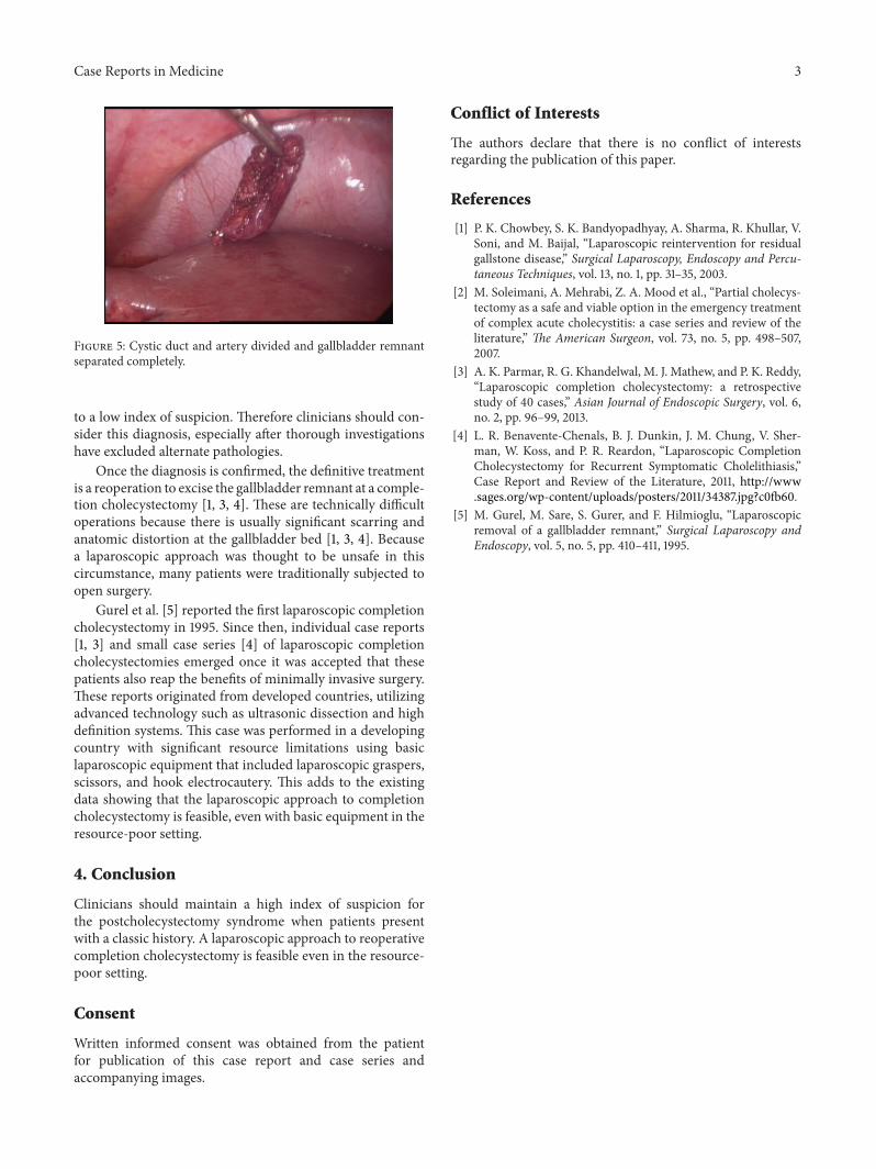
cystic duct to demonstrate Strasberg’s critical view of safety(Figure 4). The cystic duct and artery were then individuallyligated and the gallbladder separated completely (Figure 5).Her recovery was uneventful and she was discharged homeafter 8 hours.
3. Discussion
A subtotal cholecystectomy is a safe option in the face ofsevere inflammation at Calot’s triangle because it reducesthe potential for common duct injury [2]. However, thiscarries the risk of developing stump cholecystitis when thegallbladder remnant becomes inflamed due to stone disease[1, 3].
The reported incidence of stump cholecystitis varies buthas been reported to occur in as many as 5% [3] of patientsafter emergent cholecystectomy, and it is rare after electiveoperations. It tends to occur in middle-aged women whoare usually quite confident that their symptoms are similarto those that prompted their original cholecystectomy [4].Despite a suggestive history, the diagnosis is often delayed due
Case Reports in Medicine 3
Figure 5: Cystic duct and artery divided and gallbladder remnantseparated completely.
to a low index of suspicion. Therefore clinicians should con-sider this diagnosis, especially after thorough investigationshave excluded alternate pathologies.
Once the diagnosis is confirmed, the definitive treatmentis a reoperation to excise the gallbladder remnant at a comple-tion cholecystectomy [1, 3, 4]. These are technically difficultoperations because there is usually significant scarring andanatomic distortion at the gallbladder bed [1, 3, 4]. Becausea laparoscopic approach was thought to be unsafe in thiscircumstance, many patients were traditionally subjected toopen surgery.
Gurel et al. [5] reported the first laparoscopic completioncholecystectomy in 1995. Since then, individual case reports[1, 3] and small case series [4] of laparoscopic completioncholecystectomies emerged once it was accepted that thesepatients also reap the benefits of minimally invasive surgery.These reports originated from developed countries, utilizingadvanced technology such as ultrasonic dissection and highdefinition systems. This case was performed in a developingcountry with significant resource limitations using basiclaparoscopic equipment that included laparoscopic graspers,scissors, and hook electrocautery. This adds to the existingdata showing that the laparoscopic approach to completioncholecystectomy is feasible, even with basic equipment in theresource-poor setting.
4. Conclusion
Clinicians should maintain a high index of suspicion forthe postcholecystectomy syndrome when patients presentwith a classic history. A laparoscopic approach to reoperativecompletion cholecystectomy is feasible even in the resource-poor setting.
Consent
Written informed consent was obtained from the patientfor publication of this case report and case series andaccompanying images.
Conflict of Interests
The authors declare that there is no conflict of interestsregarding the publication of this paper.
References
[1] P. K. Chowbey, S. K. Bandyopadhyay, A. Sharma, R. Khullar, V.Soni, and M. Baijal, “Laparoscopic reintervention for residualgallstone disease,” Surgical Laparoscopy, Endoscopy and Percu-taneous Techniques, vol. 13, no. 1, pp. 31–35, 2003.
[2] M. Soleimani, A. Mehrabi, Z. A. Mood et al., “Partial cholecys-tectomy as a safe and viable option in the emergency treatmentof complex acute cholecystitis: a case series and review of theliterature,” The American Surgeon, vol. 73, no. 5, pp. 498–507,2007.
[3] A. K. Parmar, R. G. Khandelwal, M. J. Mathew, and P. K. Reddy,“Laparoscopic completion cholecystectomy: a retrospectivestudy of 40 cases,” Asian Journal of Endoscopic Surgery, vol. 6,no. 2, pp. 96–99, 2013.
[4] L. R. Benavente-Chenals, B. J. Dunkin, J. M. Chung, V. Sher-man, W. Koss, and P. R. Reardon, “Laparoscopic CompletionCholecystectomy for Recurrent Symptomatic Cholelithiasis,”Case Report and Review of the Literature, 2011, http://www.sages.org/wp-content/uploads/posters/2011/34387.jpg?c0fb60.
[5] M. Gurel, M. Sare, S. Gurer, and F. Hilmioglu, “Laparoscopicremoval of a gallbladder remnant,” Surgical Laparoscopy andEndoscopy, vol. 5, no. 5, pp. 410–411, 1995.

Submit your manuscripts athttp://www.hindawi.com
Stem CellsInternational
Hindawi Publishing Corporationhttp://www.hindawi.com Volume 2014
Hindawi Publishing Corporationhttp://www.hindawi.com Volume 2014
MEDIATORSINFLAMMATION
of
Hindawi Publishing Corporationhttp://www.hindawi.com Volume 2014
Behavioural Neurology
EndocrinologyInternational Journal of
Hindawi Publishing Corporationhttp://www.hindawi.com Volume 2014
Hindawi Publishing Corporationhttp://www.hindawi.com Volume 2014
Disease Markers
Hindawi Publishing Corporationhttp://www.hindawi.com Volume 2014
BioMed Research International
OncologyJournal of
Hindawi Publishing Corporationhttp://www.hindawi.com Volume 2014
Hindawi Publishing Corporationhttp://www.hindawi.com Volume 2014
Oxidative Medicine and Cellular Longevity
Hindawi Publishing Corporationhttp://www.hindawi.com Volume 2014
PPAR Research
The Scientific World JournalHindawi Publishing Corporation http://www.hindawi.com Volume 2014
Immunology ResearchHindawi Publishing Corporationhttp://www.hindawi.com Volume 2014
Journal of
ObesityJournal of
Hindawi Publishing Corporationhttp://www.hindawi.com Volume 2014
Hindawi Publishing Corporationhttp://www.hindawi.com Volume 2014
Computational and Mathematical Methods in Medicine
OphthalmologyJournal of
Hindawi Publishing Corporationhttp://www.hindawi.com Volume 2014
Diabetes ResearchJournal of
Hindawi Publishing Corporationhttp://www.hindawi.com Volume 2014
Hindawi Publishing Corporationhttp://www.hindawi.com Volume 2014
Research and TreatmentAIDS
Hindawi Publishing Corporationhttp://www.hindawi.com Volume 2014
Gastroenterology Research and Practice
Hindawi Publishing Corporationhttp://www.hindawi.com Volume 2014
Parkinson’s Disease
Evidence-Based Complementary and Alternative Medicine
Volume 2014Hindawi Publishing Corporationhttp://www.hindawi.com