case report open access immunoglobulin g4 … · case report open access immunoglobulin...
TRANSCRIPT

WORLD JOURNAL OF SURGICAL ONCOLOGY
Cai et al. World Journal of Surgical Oncology 2014, 12:363http://www.wjso.com/content/12/1/363
CASE REPORT Open Access
Immunoglobulin G4-associated cholangitismimicking cholangiocarcinoma treated bylaparoscopic choledochectomy withintracorporeal Roux-en-Y hepaticojejunostomyJiaQin Cai, Yi-Ping Mou*, Yu Pan, Ke Chen, Xiao-Wu Xu and YuCheng Zhou
Abstract
Immunoglobulin G4 (IgG4)-associated disease is a recently recognized disease entity that is characterized by elevatedserum IgG4 concentrations, abundant IgG4 lymphoplasmacytic infiltration, and dramatic steroid responses. IgG4-associatedcholangitis is one manifestation of IgG4-associated disease. However, it is clinically challenging to make a preoperativedifferentiation between this rare disease and cholangiocarcinoma, especially for those with serum concentrations of IgG4 inthe normal range. This article reports on a 57-year-old man with jaundice and upper abdominal discomfort. Imagingexamination showed biliary stricture that closely resembled cholangiocarcinoma, and the patient’s serum IgG4concentration was normal. The patient underwent a laparoscopic choledochectomy with Roux-en-Y hepaticojejunostomyusing an intracorporeal hand-sewn technique. He recovered quickly without any complications. We also present ourexperience in laparoscopic intracorporeal hand-sewn hepaticojejunostomy.
Keywords: IgG4-associated cholangitis, Hepaticojejunostomy, Intracorporeal, Laparoscopic
BackgroundIgG4-associated systemic disease (ISD) is a systemic disorderinvolving multiple organs associated with increased IgG4serum levels or IgG4 positive plasma cell infiltrates. IgG4-associated cholangitis (IAC) is one manifestation of ISD.However, the clinical, biochemical, and imaging features ofIAC mimic cholangiocarcinoma, making preoperative accur-ate diagnosis difficult [1]; therefore, surgery remains a thera-peutic choice, so as to avoid omitting malignancy.Since the development of minimally invasive surgical ap-
proaches [2], laparoscopic surgery for biliary tract diseaseshas evolved rapidly over the past decade, and laparoscopicRoux-en-Y cholangiojejunostomy and hepaticojejunostomyare accepted for the treatment of such diseases. On thebasis of our extensive laparoscopic experience gained fromlaparoscopic gastrectomy and pancreatectomy, as well asother laparoscopic operations [3-6], we were encouragedto develop an intracorporeal hand-sewn technique for
* Correspondence: [email protected] of General Surgery, Sir Run Run Shaw Hospital, School ofMedicine, Zhejiang University, 3 East Qingchun Road, Hangzhou 310016,Zhejiang Province, China
© 2014 Cai et al.; licensee BioMed Central LtdCommons Attribution License (http://creativecreproduction in any medium, provided the orDedication waiver (http://creativecommons.orunless otherwise stated.
reconstruction. Herein, we report a case of IAC preopera-tively diagnosed as cholangiocarcinoma and successfullytreated by laparoscopic hepaticojejunostomy with our intra-corporeal hand-sewn technique.
Case presentationA 57-year-old man was admitted to our department; hehad jaundice and had been experiencing upper abdominaldiscomfort for the previous month. Laboratory data wereas follows: alkaline phosphatase, 303 U/l; alanine amino-transferase, 266 IU/l; aspartate aminotransferase, 70 IU/l;γ-glutamyltransferase, 555 U/l; total bilirubin, 126 μmol/dl;direct bilirubin, 102.0 μmol/l. The patient’s serum levels ofcarcinoembryonic antigen and α-fetoprotein were withinnormal limits; however, serum carbohydrate antigen 19-9concentration was 306 IU/ml. Other test results, includingIgG4 serum levels, were all within normal ranges.Enhanced abdominal computed tomography revealed
a mass involving the common hepatic duct (Figure 1A).Magnetic resonance cholangiopancreatography revealeda stricture at the upper middle segment of the commonhepatic duct, together with proximal bile duct dilatation
. This is an Open Access article distributed under the terms of the Creativeommons.org/licenses/by/4.0), which permits unrestricted use, distribution, andiginal work is properly credited. The Creative Commons Public Domaing/publicdomain/zero/1.0/) applies to the data made available in this article,

Figure 1 Preoperative imaging examination. (A) Computed tomography showed biliary stricture (white arrow). (B) Magnetic resonancecholangiopancreatography revealed biliary stricture (black arrow) and dilatation at the top of the stricture.
Cai et al. World Journal of Surgical Oncology 2014, 12:363 Page 2 of 7http://www.wjso.com/content/12/1/363
(Figure 1B). The mass was initially diagnosed as a chol-angiocarcinoma, resulting in biliary stenosis and jaun-dice. Therefore, laparoscopic choledochectomy withRoux-Y hepaticojejunostomy was selected.The patients’ position and the placement of trocars were
similar to our previous studies [7] (Figure 2). The hepato-duodenal ligament was divided by ultrasonic coagulatingshears (Figure 3A). Then the common hepatic artery, com-mon bile duct and portal vein were visualized (Figure 3B).Calot’s triangle was identified and the cystic artery was
Figure 2 Trocar placement.
clipped and divided while the cystic duct was clipped andleft in situ (Figure 3C). The biliary tract, including the com-mon bile duct, common hepatic duct and left and righthepatic ducts was divided further from the portal vein. Theportal vein and common hepatic artery were then sepa-rated from the surrounding tissue upward to the hilarplate. Lymphadenectomy around the pancreatic head wasperformed. The mass was found at the middle of the com-mon bile duct. The dilated left and right hepatic ductswere transected 1.0 cm above the mass (Figure 3D), and

Figure 3 Resection of mass on biliary duct. (A) Division of hepatoduodenal ligament. (B) Visualization of properhepatic artery, common bileduct, and portal vein. (C) The cystic artery was clipped and divided while the cystic duct was clipped and left in situ. (D) Transection of left andright hepatic ducts 1.0 cm above the mass. (E) Traversal of common bile duct about 0.5 cm below the mass. CBD, common bile duct; LHA, lefthepatic artery; LHD, left hepatic duct; PHA, proper hepatic artery; PV, portal vein; RHD, right hepatic duct; RPV, right portal vein.
Cai et al. World Journal of Surgical Oncology 2014, 12:363 Page 3 of 7http://www.wjso.com/content/12/1/363
the common bile duct was traversed about 0.5 cm belowthe mass (Figure 3E). The resected tissue and the gallblad-der were taken out in an endoscopic retrieval bag throughthe umbilical incision.The incision was sutured, and the pneumoperitoneum
was reestablished. After exploring the upper jejunum about25 cm distally from the ligament of Treitz, an incision wasmade in the mesentery of this loop, and a transection of thejejunum was performed with an endoscopic linear stapler(Figure 4A). The left and right hepatic ducts and the distallimb were then approximated using a draft line (Figure 4B),and a 5 mm wide incision was made at the antimesentericside of the jejunum (Figure 4C) for end-to-side hepaticoje-junostomy. At the 3 o’clock and 9 o’clock positions of theanastomotic stoma, the sutures were held as long staysutures to lift the corners (Figure 4D). The posterior wall ofthe hepaticojejunostomy was sutured using interrupted su-tures (Figure 4E,F) and the anterior wall was sutured usinga continuous suture (Figure 4G). The seromuscular layer
was strengthened with interrupted sutures to reducetension (Figure 4H). Then a side-to-side jejunojejunostomywas performed through the enlarged umbilical incision. Fi-nally, a peritoneal cavity drainage tube was placed posteriorto the bilioenteric anastomosis.The operative time was 210 min and blood loss was 60
ml. The intraoperative frozen pathological diagnosis wasprobably IAC, rather than cholangiocarcinoma, and themargins were negative. The gross finding was a 20 × 15mm mass located in the middle part of the common bileduct, in communication with a dilated proximal bile duct(Figure 5). Microscopically, lymphoplasmacytic infiltratewas identified, with a moderate CD138 positive plasmacell infiltration (Figure 6A) and abundant IgG4 positivecells (Figure 6B).The postoperative course was uneventful. The patient
started to take semi-fluid on the day after surgery. Asthere were no complications such as hemorrhage or bileleak, the drainage tube was removed on postoperative day
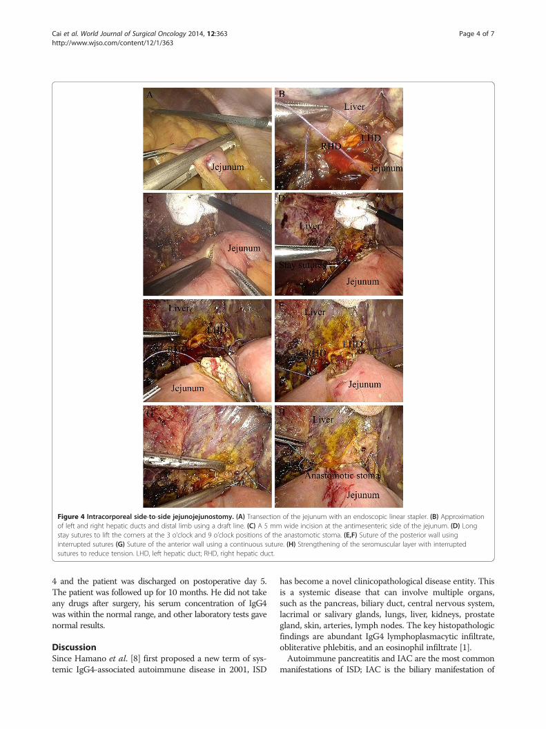
Figure 4 Intracorporeal side-to-side jejunojejunostomy. (A) Transection of the jejunum with an endoscopic linear stapler. (B) Approximationof left and right hepatic ducts and distal limb using a draft line. (C) A 5 mm wide incision at the antimesenteric side of the jejunum. (D) Longstay sutures to lift the corners at the 3 o’clock and 9 o’clock positions of the anastomotic stoma. (E,F) Suture of the posterior wall usinginterrupted sutures (G) Suture of the anterior wall using a continuous suture. (H) Strengthening of the seromuscular layer with interruptedsutures to reduce tension. LHD, left hepatic duct; RHD, right hepatic duct.
Cai et al. World Journal of Surgical Oncology 2014, 12:363 Page 4 of 7http://www.wjso.com/content/12/1/363
4 and the patient was discharged on postoperative day 5.The patient was followed up for 10 months. He did not takeany drugs after surgery, his serum concentration of IgG4was within the normal range, and other laboratory tests gavenormal results.
DiscussionSince Hamano et al. [8] first proposed a new term of sys-temic IgG4-associated autoimmune disease in 2001, ISD
has become a novel clinicopathological disease entity. Thisis a systemic disease that can involve multiple organs,such as the pancreas, biliary duct, central nervous system,lacrimal or salivary glands, lungs, liver, kidneys, prostategland, skin, arteries, lymph nodes. The key histopathologicfindings are abundant IgG4 lymphoplasmacytic infiltrate,obliterative phlebitis, and an eosinophil infiltrate [1].Autoimmune pancreatitis and IAC are the most common
manifestations of ISD; IAC is the biliary manifestation of

Figure 5 Surgical specimen, showing a mass in the commonbile duct.
Cai et al. World Journal of Surgical Oncology 2014, 12:363 Page 5 of 7http://www.wjso.com/content/12/1/363
ISD, and is often associated with autoimmune pancreatitis.Although the diagnosis of IAC is often established based ona combination of clinical, serologic, radiological, and histo-logic findings, strict criteria for IAC are lacking [9]. Severaldiagnostic criteria have been proposed, following the initialJapanese consensus criteria for the diagnosis of auto-immune pancreatitis [10]. The HISORt (histology, imaging,serology, other organ involvement, and response to cortico-steroid) criteria [11] for autoimmune pancreatitis and thevariant criteria for IAC [12], as well as the Asian consensuscriteria [13] have been the most commonly applied criteriafor diagnosing autoimmune pancreatitis and IAC. Limitedepidemiologic data exist on the rarity of IAC. Studies havereported that IAC is found mostly in men older than 50years [9]. Notwithstanding the fact that the clinical presen-tation of patients with IAC is highly variable, obstructivejaundice is most common, present in up to 75% of patients[9]. Patients may present with other symptoms, such as
Figure 6 Immunostained bile duct specimens. (A) Moderate CD138-poscells, ×100.
abdominal pain, weight loss, pruritus, and biochemicalsigns of pancreatitis and cholestasis [12].The low clinical specificity makes it difficult to distin-
guish IAC from cholangiocarcinoma. Although a serumIgG4 increase is characteristic of IAC, it may not bediagnostic of the disease. The sensitivity and accuracy ofserum IgG4 for ISD were reported as 50% and 60%,respectively [14]. According to a recent study from theMayo Clinic, which investigated the ability of IgG4 todistinguish IAC from cholangiocarcinoma reliably,serum IgG4 levels more than twice the upper limits ofnormal provided a specificity of 97% and a sensitivity of50% in distinguishing IAC from cholangiocarcinoma[15]. Nevertheless, an IgG4 increase does occur in theabsence of ISD, and hence increased serum IgG4 levelsalone should not be used to diagnose IAC. Serum CA19-9 levels can also be of use because concentrationsgreater than 100 IU/ml are less likely in IAC than incholangiocarcinoma (18% versus 60% to 80%) [16,17]. Inaddition, biliary strictures in IAC also do not have anyhighly specific diagnostic features, unlike autoimmunepancreatitis, in which typical pancreatic imagingfeatures, such as diffuse sausage-shaped pancreatic en-largement and diffuse irregular narrowing of the pancre-atic duct, have been described. Imaging often showsproximal bile duct or intrahepatic strictures with dilata-tion of the upstream biliary system, along with sclerosingchanges, which are quite similar to cholangiocarcinoma.This indicates that, while imaging modalities are usefulin determining the level of bile duct obstruction, theyare limited when it comes to establishing a definitivediagnosis, distinguishing IAC from malignant biliarystrictures reliably [18,19].An unexplained biliary stricture, not caused by trauma
or choledocholithiasis, is often presumed to be causedby malignancy. In our case, with normal serum concen-trations of IgG4 (1.86 g/l) and apparently elevated levels
itive plasma cell infiltration, ×100. (B) Abundant IgG4-positive plasma
Cai et al. World Journal of Surgical Oncology 2014, 12:363 Page 6 of 7http://www.wjso.com/content/12/1/363
of CA19-9, the images showing segmental biliary strictureand no other organ involvement were assumed to indicateISD; it is extremely difficult to distinguish IAC from chol-angiocarcinoma preoperatively. Although endoscopic retro-grade cholangiopancreatography is the most commonlyperformed procedure for cholangiocarcinoma and can pro-vide a tissue diagnosis through brush cytology of the bileduct, the sensitivity of biliary brush cytology to diagnosecholangiocarcinoma may be as low as 30% [20]. Althoughendoscopic ultrasonography can complement the role ofendoscopic retrograde cholangiopancreatography and pro-vide a tissue diagnosis through fine needle aspiration andstaging through ultrasound imaging, it can also lead totumor seeding [20]. Hence, choledochectomy with Roux-en-Y hepaticojejunostomy was performed.The laparoscopic technique is indispensable in biliary
surgery. Compared with traditional open surgery, moststudies have reported that laparoscopic techniques canachieve better cosmesis, shorter hospital stay, and fasterpostoperative recovery [21-24]. By choosing the laparo-scopic approach, not only are the incision complicationsminimized, but also the quality of visualization is sig-nificantly enhanced, leading to greater precision in thecontrol of reconstructions. However, it is important tohave an experienced hand with wonderful skills. As withopen surgery, laparoscopic ductoplasty appears to betechnically possible in experienced hands. A properwide hepaticojejunostomy must be performed for suffi-cient bile drainage. After surgery, the anastomoticstoma would become smaller because of inflammationand denuded epithelial mucosa of the bile duct. Theposterior wall anastomosis is the most challenging step.Based on our experience, keeping the long corner staysutures at the 3 o’clock and 9 o’clock positions of theanastomotic stoma was able to maintain tension to pro-vide a clear view of posterior wall and allow more pre-cise anastomosis. Maintaining the integrity of thisanastomosis is important, and lessening the tension ofthe anastomosis is also a key point in preventing theoccurrence of bile leakage. The intestinal mesenteryshould be isolated from the mesenteric root as much aspossible, to ensure appropriate mobility of the biliarylimb. In addition, the greater omentum should be sepa-rated if necessary. Moreover, interrupted sutures of ser-omuscular layer are also helpful, to reduce tension.
ConclusionsOur case suggests that totally laparoscopic Roux-en-Yhepaticojejunostomy using an intracorporeal hand-sewntechnique is a feasible procedure. Adequate preoperativeevaluation, appropriate intraoperative hand-sewn tech-niques, and highly skilled laparoscopic techniques are thekey factors of success in laparoscopic hepaticojejunostomy.
ConsentWritten informed consent was obtained from the patientfor the publication of this case report and any accom-panying images.
AbbreviationsHISORt: histology, imaging, serology, other organ involvement, and responseto corticosteroid; IAC: IgG4-associated cholangitis; IgG4: immunoglobulin G4;ISD: IgG4-associated systemic disease.
Competing interestsThe authors declare that they have no competing interests.
Authors’ contributionsYPM, XWX, and YCZ performed the operation. YP and KC collected case data.JQC wrote the manuscript. YPM proofread and revised the manuscript.All authors approved the version to be published.
AcknowledgementsThis work was supported by the Zhejiang Key Subject of Medical ScienceFoundation (grant no. 11-CX-21).
Received: 15 July 2014 Accepted: 18 November 2014Published: 29 November 2014
References1. Stone JH, Zen Y, Deshpande V: IgG4-related disease. N Engl J Med 2012,
366:539–551.2. Gawande A: Two hundred years of surgery. N Engl J Med 2012,
366:1716–1723.3. Xu X, Chen K, Zhou W, Zhang R, Wang J, Wu D, Mou Y: Laparoscopic
transgastric resection of gastric submucosal tumors located near theesophagogastric junction. J Gastrointest Surg 2013, 17:1570–1575.
4. Chen K, Xu X, Mou Y, Pan Y, Zhang R, Zhou Y, Wu D, Huang C: Totallylaparoscopic distal gastrectomy with D2 lymphadenectomy and BillrothII gastrojejunostomy for gastric cancer: short- and medium-term resultsof 139 consecutive cases from a single institution. Int J Med Sci 2013,10:1462–1470.
5. Chen K, Mou YP, Xu XW, Cai JQ, Wu D, Pan Y, Zhang RC: Short-termsurgical and long-term survival outcomes after laparoscopic distalgastrectomy with D2 lymphadenectomy for gastric cancer.BMC Gastroenterol 2014, 14:41.
6. Zhang RC, Yan JF, Xu XW, Chen K, Ajoodhea H, Mou YP: Laparoscopic vsopen distal pancreatectomy for solid pseudopapillary tumor of thepancreas. World J Gastroenterol 2013, 19:6272–6277.
7. Xu XW, Li RH, Zhou W, Wang J, Zhang RC, Chen K, Mou YP: Laparoscopicresection of synchronous intraductal papillary mucinous neoplasms: acase report. World J Gastroenterol 2012, 18:6510–6514.
8. Hamano H, Kawa S, Horiuchi A, Unno H, Furuya N, Akamatsu T, FukushimaM, Nikaido T, Nakayama K, Usuda N, Kiyosawa K: High serum IgG4concentrations in patients with sclerosing pancreatitis. N Engl J Med2001, 344:732–738.
9. Björnsson E, Chari ST, Smyrk TC, Lindor K: Immunoglobulin G4 associatedcholangitis: description of an emerging clinical entity based on reviewof the literature. Hepatology 2007, 45:1547–1554.
10. Kamisawa T, Okazaki K, Kawa S: Diagnostic criteria for autoimmunepancreatitis in Japan. World J Gastroenterol 2008, 14:4992–4994.
11. Chari ST, Smyrk TC, Levy MJ, Topazian MD, Takahashi N, Zhang L, Clain JE,Pearson RK, Petersen BT, Vege SS, Farnell MB: Diagnosis of autoimmunepancreatitis: the Mayo Clinic experience. Clin Gastroenterol Hepatol 2006,4:1010–1016. quiz 934.
12. Ghazale A, Chari ST, Zhang L, Smyrk TC, Takahashi N, Levy MJ, Topazian MD,Clain JE, Pearson RK, Petersen BT, Vege SS, Lindor K, Farnell MB:Immunoglobulin G4-associated cholangitis: clinical profile and responseto therapy. Gastroenterology 2008, 134:706–715.
13. Otsuki M, Chung JB, Okazaki K, Kim MH, Kamisawa T, Kawa S, Park SW,Shimosegawa T, Lee K, Ito T, Nishimori I, Notohara K, Naruse S, Ko SB, KiharaY, Research Committee of Intractable Pancreatic Diseases provided by theMinistry of Health, Labour and Welfare of Japan and the Korean Society ofPancreatobiliary Diseases: Asian diagnostic criteria for autoimmune
Cai et al. World Journal of Surgical Oncology 2014, 12:363 Page 7 of 7http://www.wjso.com/content/12/1/363
pancreatitis: consensus of the Japan-Korea Symposium on AutoimmunePancreatitis. J Gastroenterol 2008, 43:403–408.
14. Lytras D, Kalaitzakis E, Webster GJ, Imber CJ, Amin Z, Rodriguez-Justo M,Pereira SP, OldeDamink SW, Malago M: Cholangiocarcinoma or IgG4-associated cholangitis: how feasible it is to avoid unnecessary surgicalinterventions? Ann Surg 2012, 256:1059–1067.
15. Oseini AM, Chaiteerakij R, Shire AM, Ghazale A, Kaiya J, Moser CD, Aderca I,Mettler TA, Therneau TM, Zhang L, Takahashi N, Chari ST, Roberts LR: Utilityof serum immunoglobulin G4 in distinguishing immunoglobulin G4-associated cholangitis from cholangiocarcinoma. Hepatology 2011,54:940–948.
16. Tangkijvanich P, Thong-ngam D, Theamboonlers A, Hanvivatvong O,Kullavanijaya P, Poovorawan Y: Diagnostic role of serum interleukin 6 andCA 19-9 in patients with cholangiocarcinoma. Hepatogastroenterology2004, 51:15–19.
17. Qin XL, Wang ZR, Shi JS, Lu M, Wang L, He QR: Utility of serum CA19-9 indiagnosis of cholangiocarcinoma: in comparison with CEA. World JGastroenterol 2004, 10:427–432.
18. Knoefel WT, Prenzel KL, Peiper M, Hosch SB, Gundlach M, Eisenberger CF,Strate T, Scheunemann P, Rogiers X, Izbicki JR: Klatskin tumors and Klatskinmimicking lesions of the biliary tree. Eur J SurgOncol 2003, 29:658–661.
19. van Gulik TM, Gouma DJ: Changing perspectives in the assessment ofresectability of hilarcholangiocarcinoma. Ann SurgOncol 2007,14:1969–1971.
20. Strongin A, Singh H, Eloubeidi MA, Siddiqui AA: Role of endoscopicultrasonography in the evaluation of extrahepaticcholangiocarcinoma.Endosc Ultrasound 2013, 2:71–76.
21. Liem NT, Pham HD, Vu HM: Is the laparoscopic operation as safe as openoperation for choledochal cyst in children? J Laparoendosc Adv Surg TechA 2011, 21:367–370.
22. Diao M, Li L, Cheng W: Laparoscopic versus open Roux-en-Y hepatojeju-nostomy for children with choledochal cysts: intermediate-term follow-up results. SurgEndosc 2011, 25:1567–1573.
23. Wang J, Zhang W, Sun D, Zhang Q, Liu H, Xi D, Li A: Laparoscopictreatment for choledochal cysts with stenosis of the common hepaticduct. J Am Coll Surg 2012, 214:e47–e52.
24. Liu Y, Yao X, Li S, Liu W, Liu L, Liu J: Comparison of therapeutic effects oflaparoscopic and open operation for congenital choledochal cysts inadults. Gastroenterol Res Pract 2014, 2014:670260.
doi:10.1186/1477-7819-12-363Cite this article as: Cai et al.: Immunoglobulin G4-associated cholangitismimicking cholangiocarcinoma treated by laparoscopic choledochectomywith intracorporeal Roux-en-Y hepaticojejunostomy. World Journal ofSurgical Oncology 2014 12:363.
Submit your next manuscript to BioMed Centraland take full advantage of:
• Convenient online submission
• Thorough peer review
• No space constraints or color figure charges
• Immediate publication on acceptance
• Inclusion in PubMed, CAS, Scopus and Google Scholar
• Research which is freely available for redistribution
Submit your manuscript at www.biomedcentral.com/submit