case report...
TRANSCRIPT
Hindawi Publishing CorporationCase Reports in Neurological MedicineVolume 2011, Article ID 329738, 2 pagesdoi:10.1155/2011/329738
Case Report
Moya Moya Disease in a Child: A Case Report
Jagdish P. Goyal, Sanjeev S. Rao, and Sangita Trivedi
Department of Pediatrics, Government Medical College & New Civil Hospital, Surat, Gujarat 395001, India
Correspondence should be addressed to Jagdish P. Goyal, [email protected]
Received 24 May 2011; Accepted 14 July 2011
Academic Editor: D. Long
Copyright © 2011 Jagdish P. Goyal et al. This is an open access article distributed under the Creative Commons AttributionLicense, which permits unrestricted use, distribution, and reproduction in any medium, provided the original work is properlycited.
We report a case of 8-year-old female child who was admitted at our hospital with complaints of transient ischemic attacks and left-sided hemiparesis. On MR angiography, the child was diagnosed to have Moya Moya disease. Moya Moya disease is a rare causeof cerebral stroke in children. The patient was treated conservatively and referred to a higher centre for specific neurosurgery.Neurosurgical revascularization process leads to favourable outcome.
1. Introduction
Acute stroke is an infrequent disease of paediatric age grouppatients. Moya Moya is a rare cerebrovascular disease of un-known aetiology. We report a case of 8-year-old female childwho presented with left-sided hemiplegia to a tertiary carehospital in India and diagnosed as Moya Moya disease. Cere-bral revascularization surgery leads to favourable outcome.
2. Case History
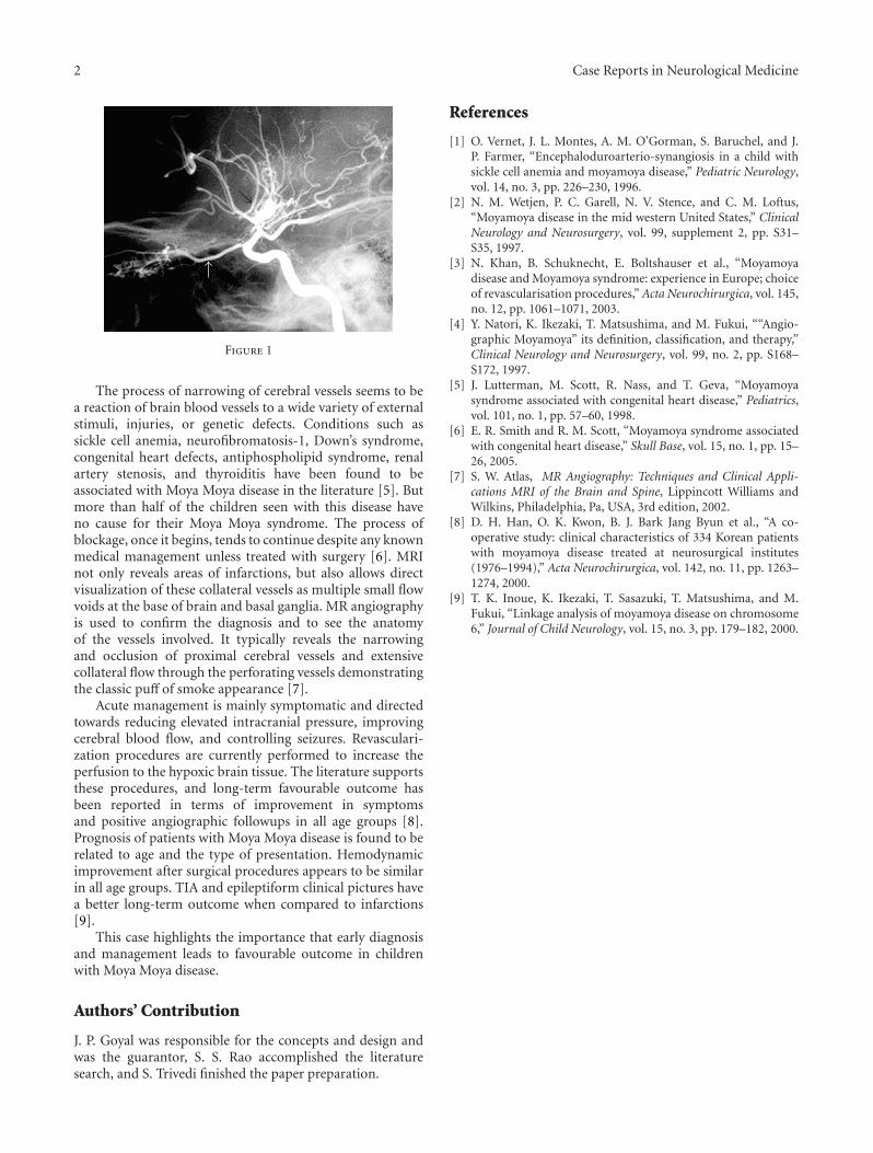
An 8-year-old female child was admitted at our institutewith complaints of weakness over left upper limb followedby lower limb for 20 days. There was also history of suddenfall 3 times probably indicating transient ischemic attacks.There was no history of fever, convulsion, head injury, andear discharge. There was no history of delayed mile stones.There were no neurocutaneous markers or asymmetry offace. On neurological examination, gait was hemiplegic, tonewas decreased over left side, power was 2/5 over left upperand lower limb, deep tendon reflexes were exaggerated, andplanter was extensor over left side. Haematological and CSFexamination was normal. MRI brain was suggestive of rightsided hemiatrophy with frontal gliotic changes and subacuteright occipital MCA territory/watershed zone infarct. MRangiography brain revealed generalized irregularity andbeaded appearance in both posterior cerebral arteries (right> left). There were few Moya Moya collaterals detected
in right basal ganglia region, which gives “puff of smoke”appearance (Figure 1). Reduced calibre of right CCA is alsoseen as compared to left side.
We treated the patient conservatively with Glycerol1 mL/kg/day and Aspirin 5 mg/kg/day. Patient showed slightimprovement on power over left side (3/5). As our centre isnot equipped with such kind of neurosurgery, so we referredthe child to higher centre for surgery where cerebral revas-cularization surgery using encephaloduroarteriosynangiosis(EDAS) was done. The child showed marked improvementafter surgery. Her hemiparesis was improved after 6 months,and repeat MRI was also found to be normal.
3. Discussion
Moya Moya disease is a rare disease characterized by multipleocclusions of the cerebral circulation with an unusual net likesystem of collaterals. In Japanese, Moya Moya means “hazy”.The disease derives its peculiar name from the angiographicappearance of cerebral vessels in the disease that resembles a“puff of smoke”. In children, the most common presentationis that of recurrent episodes of cerebral ischemia manifestingclinically as focal deficits, paresthesiae, and seizures [1].Previously thought to be prevalent only in Japan, cases havenow been reported from across the globe [2, 3]. However,majority of the cases are reported in Asia and other non-Caucasian regions [4].
2 Case Reports in Neurological Medicine
Figure 1
The process of narrowing of cerebral vessels seems to bea reaction of brain blood vessels to a wide variety of externalstimuli, injuries, or genetic defects. Conditions such assickle cell anemia, neurofibromatosis-1, Down’s syndrome,congenital heart defects, antiphospholipid syndrome, renalartery stenosis, and thyroiditis have been found to beassociated with Moya Moya disease in the literature [5]. Butmore than half of the children seen with this disease haveno cause for their Moya Moya syndrome. The process ofblockage, once it begins, tends to continue despite any knownmedical management unless treated with surgery [6]. MRInot only reveals areas of infarctions, but also allows directvisualization of these collateral vessels as multiple small flowvoids at the base of brain and basal ganglia. MR angiographyis used to confirm the diagnosis and to see the anatomyof the vessels involved. It typically reveals the narrowingand occlusion of proximal cerebral vessels and extensivecollateral flow through the perforating vessels demonstratingthe classic puff of smoke appearance [7].
Acute management is mainly symptomatic and directedtowards reducing elevated intracranial pressure, improvingcerebral blood flow, and controlling seizures. Revasculari-zation procedures are currently performed to increase theperfusion to the hypoxic brain tissue. The literature supportsthese procedures, and long-term favourable outcome hasbeen reported in terms of improvement in symptomsand positive angiographic followups in all age groups [8].Prognosis of patients with Moya Moya disease is found to berelated to age and the type of presentation. Hemodynamicimprovement after surgical procedures appears to be similarin all age groups. TIA and epileptiform clinical pictures havea better long-term outcome when compared to infarctions[9].
This case highlights the importance that early diagnosisand management leads to favourable outcome in childrenwith Moya Moya disease.
Authors’ Contribution
J. P. Goyal was responsible for the concepts and design andwas the guarantor, S. S. Rao accomplished the literaturesearch, and S. Trivedi finished the paper preparation.
References
[1] O. Vernet, J. L. Montes, A. M. O’Gorman, S. Baruchel, and J.P. Farmer, “Encephaloduroarterio-synangiosis in a child withsickle cell anemia and moyamoya disease,” Pediatric Neurology,vol. 14, no. 3, pp. 226–230, 1996.
[2] N. M. Wetjen, P. C. Garell, N. V. Stence, and C. M. Loftus,“Moyamoya disease in the mid western United States,” ClinicalNeurology and Neurosurgery, vol. 99, supplement 2, pp. S31–S35, 1997.
[3] N. Khan, B. Schuknecht, E. Boltshauser et al., “Moyamoyadisease and Moyamoya syndrome: experience in Europe; choiceof revascularisation procedures,” Acta Neurochirurgica, vol. 145,no. 12, pp. 1061–1071, 2003.
[4] Y. Natori, K. Ikezaki, T. Matsushima, and M. Fukui, ““Angio-graphic Moyamoya” its definition, classification, and therapy,”Clinical Neurology and Neurosurgery, vol. 99, no. 2, pp. S168–S172, 1997.
[5] J. Lutterman, M. Scott, R. Nass, and T. Geva, “Moyamoyasyndrome associated with congenital heart disease,” Pediatrics,vol. 101, no. 1, pp. 57–60, 1998.
[6] E. R. Smith and R. M. Scott, “Moyamoya syndrome associatedwith congenital heart disease,” Skull Base, vol. 15, no. 1, pp. 15–26, 2005.
[7] S. W. Atlas, MR Angiography: Techniques and Clinical Appli-cations MRI of the Brain and Spine, Lippincott Williams andWilkins, Philadelphia, Pa, USA, 3rd edition, 2002.
[8] D. H. Han, O. K. Kwon, B. J. Bark Jang Byun et al., “A co-operative study: clinical characteristics of 334 Korean patientswith moyamoya disease treated at neurosurgical institutes(1976–1994),” Acta Neurochirurgica, vol. 142, no. 11, pp. 1263–1274, 2000.
[9] T. K. Inoue, K. Ikezaki, T. Sasazuki, T. Matsushima, and M.Fukui, “Linkage analysis of moyamoya disease on chromosome6,” Journal of Child Neurology, vol. 15, no. 3, pp. 179–182, 2000.

Submit your manuscripts athttp://www.hindawi.com
Stem CellsInternational
Hindawi Publishing Corporationhttp://www.hindawi.com Volume 2014
Hindawi Publishing Corporationhttp://www.hindawi.com Volume 2014
MEDIATORSINFLAMMATION
of
Hindawi Publishing Corporationhttp://www.hindawi.com Volume 2014
Behavioural Neurology
EndocrinologyInternational Journal of
Hindawi Publishing Corporationhttp://www.hindawi.com Volume 2014
Hindawi Publishing Corporationhttp://www.hindawi.com Volume 2014
Disease Markers
Hindawi Publishing Corporationhttp://www.hindawi.com Volume 2014
BioMed Research International
OncologyJournal of
Hindawi Publishing Corporationhttp://www.hindawi.com Volume 2014
Hindawi Publishing Corporationhttp://www.hindawi.com Volume 2014
Oxidative Medicine and Cellular Longevity
Hindawi Publishing Corporationhttp://www.hindawi.com Volume 2014
PPAR Research
The Scientific World JournalHindawi Publishing Corporation http://www.hindawi.com Volume 2014
Immunology ResearchHindawi Publishing Corporationhttp://www.hindawi.com Volume 2014
Journal of
ObesityJournal of
Hindawi Publishing Corporationhttp://www.hindawi.com Volume 2014
Hindawi Publishing Corporationhttp://www.hindawi.com Volume 2014
Computational and Mathematical Methods in Medicine
OphthalmologyJournal of
Hindawi Publishing Corporationhttp://www.hindawi.com Volume 2014
Diabetes ResearchJournal of
Hindawi Publishing Corporationhttp://www.hindawi.com Volume 2014
Hindawi Publishing Corporationhttp://www.hindawi.com Volume 2014
Research and TreatmentAIDS
Hindawi Publishing Corporationhttp://www.hindawi.com Volume 2014
Gastroenterology Research and Practice
Hindawi Publishing Corporationhttp://www.hindawi.com Volume 2014
Parkinson’s Disease
Evidence-Based Complementary and Alternative Medicine
Volume 2014Hindawi Publishing Corporationhttp://www.hindawi.com