case report management of cervical fibroid during the...
TRANSCRIPT

Hindawi Publishing CorporationCase Reports in Obstetrics and GynecologyVolume 2013, Article ID 984030, 3 pageshttp://dx.doi.org/10.1155/2013/984030
Case ReportManagement of Cervical Fibroid during the Reproductive Period
Remon Keriakos and Mark Maher
Department of Obstetrics and Gynaecology, Sheffield Teaching Hospitals, Royal Hallamshire Hospital,Jessop Wing, Sheffield S10 3QZ, UK
Correspondence should be addressed to Remon Keriakos; [email protected]
Received 3 July 2013; Accepted 10 August 2013
Academic Editors: E. Cosmi, A. Ohkuchi, B. Piura, and E. Vaisbuch
Copyright © 2013 R. Keriakos and M. Maher. This is an open access article distributed under the Creative Commons AttributionLicense, which permits unrestricted use, distribution, and reproduction in any medium, provided the original work is properlycited.
This is a case report of a 29-year-old lady who presented with excessive vaginal discharge and sessile cervical fibroid arising fromthe vaginal portion of the cervix. She was not suitable for uterine artery embolization as she has never previously been pregnantbefore. She was encouraged to get pregnant and to avoid surgical excision which can lead to hysterectomy. Shortly after, shebecame pregnant. She had many admissions during pregnancy due to bleeding from the fibroid, and in one occasion she hadblood transfusions. The fibroid increased in size to become larger than the head of the baby. An emergency caesarean section wasperformed at 37weekswhen she attended in labour before the date of her elective caesarean section. Shewasmanaged conservativelyfollowing delivery in the hope that the fibroid becomes smaller making surgery easier.The fibroid degenerated and reduced in size.Vaginal myomectomy was carried out. The patient is now pregnant for the second time and had a cervical suture at 20 weeksgestation. In this educational case report we discuss the different management options of cervical fibroids and review the literatureof other similar cases and their outcome.
1. Introduction
A cervical fibroid during pregnancy is rare. There are onlyvery few reported cases in the literature. Sessile cervicalfibroid, arising from the vaginal portion of the cervix, in preg-nancy is extremely rare, and to our knowledge there are onlythree cases reported in the literature.
Although the majority of fibroids (60%–78%) show nosignificant change in size during pregnancy [1], some mayrapidly increase in volume due to the increased blood flow tothe uterus and the high levels of steroid hormone [2]. Ante-natal and postnatal complications can arise depending on thesize and type of the fibroid. Surgical interventions during pre-gnancy have been described in certain types of cervical fib-roids.
In this case report we discuss management of sessilecervical fibroid arising from the vaginal portion of the cervixbefore pregnancy, during pregnancy, during delivery, andafter delivery and management of patients’ future preg-nancy.
2. Case Report
A 29-year-old nulligravida lady was referred to gynaecologyclinic complaining of significant vaginal discharge for 8months sufficiently heavy towarrant frequent changing of herpanty liner. During the same period, the patient had severalepisodes of intermenstrual light vaginal bleedings. Shewas onoral contraceptive pills for 3 years, and the bleeding and thevaginal discharge did not change with cessation of the pills.She was screened for sexually transmitted diseases by hergeneral practitioner, and the results were normal. Two largeloop excisions of the transformation zone for intraepithelialneoplasia had previously taken place. On clinical examina-tion, she was found to have a 50mm sessile cervical fibroidarising from the posterior wall of the left cervical lip of thecervix. This was confirmed by vaginal ultrasound scanning.The patient was put off uterine artery embolization due tothe small risk of ovarian failure and risk of bleeding andinfection which could lead to a hysterectomy. Hence, shewas advised to consider starting to try and conceive sooner
2 Case Reports in Obstetrics and Gynecology
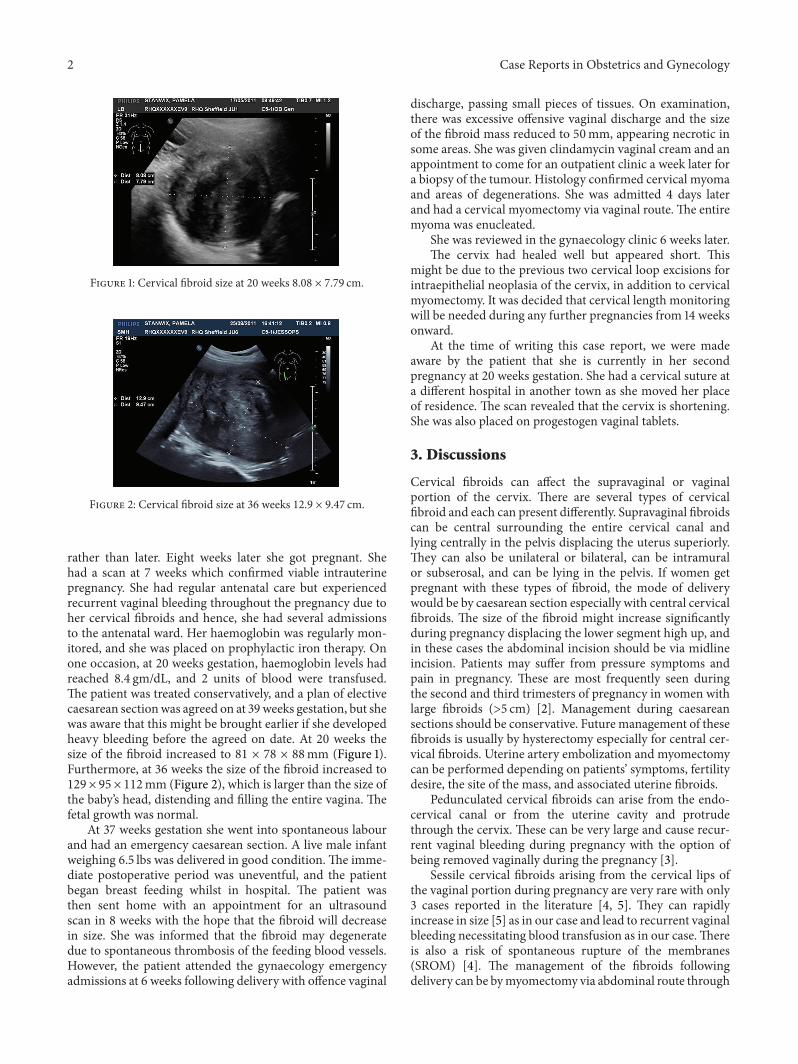
Figure 1: Cervical fibroid size at 20 weeks 8.08 × 7.79 cm.
Figure 2: Cervical fibroid size at 36 weeks 12.9 × 9.47 cm.
rather than later. Eight weeks later she got pregnant. Shehad a scan at 7 weeks which confirmed viable intrauterinepregnancy. She had regular antenatal care but experiencedrecurrent vaginal bleeding throughout the pregnancy due toher cervical fibroids and hence, she had several admissionsto the antenatal ward. Her haemoglobin was regularly mon-itored, and she was placed on prophylactic iron therapy. Onone occasion, at 20 weeks gestation, haemoglobin levels hadreached 8.4 gm/dL, and 2 units of blood were transfused.The patient was treated conservatively, and a plan of electivecaesarean sectionwas agreed on at 39weeks gestation, but shewas aware that this might be brought earlier if she developedheavy bleeding before the agreed on date. At 20 weeks thesize of the fibroid increased to 81 × 78 × 88mm (Figure 1).Furthermore, at 36 weeks the size of the fibroid increased to129 × 95 × 112mm (Figure 2), which is larger than the size ofthe baby’s head, distending and filling the entire vagina. Thefetal growth was normal.
At 37 weeks gestation she went into spontaneous labourand had an emergency caesarean section. A live male infantweighing 6.5 lbs was delivered in good condition.The imme-diate postoperative period was uneventful, and the patientbegan breast feeding whilst in hospital. The patient wasthen sent home with an appointment for an ultrasoundscan in 8 weeks with the hope that the fibroid will decreasein size. She was informed that the fibroid may degeneratedue to spontaneous thrombosis of the feeding blood vessels.However, the patient attended the gynaecology emergencyadmissions at 6 weeks following delivery with offence vaginal
discharge, passing small pieces of tissues. On examination,there was excessive offensive vaginal discharge and the sizeof the fibroid mass reduced to 50mm, appearing necrotic insome areas. She was given clindamycin vaginal cream and anappointment to come for an outpatient clinic a week later fora biopsy of the tumour. Histology confirmed cervical myomaand areas of degenerations. She was admitted 4 days laterand had a cervical myomectomy via vaginal route. The entiremyoma was enucleated.
She was reviewed in the gynaecology clinic 6 weeks later.The cervix had healed well but appeared short. This
might be due to the previous two cervical loop excisions forintraepithelial neoplasia of the cervix, in addition to cervicalmyomectomy. It was decided that cervical length monitoringwill be needed during any further pregnancies from 14 weeksonward.
At the time of writing this case report, we were madeaware by the patient that she is currently in her secondpregnancy at 20 weeks gestation. She had a cervical suture ata different hospital in another town as she moved her placeof residence. The scan revealed that the cervix is shortening.She was also placed on progestogen vaginal tablets.
3. Discussions
Cervical fibroids can affect the supravaginal or vaginalportion of the cervix. There are several types of cervicalfibroid and each can present differently. Supravaginal fibroidscan be central surrounding the entire cervical canal andlying centrally in the pelvis displacing the uterus superiorly.They can also be unilateral or bilateral, can be intramuralor subserosal, and can be lying in the pelvis. If women getpregnant with these types of fibroid, the mode of deliverywould be by caesarean section especially with central cervicalfibroids. The size of the fibroid might increase significantlyduring pregnancy displacing the lower segment high up, andin these cases the abdominal incision should be via midlineincision. Patients may suffer from pressure symptoms andpain in pregnancy. These are most frequently seen duringthe second and third trimesters of pregnancy in women withlarge fibroids (>5 cm) [2]. Management during caesareansections should be conservative. Future management of thesefibroids is usually by hysterectomy especially for central cer-vical fibroids. Uterine artery embolization and myomectomycan be performed depending on patients’ symptoms, fertilitydesire, the site of the mass, and associated uterine fibroids.
Pedunculated cervical fibroids can arise from the endo-cervical canal or from the uterine cavity and protrudethrough the cervix. These can be very large and cause recur-rent vaginal bleeding during pregnancy with the option ofbeing removed vaginally during the pregnancy [3].
Sessile cervical fibroids arising from the cervical lips ofthe vaginal portion during pregnancy are very rare with only3 cases reported in the literature [4, 5]. They can rapidlyincrease in size [5] as in our case and lead to recurrent vaginalbleeding necessitating blood transfusion as in our case.Thereis also a risk of spontaneous rupture of the membranes(SROM) [4]. The management of the fibroids followingdelivery can be bymyomectomy via abdominal route through

Case Reports in Obstetrics and Gynecology 3
an incision in the vagina during the caesarean section [5] orvaginal myomectomy as in our case. In our case we initiallyplanned to manage the patient conservatively hoping that thefibroid degenerates by spontaneous thrombosis of its feedingblood vessels making myomectomy easier to perform. Aswe anticipated, this is exactly what had happened with thesize of the fibroid reducing from 12 cm to 5 cm making iteasier to remove with insignificant amount of bleeding.There is a risk of infection with degenerating fibroids, andthese patients need monitoring for such risks in order toinstigate early treatment as happened in our case. In twoearlier similar case reports from 1958 [4], they were managedby abdominal hysterectomy; one had SROM at 20 weeksgestation with cord prolapse and intrauterine fetal death.She had abdominal hysterectomy following infection. Theother case was delivered by caesarean section at 37 weeksgestation followed by caesarean hysterectomy. Uterine arteryembolization is a possibility in these cases to help reducingthe size of the fibroid before the myomectomy.
In future pregnancy, women should be scanned forcervical length and to insert cervical suture to reduce the riskof miscarriage as in our case.
4. Conclusion
This case report has shown that women with sessile cervicalfibroids can be managed conservatively before pregnancy,during pregnancy, and after delivery with very good out-comes. Myomectomy can be delayed following delivery asthese fibroids decrease in size making it easier to removethem vaginally. Future pregnancies should bemonitoredwithcervical length measurement.
Conflict of Interests
The authors declare that there is no conflict of interest andthat the patient has consented to publish this case report.
References
[1] A. Aharoni, A. Reiter, D. Golan, Y. Paltiely, and M. Sharf, “Pat-terns of growth of uterine leiomyomas during pregnancy. A pro-spective longitudinal study,”TheBritish Journal of Obstetrics andGynaecology, vol. 95, no. 5, pp. 510–513, 1988.
[2] V. L. Katz, D. J. Dotters, andW. Droegemueller, “Complicationsof uterine leiomyomas in pregnancy,” Obstetrics and Gynecol-ogy, vol. 73, no. 4, pp. 593–596, 1989.
[3] S. Oruc, O. Karaen, and O. Kurtul, “Coexistence of a prolapsedpedunculated cervical myoma and pregnancy complications: acase study,” Journal of Reproductive Medicine, vol. 49, no. 7, pp.575–577, 2004.
[4] M. M. Abitbol and R. L. Madison, “Cervical fibroids complicat-ing pregnancy,”Obstetrics andGynecology, vol. 12, no. 4, pp. 397–398, 1958.
[5] J. Erian, T. El-Toukhy, S. Chandakas, O. Kazal, and N. Hill,“Rapidly enlarging cervical fibroids during pregnancy: a casereport,” Journal of Obstetrics and Gynaecology, vol. 24, no. 5, pp.578–579, 2004.

Submit your manuscripts athttp://www.hindawi.com
Stem CellsInternational
Hindawi Publishing Corporationhttp://www.hindawi.com Volume 2014
Hindawi Publishing Corporationhttp://www.hindawi.com Volume 2014
MEDIATORSINFLAMMATION
of
Hindawi Publishing Corporationhttp://www.hindawi.com Volume 2014
Behavioural Neurology
EndocrinologyInternational Journal of
Hindawi Publishing Corporationhttp://www.hindawi.com Volume 2014
Hindawi Publishing Corporationhttp://www.hindawi.com Volume 2014
Disease Markers
Hindawi Publishing Corporationhttp://www.hindawi.com Volume 2014
BioMed Research International
OncologyJournal of
Hindawi Publishing Corporationhttp://www.hindawi.com Volume 2014
Hindawi Publishing Corporationhttp://www.hindawi.com Volume 2014
Oxidative Medicine and Cellular Longevity
Hindawi Publishing Corporationhttp://www.hindawi.com Volume 2014
PPAR Research
The Scientific World JournalHindawi Publishing Corporation http://www.hindawi.com Volume 2014
Immunology ResearchHindawi Publishing Corporationhttp://www.hindawi.com Volume 2014
Journal of
ObesityJournal of
Hindawi Publishing Corporationhttp://www.hindawi.com Volume 2014
Hindawi Publishing Corporationhttp://www.hindawi.com Volume 2014
Computational and Mathematical Methods in Medicine
OphthalmologyJournal of
Hindawi Publishing Corporationhttp://www.hindawi.com Volume 2014
Diabetes ResearchJournal of
Hindawi Publishing Corporationhttp://www.hindawi.com Volume 2014
Hindawi Publishing Corporationhttp://www.hindawi.com Volume 2014
Research and TreatmentAIDS
Hindawi Publishing Corporationhttp://www.hindawi.com Volume 2014
Gastroenterology Research and Practice
Hindawi Publishing Corporationhttp://www.hindawi.com Volume 2014
Parkinson’s Disease
Evidence-Based Complementary and Alternative Medicine
Volume 2014Hindawi Publishing Corporationhttp://www.hindawi.com