case report - journal of clinical pathologyjcp.bmj.com/content/jclinpath/61/6/777.full.pdf ·...
TRANSCRIPT
Adenomatoid tumour of the liver
S Nagata,1 S Aishima,2 K Fukuzawa,3 H Takagi,4 H Yonemasu,5 Y Iwashita,3
T Kinoshita,3 K Wakasugi3
1 Department of Surgery,Nakabaru Hospital, Fukuoka,Japan; 2 Department ofAnatomic Pathology,Hamanomachi Hospital,Fukuoka, Japan; 3 Department ofSurgery, Oita Red CrossHospital, Oita, Japan;4 Department of Roentgenology,Oita Red Cross Hospital, Oita,Japan; 5 Department ofAnatomic Pathology, Oita RedCross Hospital, Oita, Japan
Correspondence to:Dr S Nagata, Department ofSurgery, Nakabaru Hospital, 6Mitarai, Kasuya 811-2206,Japan; [email protected]
Accepted 16 January 2008
This paper is freely availableonline under the BMJ Journalsunlocked scheme, see http://jcp.bmj.com/info/unlocked.dtl
ABSTRACTAn unusual primary adenomatoid tumour arising in thenormal liver is described. Hepatectomy was performed,and the patient is alive and free of disease 1 yearpostsurgery. Grossly, the tumour showed a haemorrhagiccut surface with numerous microcystic structures.Histological examination revealed cystic or angiomatoidspaces of various sizes lined by cuboidal, low-columnar, orflattened epithelioid cells with vacuolated cytoplasm andround to oval nuclei. The epithelioid cells were entirelysupported by proliferated capillaries and arteries togetherwith collagenous stroma. Immunohistochemical studiesshowed that the epithelioid cells were strongly positive fora broad spectrum of cytokeratins (AE1/AE3, CAM5.2,epithelial membrane antigen and cytokeratin 7) andmesothelial markers (calretinin, Wilms’ tumour 1 and D2-40). These cells were negative for Hep par-1, carci-noembryonic antigen, neural cell adhesion molecule,CD34, CD31 and HMB45. Atypically, abundant capillarieswere observed; however, the cystic proliferation ofepithelioid cells with vacuoles and immunohistochemicalprofile of the epithelioid element were consistent withhepatic adenomatoid tumour.
Adenomatoid tumours are benign neoplasms ofmesothelial origin and are most commonlyencountered in the testis, epididymis, fallopiantube, uterus and ovary.1–3 Sporadic cases have beenreported in the adrenal glands,4 pleura5 andpancreas.6 Recently, multiple adenomatoidtumours involving the liver and peritoneum havebeen reported;7 however, there have been noreports of a solitary hepatic adenomatoid tumour.
We report the case of hypervascular hepaticadenomatoid tumour incidentally discovered anddiagnosed based on the findings of light micro-scopy and immunohistochemistry.
CASE PRESENTATION
Clinical findingsA 39-year-old man without any remarkable pre-vious medical history was introduced to our unitfrom another hospital for the treatment of a2.0 cm hypervascular hepatic tumour in segmentV; the tumour had been recently discovered byabdominal ultrasonography (US) performed duringa general check-up. Physical examination wasnormal, and his carcinoembryonic antigen (CEA),carbohydrate antigen 19-9, a-fetoprotein, andprotein induced by vitamin K absence-II levelswere within the normal range. The patient wasnegative for both the hepatitis B virus surfaceantigen and the hepatitis C virus antibody.
Abdominal US demonstrated a hyperecho-genic round tumour in hepatic segment V.Abdominal CT demonstrated that the tumour
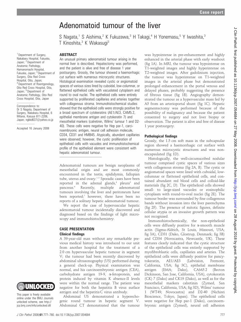
was hypointense in pre-enhancement and highlyenhanced in the arterial phase with early washout(fig 1A). In MRI, the tumour was hypointense onT1-weighted images and highly hyperintense onT2-weighted images. After gadolinium injection,the tumour was hyperintense on T1-weightedimages in the arterial phase but demonstratedprolonged enhancement in the portal venous anddelayed phases, probably suggesting the presenceof fibrous tissue (fig 1B). Angiography demon-strated the tumour as a hypervascular mass fed byA5 from an arterioportal shunt (fig 1C). Hepaticsegmentectomy was performed because of thepossibility of malignancy and because the patientconsented to surgery and not liver biopsy orobservation. The patient is alive and free of disease1 year postsurgery.
Pathological findingsGrossly, the 1.5 cm soft mass in the subcapsularregion showed a haemorrhagic cut surface withnumerous microcystic structures and was non-encapsulated (fig 1D).
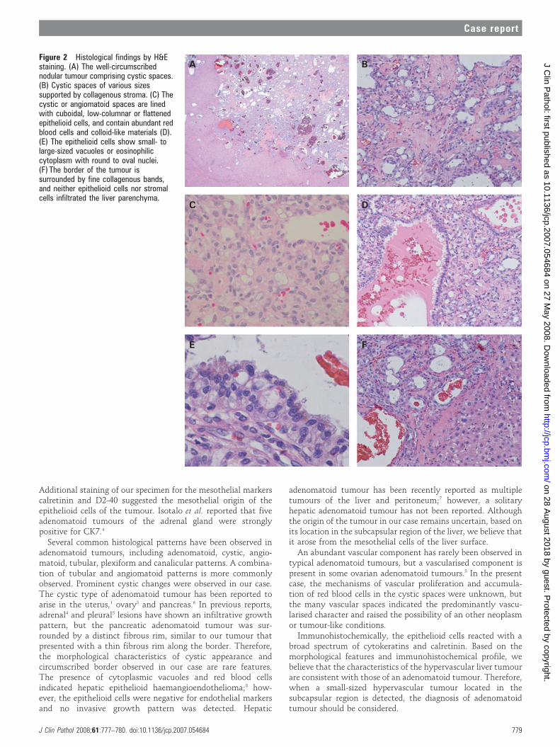
Histologically, the well-circumscribed nodulartumour comprised cystic spaces of various sizeswith collagenous stroma (fig 2A, B). The cystic orangiomatoid spaces were lined with cuboidal, low-columnar or flattened epithelioid cells, and con-tained abundant red blood cells and colloid-likematerials (fig 2C, D). The epithelioid cells showedsmall- to large-sized vacuoles or eosinophiliccytoplasm with round-to-oval nuclei (fig 2E). Thetumour border was surrounded by fine collagenousbands without invasion into the liver parenchyma(fig 2F). The presence of mitotic figures, definitecellular atypia or an invasive growth pattern wasnot recognised.
Immunohistochemically, the non-epithelioidcells were diffusely positive for a-smooth muscleactin (Sigma-Aldrich, St Louis, Missouri, USA;fig 3A), CD31 (Dako, Glostrup, Denmark; fig 3B)and CD34 (Novocastra, Newcastle, UK). Thesefeatures clearly indicated that the cystic structureof the epithelioid cells was entirely supported bymyofibroblastic cells, capillaries and arteries. Theepithelioid cells were diffusely positive for pancy-tokeratin, AE1/AE3 (Labvision, Fremont,California, USA; fig 3C), epithelial membraneantigen (EMA; Dako), CAM5.2 (BectonDickinson, San Jose, California, USA), cytokeratin(CK) 7 (Dako) and CK19 (Dako), as well as for themesothelial markers calretinin (Zymed, SanFrancisco, California, USA; fig 3D), Wilms’ tumour1 (WT49; Novocastra) and D2-40 (NichireiBioscience, Tokyo, Japan). The epithelioid cellswere negative for Hep par-1 (Dako), carcinoem-bryonic antigen (Zymed), neural cell adhesion
Case report
J Clin Pathol 2008;61:777–780. doi:10.1136/jcp.2007.054684 777
on 28 August 2018 by guest. P
rotected by copyright.http://jcp.bm
j.com/
J Clin P
athol: first published as 10.1136/jcp.2007.054684 on 27 May 2008. D
ownloaded from

molecule (NCAM; Novocastra Laboratories), CD31, CD34 andHMB45 (Enzo, Farmingdale, New York, USA).
DISCUSSIONWe describe an unusual case of a hepatic adenomatoid tumourthat was incidentally detected by radiography. Thoroughclinical and radiological examinations did not reveal anyevidence of another tumour. During preoperative diagnosis ofthe hypervascular hepatic mass, we suspected haemangioma orhaemangioma-like tumour, epithelioid haemangioendotheliomaand haemangiopericytoma, both of which occasionally demon-strate malignant characteristics. Hypervascular hepatic tumourssuch as hepatocellular carcinoma, haemangioma, focal nodularhyperplasia, hepatic adenoma and hepatic angiomyolipomawere also included in the differential diagnosis.8–10 However, the
possibility of hepatocellular carcinoma was undeniable, andsurgery was performed.
Grossly, the tumour resembled a haemangioma with regardto blood-related features and cystic, spongiotic appearance.However, the microscopic features of the tumour in our casewere not consistent with those of any of the above-mentionedtumours. Neither hepatocellular proliferation nor simpleendothelial proliferation was observed. This tumour was a soft,well-circumscribed, but non-capsulated mass with a promi-nently haemorrhagic and multicystic surface. Histologically, thetumour comprised cuboidal or flattened epithelioid cellsarranged in a cystic or angiomatoid structure, supported byprominent capillaries and arteries. These epithelioid cells, whichdid not look like bile duct, were positive for a broad spectrum ofcytokeratins such as AE1/AE3, CAM5.2, EMA, CK7 and CK19.
Figure 1 (A) CT revealed that thetumour was hypointense pre-enhancement and highly enhanced in thearterial phase with early washout(arrows). (B) Enhanced MRI aftergadolinium injection. The tumour washyperintense on T1-weighted images inthe arterial phase but demonstratedprolonged enhancement in the portalvenous and delayed phases (arrows).(C) Angiography showed a definitetumour with an arterioportal shunt (theartery (A5, filled arrow) and the portalvein (P5, open arrow)). (D) Grossly, the1.5 cm soft mass in the subcapsularregion showed a haemorrhagic cutsurface with numerous microcysticspongiotic structures but was non-encapsulated.
Case report
778 J Clin Pathol 2008;61:777–780. doi:10.1136/jcp.2007.054684
on 28 August 2018 by guest. P
rotected by copyright.http://jcp.bm
j.com/
J Clin P
athol: first published as 10.1136/jcp.2007.054684 on 27 May 2008. D
ownloaded from
Additional staining of our specimen for the mesothelial markerscalretinin and D2-40 suggested the mesothelial origin of theepithelioid cells of the tumour. Isotalo et al. reported that fiveadenomatoid tumours of the adrenal gland were stronglypositive for CK7.4
Several common histological patterns have been observed inadenomatoid tumours, including adenomatoid, cystic, angio-matoid, tubular, plexiform and canalicular patterns. A combina-tion of tubular and angiomatoid patterns is more commonlyobserved. Prominent cystic changes were observed in our case.The cystic type of adenomatoid tumour has been reported toarise in the uterus,1 ovary3 and pancreas.6 In previous reports,adrenal4 and pleural5 lesions have shown an infiltrative growthpattern, but the pancreatic adenomatoid tumour was sur-rounded by a distinct fibrous rim, similar to our tumour thatpresented with a thin fibrous rim along the border. Therefore,the morphological characteristics of cystic appearance andcircumscribed border observed in our case are rare features.The presence of cytoplasmic vacuoles and red blood cellsindicated hepatic epithelioid haemangioendothelioma;8 how-ever, the epithelioid cells were negative for endothelial markersand no invasive growth pattern was detected. Hepatic
adenomatoid tumour has been recently reported as multipletumours of the liver and peritoneum;7 however, a solitaryhepatic adenomatoid tumour has not been reported. Althoughthe origin of the tumour in our case remains uncertain, based onits location in the subcapsular region of the liver, we believe thatit arose from the mesothelial cells of the liver surface.
An abundant vascular component has rarely been observed intypical adenomatoid tumours, but a vascularised component ispresent in some ovarian adenomatoid tumours.3 In the presentcase, the mechanisms of vascular proliferation and accumula-tion of red blood cells in the cystic spaces were unknown, butthe many vascular spaces indicated the predominantly vascu-larised character and raised the possibility of an other neoplasmor tumour-like conditions.
Immunohistochemically, the epithelioid cells reacted with abroad spectrum of cytokeratins and calretinin. Based on themorphological features and immunohistochemical profile, webelieve that the characteristics of the hypervascular liver tumourare consistent with those of an adenomatoid tumour. Therefore,when a small-sized hypervascular tumour located in thesubcapsular region is detected, the diagnosis of adenomatoidtumour should be considered.
Figure 2 Histological findings by H&Estaining. (A) The well-circumscribednodular tumour comprising cystic spaces.(B) Cystic spaces of various sizessupported by collagenous stroma. (C) Thecystic or angiomatoid spaces are linedwith cuboidal, low-columnar or flattenedepithelioid cells, and contain abundant redblood cells and colloid-like materials (D).(E) The epithelioid cells show small- tolarge-sized vacuoles or eosinophiliccytoplasm with round to oval nuclei.(F) The border of the tumour issurrounded by fine collagenous bands,and neither epithelioid cells nor stromalcells infiltrated the liver parenchyma.
Case report
J Clin Pathol 2008;61:777–780. doi:10.1136/jcp.2007.054684 779
on 28 August 2018 by guest. P
rotected by copyright.http://jcp.bm
j.com/
J Clin P
athol: first published as 10.1136/jcp.2007.054684 on 27 May 2008. D
ownloaded from

Acknowledgements: We thank the Editage team for providing critical comments onthe manuscript.
Competing interests: None.
Patient consent: Informed consent has been obtained for the publication of thedetails in this report.
REFERENCES1. Stephenson TJ, Mills PM. Adenomatoid tumours: an immunohistochemical and
ultrastructural appraisal of their histogenesis. J Pathol 1986;148:327–35.2. de Klerk DP, Nime F. Adenomatoid tumors (mesothelioma) of testicular and
paratesticular tissue. Urology 1975;6:635–41.3. Ghossain MA, Chucrallah A, Kanso H, et al. Multilocular adenomatoid tumor of the
ovary: ultrasonographic findings. J Clin Ultrasound 2005;33:233–6.4. Isotalo PA, Keeney GL, Sebo TJ, et al. Adenomatoid tumor of the adrenal gland: a
clinicopathologic study of five cases and review of the literature. Am J Surg Pathol2003;27:969–77.
5. Kaplan MA, Tazelaar HD, Hayashi T, et al. Adenomatoid tumors of the pleura.Am J Surg Pathol 1996;20:1219–23.
6. Overstreet K, Wixom C, Shabaik A, et al. Adenomatoid tumor of the pancreas: acase report with comparison of histology and aspiration cytology. Mod Pathol2003;16:613–7.
7. Hayes SJ, Clark P, Mathias R, et al. Multiple adenomatoid tumours in the liver andperitoneum. J Clin Pathol 2007;60:722–4.
8. Makhlouf HR, Ishak KG, Goodman ZD. Epithelioid haemangioendothelioma of theliver: a clinicopathologic study of 137 cases. Cancer 1999;85:562–82.
9. Namasivvayam S, Salman K, Mittal PK, et al. Hypervascular hepatic focal lesions:spectrum of imaging features. Curr Probl Diagn Radiol 2007;36:107–23.
10. Terkivatan T, Hussain SM, Man RAD, et al. Diagnosis and treatment of benign focalliver lesions. Scand J Gastroenterol 2006;41:102–15.
Figure 3 Histological findings byimmunohistochemical staining. (A) Non-epithelioid cells are diffusely positive fora-smooth muscle actin and show thearterial wall (arrow). (B) CD31-positivecapillaries surround the epithelioid cells.The epithelioid cells are diffusely positivefor AE1/AE3 (C), and calretinin (D).
Take-home messages
c This is believed to be the first case report describing a solitaryhepatic adenomatoid tumour.
c The hepatic adenomatoid tumour showed prominent cysticchanges with an abundant vascular component, andimmunohistochemical studies showed that it reacted with abroad spectrum of cytokeratins and mesothelial markers.
c When a hypervascular hepatic tumour is detected, adenomatoidtumour needs to be considered in the differential diagnosis.
Case report
780 J Clin Pathol 2008;61:777–780. doi:10.1136/jcp.2007.054684
on 28 August 2018 by guest. P
rotected by copyright.http://jcp.bm
j.com/
J Clin P
athol: first published as 10.1136/jcp.2007.054684 on 27 May 2008. D
ownloaded from