case report inadvertent apical extrusion of sodium...
TRANSCRIPT

Case ReportInadvertent Apical Extrusion of Sodium Hypochlorite withEvaluation by Dental Volumetric Tomography
Elif Delve BaGer Can, Meriç KarapJnar KazandaL, and Rabia Figen Kaptan
Department of Endodontics, Faculty of Dentistry, Yeditepe University, Bagdat Caddesi 238, Goztepe, 34728 Istanbul, Turkey
Correspondence should be addressed to Elif Delve Baser Can; [email protected]
Received 22 January 2015; Revised 10 March 2015; Accepted 15 March 2015
Academic Editor: Andrea Scribante
Copyright © 2015 Elif Delve Baser Can et al. This is an open access article distributed under the Creative Commons AttributionLicense, which permits unrestricted use, distribution, and reproduction in any medium, provided the original work is properlycited.
This case report describes the tissue injury caused by inadvertently extruded NaOCl through the apical constriction. A 56-year-oldfemale patient with complaints of pain, swelling, and ecchymosis on the left side of her face was referred to our clinic.The symptomshad emerged following root canal treatment of themaxillary left first premolar, and a soft tissue complication due to apical extrusionof NaOCl was diagnosed. Antibiotics and analgesics were prescribed. DVT images revealed that the buccal root apex had perforatedthe maxillary bone. The patient was followed up every other day and became asymptomatic on the 10th day. Endodontic therapywas completed with routine procedures. Determining working length precisely and following irrigation protocols meticulously areindispensable to prevent this type of complication. 3D visualization of the affected area may reveal the cause of the incident.
1. Introduction
Removal of bacteria and bacterial toxins from the root canalsystem during shaping and cleaning is the key factor for thesuccess of endodontic therapy [1, 2]. Mechanical instrumen-tation is known to be insufficient to clean ramifications andanatomical irregularities [3, 4], and one-third of root canalsremain untouched despite the advanced technology utilizedin root canal instruments [5].Therefore, mechanical prepara-tion should be supported by a chemically active antibacterialirrigation solution [6]. The mechanical effects of irrigationare removal of microorganisms/biofilm, dentin debris, pulptissue, and instrumentation products, whereas the chemicaleffects are dissolution of soft tissue remnants and smear layerand elimination of bacteria and their byproducts. A chemi-cally active agent is required to achieve the chemical effects[7, 8].
Sodium hypochlorite (NaOCl) is themost popular irriga-tion solution to date, as it fulfills the majority of the requiredcriteria [1, 9]. In contemporary practice, various concentra-tions of NaOCl (0.5%–6%) are used for root canal irrigation.The antibacterial and tissue dissolving effects of NaOCl areknown to occur faster at higher concentrations [10]. It hasbeen reported that 5.25% NaOCl was strong enough to kill
the bacteria commonly present in the canal; however thisconcentration of NaOCl was highly toxic and irritating. Fur-thermore, 0.5% NaOCl dissolved the necrotic tissue but hadno effect on Staphylococcus aureus [11].The toxicity of NaOClis due to its high alkalinity (pH 10.8–12.9) and hypertonicity[12, 13]. It causes oxidation of protein and lipidmembrane andcauses necrosis, hemolysis, and dermal ulcerations [14]. Dis-coloration of fabrics, ophthalmic injuries due to eye contact,apical extrusion, tissue emphysema, and allergic reactions arepossible complications during irrigation of root canals withNaOCl [15].
Apical extrusion of an irrigation solution can occur whenthe pressure of the solution is excessive or if an irrigatingneedle is stuck in the root canal during shaping. Apical extru-sion is likely to occur in teeth with larger apical diametersas well as a lack of apical constriction due to root resorption[15]. Common symptoms reportedly associated with NaOClaccidents are pain, swelling, ecchymosis, hemorrhage, andallergic reactions [16–21].
The purpose of this report is to describe the destructiveeffect ofNaOCl solution on soft tissues following its extrusionduring endodontic therapy and to analyze the cause of injuryusing dental volumetric tomography (DVT).
Hindawi Publishing CorporationCase Reports in DentistryVolume 2015, Article ID 247547, 5 pageshttp://dx.doi.org/10.1155/2015/247547

2 Case Reports in Dentistry
Figure 1: Infraorbital ecchymosis and slight bruising near the naso-labial fold.
2. Case Presentation
A 56-year-old female patient with an unremarkable medicalhistory visited our clinic with complaints of swelling, ecchy-mosis, and pain on the left side of her face.The dental historyrevealed that an endodontic retreatment therapy had beeninitiated on her maxillary left first premolar tooth 10 dayspreviously, which was followed by a second session on theprevious day. During the first session, the patient had felt lightswelling, which resolved spontaneously a few hours later. Atthe second appointment, the treatment had to be stoppedbecause of severe pain and hemorrhage from the root canalduring irrigation of the root canal. No attempt had beenmadeto restore the tooth other than placing a cotton pellet into theendodontic cavity. Another appointment had been scheduledby the general dentist to complete the treatment. The patientnoticed swelling in her cheek several hours following theprocedure, but she did not contact her doctor. However, herfacial swelling worsened significantly over the next 24 hours,and the general dentist referred her to our clinic.
Approximately 48 hours after the incident, extraoral exa-mination revealed significant soft tissue swelling extendingfrom the left infraorbital region to the mandibular border.Infraorbital ecchymosis and slight bruising near the nasola-bial fold were observed (Figure 1). Intraorally, there wereno signs of mucosal ulceration or necrosis. The tooth beingtreated was tender to vertical and horizontal percussion.Panoramic radiography showed evidence of a previous rootcanal therapy with periapical radiolucency (Figure 2).
The root canals were irrigated with saline solution, andthe tooth was restored temporarily. To prevent the risk ofinfection, intramuscular clindamycin (600mg twice a day)was administered for three days, and ibuprofen (400mg) wasprescribed for painmanagement, to be taken as required. Useof cold packs externally for the first day was replaced bywarmcompresses on the second day for treatment of the swelling.On the first recall, an increase in the ecchymosis was noticed;however, the swelling had decreased significantly (Figure 3).Both swelling and ecchymosis kept progressively decreasing
Figure 2: Panoramic radiography showed evidence of a previousroot canal therapy with periapical radiolucency.
Figure 3: An increase in the ecchymosis was noticed on the firstrecall.
Figure 4: 10 days following the incident the patient became asymp-tomatic.
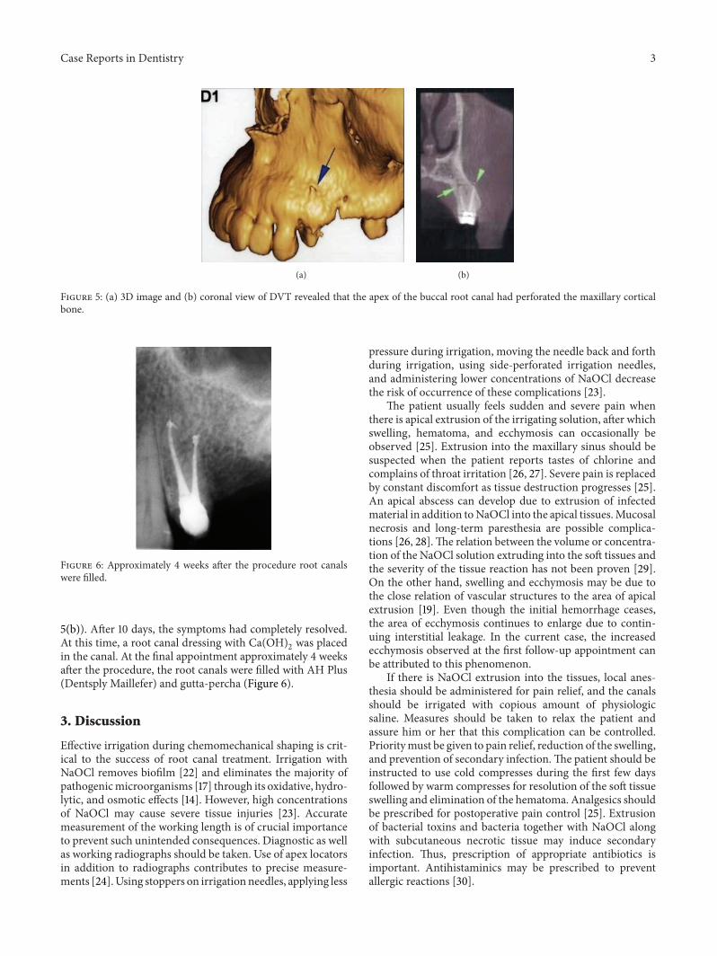
during the follow-up period, and the patient became asymp-tomatic on the 10th day after the procedure (Figure 4). Forthe evaluation of the relationship between the tooth, alveolarbone, and themaxillary sinus, dental volumetric tomography(DVT) (Newtom 3G, QR s.r.l., Verona, Italy) was performed.DVT images revealed that the apex of the buccal rootcanal had perforated the maxillary cortical bone, creating apathway for the solution into the soft tissues (Figures 5(a) and
Case Reports in Dentistry 3
(a) (b)
Figure 5: (a) 3D image and (b) coronal view of DVT revealed that the apex of the buccal root canal had perforated the maxillary corticalbone.
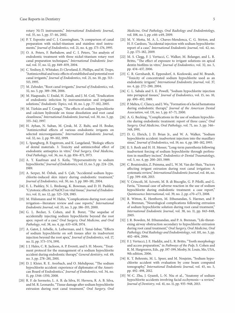
Figure 6: Approximately 4 weeks after the procedure root canalswere filled.
5(b)). After 10 days, the symptoms had completely resolved.At this time, a root canal dressing with Ca(OH)
2was placed
in the canal. At the final appointment approximately 4 weeksafter the procedure, the root canals were filled with AH Plus(Dentsply Maillefer) and gutta-percha (Figure 6).
3. Discussion
Effective irrigation during chemomechanical shaping is crit-ical to the success of root canal treatment. Irrigation withNaOCl removes biofilm [22] and eliminates the majority ofpathogenicmicroorganisms [17] through its oxidative, hydro-lytic, and osmotic effects [14]. However, high concentrationsof NaOCl may cause severe tissue injuries [23]. Accuratemeasurement of the working length is of crucial importanceto prevent such unintended consequences. Diagnostic as wellas working radiographs should be taken. Use of apex locatorsin addition to radiographs contributes to precise measure-ments [24].Using stoppers on irrigationneedles, applying less
pressure during irrigation, moving the needle back and forthduring irrigation, using side-perforated irrigation needles,and administering lower concentrations of NaOCl decreasethe risk of occurrence of these complications [23].
The patient usually feels sudden and severe pain whenthere is apical extrusion of the irrigating solution, after whichswelling, hematoma, and ecchymosis can occasionally beobserved [25]. Extrusion into the maxillary sinus should besuspected when the patient reports tastes of chlorine andcomplains of throat irritation [26, 27]. Severe pain is replacedby constant discomfort as tissue destruction progresses [25].An apical abscess can develop due to extrusion of infectedmaterial in addition toNaOCl into the apical tissues.Mucosalnecrosis and long-term paresthesia are possible complica-tions [26, 28].The relation between the volume or concentra-tion of the NaOCl solution extruding into the soft tissues andthe severity of the tissue reaction has not been proven [29].On the other hand, swelling and ecchymosis may be due tothe close relation of vascular structures to the area of apicalextrusion [19]. Even though the initial hemorrhage ceases,the area of ecchymosis continues to enlarge due to contin-uing interstitial leakage. In the current case, the increasedecchymosis observed at the first follow-up appointment canbe attributed to this phenomenon.
If there is NaOCl extrusion into the tissues, local anes-thesia should be administered for pain relief, and the canalsshould be irrigated with copious amount of physiologicsaline. Measures should be taken to relax the patient andassure him or her that this complication can be controlled.Prioritymust be given to pain relief, reduction of the swelling,and prevention of secondary infection.The patient should beinstructed to use cold compresses during the first few daysfollowed by warm compresses for resolution of the soft tissueswelling and elimination of the hematoma. Analgesics shouldbe prescribed for postoperative pain control [25]. Extrusionof bacterial toxins and bacteria together with NaOCl alongwith subcutaneous necrotic tissue may induce secondaryinfection. Thus, prescription of appropriate antibiotics isimportant. Antihistaminics may be prescribed to preventallergic reactions [30].

4 Case Reports in Dentistry
Surgical intervention may be considered in some casesdepending on the level of injury and the response to treat-ment. The goal of surgical intervention is to achieve decom-pression, ease drainage, and improve prognosis. Hematomaand/or infection may not be restricted within the anatomicalborders, as NaOCl causes tissue lysis and spreads along adimension of its own planes [25].Themajority of the patientsexperience relief after a few days, following edema, ecchy-mosis, hematoma, paralysis, and rarely secondary infection.Long-termparesthesia, scars, and aesthetic defectsmay occurin some cases [27].There are reports of neurologic injury dueto extrusion of NaOCl into soft tissues [28, 31]. There wereneither residual tissue defects nor neurologic injuries in thepresented case.
The majority of NaOCl extrusions into the periapicalarea are attributed to incorrect determination of the workinglength, excessive enlargement of the apical foramen, lateralperforations, needle stuck within the root canal, or verticalroot fractures as well [15, 20, 31]. Destruction of periapicalalveolar bone due to chronic infection as well as use of highpressure during injection facilitatesNaOCl extrusion into softtissues [32]. In a survey of 23 cases, 18 were female, whereasonly 5weremale [19]. In the same report, 20were inmaxillaryregion and only 3 were in the mandibular jaw. Mandibularteeth are centrally located in more dense cortical bone ascompared to maxillary teeth. A thin layer of cortical bonesuperficially covers the buccal roots of maxillary premolarand molar teeth. Therefore, maxillary teeth are thought tobe prone to NaOCl extrusion into soft tissues more thanmandibular teeth [19, 33]. Behrents et al. [34] also reported asodium hypochlorite accident derived from maxillary uppersecond premolar. With cone beam computed tomography(CBCT) images they demonstrated that the buccal root apexhad perforated the buccal plate as was the case with thisreport. In the present case, chronic infection preexisted peri-apically, and, additionally, DVT images revealed that thebuccal root apex had perforated the maxillary bone, whichmay have facilitated extrusion of the NaOCl solution. Inaddition, it is very likely that the operator applied excessivepressure on the needle or wedged the needle in the root canal,making the situation worse. Furthermore, in doubtful casesan initial CBCT would have identified the risk factors foraccidents NaOCl.
Some patientsmay bemore sensitive toNaOCl.This issueis supposed to be highlighted during medical anamnesis.Thepatient should be asked about any history of discomfort duetoNaOCl used for household bleach or chlorine in swimmingpools before beginning root canal treatment. If there ishistory of sensitivity, usage of NaOCl must be avoided unlessproven that there is no sensitivity provided by the patient.Dermatologic tests for NaOCl are available for chairsideapplication. Irrigation solutions containing either chlorhex-idine or ethylenediaminetetraacetic acid (EDTA) should beselected if there are no test results available and there is aprevious history of hypochlorous ion sensitivity [34].
Zhu et al. [35] indicated in their review that appearanceof facial ecchymosis after NaOCl accident follows the courseof superficial venous vasculature. Thus similarity of the loca-tions at which ecchymosis was manifested in the case reports
involved in the literature was not an unexpected result. Theextent of the ecchymosis is determined by some factors likethe amount and concentration of NaOCl entering the venouscomplex and the specific location of the venous elements andassociated tissues [35].
When the literature was reviewed it could be clearlyseen that there were limited number of case reports aboutNaOCl accidents; in fact millions of root canal therapies areperformed in a year using NaOCl for irrigation as Behrentset al. [34] have indicated. The authors related this occasionto several conditions that have to exist for developing ecchy-mosis after NaOCl accident.The apical foramen of the relatedtooth has to be open to the periapical tissues, an anatomicalvariation has to exist for drainage of the infused NaOCldirectly into the anterior facial vein to result in subcuta-neous facial haemorrhage, and apical pressure generated bypositive-pressure irrigation delivery system at the periapexhas to exceed the venous pressure in the superficial veins ofthe neck [35].These factors should be considered during rootcanal therapy procedures in order to prevent severe NaOClaccidents.
NaOCl accidents may cause a variety of problems, fromdiscoloration of clothes to serious complications requiringsurgical intervention. Meticulous attention to detail andefforts to follow irrigation rules are required to prevent suchan unexpected and unpleasant event. Syringes as well asNaOCl containers should be labeled to prevent inadvertentinjection of NaOCl into the soft tissues instead of anestheticsolution. In a better way anesthetic cartridges should neverbe filled with any of the irrigation solutions. For cases ofNaOCl extrusion into the periradicular area for any reason,measures should be taken to relax the patient, and appropriatetreatment following pain management should be initiated. Itis reasonable to employ advanced imaging techniques, as theyallow 3D visualization of the affected area and may reveal thecause of the incident.
Consent
Written informed consent was obtained from the patient forpublication of this case report and any accompanying images.A copy of the written consent is available for review.
Conflict of Interests
The authors declare that there is no conflict of interestsregarding the publication of this paper.
References
[1] M. Shih, F. J.Marshall, and S. Rosen, “The bactericidal efficiencyof sodiumhypochlorite as an endodontic irrigant,”Oral Surgery,Oral Medicine, and Oral Pathology, vol. 29, no. 4, pp. 613–619,1970.
[2] R. R.White, G. L. Hays, and L. R. Janer, “Residual antimicrobialactivity after canal irrigation with chlorhexidine,” Journal ofEndodontics, vol. 23, no. 4, pp. 229–231, 1997.
[3] J. Versumer, M. Hulsmann, and F. Schafers, “A comparativestudy of root canal preparation using ProFile .04 andLightspeed
Case Reports in Dentistry 5
rotary Ni-Ti instruments,” International Endodontic Journal,vol. 35, no. 1, pp. 37–46, 2002.
[4] P. T. Esposito and C. J. Cunningham, “A comparison of canalpreparation with nickel-titanium and stainless steel instru-ments,” Journal of Endodontics, vol. 21, no. 4, pp. 173–176, 1995.
[5] O. A. Peters, F. Barbakow, and C. I. Peters, “An analysis ofendodontic treatment with three nickel-titanium rotary rootcanal preparation techniques,” International Endodontic Jour-nal, vol. 37, no. 12, pp. 849–859, 2004.
[6] C. Yesilsoy, E.Whitaker, D. Cleveland, E. Phillips, andM. Trope,“Antimicrobial and toxic effects of established andpotential rootcanal irrigants,” Journal of Endodontics, vol. 21, no. 10, pp. 513–515, 1995.
[7] M. Zehnder, “Root canal irrigants,” Journal of Endodontics, vol.32, no. 5, pp. 389–398, 2006.
[8] M. Haapasalo, U. Endal, H. Zandi, and J. M. Coil, “Eradicationof endodontic infection by instrumentation and irrigationsolutions,” Endodontic Topics, vol. 10, no. 1, pp. 77–102, 2005.
[9] M. Turkun and T. Cengiz, “The effects of sodium hypochloriteand calcium hydroxide on tissue dissolution and root canalcleanliness,” International Endodontic Journal, vol. 30, no. 5, pp.335–342, 1997.
[10] H. Ayhan, N. Sultan, M. Cirak, M. Z. Ruhi, and H. Bodur,“Antimicrobial effects of various endodontic irrigants onselected microorganisms,” International Endodontic Journal,vol. 32, no. 2, pp. 99–102, 1999.
[11] L. Spangberg, B. Engstrom, and K. Langeland, “Biologic effectsof dental materials. 3. Toxicity and antimicrobial effect ofendodontic antiseptics in vitro,” Oral Surgery, Oral Medicine,Oral Pathology, vol. 36, no. 6, pp. 856–871, 1973.
[12] A. Y. Kaufman and S. Keila, “Hypersensitivity to sodiumhypochlorite,” Journal of Endodontics, vol. 15, no. 5, pp. 224–226,1989.
[13] A. Serper, M. Ozbek, and S. Calt, “Accidental sodium hypo-chlorite-induced skin injury during endodontic treatment,”Journal of Endodontics, vol. 30, no. 3, pp. 180–181, 2004.
[14] E. L. Pashley, N. L. Birdsong, K. Bowman, and D. H. Pashley,“Cytotoxic effects of NaOCl on vital tissue,” Journal of Endodon-tics, vol. 11, no. 12, pp. 525–528, 1985.
[15] M. Hulsmann and W. Hahn, “Complications during root canalirrigation—literature review and case reports,” InternationalEndodontic Journal, vol. 33, no. 3, pp. 186–193, 2000.
[16] G. L. Becker, S. Cohen, and R. Borer, “The sequelae ofaccidentally injecting sodium hypochlorite beyond the rootapex: report of a case,” Oral Surgery, Oral Medicine, and OralPathology, vol. 38, no. 4, pp. 633–638, 1974.
[17] A. Gatot, J. Arbelle, A. Leiberman, and I. Yanai-Inbar, “Effectsof sodium hypochlorite on soft tissues after its inadvertentinjection beyond the root apex,” Journal of Endodontics, vol. 17,no. 11, pp. 573–574, 1991.
[18] J. J. Hales, C. R. Jackson, A. P. Everett, and S. H. Moore, “Treat-ment protocol for the management of a sodium hypochloriteaccident during endodontic therapy,” General dentistry, vol. 49,no. 3, pp. 278–281, 2001.
[19] D. J. Kleier, R. E. Averbach, and O. Mehdipour, “The sodiumhypochlorite accident: experience of diplomates of the Ameri-can Board of Endodontics,” Journal of Endodontics, vol. 34, no.11, pp. 1346–1350, 2008.
[20] R. F. de Sermeno, L. A. B. da Silva, H. Herrera, R. A. B. Silva,andM. R. Leonardo, “Tissue damage after sodium hypochloriteextrusion during root canal treatment,” Oral Surgery, Oral
Medicine, Oral Pathology, Oral Radiology and Endodontology,vol. 108, no. 1, pp. e46–e49, 2009.
[21] M. V. Motta, M. A. L. Chaves-Mendonca, C. G. Stirton, andH. F. Cardozo, “Accidental injection with sodium hypochlorite:report of a case,” International Endodontic Journal, vol. 42, no.2, pp. 175–182, 2009.
[22] M. S. Clegg, F. J. Vertucci, C. Walker, M. Belanger, and L. R.Britto, “The effect of exposure to irrigant solutions on apicaldentin biofilms in vitro,” Journal of Endodontics, vol. 32, no. 5,pp. 434–437, 2006.
[23] C. R. Gernhardt, K. Eppendorf, A. Kozlowski, and M. Brandt,“Toxicity of concentrated sodium hypochlorite used as anendodontic irrigant,” International Endodontic Journal, vol. 37,no. 4, pp. 272–280, 2004.
[24] C. L. Sabala and S. E. Powell, “Sodium hypochlorite injectioninto periapical tissues,” Journal of Endodontics, vol. 15, no. 10,pp. 490–492, 1989.
[25] P.Mehra, C. Clancy, and J.Wu, “Formation of a facial hematomaduring endodontic therapy,” Journal of the American DentalAssociation, vol. 131, no. 1, pp. 67–71, 2000.
[26] A. G. Becking, “Complications in the use of sodium hypochlo-rite during endodontic treatment: report of three cases,” OralSurgery, Oral Medicine, Oral Pathology, vol. 71, no. 3, pp. 346–348, 1991.
[27] D. G. Ehrich, J. D. Brian Jr., and W. A. Walker, “Sodiumhypochlorite accident: inadvertent injection into the maxillarysinus,” Journal of Endodontics, vol. 19, no. 4, pp. 180–182, 1993.
[28] E. S. Reeh and H. H. Messer, “Long-term paresthesia followinginadvertent forcing of sodium hypochlorite through perfora-tion in maxillary incisor,” Endodontics & Dental Traumatology,vol. 5, no. 4, pp. 200–203, 1989.
[29] C. Boutsioukis, Z. Psimma, and L.W.M. Van der Sluis, “Factorsaffecting irrigant extrusion during root canal irrigation: Asystematic review,” International Endodontic Journal, vol. 46, no.7, pp. 599–618, 2013.
[30] V. Crincoli, M. Scivetti, M. B. di Bisceglie, G. P. Pilolli, and G.Favia, “Unusual case of adverse reaction in the use of sodiumhypochlorite during endodontic treatment: a case report,”Quintessence International, vol. 39, no. 2, pp. e70–e73, 2008.
[31] R. Witton, K. Henthorn, M. Ethunandan, S. Harmer, and P.A. Brennan, “Neurological complications following extrusionof sodium hypochlorite solution during root canal treatment,”International Endodontic Journal, vol. 38, no. 11, pp. 843–848,2005.
[32] J. R. Bowden, M. Ethunandan, and P. A. Brennan, “Life-threat-ening airway obstruction secondary to hypochlorite extrusionduring root canal treatment,” Oral Surgery, Oral Medicine, OralPathology, Oral Radiology and Endodontology, vol. 101, no. 3, pp.402–404, 2006.
[33] F. J. Vertucci, J. E. Haddix, and L. R. Britto, “Tooth morphologyand access preparation,” in Pathways of the Pulp, S. Cohen andK.M. Hargreaves, Eds., pp. 197–199, Mosby, St. Louis, Mo, USA,9th edition, 2006.
[34] K. T. Behrents, M. L. Speer, and M. Noujeim, “Sodium hypo-chlorite accident with evaluation by cone beam computedtomography,” International Endodontic Journal, vol. 45, no. 5,pp. 492–498, 2012.
[35] W.-C. Zhu, J. Gyamfi, L.-N. Niu et al., “Anatomy of sodiumhypochlorite accidents involving facial ecchymosis—a review,”Journal of Dentistry, vol. 41, no. 11, pp. 935–948, 2013.

Submit your manuscripts athttp://www.hindawi.com
Hindawi Publishing Corporationhttp://www.hindawi.com Volume 2014
Oral OncologyJournal of
DentistryInternational Journal of
Hindawi Publishing Corporationhttp://www.hindawi.com Volume 2014
Hindawi Publishing Corporationhttp://www.hindawi.com Volume 2014
International Journal of
Biomaterials
Hindawi Publishing Corporationhttp://www.hindawi.com Volume 2014
BioMed Research International
Hindawi Publishing Corporationhttp://www.hindawi.com Volume 2014
Case Reports in Dentistry
Hindawi Publishing Corporationhttp://www.hindawi.com Volume 2014
Oral ImplantsJournal of
Hindawi Publishing Corporationhttp://www.hindawi.com Volume 2014
Anesthesiology Research and Practice
Hindawi Publishing Corporationhttp://www.hindawi.com Volume 2014
Radiology Research and Practice
Environmental and Public Health
Journal of
Hindawi Publishing Corporationhttp://www.hindawi.com Volume 2014
The Scientific World JournalHindawi Publishing Corporation http://www.hindawi.com Volume 2014
Hindawi Publishing Corporationhttp://www.hindawi.com Volume 2014
Dental SurgeryJournal of
Drug DeliveryJournal of
Hindawi Publishing Corporationhttp://www.hindawi.com Volume 2014
Hindawi Publishing Corporationhttp://www.hindawi.com Volume 2014
Oral DiseasesJournal of
Hindawi Publishing Corporationhttp://www.hindawi.com Volume 2014
Computational and Mathematical Methods in Medicine
ScientificaHindawi Publishing Corporationhttp://www.hindawi.com Volume 2014
PainResearch and TreatmentHindawi Publishing Corporationhttp://www.hindawi.com Volume 2014
Preventive MedicineAdvances in
Hindawi Publishing Corporationhttp://www.hindawi.com Volume 2014
EndocrinologyInternational Journal of
Hindawi Publishing Corporationhttp://www.hindawi.com Volume 2014
Hindawi Publishing Corporationhttp://www.hindawi.com Volume 2014
OrthopedicsAdvances in