case report - hindawi · case report postoperativeasepticintracranialgranuloma: ... for removal of...
TRANSCRIPT
Hindawi Publishing CorporationCase Reports in SurgeryVolume 2012, Article ID 614321, 5 pagesdoi:10.1155/2012/614321
Case Report
Postoperative Aseptic Intracranial Granuloma: The PossibleInfluence of Fluid Hemostatics
Mario Ganau, Nicola Nicassio, and Leonello Tacconi
Department of Neurosurgery, University Hospital Trieste, Strada di Fiume 447, 34149 Trieste, Italy
Correspondence should be addressed to Mario Ganau, [email protected]
Received 11 June 2012; Accepted 10 July 2012
Academic Editors: K. L. Chaichana and T. Sorimachi
Copyright © 2012 Mario Ganau et al. This is an open access article distributed under the Creative Commons Attribution License,which permits unrestricted use, distribution, and reproduction in any medium, provided the original work is properly cited.
Background. Numerous reports have demonstrated how postoperative intracranial granulomas can often mimic neoplasmclinically, radiologically, and even macroscopically. Herein we present an unusual case of postsurgical intracranial asepticgranuloma secondary to a chronic inflammatory reaction without any identifiable retained foreign body. Case Description. A71-year-old patient started complaining of severe headache seven months after surgical excision of WHO Grade I right frontalfalx meningioma. CT and MRI scans disclosed a contrast-enhanced lesion with diffuse mass effect in the previous surgicalsite. The lesion was resected; intraoperative finding and histological specimens led to the diagnosis of postoperative granuloma,likely expression of a glial reaction to the fluid absorbable hemostatics applied in the surgical site after meningioma excision.The possible granuloma-inducing materials and the timing of granuloma formation are discussed. Conclusion. A comprehensiveanalysis of clinical and neuroradiological data, as well as results of blood tests including positive and negative acute phase proteins,is mandatory to raise the suspicion of postoperative granuloma. The treatment options should be evaluated on a case-by-casebasis, with a conservative attitude being the one of choice only for patients without progressive neurological deficit. Alternatively,aggressive surgical treatment and histopathological examination should be advocated.
1. Introduction
Intracranial granulomas are a rare pathologic finding; theirformation is expression of chronic inflammation charac-terized by accumulation of modified macrophages and isinitiated by a variety of infectious and noninfectious agents[1]. Infectious granulomas are the most frequent, generallyrelated to tuberculosis or sarcoidosis. Noninfectious granu-lomas are instead a reaction to a foreign body. The latter arereported to happen anywhere from months to decades aftersurgical procedures, and numerous reports have demon-strated how granulomas can often mimic neoplasm clinically,radiologically, and even grossly [2].
In this paper we present an unusual case of delayedintracranial granuloma, occurring seven months after totalresection of a right frontal falx transitional meningioma(WHO Grade I). The mass mimicking a relapse of tumorgrowth was microscopically excised: interestingly, histolog-ical examination showed a chronic aseptic inflammatoryprocess inconsistent with tumor recurrence, abscess, or
plasma cell granuloma. After a revision of all the clinical andradiological data and supported by the fact that no evidenceof foreign body was histologically found into the granuloma,but elements of advanced degradation of the fluid absorbablehemostatics applied in the surgical site after the meningiomaexcision, we have speculated that this space-occupying massmight likely result as a glial reaction to their intraoperativeuse.
Identification of postoperative granulomas is importantto prevent inappropriate treatment of presumed tumorrecurrence. A discussion of the relevant literature is providedwith special attention to possible granuloma-inducing mate-rials, diagnostic flow chart, and therapeutic options.
2. Case Report
A 71-year-old woman was rehospitalized in ourNeurosurgical Department with an irregular intracranial
2 Case Reports in Surgery
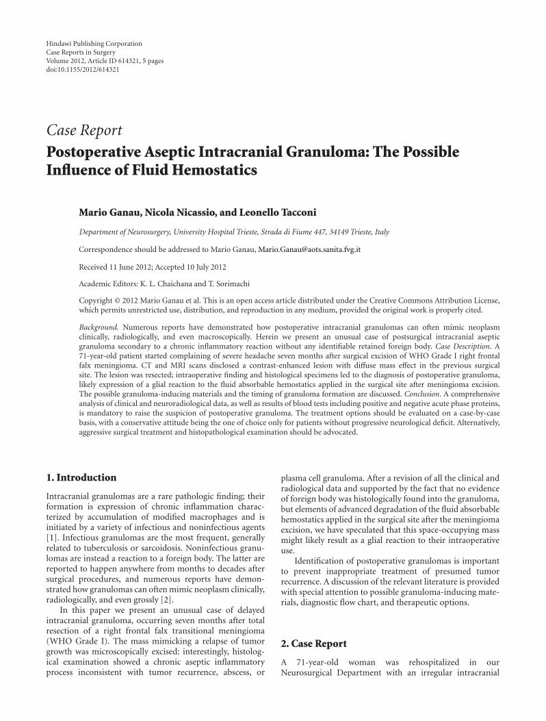
Figure 1: Preoperative contrast-enhanced axial MRI of the rightfrontal falx meningioma.
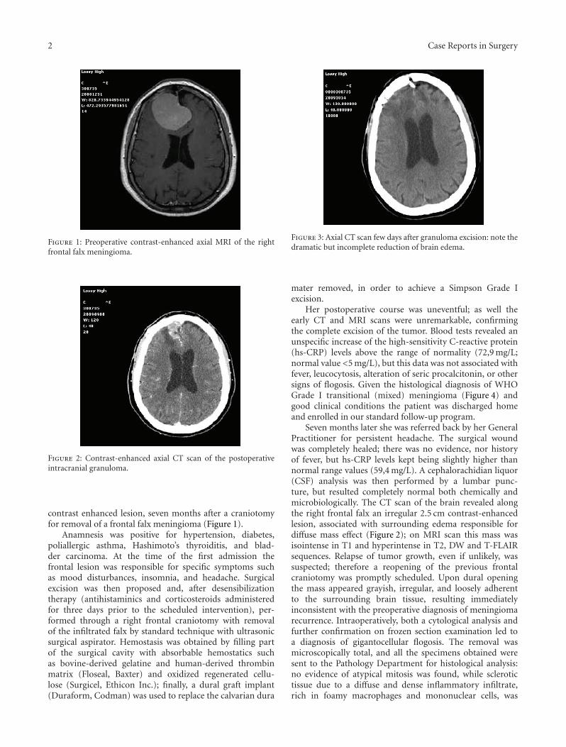
Figure 2: Contrast-enhanced axial CT scan of the postoperativeintracranial granuloma.
contrast enhanced lesion, seven months after a craniotomyfor removal of a frontal falx meningioma (Figure 1).
Anamnesis was positive for hypertension, diabetes,poliallergic asthma, Hashimoto’s thyroiditis, and blad-der carcinoma. At the time of the first admission thefrontal lesion was responsible for specific symptoms suchas mood disturbances, insomnia, and headache. Surgicalexcision was then proposed and, after desensibilizationtherapy (antihistaminics and corticosteroids administeredfor three days prior to the scheduled intervention), per-formed through a right frontal craniotomy with removalof the infiltrated falx by standard technique with ultrasonicsurgical aspirator. Hemostasis was obtained by filling partof the surgical cavity with absorbable hemostatics suchas bovine-derived gelatine and human-derived thrombinmatrix (Floseal, Baxter) and oxidized regenerated cellu-lose (Surgicel, Ethicon Inc.); finally, a dural graft implant(Duraform, Codman) was used to replace the calvarian dura
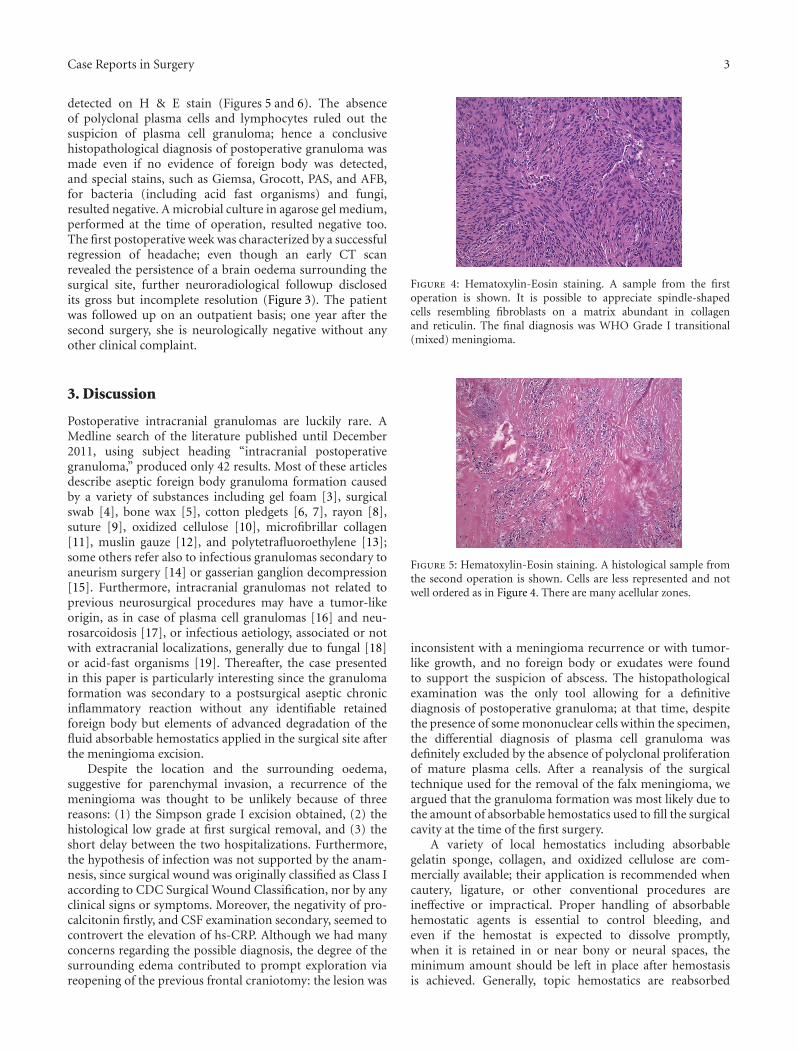
Figure 3: Axial CT scan few days after granuloma excision: note thedramatic but incomplete reduction of brain edema.
mater removed, in order to achieve a Simpson Grade Iexcision.
Her postoperative course was uneventful; as well theearly CT and MRI scans were unremarkable, confirmingthe complete excision of the tumor. Blood tests revealed anunspecific increase of the high-sensitivity C-reactive protein(hs-CRP) levels above the range of normality (72,9 mg/L;normal value <5 mg/L), but this data was not associated withfever, leucocytosis, alteration of seric procalcitonin, or othersigns of flogosis. Given the histological diagnosis of WHOGrade I transitional (mixed) meningioma (Figure 4) andgood clinical conditions the patient was discharged homeand enrolled in our standard follow-up program.
Seven months later she was referred back by her GeneralPractitioner for persistent headache. The surgical woundwas completely healed; there was no evidence, nor historyof fever, but hs-CRP levels kept being slightly higher thannormal range values (59,4 mg/L). A cephalorachidian liquor(CSF) analysis was then performed by a lumbar punc-ture, but resulted completely normal both chemically andmicrobiologically. The CT scan of the brain revealed alongthe right frontal falx an irregular 2.5 cm contrast-enhancedlesion, associated with surrounding edema responsible fordiffuse mass effect (Figure 2); on MRI scan this mass wasisointense in T1 and hyperintense in T2, DW and T-FLAIRsequences. Relapse of tumor growth, even if unlikely, wassuspected; therefore a reopening of the previous frontalcraniotomy was promptly scheduled. Upon dural openingthe mass appeared grayish, irregular, and loosely adherentto the surrounding brain tissue, resulting immediatelyinconsistent with the preoperative diagnosis of meningiomarecurrence. Intraoperatively, both a cytological analysis andfurther confirmation on frozen section examination led toa diagnosis of gigantocellular flogosis. The removal wasmicroscopically total, and all the specimens obtained weresent to the Pathology Department for histological analysis:no evidence of atypical mitosis was found, while sclerotictissue due to a diffuse and dense inflammatory infiltrate,rich in foamy macrophages and mononuclear cells, was
Case Reports in Surgery 3
detected on H & E stain (Figures 5 and 6). The absenceof polyclonal plasma cells and lymphocytes ruled out thesuspicion of plasma cell granuloma; hence a conclusivehistopathological diagnosis of postoperative granuloma wasmade even if no evidence of foreign body was detected,and special stains, such as Giemsa, Grocott, PAS, and AFB,for bacteria (including acid fast organisms) and fungi,resulted negative. A microbial culture in agarose gel medium,performed at the time of operation, resulted negative too.The first postoperative week was characterized by a successfulregression of headache; even though an early CT scanrevealed the persistence of a brain oedema surrounding thesurgical site, further neuroradiological followup disclosedits gross but incomplete resolution (Figure 3). The patientwas followed up on an outpatient basis; one year after thesecond surgery, she is neurologically negative without anyother clinical complaint.
3. Discussion
Postoperative intracranial granulomas are luckily rare. AMedline search of the literature published until December2011, using subject heading “intracranial postoperativegranuloma,” produced only 42 results. Most of these articlesdescribe aseptic foreign body granuloma formation causedby a variety of substances including gel foam [3], surgicalswab [4], bone wax [5], cotton pledgets [6, 7], rayon [8],suture [9], oxidized cellulose [10], microfibrillar collagen[11], muslin gauze [12], and polytetrafluoroethylene [13];some others refer also to infectious granulomas secondary toaneurism surgery [14] or gasserian ganglion decompression[15]. Furthermore, intracranial granulomas not related toprevious neurosurgical procedures may have a tumor-likeorigin, as in case of plasma cell granulomas [16] and neu-rosarcoidosis [17], or infectious aetiology, associated or notwith extracranial localizations, generally due to fungal [18]or acid-fast organisms [19]. Thereafter, the case presentedin this paper is particularly interesting since the granulomaformation was secondary to a postsurgical aseptic chronicinflammatory reaction without any identifiable retainedforeign body but elements of advanced degradation of thefluid absorbable hemostatics applied in the surgical site afterthe meningioma excision.
Despite the location and the surrounding oedema,suggestive for parenchymal invasion, a recurrence of themeningioma was thought to be unlikely because of threereasons: (1) the Simpson grade I excision obtained, (2) thehistological low grade at first surgical removal, and (3) theshort delay between the two hospitalizations. Furthermore,the hypothesis of infection was not supported by the anam-nesis, since surgical wound was originally classified as Class Iaccording to CDC Surgical Wound Classification, nor by anyclinical signs or symptoms. Moreover, the negativity of pro-calcitonin firstly, and CSF examination secondary, seemed tocontrovert the elevation of hs-CRP. Although we had manyconcerns regarding the possible diagnosis, the degree of thesurrounding edema contributed to prompt exploration viareopening of the previous frontal craniotomy: the lesion was
Figure 4: Hematoxylin-Eosin staining. A sample from the firstoperation is shown. It is possible to appreciate spindle-shapedcells resembling fibroblasts on a matrix abundant in collagenand reticulin. The final diagnosis was WHO Grade I transitional(mixed) meningioma.
Figure 5: Hematoxylin-Eosin staining. A histological sample fromthe second operation is shown. Cells are less represented and notwell ordered as in Figure 4. There are many acellular zones.
inconsistent with a meningioma recurrence or with tumor-like growth, and no foreign body or exudates were foundto support the suspicion of abscess. The histopathologicalexamination was the only tool allowing for a definitivediagnosis of postoperative granuloma; at that time, despitethe presence of some mononuclear cells within the specimen,the differential diagnosis of plasma cell granuloma wasdefinitely excluded by the absence of polyclonal proliferationof mature plasma cells. After a reanalysis of the surgicaltechnique used for the removal of the falx meningioma, weargued that the granuloma formation was most likely due tothe amount of absorbable hemostatics used to fill the surgicalcavity at the time of the first surgery.
A variety of local hemostatics including absorbablegelatin sponge, collagen, and oxidized cellulose are com-mercially available; their application is recommended whencautery, ligature, or other conventional procedures areineffective or impractical. Proper handling of absorbablehemostatic agents is essential to control bleeding, andeven if the hemostat is expected to dissolve promptly,when it is retained in or near bony or neural spaces, theminimum amount should be left in place after hemostasisis achieved. Generally, topic hemostatics are reabsorbed
4 Case Reports in Surgery
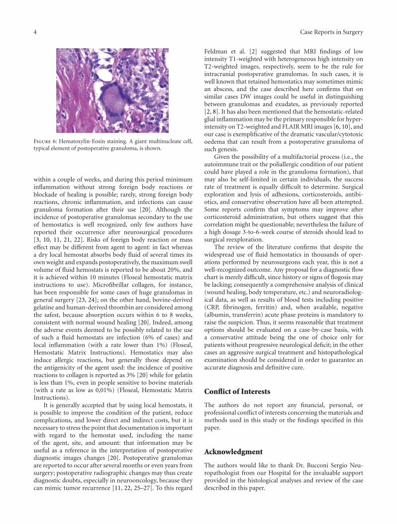
Figure 6: Hematoxylin-Eosin staining. A giant multinucleate cell,typical element of postoperative granuloma, is shown.
within a couple of weeks, and during this period minimuminflammation without strong foreign body reactions orblockade of healing is possible; rarely, strong foreign bodyreactions, chronic inflammation, and infections can causegranuloma formation after their use [20]. Although theincidence of postoperative granulomas secondary to the useof hemostatics is well recognized, only few authors havereported their occurrence after neurosurgical procedures[3, 10, 11, 21, 22]. Risks of foreign body reaction or masseffect may be different from agent to agent: in fact whereasa dry local hemostat absorbs body fluid of several times itsown weight and expands postoperatively, the maximum swellvolume of fluid hemostats is reported to be about 20%, andit is achieved within 10 minutes (Floseal hemostatic matrixinstructions to use). Microfibrillar collagen, for instance,has been responsible for some cases of huge granulomas ingeneral surgery [23, 24]; on the other hand, bovine-derivedgelatine and human-derived thrombin are considered amongthe safest, because absorption occurs within 6 to 8 weeks,consistent with normal wound healing [20]. Indeed, amongthe adverse events deemed to be possibly related to the useof such a fluid hemostats are infection (6% of cases) andlocal inflammation (with a rate lower than 1%) (Floseal,Hemostatic Matrix Instructions). Hemostatics may alsoinduce allergic reactions, but generally those depend onthe antigenicity of the agent used: the incidence of positivereactions to collagen is reported as 3% [20] while for gelatinis less than 1%, even in people sensitive to bovine materials(with a rate as low as 0,01%) (Floseal, Hemostatic MatrixInstructions).
It is generally accepted that by using local hemostats, itis possible to improve the condition of the patient, reducecomplications, and lower direct and indirect costs, but it isnecessary to stress the point that documentation is importantwith regard to the hemostat used, including the nameof the agent, site, and amount: that information may beuseful as a reference in the interpretation of postoperativediagnostic images changes [20]. Postoperative granulomasare reported to occur after several months or even years fromsurgery; postoperative radiographic changes may thus creatediagnostic doubts, especially in neurooncology, because theycan mimic tumor recurrence [11, 22, 25–27]. To this regard
Feldman et al. [2] suggested that MRI findings of lowintensity T1-weighted with heterogeneous high intensity onT2-weighted images, respectively, seem to be the rule forintracranial postoperative granulomas. In such cases, it iswell known that retained hemostatics may sometimes mimican abscess, and the case described here confirms that onsimilar cases DW images could be useful in distinguishingbetween granulomas and exudates, as previously reported[2, 8]. It has also been mentioned that the hemostatic-relatedglial inflammation may be the primary responsible for hyper-intensity on T2-weighted and FLAIR MRI images [6, 10], andour case is exemplificative of the dramatic vascular/cytotoxicoedema that can result from a postoperative granuloma ofsuch genesis.
Given the possibility of a multifactorial process (i.e., theautoimmune trait or the poliallergic condition of our patientcould have played a role in the granuloma formation), thatmay also be self-limited in certain individuals, the successrate of treatment is equally difficult to determine. Surgicalexploration and lysis of adhesions, corticosteroids, antibi-otics, and conservative observation have all been attempted.Some reports confirm that symptoms may improve aftercorticosteroid administration, but others suggest that thiscorrelation might be questionable; nevertheless the failure ofa high dosage 3-to-6-week course of steroids should lead tosurgical reexploration.
The review of the literature confirms that despite thewidespread use of fluid hemostatics in thousands of oper-ations performed by neurosurgeons each year, this is not awell-recognized outcome. Any proposal for a diagnostic flowchart is merely difficult, since history or signs of flogosis maybe lacking; consequently a comprehensive analysis of clinical(wound healing, body temperature, etc.) and neuroradiolog-ical data, as well as results of blood tests including positive(CRP, fibrinogen, ferritin) and, when available, negative(albumin, transferrin) acute phase proteins is mandatory toraise the suspicion. Thus, it seems reasonable that treatmentoptions should be evaluated on a case-by-case basis, witha conservative attitude being the one of choice only forpatients without progressive neurological deficit; in the othercases an aggressive surgical treatment and histopathologicalexamination should be considered in order to guarantee anaccurate diagnosis and definitive cure.
Conflict of Interests
The authors do not report any financial, personal, orprofessional conflict of interests concerning the materials andmethods used in this study or the findings specified in thispaper.
Acknowledgment
The authors would like to thank Dr. Bucconi Sergio Neu-ropathologist from our Hospital for the invaluable supportprovided in the histological analyses and review of the casedescribed in this paper.
Case Reports in Surgery 5
References
[1] R. Cotran, V. Kumar, and S. Robbins, Pathologic Basis ofDisease, W.B. Saunders, Philadelphia, Pa, USA, 4th edition,1989.
[2] R. P. Feldman, A. Marcovici, M. Suarez, and J. T. Goodrich,“Foreign body granuloma mimicking intracranial menin-gioma: case report and review of the literature,” Neurosurgery,vol. 44, no. 4, pp. 855–858, 1999.
[3] G. T. G. Knowlson, “Gel foam granuloma in the brain,” Journalof Neurology Neurosurgery and Psychiatry, vol. 37, no. 8, pp.971–973, 1974.
[4] M. Djindjian, P. Brugieres, F. Razavi-Encha, C. Allegret, and J.Poirier, “Post-operative intracranial foreign body granuloma:a case report,” Neuroradiology, vol. 29, no. 5, pp. 497–499,1987.
[5] J. M. Lainez, F. Fontana, M. del Rosario Martı́n, J. Sancho, andC. Barcia, “Intracranial granuloma as a late complication ofsubdural hematoma,” Archivos de Neurobiologia, vol. 52, no. 2,pp. 105–107, 1989.
[6] B. Bilginer, K. Yavuz, K. Agayev, A. Akbay, and I. M. Ziyal,“Existence of cotton granuloma after removal of a parasagittalmeningioma: clinical and radiological evaluation—A casereport,” Kobe Journal of Medical Sciences, vol. 53, no. 2, pp. 43–47, 2007.
[7] M. Fujimura, M. Nishijima, K. Umezawa et al., “Optochi-asmal arachnoiditis following cotton wrapping of anteriorcommunicating artery aneurysm treated by surgical removalof granuloma,” Journal of Clinical Neuroscience, vol. 10, no. 2,pp. 254–257, 2003.
[8] A. G. Vishteh, P. J. Apostolides, B. Dean, and R. F. Spetzler,“Magnetic resonance image of postcraniotomy retained cottonor rayon,” Journal of Neurosurgery, vol. 88, no. 5, article 928,1998.
[9] A. J. Epstein, E. J. Russell, and L. Berlin, “Suture granuloma: anunusual cause of an enhancing ring lesion in the postoperativebrain,” Journal of Computer Assisted Tomography, vol. 6, no. 4,pp. 815–817, 1982.
[10] N. Aoki, T. Sakai, and A. Oikawa, “Postoperative inflammatoryreaction developing focal but severe brain edema: a possiblecomplication of topical application of Biobond-soaked oxycel-lulose,” Acta Neurologica Scandinavica, vol. 98, no. 4, pp. 288–291, 1998.
[11] B. A. O’Shaughnessy, K. T. Schafernak, A. J. DiPatri, S.Goldman, and T. Tomita, “A granulomatous reaction toAvitene mimicking recurrence of a medulloblastoma: casereport,” Journal of Neurosurgery, vol. 104, supplement 1, pp.33–36, 2006.
[12] I. Chambi, R. R. Tasker, F. Gentili et al., “Gauze-induced gran-uloma (“gauzoma”): an uncommon complication of gauzereinforcement of berry aneurysms,” Journal of Neurosurgery,vol. 72, no. 2, pp. 163–170, 1990.
[13] J. F. Chen, S. T. Lee, T. N. Lui, Y. S. Yeh, T. Y. Chen, and W. C.Tzaan, “Teflon granuloma after microvascular decompressionfor trigeminal neuralgia,” Surgical Neurology, vol. 53, no. 3, pp.281–287, 2000.
[14] R. W. Kirollos, A. K. Tyagi, P. V. Marks, and P. T. VanHille, “Muslin induced granuloma following wrapping ofintracranial aneurysms: the role of infection as an additionalprecipitating factor. Report of two cases and review of theliterature,” Acta Neurochirurgica, vol. 139, no. 5, pp. 411–415,1997.
[15] A. L. De Sousa, J. E. Kalsbeck, and S. Batnitzky, “Anunusual late complication of Gasserian ganglion decompres-sion surgery,” Journal of Neurosurgery, vol. 48, no. 5, pp. 834–837, 1978.
[16] D. M. Cannella, A. P. Prezyna, and J. P. Kapp, “Primaryintracranial plasma-cell granuloma. Case report,” Journal ofNeurosurgery, vol. 69, no. 5, pp. 785–788, 1988.
[17] V. M. Sattelmeyer, O. Vernet, R. Janzer, and N. De Tribolet,“Neurosarcoidosis presenting as an isolated mass of thequadrigeminal plate,” Journal of Clinical Neuroscience, vol. 6,no. 3, pp. 259–261, 1999.
[18] B. S. Sharma, V. K. Khosla, A. K. Banerjee et al., “Intracranialfungal granuloma,” Surgical Neurology, vol. 47, no. 5, pp. 489–497, 1997.
[19] M. Boutarbouch, Y. Arkha, R. Gana, M. R. El Maquili, and F.Bellakhdar, “Tuberculoma of the cavernous sinus mimicking ameningioma: case report and review of the literature,” Journalof the Neurological Sciences, vol. 278, no. 1-2, pp. 123–126,2009.
[20] Y. Tomizawa, “Clinical benefits and risk analysis of topicalhemostats: a review,” Journal of Artificial Organs, vol. 8, no.3, pp. 137–142, 2005.
[21] H. Ito, H. Onishi, K. Shoin, and H. Nagatani, “Granulomacaused by oxidized cellulose. Following craniotomy,” ActaNeurochirurgica, vol. 100, no. 1-2, pp. 70–73, 1989.
[22] G. S. Sandhu, J. A. Elexpuru-Camiruaga, and S. Buckley, “Oxi-dized cellulose (Surgicel�) granulomata mimicking tumourrecurrence,” British Journal of Neurosurgery, vol. 10, no. 6, pp.617–619, 1996.
[23] D. H. McGregor, R. I. MacArthur, and T. Carter, “Avitene gran-ulomas of colonic serosa,” Annals of Clinical and LaboratoryScience, vol. 16, no. 4, pp. 296–302, 1986.
[24] M. Nakajima, T. Kamei, K. Tomimatu, and T. Manabe,“An intraperitoneal tumorous mass caused by granulomasof microfibrillar collagen hemostat (Avitene),” Archives ofPathology and Laboratory Medicine, vol. 119, no. 12, pp. 1161–1163, 1995.
[25] K. F. Kothbauer, G. I. Jallo, J. Siffert, E. Jimenez, J. C. Allen, andF. J. Epstein, “Foreign body reaction to hemostatic materialsmimicking recurrent brain tumor. Report of three cases,”Journal of Neurosurgery, vol. 95, no. 3, pp. 503–506, 2001.
[26] A. A. Razzaq and M. K. N. Chishti, “Foreign body granulomaafter craniotomy for tumor: a diagnostic dilemma,” BritishJournal of Neurosurgery, vol. 14, no. 6, pp. 591–592, 2000.
[27] S. Shimosaka and S. Waga, “Foreign-body granuloma simu-lating recurrence of falx meningioma. Case report,” Journal ofNeurosurgery, vol. 59, no. 6, pp. 1085–1087, 1983.

Submit your manuscripts athttp://www.hindawi.com
Stem CellsInternational
Hindawi Publishing Corporationhttp://www.hindawi.com Volume 2014
Hindawi Publishing Corporationhttp://www.hindawi.com Volume 2014
MEDIATORSINFLAMMATION
of
Hindawi Publishing Corporationhttp://www.hindawi.com Volume 2014
Behavioural Neurology
EndocrinologyInternational Journal of
Hindawi Publishing Corporationhttp://www.hindawi.com Volume 2014
Hindawi Publishing Corporationhttp://www.hindawi.com Volume 2014
Disease Markers
Hindawi Publishing Corporationhttp://www.hindawi.com Volume 2014
BioMed Research International
OncologyJournal of
Hindawi Publishing Corporationhttp://www.hindawi.com Volume 2014
Hindawi Publishing Corporationhttp://www.hindawi.com Volume 2014
Oxidative Medicine and Cellular Longevity
Hindawi Publishing Corporationhttp://www.hindawi.com Volume 2014
PPAR Research
The Scientific World JournalHindawi Publishing Corporation http://www.hindawi.com Volume 2014
Immunology ResearchHindawi Publishing Corporationhttp://www.hindawi.com Volume 2014
Journal of
ObesityJournal of
Hindawi Publishing Corporationhttp://www.hindawi.com Volume 2014
Hindawi Publishing Corporationhttp://www.hindawi.com Volume 2014
Computational and Mathematical Methods in Medicine
OphthalmologyJournal of
Hindawi Publishing Corporationhttp://www.hindawi.com Volume 2014
Diabetes ResearchJournal of
Hindawi Publishing Corporationhttp://www.hindawi.com Volume 2014
Hindawi Publishing Corporationhttp://www.hindawi.com Volume 2014
Research and TreatmentAIDS
Hindawi Publishing Corporationhttp://www.hindawi.com Volume 2014
Gastroenterology Research and Practice
Hindawi Publishing Corporationhttp://www.hindawi.com Volume 2014
Parkinson’s Disease
Evidence-Based Complementary and Alternative Medicine
Volume 2014Hindawi Publishing Corporationhttp://www.hindawi.com