case report - hindawi publishing corporationdownloads.hindawi.com/journals/crid/2019/8246129.pdf ·...
TRANSCRIPT
Case ReportBiomechanics for Orthodontic Intrusion of Severely ExtrudedMaxillary Molars for Functional Prosthetic Rehabilitation
Ivan Pedro Taffarel,1,2 Thiago Martins Meira,1,3 Lara Karolina Guimarães,1
Oscar Mario Antelo,1,4 and Orlando Motohiro Tanaka 1,2,5
1School of Life Sciences, Pontifícia Universidade Católica do Paraná, Curitiba, Brazil2Brazilian Board of Orthodontics and Dentofacial Orthopedics, Brazil3Bahia State University (UNEB), Guanambi, Bahia, Brazil4Universidad Intercontinental, Cidade de Mexico, Mexico5The Center for Advanced Dental Education, Saint Louis University, USA
Correspondence should be addressed to Orlando Motohiro Tanaka; [email protected]
Received 29 April 2019; Accepted 26 October 2019; Published 15 November 2019
Academic Editor: Wasiu L. Adeyemo
Copyright © 2019 Ivan Pedro Taffarel et al. This is an open access article distributed under the Creative Commons AttributionLicense, which permits unrestricted use, distribution, and reproduction in any medium, provided the original work isproperly cited.
The objective of this clinical case is thus to present a Class II, division 1, subdivision malocclusion with a severely extrudedmaxillary left hemiarch, which, due to the loss of mandibular teeth, makes prosthetic rehabilitation of the edentulous spacesimpossible. A significant intrusion was performed with mini-implants followed by miniplates associated with fixed applianceand elastomeric chains. The results of this process showed that the biological responses of the teeth and the surrounding bonystructure to the intrusion were demonstrated to be normal and acceptable. A clinically significant intrusion of the left maxillarymolars, along with the recovery of the interocclusal space and the prosthetic rehabilitation, was obtained with a fixedorthodontic appliance that was associated to the biomechanics with TADs. It also allowed the obtaining of Class I caninerelationship, demonstrated periodontal health and favored the prosthetic rehabilitation with good occlusion, aesthetics, andsatisfactory function.
1. Introduction
Posterior teeth that are supraerupted due to the early loss oftheir antagonists are commonly seen in adults that havelimited or no access to dentistry during childhood andadolescence [1]. An early loss of any molar is bound to causesupraeruption of the opposing molar into the available space.Overeruption of such a molar can lead to occlusal interfer-ence and functional disturbances and cause great difficultyduring prosthetic reconstruction [2].
Orthodontic treatment of overerupted molars has alwaysbeen considered challenging for most orthodontists, as it isone of the most difficult movements to achieve during ortho-dontic treatment [3]. This is primarily due to the greater rootvolume of these teeth [4]. Pure intrusion can only be achieved
when an adequate anchorage system is available to supportthe light and continuous forces that are directed throughthe tooth’s center of resistance [5].
Various approaches have been proposed to intrude over-erupted molars, including the use of removable applianceswith elastics [6], modified palatal arches [7], elastomericchains [8], magnets [9], and skeletal anchorage systems [10].
Currently, temporary anchorage devices (TADs), such asmini-implants and miniplates, are the treatment of choice forenhancing orthodontic tooth movement. These devicespromote absolute anchorage, and the teeth can be movedimmediately after the placement, with no need for patientcollaboration. TADs reduce the orthodontic treatmentperiod, minimize the discomfort during treatment, favoraesthetics, and increase the predictability of the final result.
HindawiCase Reports in DentistryVolume 2019, Article ID 8246129, 8 pageshttps://doi.org/10.1155/2019/8246129
The purpose of this clinical report is to describe a caseof Class II, division 1, subdivision malocclusion that hadsevere maxillary left molar extrusion, and the direct useof mini-implants and miniplate for maxillary molar intru-sion was chosen to create the necessary space for pros-thetic rehabilitation.
2. Case Report
2.1. Diagnosis and Etiology. The patient was a 26.2-year-oldfemale, who had presented for an initial consultation at theorthodontic office with the chief complaint that “the top teethhave gone down.”
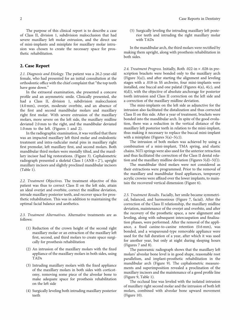
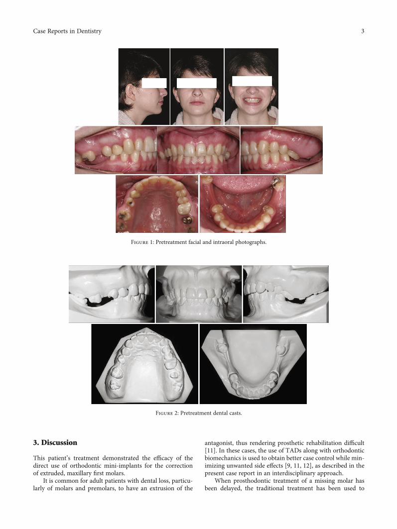
In the extraoral examination, she presented a concaveprofile and an asymmetric smile. Clinically presented, shehad a Class II, division 1, subdivision malocclusion(4.0mm), overjet, moderate overbite, and an absence ofthe first and second mandibular molars and maxillaryright first molar. With severe extrusion of the maxillarymolars, more severe on the left side, the maxillary midlinedeviated 2.0mm to the right, and the mandibular deviated1.0mm to the left. (Figures 1 and 2).
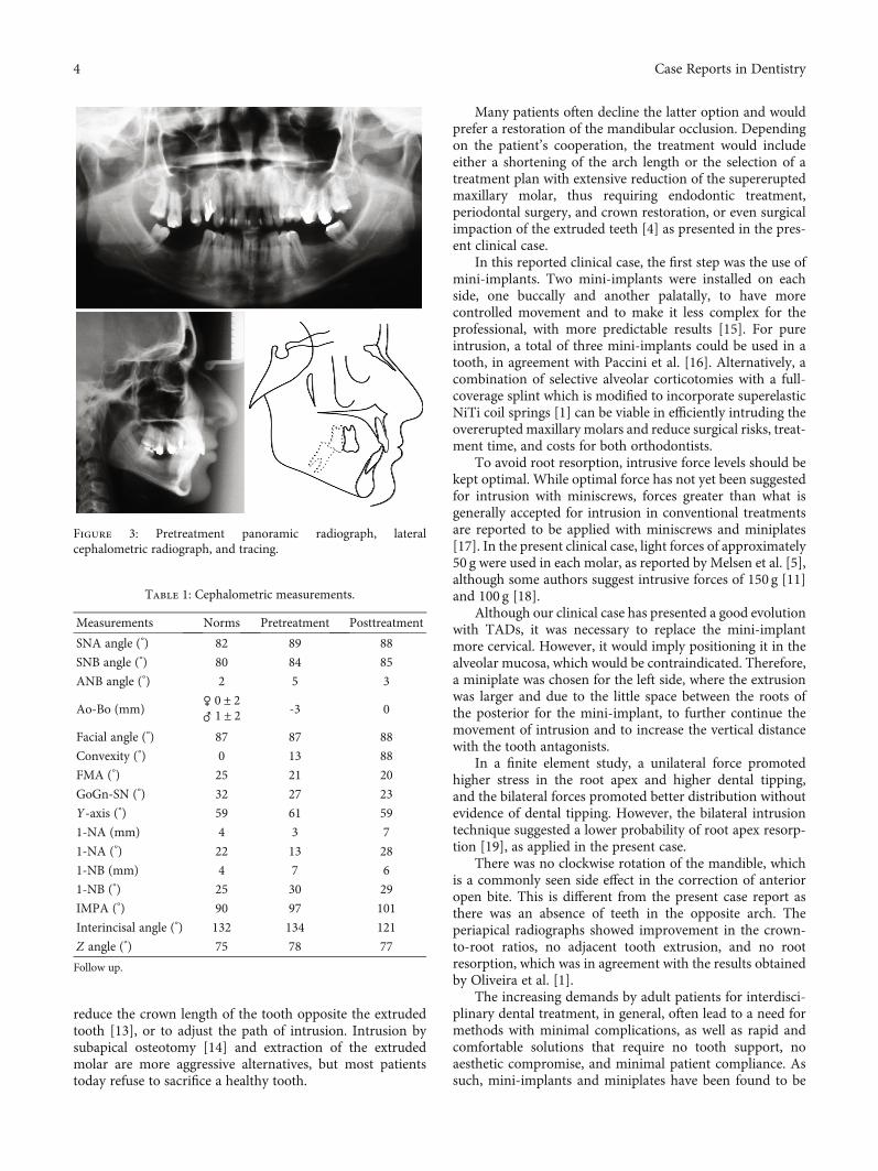
In the radiographic examination, it was verified that therewas an impacted maxillary left third molar and endodontictreatment and intra-radicular metal pins in maxillary rightfirst premolar, left maxillary first, and second molars. Bothmandibular third molars were mesially tilted, and the maxil-lary incisor had big restorations. (Figure 3). Cephalometricradiograph presented a skeletal Class I (ANB = 2°), uprightmaxillary incisors and slightly proclined mandibular incisors(Table 1).
2.2. Treatment Objectives. The treatment objective of thispatient was thus to correct Class II on the left side, attainan ideal overjet and overbite, correct the midline deviation,intrude maxillary posterior teeth, and recover space for pros-thetic rehabilitation. This was in addition to maintaining theoptimal facial balance and aesthetics.
2.3. Treatment Alternatives. Alternative treatments are asfollows:
(1) Reduction of the crown height of the second rightmaxillary molar or an extraction of the maxillary leftfirst, second, and third molars to create space surgi-cally for prosthesis rehabilitation
(2) An intrusion of the maxillary molars with the fixedappliance of the maxillary molars in both sides, usingTADs
(3) Intruding maxillary molars with the fixed applianceof the maxillary molars in both sides with corticot-omy, removing some piece of the alveolar bone tomake adequate space for prosthesis rehabilitationon the left side
(4) Surgically leveling both intruding maxillary posteriorteeth
(5) Surgically leveling the intruding maxillary left poste-rior teeth and intruding the right maxillary molarwith TADs
In the mandibular arch, the third molars were rectified bymaking them upright, along with prosthesis rehabilitation inboth sides.
2.4. Treatment Progress. Initially, Roth :022‐in × :028‐in pre-scription brackets were bonded only to the maxillary arch(Figure 3(a)), and after starting the alignment and levelingstages with a .018-in SS archwire, four mini-implants wereinstalled, one buccal and one palatal (Figures 4(a), 4(c), and4(d)), with the objective of absolute anchorage for posteriortooth intrusion and Class II correction on the left side anda correction of the maxillary midline deviation.
The mini-implants on the left side as adjunctive for theintrusion also facilitated the distalization and thus correctedClass II on this side. After a year of treatment, brackets werebonded into the mandibular arch. In spite of the good evolu-tion, there was a reduction in the vertical distance of themaxillary left posterior teeth in relation to the mini-implant,thus making it necessary to replace the buccal mini-implantwith a miniplate (Figures 5(a)–5(c)).
The intrusion of both molars was achieved by using acombination of a mini-implant, TMA spring, and elasticchains. NiTi springs were also used for the anterior retractionand thus facilitated the correction of the Class II dental rela-tion and the maxillary midline deviation (Figures 5(d)–5(f)).
The mandibular third molars were not considered astheir extractions were programmed. Prior to the removal ofthe maxillary and mandibular fixed appliances, temporaryacrylic crowns were affixed over the lower implants, to main-tain the recovered vertical dimension (Figure 6).
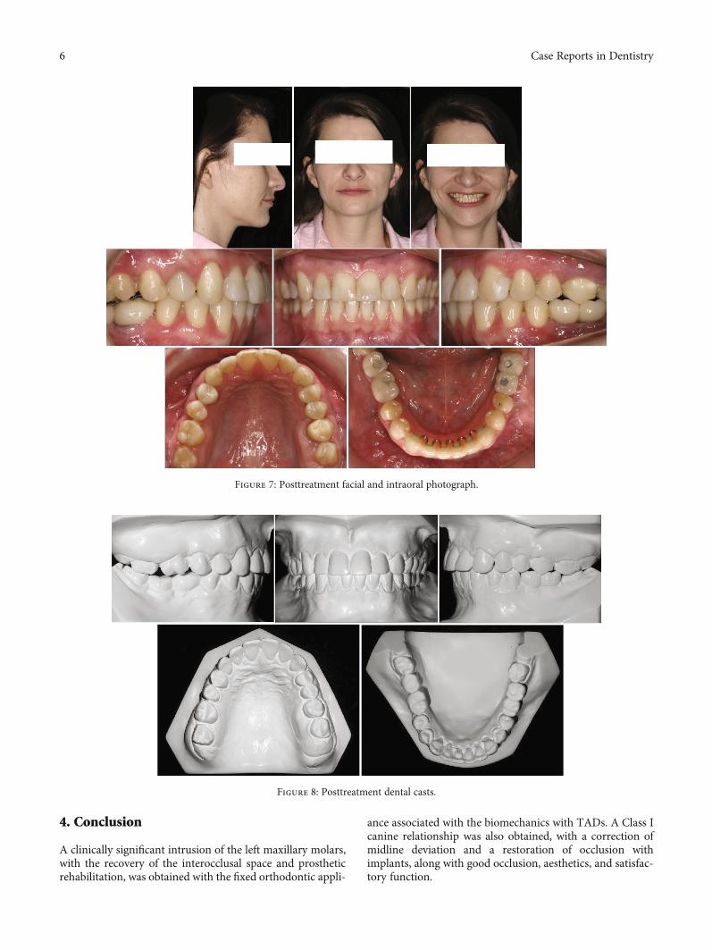
2.5. Treatment Results. Facially, her smile became symmetri-cal, balanced, and harmonious (Figure 7, facial). After thecorrection of the Class II relationship, the maxillary midlinedeviation, maintenance of the overjet and overbite, and afterthe recovery of the prosthetic space, a new alignment andleveling, along with subsequent intercuspation and finaliza-tion phases, were performed. After the removal of the appli-ance, a fixed canine-to-canine retention (0.6mm), wasbonded, and a wraparound-type removable appliance wereused for the full duration of a year, after which it was usedfor another year, but only at night during sleeping hours(Figures 7 and 8).
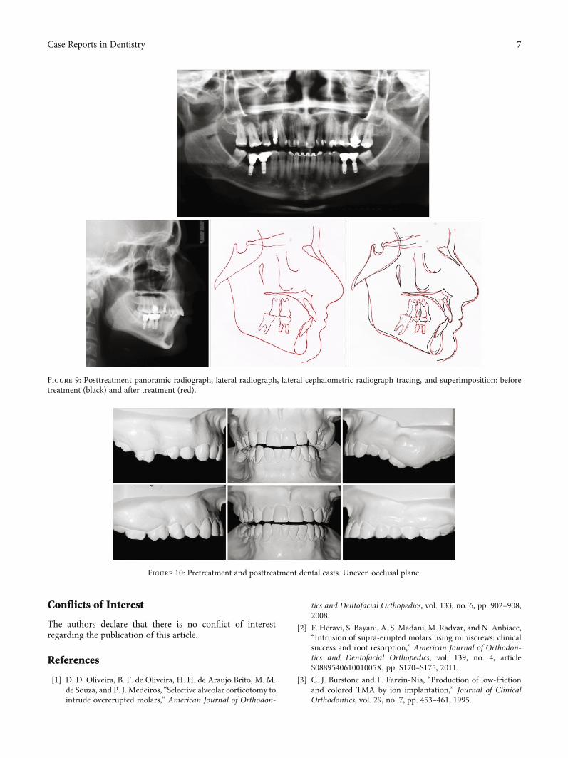
The panoramic radiograph shows that the maxillary leftmolars’ alveolar bone level is in good shape, reasonable rootparallelism, and implant-prosthetic rehabilitation in themandibular arch (Figure 9). The cephalometric measure-ments and superimposition revealed a proclination of themaxillary incisors and the maintenance of a good profile line(Figure 9, Table 1).
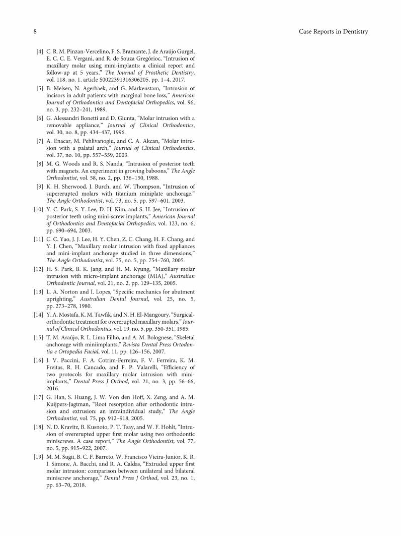
The occlusal line was leveled with the isolated intrusionof maxillary right second molar and the intrusion of both leftmolars, combined with alveolar bone upward movement(Figure 10).
2 Case Reports in Dentistry
3. Discussion
This patient’s treatment demonstrated the efficacy of thedirect use of orthodontic mini-implants for the correctionof extruded, maxillary first molars.
It is common for adult patients with dental loss, particu-larly of molars and premolars, to have an extrusion of the
antagonist, thus rendering prosthetic rehabilitation difficult[11]. In these cases, the use of TADs along with orthodonticbiomechanics is used to obtain better case control while min-imizing unwanted side effects [9, 11, 12], as described in thepresent case report in an interdisciplinary approach.
When prosthodontic treatment of a missing molar hasbeen delayed, the traditional treatment has been used to
Figure 2: Pretreatment dental casts.
Figure 1: Pretreatment facial and intraoral photographs.
3Case Reports in Dentistry
reduce the crown length of the tooth opposite the extrudedtooth [13], or to adjust the path of intrusion. Intrusion bysubapical osteotomy [14] and extraction of the extrudedmolar are more aggressive alternatives, but most patientstoday refuse to sacrifice a healthy tooth.
Many patients often decline the latter option and wouldprefer a restoration of the mandibular occlusion. Dependingon the patient’s cooperation, the treatment would includeeither a shortening of the arch length or the selection of atreatment plan with extensive reduction of the supereruptedmaxillary molar, thus requiring endodontic treatment,periodontal surgery, and crown restoration, or even surgicalimpaction of the extruded teeth [4] as presented in the pres-ent clinical case.
In this reported clinical case, the first step was the use ofmini-implants. Two mini-implants were installed on eachside, one buccally and another palatally, to have morecontrolled movement and to make it less complex for theprofessional, with more predictable results [15]. For pureintrusion, a total of three mini-implants could be used in atooth, in agreement with Paccini et al. [16]. Alternatively, acombination of selective alveolar corticotomies with a full-coverage splint which is modified to incorporate superelasticNiTi coil springs [1] can be viable in efficiently intruding theovererupted maxillary molars and reduce surgical risks, treat-ment time, and costs for both orthodontists.
To avoid root resorption, intrusive force levels should bekept optimal. While optimal force has not yet been suggestedfor intrusion with miniscrews, forces greater than what isgenerally accepted for intrusion in conventional treatmentsare reported to be applied with miniscrews and miniplates[17]. In the present clinical case, light forces of approximately50 g were used in each molar, as reported by Melsen et al. [5],although some authors suggest intrusive forces of 150 g [11]and 100 g [18].
Although our clinical case has presented a good evolutionwith TADs, it was necessary to replace the mini-implantmore cervical. However, it would imply positioning it in thealveolar mucosa, which would be contraindicated. Therefore,a miniplate was chosen for the left side, where the extrusionwas larger and due to the little space between the roots ofthe posterior for the mini-implant, to further continue themovement of intrusion and to increase the vertical distancewith the tooth antagonists.
In a finite element study, a unilateral force promotedhigher stress in the root apex and higher dental tipping,and the bilateral forces promoted better distribution withoutevidence of dental tipping. However, the bilateral intrusiontechnique suggested a lower probability of root apex resorp-tion [19], as applied in the present case.
There was no clockwise rotation of the mandible, whichis a commonly seen side effect in the correction of anterioropen bite. This is different from the present case report asthere was an absence of teeth in the opposite arch. Theperiapical radiographs showed improvement in the crown-to-root ratios, no adjacent tooth extrusion, and no rootresorption, which was in agreement with the results obtainedby Oliveira et al. [1].
The increasing demands by adult patients for interdisci-plinary dental treatment, in general, often lead to a need formethods with minimal complications, as well as rapid andcomfortable solutions that require no tooth support, noaesthetic compromise, and minimal patient compliance. Assuch, mini-implants and miniplates have been found to be
Figure 3: Pretreatment panoramic radiograph, lateralcephalometric radiograph, and tracing.
Table 1: Cephalometric measurements.
Measurements Norms Pretreatment Posttreatment
SNA angle (°) 82 89 88
SNB angle (°) 80 84 85
ANB angle (°) 2 5 3
Ao-Bo (mm)♀ 0 ± 2♂ 1 ± 2 -3 0
Facial angle (°) 87 87 88
Convexity (°) 0 13 88
FMA (°) 25 21 20
GoGn-SN (°) 32 27 23
Y-axis (°) 59 61 59
1-NA (mm) 4 3 7
1-NA (°) 22 13 28
1-NB (mm) 4 7 6
1-NB (°) 25 30 29
IMPA (°) 90 97 101
Interincisal angle (°) 132 134 121
Z angle (°) 75 78 77
Follow up.
4 Case Reports in Dentistry
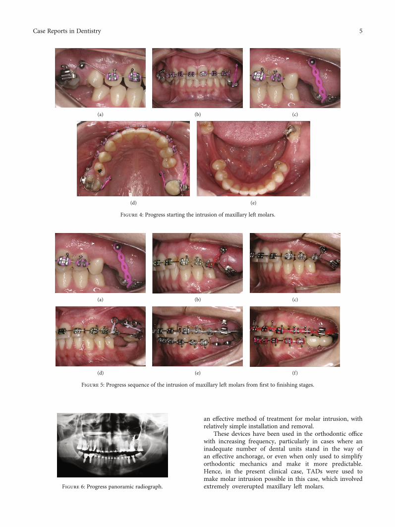
an effective method of treatment for molar intrusion, withrelatively simple installation and removal.
These devices have been used in the orthodontic officewith increasing frequency, particularly in cases where aninadequate number of dental units stand in the way ofan effective anchorage, or even when only used to simplifyorthodontic mechanics and make it more predictable.Hence, in the present clinical case, TADs were used tomake molar intrusion possible in this case, which involvedextremely overerupted maxillary left molars.
(a) (b) (c)
(d) (e)
Figure 4: Progress starting the intrusion of maxillary left molars.
(a) (b) (c)
(d) (e) (f)
Figure 5: Progress sequence of the intrusion of maxillary left molars from first to finishing stages.
Figure 6: Progress panoramic radiograph.
5Case Reports in Dentistry
4. Conclusion
A clinically significant intrusion of the left maxillary molars,with the recovery of the interocclusal space and prostheticrehabilitation, was obtained with the fixed orthodontic appli-
ance associated with the biomechanics with TADs. A Class Icanine relationship was also obtained, with a correction ofmidline deviation and a restoration of occlusion withimplants, along with good occlusion, aesthetics, and satisfac-tory function.
Figure 7: Posttreatment facial and intraoral photograph.
Figure 8: Posttreatment dental casts.
6 Case Reports in Dentistry
Conflicts of Interest
The authors declare that there is no conflict of interestregarding the publication of this article.
References
[1] D. D. Oliveira, B. F. de Oliveira, H. H. de Araujo Brito, M. M.de Souza, and P. J. Medeiros, “Selective alveolar corticotomy tointrude overerupted molars,” American Journal of Orthodon-
tics and Dentofacial Orthopedics, vol. 133, no. 6, pp. 902–908,2008.
[2] F. Heravi, S. Bayani, A. S. Madani, M. Radvar, and N. Anbiaee,“Intrusion of supra-erupted molars using miniscrews: clinicalsuccess and root resorption,” American Journal of Orthodon-tics and Dentofacial Orthopedics, vol. 139, no. 4, articleS088954061001005X, pp. S170–S175, 2011.
[3] C. J. Burstone and F. Farzin-Nia, “Production of low-frictionand colored TMA by ion implantation,” Journal of ClinicalOrthodontics, vol. 29, no. 7, pp. 453–461, 1995.
Figure 9: Posttreatment panoramic radiograph, lateral radiograph, lateral cephalometric radiograph tracing, and superimposition: beforetreatment (black) and after treatment (red).
Figure 10: Pretreatment and posttreatment dental casts. Uneven occlusal plane.
7Case Reports in Dentistry
[4] C. R. M. Pinzan-Vercelino, F. S. Bramante, J. de Araújo Gurgel,E. C. C. E. Vergani, and R. de Souza Gregórioc, “Intrusion ofmaxillary molar using mini-implants: a clinical report andfollow-up at 5 years,” The Journal of Prosthetic Dentistry,vol. 118, no. 1, article S0022391316306205, pp. 1–4, 2017.
[5] B. Melsen, N. Agerbaek, and G. Markenstam, “Intrusion ofincisors in adult patients with marginal bone loss,” AmericanJournal of Orthodontics and Dentofacial Orthopedics, vol. 96,no. 3, pp. 232–241, 1989.
[6] G. Alessandri Bonetti and D. Giunta, “Molar intrusion with aremovable appliance,” Journal of Clinical Orthodontics,vol. 30, no. 8, pp. 434–437, 1996.
[7] A. Enacar, M. Pehlivanoglu, and C. A. Akcan, “Molar intru-sion with a palatal arch,” Journal of Clinical Orthodontics,vol. 37, no. 10, pp. 557–559, 2003.
[8] M. G. Woods and R. S. Nanda, “Intrusion of posterior teethwith magnets. An experiment in growing baboons,” The AngleOrthodontist, vol. 58, no. 2, pp. 136–150, 1988.
[9] K. H. Sherwood, J. Burch, and W. Thompson, “Intrusion ofsupererupted molars with titanium miniplate anchorage,”The Angle Orthodontist, vol. 73, no. 5, pp. 597–601, 2003.
[10] Y. C. Park, S. Y. Lee, D. H. Kim, and S. H. Jee, “Intrusion ofposterior teeth using mini-screw implants,” American Journalof Orthodontics and Dentofacial Orthopedics, vol. 123, no. 6,pp. 690–694, 2003.
[11] C. C. Yao, J. J. Lee, H. Y. Chen, Z. C. Chang, H. F. Chang, andY. J. Chen, “Maxillary molar intrusion with fixed appliancesand mini-implant anchorage studied in three dimensions,”The Angle Orthodontist, vol. 75, no. 5, pp. 754–760, 2005.
[12] H. S. Park, B. K. Jang, and H. M. Kyung, “Maxillary molarintrusion with micro-implant anchorage (MIA),” AustralianOrthodontic Journal, vol. 21, no. 2, pp. 129–135, 2005.
[13] L. A. Norton and I. Lopes, “Specific mechanics for abutmentuprighting,” Australian Dental Journal, vol. 25, no. 5,pp. 273–278, 1980.
[14] Y. A.Mostafa, K.M. Tawfik, andN.H. El-Mangoury, “Surgical-orthodontic treatment for overeruptedmaxillarymolars,” Jour-nal of Clinical Orthodontics, vol. 19, no. 5, pp. 350-351, 1985.
[15] T. M. Araújo, R. L. Lima Filho, and A. M. Bolognese, “Skeletalanchorage with miniimplants,” Revista Dental Press Ortodon-tia e Ortopedia Facial, vol. 11, pp. 126–156, 2007.
[16] J. V. Paccini, F. A. Cotrim-Ferreira, F. V. Ferreira, K. M.Freitas, R. H. Cancado, and F. P. Valarelli, “Efficiency oftwo protocols for maxillary molar intrusion with mini-implants,” Dental Press J Orthod, vol. 21, no. 3, pp. 56–66,2016.
[17] G. Han, S. Huang, J. W. Von den Hoff, X. Zeng, and A. M.Kuijpers-Jagtman, “Root resorption after orthodontic intru-sion and extrusion: an intraindividual study,” The AngleOrthodontist, vol. 75, pp. 912–918, 2005.
[18] N. D. Kravitz, B. Kusnoto, P. T. Tsay, and W. F. Hohlt, “Intru-sion of overerupted upper first molar using two orthodonticminiscrews. A case report,” The Angle Orthodontist, vol. 77,no. 5, pp. 915–922, 2007.
[19] M. M. Sugii, B. C. F. Barreto, W. Francisco Vieira-Junior, K. R.I. Simone, A. Bacchi, and R. A. Caldas, “Extruded upper firstmolar intrusion: comparison between unilateral and bilateralminiscrew anchorage,” Dental Press J Orthod, vol. 23, no. 1,pp. 63–70, 2018.
8 Case Reports in Dentistry

DentistryInternational Journal of
Hindawiwww.hindawi.com Volume 2018
Environmental and Public Health
Journal of
Hindawiwww.hindawi.com Volume 2018
Hindawi Publishing Corporation http://www.hindawi.com Volume 2013Hindawiwww.hindawi.com
The Scientific World Journal
Volume 2018Hindawiwww.hindawi.com Volume 2018
Public Health Advances in
Hindawiwww.hindawi.com Volume 2018
Case Reports in Medicine
Hindawiwww.hindawi.com Volume 2018
International Journal of
Biomaterials
Scienti�caHindawiwww.hindawi.com Volume 2018
PainResearch and TreatmentHindawiwww.hindawi.com Volume 2018
Preventive MedicineAdvances in
Hindawiwww.hindawi.com Volume 2018
Hindawiwww.hindawi.com Volume 2018
Case Reports in Dentistry
Hindawiwww.hindawi.com Volume 2018
Surgery Research and Practice
Hindawiwww.hindawi.com Volume 2018
BioMed Research International Medicine
Advances in
Hindawiwww.hindawi.com Volume 2018
Hindawiwww.hindawi.com Volume 2018
Anesthesiology Research and Practice
Hindawiwww.hindawi.com Volume 2018
Radiology Research and Practice
Hindawiwww.hindawi.com Volume 2018
Computational and Mathematical Methods in Medicine
EndocrinologyInternational Journal of
Hindawiwww.hindawi.com Volume 2018
Hindawiwww.hindawi.com Volume 2018
OrthopedicsAdvances in
Drug DeliveryJournal of
Hindawiwww.hindawi.com Volume 2018
Submit your manuscripts atwww.hindawi.com