case report endometrioid paraovarian borderline cystic tumor...
TRANSCRIPT

Case ReportEndometrioid Paraovarian Borderline Cystic Tumor in an Infantwith Proteus Syndrome
Liliana Vasquez,1 Mariela Tello,1 Ivan Maza,1 Monica Oscanoa,1 Milagros Dueñas,2
Haydee Castro,3 and Alan Latorre4
1Department of Oncology and Radiotherapy, Rebagliati Hospital, Lima, Peru2Department of Genetics, Rebagliati Hospital, Lima, Peru3Department of Gynecology-Oncology, Rebagliati Hospital, Lima, Peru4Department of Pathology, Rebagliati Hospital, Lima, Peru
Correspondence should be addressed to Liliana Vasquez; [email protected]
Received 21 August 2015; Revised 26 September 2015; Accepted 30 September 2015
Academic Editor: Yoshihito Yokoyama
Copyright © 2015 Liliana Vasquez et al.This is an open access article distributed under the Creative Commons Attribution License,which permits unrestricted use, distribution, and reproduction in any medium, provided the original work is properly cited.
Ovarian and paraovarian neoplasms are uncommon in children, mainly originating from germ cell tumors and, least frequently,epithelial tumors. There is an association between genital tract tumors and Proteus syndrome, a rare, sporadic, and progressiveentity, characterized by a postnatal overgrowth in several tissues caused by a mosaic mutation in the AKT1 gene. We describe a20-month-old asymptomatic infant with Proteus syndrome who developed an endometrioid paraovarian borderline cystic tumor.This is the youngest patient so far reported in the literature with this rare syndrome and an adnexal tumor of borderlinemalignancy.A total of nine patients have been described with female tract tumors and associated Proteus syndrome, which includes bilateralovarian cystadenomas and other benign masses. A paraovarian neoplasm is extremely rare in children and could be considered acriterion for Proteus syndrome. Standardized staging and treatment of these tumors are not well established; however, most authorsconclude that these neoplasms must be treated as their ovarian counterparts.
1. Introduction
Ovarian and paraovarian neoplasms are rare in childrenand represent less than 5% of solid tumors [1–3]. The mostcommon age at presentation is the second decade of lifeand usually older girls are at greatest risk of malignancy [4].Incidence of ovarian neoplasms in the pediatric age groupis unknown, but it is estimated at 2.6 cases per 100,000girls per year [2]. The most common histological subtypeof pediatric ovarian cancer is derived from germ cells [5–7], followed by epithelial tumors, such as cystadenomas oradenocarcinomas. Primary malignant paraovarian epithelialtumors are even more rare, previously described in literatureas cystadenocarcinomas with low malignant potential [8, 9].
Proteus syndrome (PS) is a rare and sporadic disorderthat causes postnatal overgrowth of multiple tissues in amosaic pattern [10].
Cohen Jr. and Hayden first described it in 1979 as “a newhamartomatous syndrome” [11]. Wiedemann et al. furtherexplained the syndrome and named it Proteus syndrome [12].Since its first description until today, there have only beenapproximately 200 diagnosed cases in developed countries.PS is a progressive disorder that most commonly affects theskeleton, skin, adipose, and central nervous systems, causingsevere overgrowth and disfigurement, physical disability, anda decline in the patient’s quality of life [13]. It is associatedwitha range of tumors, pulmonary complications, and a strikingpredisposition to deep vein thrombosis and pulmonaryembolism [14]. Among such tumors are those derived fromlymphatic tissue and ovarian tumors, which are also includedin the diagnostic criteria for this syndrome.
We report an infant with PS, who developed anendometrioid borderline paraovarian cystic tumor.This is thetenth case reported worldwide presenting an adnexal mass
Hindawi Publishing CorporationCase Reports in Oncological MedicineVolume 2015, Article ID 392576, 6 pageshttp://dx.doi.org/10.1155/2015/392576
2 Case Reports in Oncological Medicine
(a) (b)
(c)
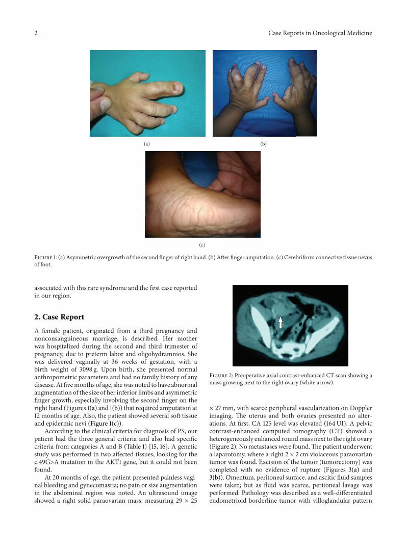
Figure 1: (a) Asymmetric overgrowth of the second finger of right hand. (b) After finger amputation. (c) Cerebriform connective tissue nevusof foot.
associated with this rare syndrome and the first case reportedin our region.
2. Case Report
A female patient, originated from a third pregnancy andnonconsanguineous marriage, is described. Her motherwas hospitalized during the second and third trimester ofpregnancy, due to preterm labor and oligohydramnios. Shewas delivered vaginally at 36 weeks of gestation, with abirth weight of 3098 g. Upon birth, she presented normalanthropometric parameters and had no family history of anydisease. At fivemonths of age, shewas noted to have abnormalaugmentation of the size of her inferior limbs and asymmetricfinger growth, especially involving the second finger on theright hand (Figures 1(a) and 1(b)) that required amputation at12 months of age. Also, the patient showed several soft tissueand epidermic nevi (Figure 1(c)).
According to the clinical criteria for diagnosis of PS, ourpatient had the three general criteria and also had specificcriteria from categories A and B (Table 1) [15, 16]. A geneticstudy was performed in two affected tissues, looking for thec.49G>A mutation in the AKT1 gene, but it could not beenfound.
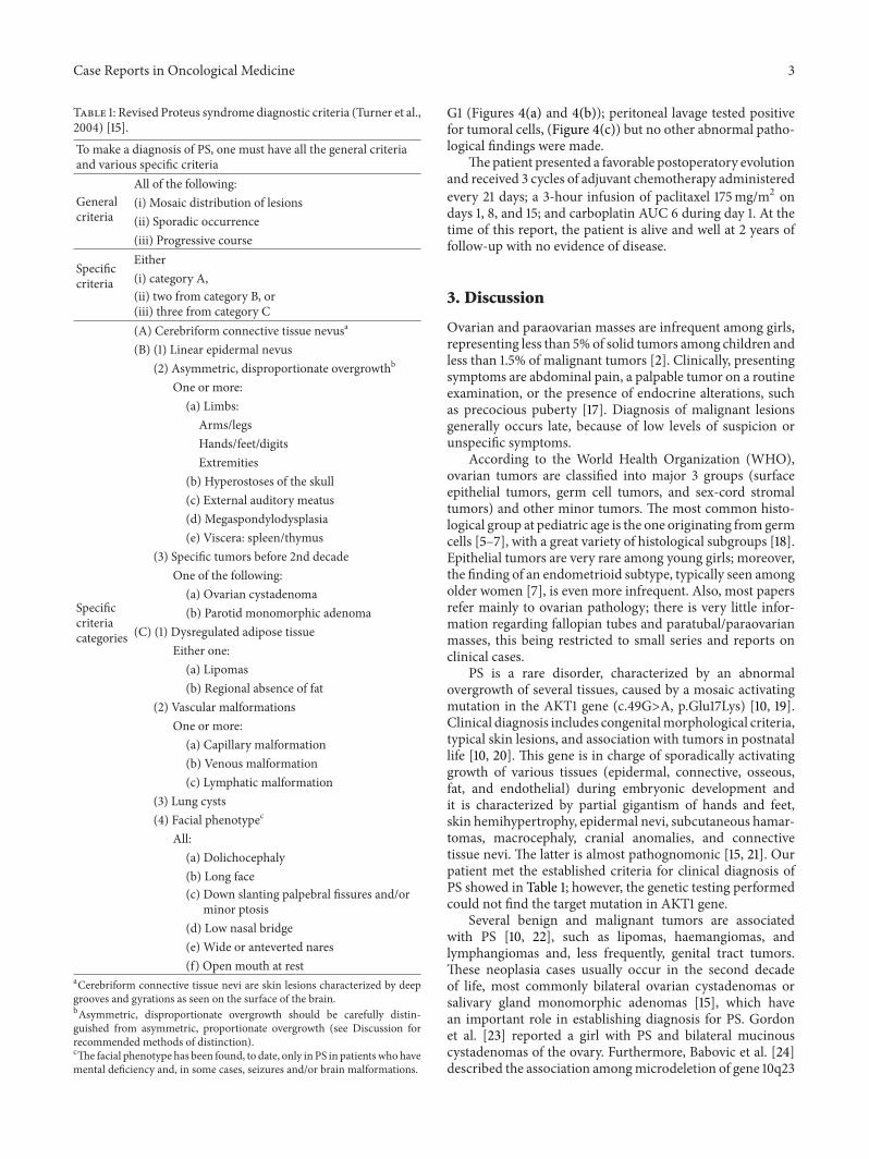
At 20 months of age, the patient presented painless vagi-nal bleeding and gynecomastia; no pain or size augmentationin the abdominal region was noted. An ultrasound imageshowed a right solid paraovarian mass, measuring 29 × 25
Figure 2: Preoperative axial contrast-enhanced CT scan showing amass growing next to the right ovary (white arrow).
× 27mm, with scarce peripheral vascularization on Dopplerimaging. The uterus and both ovaries presented no alter-ations. At first, CA 125 level was elevated (164UI). A pelviccontrast-enhanced computed tomography (CT) showed aheterogeneously enhanced roundmass next to the right ovary(Figure 2). Nometastases were found.The patient underwenta laparotomy, where a right 2 × 2 cm violaceous paraovariantumor was found. Excision of the tumor (tumorectomy) wascompleted with no evidence of rupture (Figures 3(a) and3(b)). Omentum, peritoneal surface, and ascitic fluid sampleswere taken; but as fluid was scarce, peritoneal lavage wasperformed. Pathology was described as a well-differentiatedendometrioid borderline tumor with villoglandular pattern
Case Reports in Oncological Medicine 3
Table 1: Revised Proteus syndrome diagnostic criteria (Turner et al.,2004) [15].To make a diagnosis of PS, one must have all the general criteriaand various specific criteria
Generalcriteria
All of the following:(i) Mosaic distribution of lesions(ii) Sporadic occurrence(iii) Progressive course
Specificcriteria
Either(i) category A,(ii) two from category B, or(iii) three from category C
Specificcriteriacategories
(A) Cerebriform connective tissue nevusa
(B) (1) Linear epidermal nevus(2) Asymmetric, disproportionate overgrowthb
One or more:(a) Limbs:
Arms/legsHands/feet/digitsExtremities
(b) Hyperostoses of the skull(c) External auditory meatus(d) Megaspondylodysplasia(e) Viscera: spleen/thymus
(3) Specific tumors before 2nd decadeOne of the following:(a) Ovarian cystadenoma(b) Parotid monomorphic adenoma
(C) (1) Dysregulated adipose tissueEither one:(a) Lipomas(b) Regional absence of fat
(2) Vascular malformationsOne or more:(a) Capillary malformation(b) Venous malformation(c) Lymphatic malformation
(3) Lung cysts(4) Facial phenotypec
All:(a) Dolichocephaly(b) Long face(c) Down slanting palpebral fissures and/or
minor ptosis(d) Low nasal bridge(e) Wide or anteverted nares(f) Open mouth at rest
aCerebriform connective tissue nevi are skin lesions characterized by deepgrooves and gyrations as seen on the surface of the brain.bAsymmetric, disproportionate overgrowth should be carefully distin-guished from asymmetric, proportionate overgrowth (see Discussion forrecommended methods of distinction).cThe facial phenotype has been found, to date, only in PS in patientswhohavemental deficiency and, in some cases, seizures and/or brain malformations.
G1 (Figures 4(a) and 4(b)); peritoneal lavage tested positivefor tumoral cells, (Figure 4(c)) but no other abnormal patho-logical findings were made.
Thepatient presented a favorable postoperatory evolutionand received 3 cycles of adjuvant chemotherapy administeredevery 21 days; a 3-hour infusion of paclitaxel 175mg/m2 ondays 1, 8, and 15; and carboplatin AUC 6 during day 1. At thetime of this report, the patient is alive and well at 2 years offollow-up with no evidence of disease.
3. Discussion
Ovarian and paraovarian masses are infrequent among girls,representing less than 5%of solid tumors among children andless than 1.5% of malignant tumors [2]. Clinically, presentingsymptoms are abdominal pain, a palpable tumor on a routineexamination, or the presence of endocrine alterations, suchas precocious puberty [17]. Diagnosis of malignant lesionsgenerally occurs late, because of low levels of suspicion orunspecific symptoms.
According to the World Health Organization (WHO),ovarian tumors are classified into major 3 groups (surfaceepithelial tumors, germ cell tumors, and sex-cord stromaltumors) and other minor tumors. The most common histo-logical group at pediatric age is the one originating fromgermcells [5–7], with a great variety of histological subgroups [18].Epithelial tumors are very rare among young girls; moreover,the finding of an endometrioid subtype, typically seen amongolder women [7], is even more infrequent. Also, most papersrefer mainly to ovarian pathology; there is very little infor-mation regarding fallopian tubes and paratubal/paraovarianmasses, this being restricted to small series and reports onclinical cases.
PS is a rare disorder, characterized by an abnormalovergrowth of several tissues, caused by a mosaic activatingmutation in the AKT1 gene (c.49G>A, p.Glu17Lys) [10, 19].Clinical diagnosis includes congenitalmorphological criteria,typical skin lesions, and association with tumors in postnatallife [10, 20]. This gene is in charge of sporadically activatinggrowth of various tissues (epidermal, connective, osseous,fat, and endothelial) during embryonic development andit is characterized by partial gigantism of hands and feet,skin hemihypertrophy, epidermal nevi, subcutaneous hamar-tomas, macrocephaly, cranial anomalies, and connectivetissue nevi. The latter is almost pathognomonic [15, 21]. Ourpatient met the established criteria for clinical diagnosis ofPS showed in Table 1; however, the genetic testing performedcould not find the target mutation in AKT1 gene.
Several benign and malignant tumors are associatedwith PS [10, 22], such as lipomas, haemangiomas, andlymphangiomas and, less frequently, genital tract tumors.These neoplasia cases usually occur in the second decadeof life, most commonly bilateral ovarian cystadenomas orsalivary gland monomorphic adenomas [15], which havean important role in establishing diagnosis for PS. Gordonet al. [23] reported a girl with PS and bilateral mucinouscystadenomas of the ovary. Furthermore, Babovic et al. [24]described the association amongmicrodeletion of gene 10q23

4 Case Reports in Oncological Medicine
(a) (b)
Figure 3: Intraoperative findings of laparotomy. (a) Paraovarianmass (blue arrow) and right tube (white arrow). Right ovary is located behindtumor. (b) After tumor excision, left and right tubes (white arrows) and normal uterus (green arrow) are shown.
(a) (b)
(c)
Figure 4: H&E stain, microscopic image. (a) 10x magnification. Architecture of paraovarian endometrioid borderline cystic tumor withvilloglandular pattern. (b) 40x magnification. (c) 40x peritoneal wash. Pleomorphic, well-differentiated endometrioid groups of tumor cells,found in a peritoneal fluid.
and bilateral cystadenoma of the ovary and young polyposisin a teenager. Reports of patients with adnexal tumors and PSare rare and mainly describe benign tumors [25–27]. Whenconducting a revision of reported cases worldwide, we foundonly 9 cases, and one of them, reported by Raju et al. [28],belongs to a 3-year-old girl with PS, who developed a low-or-borderline malignant endometrioid cystadenoma lesion withvilloglandular pattern. Development of a borderline ovarianneoplasia at a very young age supports the geneticmechanismof overgrowth seen in this syndrome and also represents theyoungest patient suffering from endometrioid paraovariancystic tumor.
Staging and treatment of epithelial paraovarian tumorsamong adult women are not adequately described [29] andthere is even less evidence regarding the pediatric population.Its rarity in children has not allowed publishing multicentricprospective series; therefore, management is mainly basedon studies involving adult patients. The majority of authorsdefend the fact that these tumors must be handled like theirovarian counterparts [8], that is, performing an adequatesurgical staging, whether it is conservative or radical. Inthe present case, conservative surgery (tumorectomy) wasperformed due to the patient’s young age, desired futurefertility, and lack of unfavorable intraoperative findings, such

Case Reports in Oncological Medicine 5
as tumoral adherences or rupture. Surgical treatment isvital and could be curative; chemotherapy is only used incases of patients with inoperable disease or a microscopicor macroscopic residual disease but remains controversial.Due to the presence of ascitic fluid (testing positive fortumoral cells), stage IC was established, employing adjuvantchemotherapy. Due to the fact that this disorder is rare, thereis no consensus on follow-up [30], although it is acceptedworldwide that this should be frequent and based on tumoralmarkers and pelvic imaging.
In conclusion, the finding of a paraovarian neoplasmis extremely rare in children and could be considered aclinical criterion for PS. Standardized staging and treatmentof these tumors are not well established; however, mostauthors conclude that these neoplasms must be treated astheir ovarian counterparts.
Consent
Written informed consent was obtained from the patient’sparents for publication of this case report and accompanyingimages. A copy of the written consent is available for review.
Conflict of Interests
The authors declare that there is no conflict of interestsregarding the publication of this paper.
References
[1] E. Gonzalo, I. Merino, A. Fdez-Teijeiro, and A. Navajas,“Tumores ovaricos en la infancia: a proposito de una revisioncasuıstica,”Anales Espanoles de Pediatrıa, vol. 49, no. 5, pp. 491–494, 1998.
[2] K. A. P. Schultz, S. F. Sencer, Y. Messinger, J. P. Neglia, andM. E. Steiner, “Pediatric ovarian tumors: a review of 67 cases,”Pediatric Blood and Cancer, vol. 44, no. 2, pp. 167–173, 2005.
[3] D. L. Cass, E. Hawkins, M. L. Brandt et al., “Surgery for ovarianmasses in infants, children, and adolescents: 102 consecutivepatients treated in a 15-year period,” Journal of Pediatric Surgery,vol. 36, no. 5, pp. 693–699, 2001.
[4] M. F. Brown, A. Hebra, K. McGeehin, and A. J. Ross III,“Ovarian masses in children: a review of 91 cases of malignantand benign masses,” Journal of Pediatric Surgery, vol. 28, no. 7,pp. 930–932, 1993.
[5] M. H. Malogolowkin, G. H. Mahour, M. Krailo, and J. A.Ortega, “Germ cell tumors in infancy and childhood: a 45-yearexperience,” Fetal and Pediatric Pathology, vol. 10, no. 1-2, pp.231–241, 1990.
[6] M. Gribbon, S. H. Ein, and K. Mancer, “Pediatric malignantovarian tumors: a 43-year review,” Journal of Pediatric Surgery,vol. 27, no. 4, pp. 480–484, 1992.
[7] A. Lopez Saiz, M. S. Fernandez, V. Segarra et al., “Solid ovariantumors in childhood,” Cirugıa Pediatrica, vol. 10, no. 3, pp. 104–107, 1997.
[8] M. M. Altaras, R. Jaffe, M. Corduba, M. Holtzinger, and C.Bahary, “Primary paraovarian cystadenocarcinoma: clinicaland management aspects and literature review,” GynecologicOncology, vol. 38, no. 2, pp. 268–272, 1990.
[9] K. Kaur, S. Gopalan, S. K. Gupta, and L. K. Dhaliwal, “Parovar-ian cystadenocarcinoma: a case report,”Asia-Oceania Journal ofObstetrics and Gynaecology, vol. 16, no. 2, pp. 131–135, 1990.
[10] L. G. Biesecker, “The multifaceted challenges of Proteus syn-drome,” Journal of the American Medical Association, vol. 285,no. 17, pp. 2240–2243, 2001.
[11] M.M. Cohen Jr. and P.W. Hayden, “A newly recognized hamar-tomatous syndrome,” Birth Defects: Original Article Series, vol.15, no. 5, pp. 291–296, 1979.
[12] H.-R. Wiedemann, G. R. Burgio, P. Aldenhoff, J. Kunze, H. J.Kaufmann, and E. Schirg, “The proteus syndrome,” EuropeanJournal of Pediatrics, vol. 140, no. 1, pp. 5–12, 1983.
[13] M.M. Cohen Jr., “Overgrowth syndromes: an update,”Advancesin Pediatrics, vol. 46, pp. 441–491, 1999.
[14] L. G. Biesecker and J. C. Sapp, “Proteus Syndrome,” in GeneRe-views, R. A. Pagon, M. P. Adam, H. H. Ardinger et al.,Eds., University of Washington, Seattle, Wash, USA, 2012,http://www.ncbi.nlm.nih.gov/books/NBK99495/.
[15] J. T. Turner,M.M. Cohen Jr., and L. G. Biesecker, “Reassessmentof the Proteus syndrome literature: application of diagnostic cri-teria to published cases,” American Journal of Medical Genetics,vol. 130, no. 2, pp. 111–122, 2004.
[16] M. M. Cohen Jr., “Proteus syndrome,” in Overgrowth Syn-dromes, M. M. Cohen Jr., G. Neri, and R. Weksberg, Eds.,chapter 9, pp. 75–110, Oxford University Press, New York, NY,USA, 2002.
[17] K. S. H. de Silva, S. Kanumakala, S. R. Grover, C. W. Chow, andG. L. Warne, “Ovarian lesions in children and adolescents—an 11-year review,” Journal of Pediatric Endocrinology andMetabolism, vol. 17, no. 7, pp. 951–957, 2004.
[18] C. R. Pinkerton, “Malignant germ cell tumours in childhood,”European Journal of Cancer Part A, vol. 33, no. 6, pp. 895–901,1997.
[19] M. J. Lindhurst, J. C. Sapp, J. K. Teer et al., “A mosaic activatingmutation in AKT1 associated with the proteus syndrome,” TheNew England Journal of Medicine, vol. 365, no. 7, pp. 611–619,2011.
[20] L. G. Biesecker, R. Happle, J. B. Mulliken et al., “Proteussyndrome: diagnostic criteria, differential diagnosis, and patientevaluation,”American Journal of Medical Genetics, vol. 84, no. 5,pp. 389–395, 1999.
[21] P. Redondo, “Malformaciones vasculares (I). Concepto, clasifi-cacion, fisiopatogenia ymanifestaciones clınicas,”Actas Dermo-Sifiliograficas, vol. 98, no. 3, pp. 141–158, 2007.
[22] M. M. Cohen Jr., “Proteus syndrome: clinical evidence forsomatic mosaicism and selective review,” American Journal ofMedical Genetics, vol. 47, no. 5, pp. 645–652, 1993.
[23] P. L. Gordon, R. S. Wilroy, O. E. Lasater, and M. M. Cohen Jr.,“Neoplasms in Proteus syndrome,”American Journal of MedicalGenetics, vol. 57, no. 1, pp. 74–78, 1995.
[24] N. Babovic, P. S. Simmons, C. Moir et al., “Mucinous cystade-noma of ovary in a patient with juvenile polyposis due to 10q23microdeletion: expansion of phenotype,” American Journal ofMedical Genetics Part A, vol. 152, no. 10, pp. 2623–2627, 2010.
[25] S. Smrkolj, L. Sorc, J. Sinkovec, and S. Rakar, “Mullerianpapilloma in a patient with Proteus syndrome: case report andreview of the literature,” European Journal of GynaecologicalOncology, vol. 33, no. 4, pp. 428–432, 2012.
[26] K. M. Ezzeldin, A. A. Ezzeldin, A. J. Zahrani, and M. M.Al-Zaiem, “Precocious puberty in a female with Proteus syn-drome,” Saudi Medical Journal, vol. 23, no. 3, pp. 332–334, 2002.

6 Case Reports in Oncological Medicine
[27] J. H. Hong, J. K. Lee, S. H. Song et al., “Unilateral ovariandermoid cyst accompanied by an ipsilateralparatubal cyst in agirl with Proteus Syndromediscovered by laparoscopic surgery,”Journal of Pediatric and Adolescent Gynecology, vol. 23, no. 3, pp.e107–e110, 2010.
[28] R. R. Raju, W. R. Hart, D. K. Magnuson, J. R. Reid, and D.G. Rogers, “Genital tract tumors in Proteus syndrome: reportof a case of bilateral paraovarian endometrioid cystic tumorsof borderline malignancy and review of the literature,”ModernPathology, vol. 15, no. 2, pp. 172–180, 2002.
[29] A. L. F. A. De Areia, C. Frutuoso, N. Amaral, I. Dias, and C.De Oliveira, “Paraovarian tumor of borderline malignancy—acase report,” International Journal of Gynecological Cancer, vol.14, no. 4, pp. 680–682, 2004.
[30] V. L. Seltzer, L.Molho, A. Fougner et al., “Parovarian cystadeno-carcinoma of low-malignant potential,” Gynecologic Oncology,vol. 30, no. 2, pp. 216–221, 1988.

Submit your manuscripts athttp://www.hindawi.com
Stem CellsInternational
Hindawi Publishing Corporationhttp://www.hindawi.com Volume 2014
Hindawi Publishing Corporationhttp://www.hindawi.com Volume 2014
MEDIATORSINFLAMMATION
of
Hindawi Publishing Corporationhttp://www.hindawi.com Volume 2014
Behavioural Neurology
EndocrinologyInternational Journal of
Hindawi Publishing Corporationhttp://www.hindawi.com Volume 2014
Hindawi Publishing Corporationhttp://www.hindawi.com Volume 2014
Disease Markers
Hindawi Publishing Corporationhttp://www.hindawi.com Volume 2014
BioMed Research International
OncologyJournal of
Hindawi Publishing Corporationhttp://www.hindawi.com Volume 2014
Hindawi Publishing Corporationhttp://www.hindawi.com Volume 2014
Oxidative Medicine and Cellular Longevity
Hindawi Publishing Corporationhttp://www.hindawi.com Volume 2014
PPAR Research
The Scientific World JournalHindawi Publishing Corporation http://www.hindawi.com Volume 2014
Immunology ResearchHindawi Publishing Corporationhttp://www.hindawi.com Volume 2014
Journal of
ObesityJournal of
Hindawi Publishing Corporationhttp://www.hindawi.com Volume 2014
Hindawi Publishing Corporationhttp://www.hindawi.com Volume 2014
Computational and Mathematical Methods in Medicine
OphthalmologyJournal of
Hindawi Publishing Corporationhttp://www.hindawi.com Volume 2014
Diabetes ResearchJournal of
Hindawi Publishing Corporationhttp://www.hindawi.com Volume 2014
Hindawi Publishing Corporationhttp://www.hindawi.com Volume 2014
Research and TreatmentAIDS
Hindawi Publishing Corporationhttp://www.hindawi.com Volume 2014
Gastroenterology Research and Practice
Hindawi Publishing Corporationhttp://www.hindawi.com Volume 2014
Parkinson’s Disease
Evidence-Based Complementary and Alternative Medicine
Volume 2014Hindawi Publishing Corporationhttp://www.hindawi.com