case report destructive osteoblastoma with … osteoblastoma with secondary aneurysmal bone cyst of...
TRANSCRIPT
Int J Clin Exp Med 2014;7(1):290-295www.ijcem.com /ISSN:1940-5901/IJCEM1310034
Case ReportDestructive osteoblastoma with secondary aneurysmal bone cyst of cervical vertebra in an 11-year-old boy: case report
Hongtao Hu1, Jianxin Wu2, Liang Ren1, Xianze Sun1, Feng Li1, Xiaojian Ye3
1Department of Orthopaedics, Shijiazhuang No. 3 Hospital, Shijiazhuang, China; 2Department of Orthopaedics, The No. 411 Hospital, Shanghai, China; 3Department of Orthopaedics, Changzheng Hospital, Shanghai, China
Received October 24, 2013; Accepted December 1, 2013; Epub January 15, 2014; Published January 30, 2014
Abstract: Study Design: A case report and review of previous literature are presented. Objective and Background: The objective of this manuscript was to report a case of destructive osteoblastoma with secondary aneurysmal bone cyst of cervical vertebra in a child, and discuss the pathogenesis of this disease. The combination of osteo-blastoma and aneurysmal bone cyst in the cervical spine is rare in primary bone neoplasm. To the authors’ knowl-edge, only one case in a child has been reported. Method: Plain X-rays, technetium bone scanning, CT scan and MRI indicated an expansile, partially sclerotic lesion of the C4 involving the body of vertebra and appendix. The lesion was excised through anterior and posterior approach. Results: After operation the tumor was removed completely. There has been no sign of tumor recurrence or clinical or radiologic sign of instability in the follow-up investigations. Conclusions: We report a rare case of destructive osteoblastoma with Secondary aneurysmal bone cyst of cervical vertebra in a child, a full investigation indicated that complete resection of the tumor can prevent recurrence and malignant transformation. Long-term follow-up is needed to declare a lifelong cure of the disease.
Keywords: Osteoblastoma, aneurysmal bone cyst, cervical vertebra, child
Introduction
Osteoblastoma (OTB) and aneurysmal bone cyst (ABC) are infrequent primary neoplasm of bone and individually occur in less than 1% of bone tumors [1-4]. Furthermore, the combina-tion of OTB and ABC in the cervical vertebrae is relatively rare in the subclass of primary tumors [4, 5]. We report a case of destructive osteo-blastoma with secondary aneurysmal bone cyst in the C4 of an 11-year-old child, including the clinical symptom, radiographic findings, and surgical intervention with spinal fusion. One-year clinical and radiographic follow-up is presented.
Case report
An 11-year-old boy was referred for neck skew to left side and suffered mild pain for 3 years. The cervical destruction was found during a health examination through X-ray. The past his-tory was negative for any major cervical trauma or bone tuberculosis.
On physical examination, the cervical range of motion was not limited. There was tenderness on the acantha of C4 and C5. No motor, senso-ry or reflex changes were noted at physical examination. Routine laboratory blood tests were all within the normal range. Chest X-ray was normal.
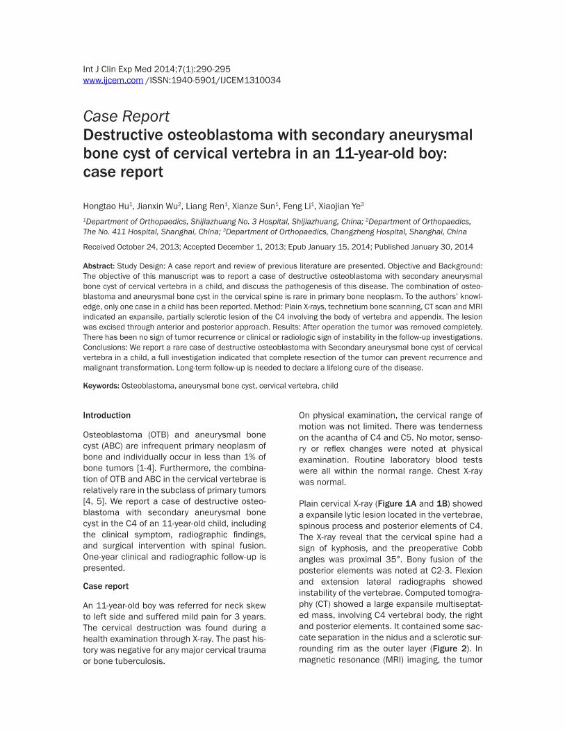
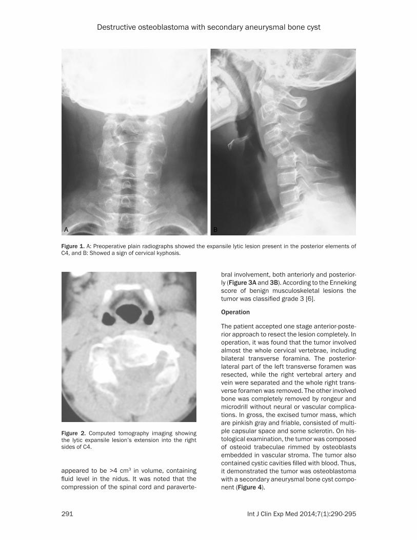
Plain cervical X-ray (Figure 1A and 1B) showed a expansile lytic lesion located in the vertebrae, spinous process and posterior elements of C4. The X-ray reveal that the cervical spine had a sign of kyphosis, and the preoperative Cobb angles was proximal 35°. Bony fusion of the posterior elements was noted at C2-3. Flexion and extension lateral radiographs showed instability of the vertebrae. Computed tomogra-phy (CT) showed a large expansile multiseptat-ed mass, involving C4 vertebral body, the right and posterior elements. It contained some sac-cate separation in the nidus and a sclerotic sur-rounding rim as the outer layer (Figure 2). In magnetic resonance (MRI) imaging, the tumor
Destructive osteoblastoma with secondary aneurysmal bone cyst
291 Int J Clin Exp Med 2014;7(1):290-295
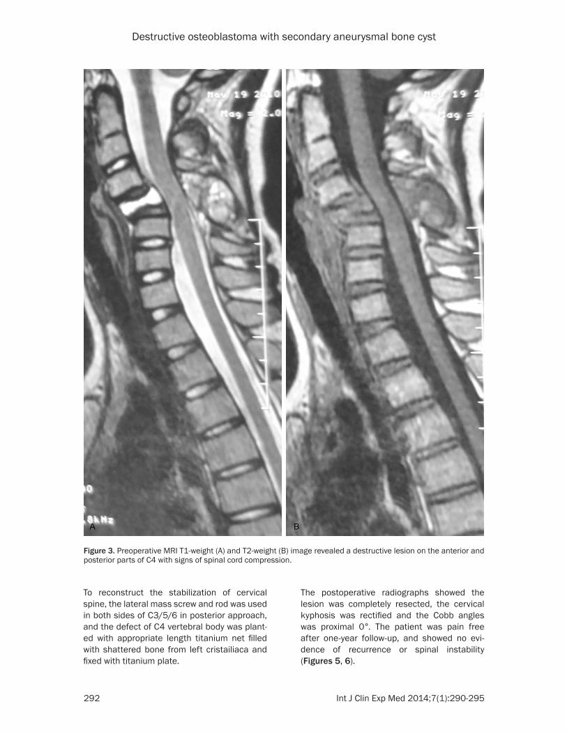
appeared to be >4 cm3 in volume, containing fluid level in the nidus. It was noted that the compression of the spinal cord and paraverte-
bral involvement, both anteriorly and posterior-ly (Figure 3A and 3B). According to the Enneking score of benign musculoskeletal lesions the tumor was classified grade 3 [6].
Operation
The patient accepted one stage anterior-poste-rior approach to resect the lesion completely. In operation, it was found that the tumor involved almost the whole cervical vertebrae, including bilateral transverse foramina. The posterior-lateral part of the left transverse foramen was resected, while the right vertebral artery and vein were separated and the whole right trans-verse foramen was removed. The other involved bone was completely removed by rongeur and microdrill without neural or vascular complica-tions. In gross, the excised tumor mass, which are pinkish gray and friable, consisted of multi-ple capsular space and some sclerotin. On his-tological examination, the tumor was composed of osteoid trabeculae rimmed by osteoblasts embedded in vascular stroma. The tumor also contained cystic cavities filled with blood. Thus, it demonstrated the tumor was osteoblastoma with a secondary aneurysmal bone cyst compo-nent (Figure 4).
Figure 1. A: Preoperative plain radiographs showed the expansile lytic lesion present in the posterior elements of C4, and B: Showed a sign of cervical kyphosis.
Figure 2. Computed tomography imaging showing the lytic expansile lesion’s extension into the right sides of C4.
Destructive osteoblastoma with secondary aneurysmal bone cyst
292 Int J Clin Exp Med 2014;7(1):290-295
To reconstruct the stabilization of cervical spine, the lateral mass screw and rod was used in both sides of C3/5/6 in posterior approach, and the defect of C4 vertebral body was plant-ed with appropriate length titanium net filled with shattered bone from left cristailiaca and fixed with titanium plate.
Figure 3. Preoperative MRI T1-weight (A) and T2-weight (B) image revealed a destructive lesion on the anterior and posterior parts of C4 with signs of spinal cord compression.
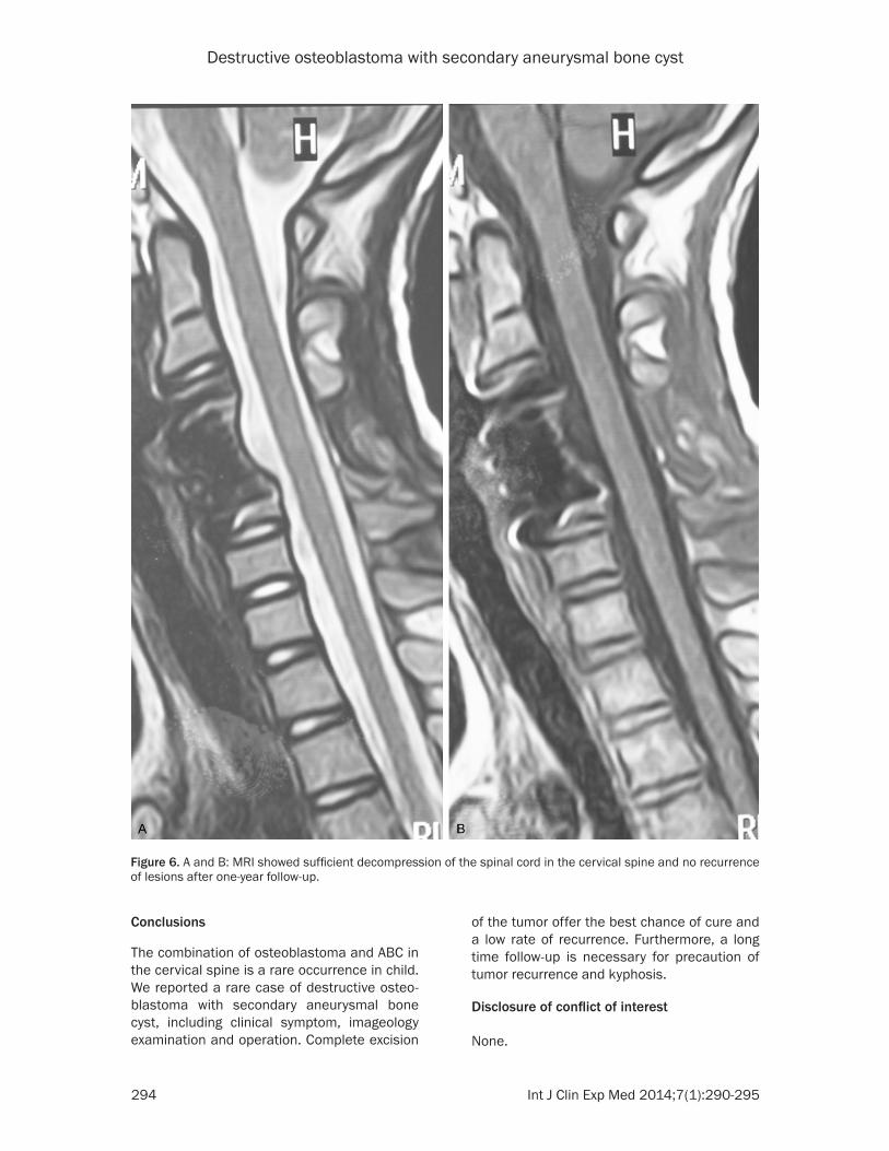
The postoperative radiographs showed the lesion was completely resected, the cervical kyphosis was rectified and the Cobb angles was proximal 0°. The patient was pain free after one-year follow-up, and showed no evi-dence of recurrence or spinal instability (Figures 5, 6).
Destructive osteoblastoma with secondary aneurysmal bone cyst
293 Int J Clin Exp Med 2014;7(1):290-295
Discussion
Osteoblastoma (OTB) is rare benign tumor that accounts for approximately 0.5% to 1% of all primary bone tumors [7, 8]. It is described that around 20% to 40% OTB affected in cervical vertebrae [9]. Lesion is most frequently found in the posterior elements with potential exten-sion to the vertebral bodies. In clinically, pro-gressive pain is the main symptom, followed by torticollis, scoliosis and neurologic manifesta-tions [10, 11]. In gross, the OTB is observed as circumscribed, friable and hemorrhagic and often forms extraskeletal bone in the soft tis-sue [2]. Osteoblastoma is similar to osteoid osteoma in histology, but osteoid osteoma is defined smaller than osteoblastoma [12].
Aneurysmal bone cysts are rare, benign lesion of the bone. It can be located in all bone types and often found in femur, tibia, and small bones of the hands and feet [13]. Approximately 8% to 30% of ABCs arise in the spine, mostly in the thoracic and the lumbar regions [14]. It has been reported to be 25% in the cervical spine [15]. Aneurysmal bone cysts frequently affect the patients younger than 20 years and seem to be slightly more frequent in females than males. In clinical, patients present with insidi-ous onset back pain, stiffness, and swelling [16]. Due to cord compression and spinal insta-bility, some patients present neurologic dys-function. In gross, the lesion consists of a soft fibrovascular core containing blood-filled cyst-like cavities surrounded by a bone shell [17]. Aneurysmal bone cysts are divided into two
classes: primary lesions developing indepen-dently and secondary lesions. The secondary aneurysmal bone cysts can be associated with osteoblastoma, giant cell tumor, osteosarco-ma, and fibrous dysplasia [15, 18].
The association between osteoblastoma and aneurysmal bone cyst is quite rare in the gen-eral population [19]. Several cases of this asso-ciation, such as the posterior cranial fossa [20], skull [21], ethmoid sinus [22], mandibular ramus and condyle [23], sacrum [24], were reported. To our knowledge, only one case of the osteoblastoma with secondary aneurysmal bone cyst of cervical vertebra in child has been reported [25]. In our case, the lesion extends to the vertebral body and posterior elements of C4 and caused to cervical deformity. Operative therapy is necessary for the patient. It has reported that osteoblastomas and aneurysmal bone cyst with incomplete resection has a high rate of recurrence [26, 27]. To avoid the recur-rence of tumor, we choose the complete exci-sion. But for large and extensive lesions com-plete resection will likely incur iatrogenic insta-bility and need instrumented fusion [28]. In our patient, both anterior and posterior approaches were used to rebuild the cervical stability. After one year follow-up, the patient has no evidence of local recurrence and rachiterata.
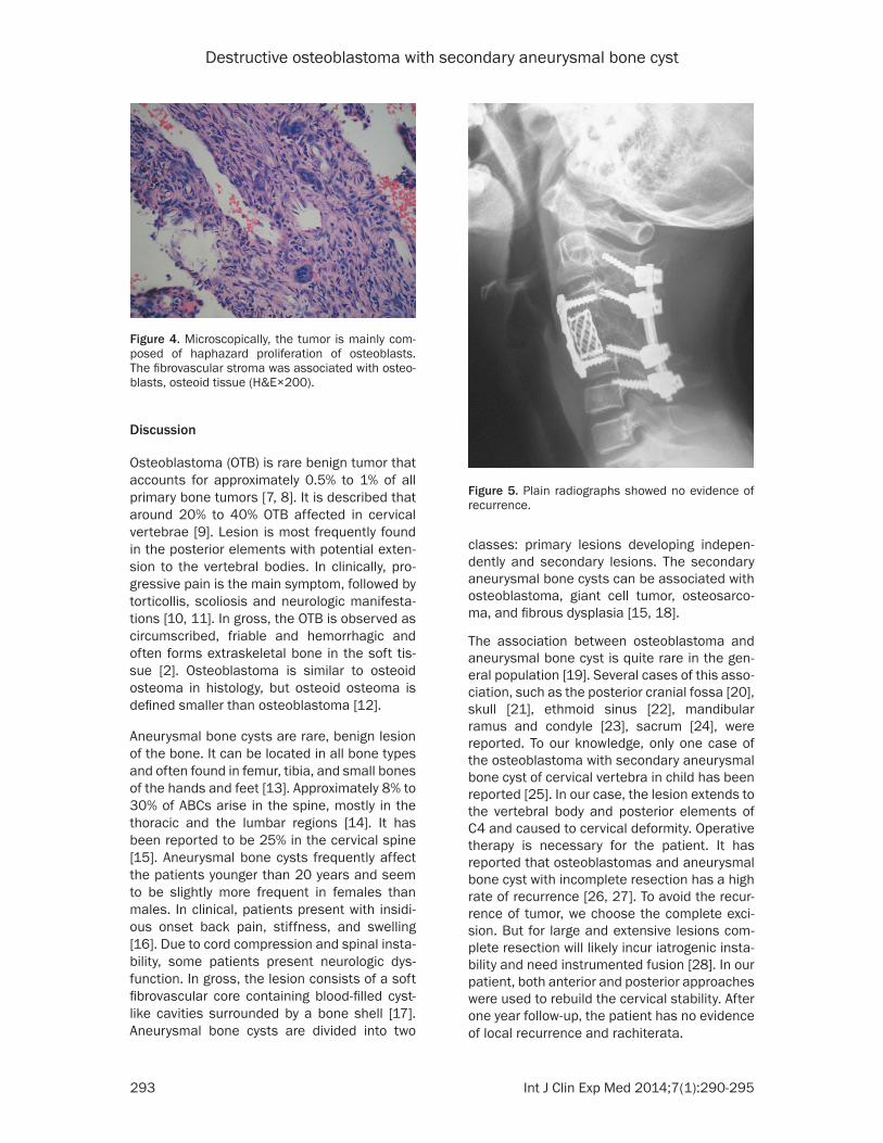
Figure 4. Microscopically, the tumor is mainly com-posed of haphazard proliferation of osteoblasts. The fibrovascular stroma was associated with osteo-blasts, osteoid tissue (H&E×200).
Figure 5. Plain radiographs showed no evidence of recurrence.
Destructive osteoblastoma with secondary aneurysmal bone cyst
294 Int J Clin Exp Med 2014;7(1):290-295
Conclusions
The combination of osteoblastoma and ABC in the cervical spine is a rare occurrence in child. We reported a rare case of destructive osteo-blastoma with secondary aneurysmal bone cyst, including clinical symptom, imageology examination and operation. Complete excision
of the tumor offer the best chance of cure and a low rate of recurrence. Furthermore, a long time follow-up is necessary for precaution of tumor recurrence and kyphosis.
Disclosure of conflict of interest
None.
Figure 6. A and B: MRI showed sufficient decompression of the spinal cord in the cervical spine and no recurrence of lesions after one-year follow-up.
Destructive osteoblastoma with secondary aneurysmal bone cyst
295 Int J Clin Exp Med 2014;7(1):290-295
Address correspondence to: Dr. Hongtao Hu, Department of Orthopaedics, Shijiazhuang No. 3 Hospital, Shijiazhuang, China. Tel: +86-311-8599-0631; Fax: +86-311-8587-6058; E-mail: hht232@- 126.com
References
[1] Unni KK. Benign osteoblastoma (Giant osteoid osteoma). In: Unni KK, editor. Dahlin’s bone tumors general aspects and Data on 11087 Cases. 5th edition. Philadelphia: Lippincott-Raven Publishers 1996; pp: 131-42.
[2] Weber KL, Heck RK Jr. Cystic and benign bone lesions. In: Schwartz HS, editor. Orthopaedic knowledge update: musculoskeletal tumors 2. Rosemont, IL: American Academy of Orthopae-dic Surgeons 2007; pp: 87-102.
[3] Cottalorda J, Bourelle S. Current treatments of primary aneurismal bone cysts. J Pediatr Or-thop B 2006; 15: 155-67.
[4] Austin J, Joseph D. Balancing spinal stability and future mobility in the cervical spine: surgi-cal treatment of a case of osteoblastoma with secondary aneurysmal bone cyst. Spine J 2011; 11: e5-e12.
[5] Tarantino R, Piccirilli M, Anichini G, Delfini R. Benign osteoblastoma of the odontoid process of the axis with secondary aneurysmal bone cyst component: a case report. Neurosurg Rev 2008; 31: 111-5.
[6] Enneking WF. Clinical Musculoskeletal Pathol-ogy. Gainesville, Florida: Storter 1986; pp: 464.
[7] Huvos AG. Bone tumors: Diagnosis, treatment and prognosis. Philadelphia: W.B. Saunders Co 1979; pp: 33-46.
[8] Healey JH, Ghelman B. Osteoid osteoma and osteoblastoma: Current concepts and recent advances. Clin Orthop 1986; 204: 76-85.
[9] Raskas DS, Graziano GP, Herzenberg JE, Hei-delberger KP, Hensinger RN. Osteoid osteoma and osteoblastoma of the spine. J Spinal Dis-ord 1992; 5: 204-211.
[10] Amacher AL, Eltomey A. Spinal osteoblastoma in children and adolescents. Childs Nerv Syst 1985; 1: 29-32.
[11] Ozaki T, Liljenqvist U, Hillmann A, Halm H, Lind-ner N, Gosheger G, Winkelmann W. Osteoid osteoma and osteoblastoma of the spine: ex-periences with 22 patients. Clin Orthop Relat Res 2002; 397: 394-402.
[12] Zileli M, Cagli S, Basdemir G, Ersahin Y. Oste-oid osteomas and osteoblastomas of the spine. Neurosurg Focus 2003; 15: E5.
[13] Vergel De Dios AM, Bond JR, Shives TC, McLeod RA, Unni KK. Aneurysmal bone cyst. A clinico-pathologic study of 238 cases. Cancer 1992; 69: 2921-31.
[14] Boriani S, De Iure F, Campanacci L, Gasbarrini A, Bandiera S, Biagini R, Bertoni F, Picci P. An-
eurysmal bone cyst of the mobile spine: report on 41 cases. Spine (Phila Pa 1976) 2001 Jan 1; 26: 27-35.
[15] Saccomanni B. Aneurysmal bone cyst of spine: a review of literature. Arch Orthop Trauma Surg 2008; 128: 1145-7.
[16] Deo SD, Fairbank JC, Wilson-Macdonald J, Richards P, Pike M, Athanasou N, Wheeler K. Aneurysmal bone cyst as a rare cause of spinal cord compression in a young child. Spine (Phi-la Pa 1976) 2005 Feb 1; 30: E80-2.
[17] Boriani S, De Iure F, Campanacci L, Gasbarrini A, Bandiera S, Biagini R, Bertoni F, Picci P. An-eurysmal bone cyst of the mobile spine: report on 41 cases. Spine (Phila Pa 1976) 2001 Jan 1; 26: 27-35.
[18] Kransdorf MJ, Sweet DE. Aneurysmal bone cyst: concept, controversy, clinical presenta-tion, and imaging. Am J Roentgenol 2005; 164: 573-80.
[19] Della Rocca C, Huvos AG. Osteoblastoma: var-ied histological presentations with a benign clinical course. An analysis of 55 cases. Am J Surg Pathol 1996; 20: 841-50.
[20] Han X, Dong Y, Sun K, Lu Y. A huge occipital osteoblastoma accompanied with aneurismal bone cyst in the posterior cranial fossa. Clin Neurol Neurosurg 2008; 110: 282-5.
[21] Wang YC, Huang JS, Wu CJ, Jeng CM, Fan JK, Resnick D. A huge osteoblastoma with aneu-rysmal bone cyst in skull base. Clin Imaging 2001; 25: 247-50.
[22] Coscina WF, Lee BC. Concurrent osteoblasto-ma and aneurysmal bone cyst of the ethmoid sinus: case report. J Comput Tomogr 1985; 9: 347-50.
[23] Svensson B, Isacsson G. Benign osteoblasto-ma associated with an aneurysmal bone cyst of the mandibular ramus and condyle. Oral Surg Oral Med Oral Pathol 1993; 76: 433-6.
[24] Vade A, Wilbur A, Pudlowski R, Ghosh L. Case report 566: osteoblastoma of sacrum with sec-ondary aneurysmal bone cyst. Skelet Radiol 1989; 18: 475-80.
[25] Tarantino R, Piccirilli M, Anichini G, Delfini R. Benign osteoblastoma of the odontoid process of the axis with secondary aneurysmal bone cyst component: a case report. Neurosurg Rev 2008; 31: 111-5.
[26] Jackson RP. Recurrent osteoblastoma: a re-view. Clin Orthop Relat Res 1978; 131: 229-33.
[27] Lucas DR, Unni KK, McLeod RA, O’Connor MI, Sim FH. Osteoblastoma: clinicopathologic stu- dy of 306 cases. Hum Pathol 1994; 25: 117-34.
[28] Harrop JS, Schmidt MH, Boriani S, Shaffrey CI. Aggressive “benign” primary spine neoplasms: osteoblastoma, aneurysmal bone cyst, and gi-ant cell tumor. Spine (Phila Pa 1976) 2009 Oct 15; 34: S39-47.