case report complex composite odontoma with characteristic...
TRANSCRIPT

Hindawi Publishing CorporationCase Reports in DentistryVolume 2013, Article ID 157614, 5 pageshttp://dx.doi.org/10.1155/2013/157614
Case ReportComplex Composite Odontoma with Characteristic Histology
Sujatha Govindrajan,1 J. Muruganandhan,1 Shaik Shamsudeen,1 Nalin Kumar,1
M. Ramasamy,2 and Srinivasa Prasad3
1 Department of Oral and Maxillofacial Pathology, Sri Venkateswara Dental College and Hospital, Thalambur, Chennai 603103, India2Department of Orthodontics, Sri Venkateswara Dental College and Hospital, Thalambur, Chennai 603103, India3 Department of Oral and Maxillofacial Surgery, Sri Venkateswara Dental College and Hospital, Thalambur, Chennai 603103, India
Correspondence should be addressed to J. Muruganandhan; [email protected]
Received 23 April 2013; Accepted 28 May 2013
Academic Editors: I. El-Hakim and S. R. Watt-Smith
Copyright © 2013 Sujatha Govindrajan et al. This is an open access article distributed under the Creative Commons AttributionLicense, which permits unrestricted use, distribution, and reproduction in any medium, provided the original work is properlycited.
Odontomas are the most commonly occurring odontogenic tumors, which are considered by many to be hamartomas ratherthan neoplasms. These clinically asymptomatic tumors are classified into complex and compound odontomas. They are usuallydiscovered in radiographs and rarely cause bony expansion or infection.This paper discusses a case report of a complex odontomaexhibiting all the structural features and defects of enamel, dentine, and cementum in succession, with an overview on its etiology.
1. Introduction
Hamartomas of tooth forming tissues are termed as odon-toma. They are the most common tumor of epithelial andmesenchymal origin and account for 22% of all odonto-genic tumors [1]. This nonaggressive benign tumor containsenamel, dentin, cementum, and pulp either arranged in anorderly manner resembling a rudimentary tooth called com-pound odontoma or arranged in a haphazard manner calledcomplex odontoma. Complex odontoma is less commonwhen compared to the compound, and they present in ratio of1 : 2 [2]. Odontomas rarely erupt in the oral cavity.We presenta case of a partially erupted odontoma in the right thirdmolarregion.
2. Case Report
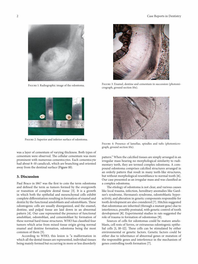
A 28-year-old male patient reported to a private clinicwith a complaint of pain in the right lower posterior toothregion for about one week. On intraoral examination, apartially erupted tooth-like structure was seen. The pain wasintermittent and was aggravated on chewing. Radiographicinvestigation revealed the presence of near-spherical opaquemass resembling calcified tissue measuring about 2 cm indiameter. The radiopaque mass with a density greater than
bone and equal or greater than that of tooth was surroundedby a radiolucent rim in all areas except the erupted portiondistal to normally erupted second molar (Figure 1).
Removal of themasswas plannedunder local anaesthesia.Mucoperiosteal flap was raised distal to 47, and the calcifiedmass was removed. The spherical mass was about 2 cm indiameter with small irregular areas of indentations. Theinferior side of the mass showed a hollow invagination givingthe appearance of a small cup (Figure 2). The specimenwas sent for histopathological examination. A diagnosis ofcomplex odontoma was made clinically.
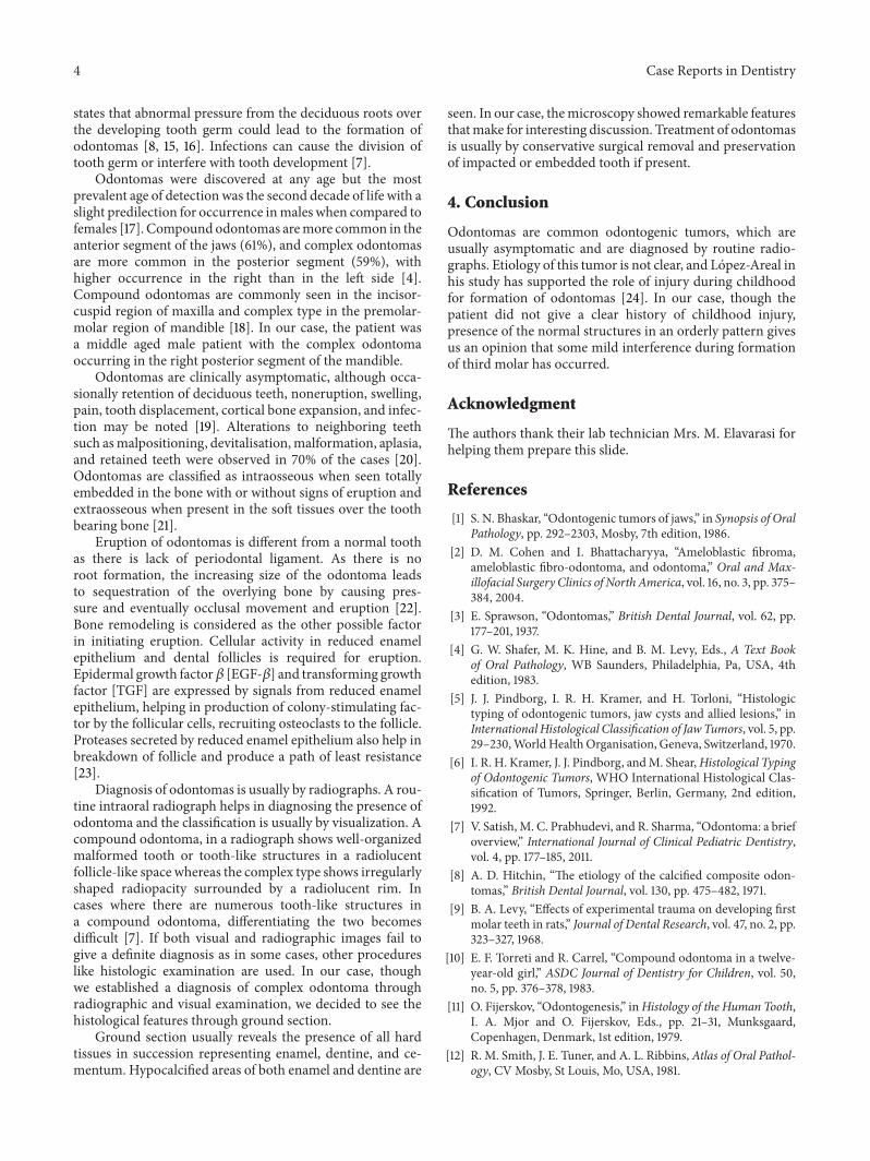
Ground sections were done on the dissected halves of thehard tissuemass.The ground section showed areas of enamel,dentine, and cementum in succession (Figure 3). The enamelshowed uneven thickness and undulating surface. Hypocalci-fied areas like lamellae, spindles, tufts, and incremental lineswere seen (Figures 4 and 5). Some areas showed irregular rodpatterns and gnarled enamel (Figure 6).
The dentinoenamel junction was regular and scallopedin some areas. The underlying dentine showed “S” shapeddentinal tubules. Primary and secondary dentine types wereobserved with clearly visible incremental lines (Figure 7).Hypocalcified areas like interglobular dentine and Tomes’granular layer were also observed (Figures 8 and 9). Deadtracts were also seen (Figure 9). Next to the dentine there
2 Case Reports in Dentistry
Figure 1: Radiographic image of the odontoma.
Figure 2: Superior and inferior surface of odontoma.
was a layer of cementum of varying thickness. Both types ofcementum were observed. The cellular cementum was moreprominent with numerous cementocytes. Each cementocytehad about 8–10 canaliculi, which are branching and orientedaway from the dentinal surface (Figure 10).
3. Discussion
Paul Braco in 1867 was the first to coin the term odontomaand defined the term as tumors formed by the overgrowthor transition of complete dental tissue [3]. It is a growthin which both the epithelial and mesenchymal cells exhibitcomplete differentiation resulting in formation of enamel anddentin by the functional ameloblasts and odontoblasts.Theseodontogenic cells are usually disorganized, and the enamel,dentine, and pulpal tissue are laid down in an abnormalpattern [4]. Our case represented the presence of functionalameloblast, odontoblast, and cementoblast by formation ofthese normal hard tissue structures. WHO has classified fourtumors which arise from mixed tissue origin giving normalenamel and dentine formation, odontoma being the mostcommon of them [5].
According to WHO, this lesion is “a malformation inwhich all the dental tissues are represented, individual tissuesbeingmainly formed but occurring inmore or less disorderly
Figure 3: Enamel, dentine and cementum in succession (photomi-crograph, ground section 10x).
Figure 4: Presence of lamellae, spindles and tufts (photomicro-graph, ground section 10x).
pattern.” When the calcified tissues are simply arranged in anirregular mass bearing no morphological similarity to rudi-mentary teeth, they are termed complex odontoma. A com-pound odontoma comprises calcified structures arranged inan orderly pattern that result in many teeth-like structures,but without morphological resemblance to normal tooth [6].Our case presented as an irregular mass and was classified asa complex odontoma.
The etiology of odontoma is not clear, and various causeslike local trauma, infection, hereditary anomalies like Gard-ner’s syndrome, Hermann’s syndrome, odontoblastic hyper-activity, and alteration in genetic components responsible fortooth development are also considered [7]. Hitchin suggestedthat odontomas are inherited (through a mutant gene) due tointerference, possibly postnatal, with genetic control of toothdevelopment [8]. Experimental studies in rats suggested therole of trauma in formation of odontomas [9].
Sources of cells for odontomas could be mature amelo-blasts, cell rests of Serres, or extraneous odontogenic epithe-lial cells [1, 10–12]. These cells can be stimulated by eitherenvironmental or genetic factors. Genetic factors could beeither due to inheritance of abnormal genes or mutation ofthe responsible genes and interference in the mechanism ofgenes controlling tooth formation [7].

Case Reports in Dentistry 3
Figure 5: Incremental lines of enamel (photomicrograph, groundsection 10x).
Figure 6: Dentinal tubules with incremental lines (photomicro-graph, ground section 10x).
Figure 7: Interglobular dentine (photomicrograph, ground section10x).
Environmental factors like trauma, growth pressure, andinfection may play a role in the pathogenesis of odontoma.A vertically directed force in the form of trauma which isdirected to the permanent tooth bud through the deciduoustooth can lead to morphological changes in the permanenttooth bud leading to formation of odontomas [13]. Studiesby Glasstone (1952) and Rushton (1957) have supported therole of trauma in the development of odontoma [8]. Levystates that the stage of development during which the trauma
Figure 8: Tomes granular layer (photomicrograph, ground section10x).
Figure 9: Dead tracts and gnarled enamel (photomicrograph,ground section 10x).
Figure 10: Cemetocytes with canaliculi (photomicrograph, groundsection 10x).
has occurred determines the development of hypoplasticteeth, odontomas, and supernumerary tooth [9]. Presence ofodontoma in sites other than tooth bearing regions suggeststhat trauma could have possibly displaced the developingtooth germ leading to its malformation [14].
Growth pressure due to inadequate space is been quotedas the etiology in some odontomas. This theory by Hitchin
4 Case Reports in Dentistry
states that abnormal pressure from the deciduous roots overthe developing tooth germ could lead to the formation ofodontomas [8, 15, 16]. Infections can cause the division oftooth germ or interfere with tooth development [7].
Odontomas were discovered at any age but the mostprevalent age of detection was the second decade of life with aslight predilection for occurrence inmales when compared tofemales [17]. Compound odontomas aremore common in theanterior segment of the jaws (61%), and complex odontomasare more common in the posterior segment (59%), withhigher occurrence in the right than in the left side [4].Compound odontomas are commonly seen in the incisor-cuspid region of maxilla and complex type in the premolar-molar region of mandible [18]. In our case, the patient wasa middle aged male patient with the complex odontomaoccurring in the right posterior segment of the mandible.
Odontomas are clinically asymptomatic, although occa-sionally retention of deciduous teeth, noneruption, swelling,pain, tooth displacement, cortical bone expansion, and infec-tion may be noted [19]. Alterations to neighboring teethsuch asmalpositioning, devitalisation,malformation, aplasia,and retained teeth were observed in 70% of the cases [20].Odontomas are classified as intraosseous when seen totallyembedded in the bone with or without signs of eruption andextraosseous when present in the soft tissues over the toothbearing bone [21].
Eruption of odontomas is different from a normal toothas there is lack of periodontal ligament. As there is noroot formation, the increasing size of the odontoma leadsto sequestration of the overlying bone by causing pres-sure and eventually occlusal movement and eruption [22].Bone remodeling is considered as the other possible factorin initiating eruption. Cellular activity in reduced enamelepithelium and dental follicles is required for eruption.Epidermal growth factor𝛽 [EGF-𝛽] and transforming growthfactor [TGF] are expressed by signals from reduced enamelepithelium, helping in production of colony-stimulating fac-tor by the follicular cells, recruiting osteoclasts to the follicle.Proteases secreted by reduced enamel epithelium also help inbreakdown of follicle and produce a path of least resistance[23].
Diagnosis of odontomas is usually by radiographs. A rou-tine intraoral radiograph helps in diagnosing the presence ofodontoma and the classification is usually by visualization. Acompound odontoma, in a radiograph shows well-organizedmalformed tooth or tooth-like structures in a radiolucentfollicle-like space whereas the complex type shows irregularlyshaped radiopacity surrounded by a radiolucent rim. Incases where there are numerous tooth-like structures ina compound odontoma, differentiating the two becomesdifficult [7]. If both visual and radiographic images fail togive a definite diagnosis as in some cases, other procedureslike histologic examination are used. In our case, thoughwe established a diagnosis of complex odontoma throughradiographic and visual examination, we decided to see thehistological features through ground section.
Ground section usually reveals the presence of all hardtissues in succession representing enamel, dentine, and ce-mentum. Hypocalcified areas of both enamel and dentine are
seen. In our case, themicroscopy showed remarkable featuresthatmake for interesting discussion. Treatment of odontomasis usually by conservative surgical removal and preservationof impacted or embedded tooth if present.
4. Conclusion
Odontomas are common odontogenic tumors, which areusually asymptomatic and are diagnosed by routine radio-graphs. Etiology of this tumor is not clear, and Lopez-Areal inhis study has supported the role of injury during childhoodfor formation of odontomas [24]. In our case, though thepatient did not give a clear history of childhood injury,presence of the normal structures in an orderly pattern givesus an opinion that some mild interference during formationof third molar has occurred.
Acknowledgment
The authors thank their lab technician Mrs. M. Elavarasi forhelping them prepare this slide.
References
[1] S. N. Bhaskar, “Odontogenic tumors of jaws,” in Synopsis of OralPathology, pp. 292–2303, Mosby, 7th edition, 1986.
[2] D. M. Cohen and I. Bhattacharyya, “Ameloblastic fibroma,ameloblastic fibro-odontoma, and odontoma,” Oral and Max-illofacial Surgery Clinics of North America, vol. 16, no. 3, pp. 375–384, 2004.
[3] E. Sprawson, “Odontomas,” British Dental Journal, vol. 62, pp.177–201, 1937.
[4] G. W. Shafer, M. K. Hine, and B. M. Levy, Eds., A Text Bookof Oral Pathology, WB Saunders, Philadelphia, Pa, USA, 4thedition, 1983.
[5] J. J. Pindborg, I. R. H. Kramer, and H. Torloni, “Histologictyping of odontogenic tumors, jaw cysts and allied lesions,” inInternational Histological Classification of JawTumors, vol. 5, pp.29–230,WorldHealthOrganisation, Geneva, Switzerland, 1970.
[6] I. R. H. Kramer, J. J. Pindborg, andM. Shear,Histological Typingof Odontogenic Tumors, WHO International Histological Clas-sification of Tumors, Springer, Berlin, Germany, 2nd edition,1992.
[7] V. Satish, M. C. Prabhudevi, and R. Sharma, “Odontoma: a briefoverview,” International Journal of Clinical Pediatric Dentistry,vol. 4, pp. 177–185, 2011.
[8] A. D. Hitchin, “The etiology of the calcified composite odon-tomas,” British Dental Journal, vol. 130, pp. 475–482, 1971.
[9] B. A. Levy, “Effects of experimental trauma on developing firstmolar teeth in rats,” Journal of Dental Research, vol. 47, no. 2, pp.323–327, 1968.
[10] E. F. Torreti and R. Carrel, “Compound odontoma in a twelve-year-old girl,” ASDC Journal of Dentistry for Children, vol. 50,no. 5, pp. 376–378, 1983.
[11] O. Fijerskov, “Odontogenesis,” inHistology of the Human Tooth,I. A. Mjor and O. Fijerskov, Eds., pp. 21–31, Munksgaard,Copenhagen, Denmark, 1st edition, 1979.
[12] R. M. Smith, J. E. Tuner, and A. L. Ribbins, Atlas of Oral Pathol-ogy, CV Mosby, St Louis, Mo, USA, 1981.

Case Reports in Dentistry 5
[13] J. O. Andreason, “Injuries to developing teeth,” in Textbookand Color Atlas of Traumatic Injuries to Teeth, J. O. Andreasonand F. M. Andreason, Eds., pp. 457–494, Mosby, Copenhagen,Denmark, 3rd edition, 1993.
[14] A. Shteyer, S. Taicher, and Y. Marmary, “Odontoma in the sub-condylar region,” British Journal of Oral Surgery, vol. 17, no. 2,pp. 161–165, 1979.
[15] P. Gurdal and T. Seckin, “Odontomas,” Quintessence Interna-tional, vol. 4, article 32, 2001.
[16] A. D. Hitchin andW. D. Mchugh, “Three coronal invaginationsin a dilated composite odontome,” British Dental Journal, vol.97, pp. 90–92, 1954.
[17] M. Vengal, H. Arora, S. Ghosh, and K. M. Pai, “Large eruptingcomplex odontoma: a case report,” Journal of the CanadianDental Association, vol. 73, no. 2, pp. 169–172, 2007.
[18] O. P. Kharbanda, C. S. Sambi, and K. Renu, “Odontoma: a casereport,” Journal of the Indian Dental Association, vol. 58, pp.269–271, 1986.
[19] M. S. Tuzum, “Orofacial pain associated with an infected com-plex odontoma—case report,” Australian Dental Journal, vol. 3,pp. 352–354, 1990.
[20] M. Kaneko, M. Fukuda, T. Sano, T. Ohnishi, and Y. Hosokawa,“Microradiographic and microscopic investigation of a rarecase of complex odontoma,” Oral Surgery, Oral Medicine, OralPathology, Oral Radiology, and Endodontics, vol. 86, no. 1, pp.131–134, 1998.
[21] L. Junquera, J. C. de Vicente, P. Roig, S. Olay, and O. Rodrıguez-Recio, “Intraosseus odontoma erupted into the oral cavity: anunusual pathology,” Medicina Oral, Patologia Oral y CirugiaBucal, vol. 10, no. 3, pp. 248–251, 2005.
[22] A. R. Ten Cate and A. Nanci, “Physiologic tooth movements:eruption and shedding,” in Ten Cate’s Oral Histology: Devel-opment, Structure and Function, A. Nanci, Ed., pp. 275–298,Mosby, St. Louis, Mo, USA, 2003.
[23] P. B. Sood, B. Patil, S. Godhi, and D. C. Shetty, “Multiple super-numerary teeth and odontoma in the maxilla: a case report,”Contemporary Clinical Dentistry, vol. 1, pp. 45–46, 2010.
[24] L. Lopez-Areal, F. Silvestre Donat, and J. Gil Lozano, “Com-pound odontoma erupting in the mouth: 4-year follow-up ofa clinical case,” Journal of Oral Pathology and Medicine, vol. 21,no. 6, pp. 285–288, 1992.

Submit your manuscripts athttp://www.hindawi.com
Hindawi Publishing Corporationhttp://www.hindawi.com Volume 2014
Oral OncologyJournal of
DentistryInternational Journal of
Hindawi Publishing Corporationhttp://www.hindawi.com Volume 2014
Hindawi Publishing Corporationhttp://www.hindawi.com Volume 2014
International Journal of
Biomaterials
Hindawi Publishing Corporationhttp://www.hindawi.com Volume 2014
BioMed Research International
Hindawi Publishing Corporationhttp://www.hindawi.com Volume 2014
Case Reports in Dentistry
Hindawi Publishing Corporationhttp://www.hindawi.com Volume 2014
Oral ImplantsJournal of
Hindawi Publishing Corporationhttp://www.hindawi.com Volume 2014
Anesthesiology Research and Practice
Hindawi Publishing Corporationhttp://www.hindawi.com Volume 2014
Radiology Research and Practice
Environmental and Public Health
Journal of
Hindawi Publishing Corporationhttp://www.hindawi.com Volume 2014
The Scientific World JournalHindawi Publishing Corporation http://www.hindawi.com Volume 2014
Hindawi Publishing Corporationhttp://www.hindawi.com Volume 2014
Dental SurgeryJournal of
Drug DeliveryJournal of
Hindawi Publishing Corporationhttp://www.hindawi.com Volume 2014
Hindawi Publishing Corporationhttp://www.hindawi.com Volume 2014
Oral DiseasesJournal of
Hindawi Publishing Corporationhttp://www.hindawi.com Volume 2014
Computational and Mathematical Methods in Medicine
ScientificaHindawi Publishing Corporationhttp://www.hindawi.com Volume 2014
PainResearch and TreatmentHindawi Publishing Corporationhttp://www.hindawi.com Volume 2014
Preventive MedicineAdvances in
Hindawi Publishing Corporationhttp://www.hindawi.com Volume 2014
EndocrinologyInternational Journal of
Hindawi Publishing Corporationhttp://www.hindawi.com Volume 2014
Hindawi Publishing Corporationhttp://www.hindawi.com Volume 2014
OrthopedicsAdvances in