case report cepacia syndrome in a non-cystic fibrosis...
TRANSCRIPT
Case ReportCepacia Syndrome in a Non-Cystic Fibrosis Patient
Naomi Hauser and Jose Orsini
Department of Medicine, New York University School of Medicine, Woodhull Medical and Mental Health Center,Brooklyn, NY 11206, USA
Correspondence should be addressed to Naomi Hauser; [email protected]
Received 29 April 2015; Accepted 3 August 2015
Academic Editor: Oguz R. Sipahi
Copyright © 2015 N. Hauser and J. Orsini. This is an open access article distributed under the Creative Commons AttributionLicense, which permits unrestricted use, distribution, and reproduction in any medium, provided the original work is properlycited.
Burkholderia (formerly Pseudomonas) cepacia complex is a known serious threat to patients with cystic fibrosis, in whom it has thepotential to cause the fatal combination of necrotizing pneumonia, worsening respiratory failure, and bacteremia, known asCepaciasyndrome.The potential for this pathogen to infect non-cystic fibrosis patients is limited and its epidemiology is poorly understood.Previously reported cases of severe Burkholderia cepacia complex lung infection in immunocompetent hosts include pneumonia,bronchiectasis, pyopneumothorax, and cavitary lesions. We present a case of a 64-year-old man with Streptococcus pneumoniaecommunity-acquired pneumonia whose hospital course was complicated by developing cavitary lung lesions, bacteremia, andacute respiratory distress syndrome. Repeated tracheal aspirate and blood cultures grew Burkholderia cepacia. Our case appearsto be the first report of Cepacia syndrome in a patient without cystic fibrosis. This report raises concern regarding the potentialseverity of pulmonary Burkholderia cepacia complex infection and the need to broaden clinicians’ suspicion for Cepacia syndrome.A framework to help diagnose and treat infected non-cystic fibrosis individuals may be useful.
1. Introduction
Burkholderia (formerly Pseudomonas) cepacia complex (Bcc)has been a recognized serious threat to patients with cysticfibrosis (CF) since it emerged as a major cause of severepulmonary infection in this population in the 1980s [1–3]. Bccis known to colonize the lungs of CF patients, occasionallycausing chronic pulmonary infection [3–8]. The greatestconcern of pulmonary Bcc infection is its potential to causeCepacia syndrome, a fatal combination of necrotizing pneu-monia, rapid respiratory decline, and bacteremia [3, 5, 8–11].The potential of Bcc to infect non-CF patients is limited andsporadic, and its epidemiology is poorly understood [1, 8].Large hospital outbreaks are infrequent [2]. Infection in thenon-CF population is most commonly nosocomial and dueto direct delivery via some clear point source, with resolutionof illness upon removing the exposure [8, 12, 13]. Casesof severe Bcc lung infection in immunocompetent hostshave been reported in the literature and include pneumonia,bronchiectasis, pyopneumothorax, and cavitary lesions [7,14–18]. Table 1 summarizes cases reported in the literatureof necrotizing pneumonia due to Bcc in non-CF individuals.
To the best of our knowledge, however, this is one very rarecase of classic Cepacia syndrome in a non-CF individual andthe only case of Cepacia syndrome due to nosocomial Bccinfection reported in the literature.
2. Case Report
A 64-year-old man presented to the emergency department(ED) complaining of right pleuritic pain, productive cough,and generalized malaise for 4 days. His past medical historywas significant for systemic arterial hypertension and type 2diabetes mellitus. Vital signs on arrival to the ED were: bloodpressure of 120/75mmHg, heart rate of 114 beats/minute,respiratory rate of 22 breaths/minute, and temperature of96.9∘F. His oxygen saturation by pulse oximetry was 89%while breathing ambient air. Coarse rales were heard onauscultation over the right lung field. The rest of the physicalexam was noncontributory.
Remarkable laboratory findings included a sodium levelof 126mmol/L (135–145), a bicarbonate level of 7mmol/L(24–31), a creatinine level of 1.9mg/L (0.8–2.0), and a glucoseof 687mg/dL (65–115). Arterial blood gas (ABG) showed
Hindawi Publishing CorporationCase Reports in Infectious DiseasesVolume 2015, Article ID 537627, 4 pageshttp://dx.doi.org/10.1155/2015/537627
2 Case Reports in Infectious Diseases
Table 1: Summary of case reports of necrotizing pneumonia due to Burkholderia cepacia complex species in non-cystic fibrosis patients.
Author, year Age, gender Exposure Bacteremia Rapid respiratory decline OutcomeBelchis et al., 2000 [18] 44, M Community Yes Yes DeathBelchis et al., 2000 [18] 40, F Community No Yes DeathBelchis et al., 2000 [18] 43, F Community No Yes DeathSuresh et al., 2013 [16] 63, M Community Unclear No Survival
paO2of 59mmHg (80–100) on room air. Chest X-ray (CXR)
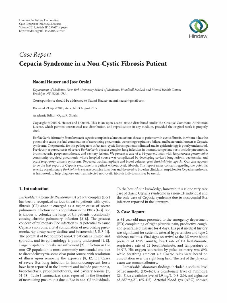
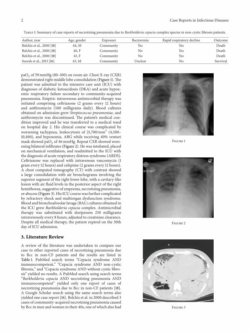
demonstrated right middle lobe consolidation (Figure 1).Thepatient was admitted to the intensive care unit (ICU) withdiagnoses of diabetic ketoacidosis (DKA) and acute hypox-emic respiratory failure secondary to community-acquiredpneumonia. Empiric intravenous antimicrobial therapy wasinitiated comprising ceftriaxone (2 grams every 12 hours)and azithromycin (500 milligrams daily). Blood culturesobtained on admission grew Streptococcus pneumoniae, andazithromycin was discontinued. The patient’s medical con-dition improved and he was transferred to a medical wardon hospital day 2. His clinical course was complicated byworsening tachypnea, leukocytosis of 21,700/mm3 (4,500–10,400), and hypoxemia. ABG while receiving 40% venturimask showed paO
2of 46mmHg. Repeat CXR showed wors-
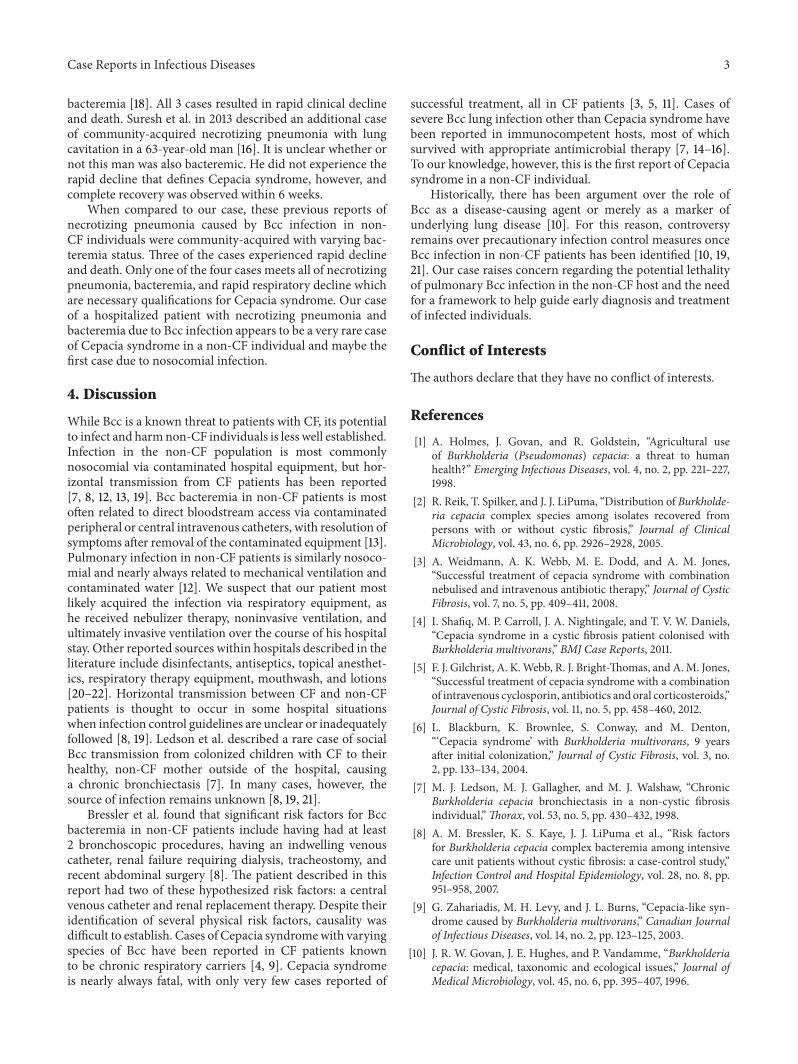
ening bilateral infiltrates (Figure 2). He was intubated, placedon mechanical ventilation, and readmitted to the ICU withthe diagnosis of acute respiratory distress syndrome (ARDS).Ceftriaxone was replaced with intravenous vancomycin (1gram every 12 hours) and cefepime (2 grams every 12 hours).A chest computed tomography (CT) with contrast showeda large consolidation with air bronchograms involving thesuperior segment of the right lower lobe, with a cavitary-likelesion with air fluid levels in the posterior aspect of the righthemithorax, suggestive of empyema, necrotizing pneumonia,or abscess (Figure 3).His ICUcoursewas further complicatedby refractory shock and multiorgan dysfunction syndrome.Blood and bronchoalveolar lavage (BAL) cultures obtained inthe ICU grew Burkholderia cepacia complex. Antimicrobialtherapy was substituted with doripenem 250 milligramsintravenously every 8 hours, adjusted to creatinine clearance.Despite all medical therapy, the patient expired on the 30thday of ICU admission.
3. Literature Review
A review of the literature was undertaken to compare ourcase to other reported cases of necrotizing pneumonia dueto Bcc in non-CF patients and the results are listed inTable 1. PubMed search terms “Cepacia syndrome ANDimmunocompetent,” “Cepacia syndrome AND non-cysticfibrosis,” and “Cepacia syndrome AND without cystic fibro-sis” yielded no results. A PubMed search using search terms“Burkholderia cepacia AND necrotizing pneumonia ANDimmunocompetent” yielded only one report of cases ofnecrotizing pneumonia due to Bcc in non-CF patients [18].A Google Scholar search using the same search terms alsoyielded one case report [16]. Belchis et al. in 2000 described 3cases of community-acquired necrotizing pneumonia causedby Bcc in men and women in their 40s, one of which also had
Figure 1
Figure 2
Figure 3
Case Reports in Infectious Diseases 3
bacteremia [18]. All 3 cases resulted in rapid clinical declineand death. Suresh et al. in 2013 described an additional caseof community-acquired necrotizing pneumonia with lungcavitation in a 63-year-old man [16]. It is unclear whether ornot this man was also bacteremic. He did not experience therapid decline that defines Cepacia syndrome, however, andcomplete recovery was observed within 6 weeks.
When compared to our case, these previous reports ofnecrotizing pneumonia caused by Bcc infection in non-CF individuals were community-acquired with varying bac-teremia status. Three of the cases experienced rapid declineand death. Only one of the four cases meets all of necrotizingpneumonia, bacteremia, and rapid respiratory decline whichare necessary qualifications for Cepacia syndrome. Our caseof a hospitalized patient with necrotizing pneumonia andbacteremia due to Bcc infection appears to be a very rare caseof Cepacia syndrome in a non-CF individual and maybe thefirst case due to nosocomial infection.
4. Discussion
While Bcc is a known threat to patients with CF, its potentialto infect and harmnon-CF individuals is less well established.Infection in the non-CF population is most commonlynosocomial via contaminated hospital equipment, but hor-izontal transmission from CF patients has been reported[7, 8, 12, 13, 19]. Bcc bacteremia in non-CF patients is mostoften related to direct bloodstream access via contaminatedperipheral or central intravenous catheters, with resolution ofsymptoms after removal of the contaminated equipment [13].Pulmonary infection in non-CF patients is similarly nosoco-mial and nearly always related to mechanical ventilation andcontaminated water [12]. We suspect that our patient mostlikely acquired the infection via respiratory equipment, ashe received nebulizer therapy, noninvasive ventilation, andultimately invasive ventilation over the course of his hospitalstay. Other reported sources within hospitals described in theliterature include disinfectants, antiseptics, topical anesthet-ics, respiratory therapy equipment, mouthwash, and lotions[20–22]. Horizontal transmission between CF and non-CFpatients is thought to occur in some hospital situationswhen infection control guidelines are unclear or inadequatelyfollowed [8, 19]. Ledson et al. described a rare case of socialBcc transmission from colonized children with CF to theirhealthy, non-CF mother outside of the hospital, causinga chronic bronchiectasis [7]. In many cases, however, thesource of infection remains unknown [8, 19, 21].
Bressler et al. found that significant risk factors for Bccbacteremia in non-CF patients include having had at least2 bronchoscopic procedures, having an indwelling venouscatheter, renal failure requiring dialysis, tracheostomy, andrecent abdominal surgery [8]. The patient described in thisreport had two of these hypothesized risk factors: a centralvenous catheter and renal replacement therapy. Despite theiridentification of several physical risk factors, causality wasdifficult to establish. Cases of Cepacia syndromewith varyingspecies of Bcc have been reported in CF patients knownto be chronic respiratory carriers [4, 9]. Cepacia syndromeis nearly always fatal, with only very few cases reported of
successful treatment, all in CF patients [3, 5, 11]. Cases ofsevere Bcc lung infection other than Cepacia syndrome havebeen reported in immunocompetent hosts, most of whichsurvived with appropriate antimicrobial therapy [7, 14–16].To our knowledge, however, this is the first report of Cepaciasyndrome in a non-CF individual.
Historically, there has been argument over the role ofBcc as a disease-causing agent or merely as a marker ofunderlying lung disease [10]. For this reason, controversyremains over precautionary infection control measures onceBcc infection in non-CF patients has been identified [10, 19,21]. Our case raises concern regarding the potential lethalityof pulmonary Bcc infection in the non-CF host and the needfor a framework to help guide early diagnosis and treatmentof infected individuals.
Conflict of Interests
The authors declare that they have no conflict of interests.
References
[1] A. Holmes, J. Govan, and R. Goldstein, “Agricultural useof Burkholderia (Pseudomonas) cepacia: a threat to humanhealth?” Emerging Infectious Diseases, vol. 4, no. 2, pp. 221–227,1998.
[2] R. Reik, T. Spilker, and J. J. LiPuma, “Distribution of Burkholde-ria cepacia complex species among isolates recovered frompersons with or without cystic fibrosis,” Journal of ClinicalMicrobiology, vol. 43, no. 6, pp. 2926–2928, 2005.
[3] A. Weidmann, A. K. Webb, M. E. Dodd, and A. M. Jones,“Successful treatment of cepacia syndrome with combinationnebulised and intravenous antibiotic therapy,” Journal of CysticFibrosis, vol. 7, no. 5, pp. 409–411, 2008.
[4] I. Shafiq, M. P. Carroll, J. A. Nightingale, and T. V. W. Daniels,“Cepacia syndrome in a cystic fibrosis patient colonised withBurkholderia multivorans,” BMJ Case Reports, 2011.
[5] F. J. Gilchrist, A. K.Webb, R. J. Bright-Thomas, and A.M. Jones,“Successful treatment of cepacia syndrome with a combinationof intravenous cyclosporin, antibiotics and oral corticosteroids,”Journal of Cystic Fibrosis, vol. 11, no. 5, pp. 458–460, 2012.
[6] L. Blackburn, K. Brownlee, S. Conway, and M. Denton,“‘Cepacia syndrome’ with Burkholderia multivorans, 9 yearsafter initial colonization,” Journal of Cystic Fibrosis, vol. 3, no.2, pp. 133–134, 2004.
[7] M. J. Ledson, M. J. Gallagher, and M. J. Walshaw, “ChronicBurkholderia cepacia bronchiectasis in a non-cystic fibrosisindividual,”Thorax, vol. 53, no. 5, pp. 430–432, 1998.
[8] A. M. Bressler, K. S. Kaye, J. J. LiPuma et al., “Risk factorsfor Burkholderia cepacia complex bacteremia among intensivecare unit patients without cystic fibrosis: a case-control study,”Infection Control and Hospital Epidemiology, vol. 28, no. 8, pp.951–958, 2007.
[9] G. Zahariadis, M. H. Levy, and J. L. Burns, “Cepacia-like syn-drome caused by Burkholderia multivorans,” Canadian Journalof Infectious Diseases, vol. 14, no. 2, pp. 123–125, 2003.
[10] J. R. W. Govan, J. E. Hughes, and P. Vandamme, “Burkholderiacepacia: medical, taxonomic and ecological issues,” Journal ofMedical Microbiology, vol. 45, no. 6, pp. 395–407, 1996.

4 Case Reports in Infectious Diseases
[11] K. Grimwood, T. J. Kidd, and M. Tweed, “Successful treatmentof cepacia syndrome,” Journal of Cystic Fibrosis, vol. 8, no. 4, pp.291–293, 2009.
[12] J. M. Conly, L. Klass, L. Larson, J. Kennedy, D. E. Low, and G.K. Harding, “Pseudomonas cepacia colonization and infectionin intensive care units,” Canadian Medical Association Journal,vol. 134, no. 4, pp. 363–366, 1986.
[13] G. W. Meyer, “Pseudomonas cepacia septicemia associated withintravenous therapy,” California Medicine, vol. 119, no. 1, pp. 15–18, 1973.
[14] G. W. Waterer, C. B. Jones, and R. G. Wunderink, “Bacteremiccommunity-acquired pneumonia in an immunocompetentadult due to Burkholderia cepacia,” Chest, vol. 116, no. 6, pp.1842–1843, 1999.
[15] S. S. Karanth, H. Regunath, K. Chawla, and M. Prabhu, “Arare case of community acquired Burkholderia cepacia infectionpresenting as pyopneumothorax in an immunocompetent indi-vidual,” Asian Pacific Journal of Tropical Biomedicine, vol. 2, no.2, pp. 166–168, 2012.
[16] G. Suresh, S. R. Prakasha, B. H. Giridhar, and K. S. Prakash,“Cavity in the lung: a rare case of Burkholderia cepacia infec-tion,” Nitte University Journal of Health Science, vol. 3, no. 2, pp.100–101, 2013.
[17] M. Pujol, X. Corbella, J. Carratala, and F. Gudiol, “Community-acquired bacteremic Pseudomonas cepacia pneumonia in animmunocompetent host,”Clinical InfectiousDiseases, vol. 15, no.5, pp. 887–888, 1992.
[18] D. A. Belchis, E. Simpson, and T. Colby, “Histopathologicfeatures of Burkholderia cepacia pneumonia in patients withoutcystic fibrosis,” Modern Pathology, vol. 13, no. 4, pp. 369–372,2000.
[19] A. Holmes, R. Nolan, R. Taylor et al., “An epidemic ofBurkholderia cepacia transmitted between patients with andwithout cystic fibrosis,” The Journal of Infectious Diseases, vol.179, no. 5, pp. 1197–1205, 1999.
[20] A. M. Jones, M. E. Dodd, and A. K. Webb, “Burkholderiacepacia: current clinical issues, environmental controversiesand ethical dilemmas,” European Respiratory Journal, vol. 17, no.2, pp. 295–301, 2001.
[21] A. H. Siddiqui, M. E. Mulligan, E. Mahenthiralingam et al., “Anepisodic outbreak of genetically related Burkholderia cepaciaamong non-cystic fibrosis patients at a university hospital,”Infection Control and Hospital Epidemiology, vol. 22, no. 7, pp.419–422, 2001.
[22] J. Zurita, L. Mejia, S. Zapata et al., “Healthcare-associatedrespiratory tract infection and colonization in an intensive careunit caused by Burkholderia cepacia isolated in mouthwash,”International Journal of Infectious Diseases, vol. 29, pp. 96–99,2014.

Submit your manuscripts athttp://www.hindawi.com
Stem CellsInternational
Hindawi Publishing Corporationhttp://www.hindawi.com Volume 2014
Hindawi Publishing Corporationhttp://www.hindawi.com Volume 2014
MEDIATORSINFLAMMATION
of
Hindawi Publishing Corporationhttp://www.hindawi.com Volume 2014
Behavioural Neurology
EndocrinologyInternational Journal of
Hindawi Publishing Corporationhttp://www.hindawi.com Volume 2014
Hindawi Publishing Corporationhttp://www.hindawi.com Volume 2014
Disease Markers
Hindawi Publishing Corporationhttp://www.hindawi.com Volume 2014
BioMed Research International
OncologyJournal of
Hindawi Publishing Corporationhttp://www.hindawi.com Volume 2014
Hindawi Publishing Corporationhttp://www.hindawi.com Volume 2014
Oxidative Medicine and Cellular Longevity
Hindawi Publishing Corporationhttp://www.hindawi.com Volume 2014
PPAR Research
The Scientific World JournalHindawi Publishing Corporation http://www.hindawi.com Volume 2014
Immunology ResearchHindawi Publishing Corporationhttp://www.hindawi.com Volume 2014
Journal of
ObesityJournal of
Hindawi Publishing Corporationhttp://www.hindawi.com Volume 2014
Hindawi Publishing Corporationhttp://www.hindawi.com Volume 2014
Computational and Mathematical Methods in Medicine
OphthalmologyJournal of
Hindawi Publishing Corporationhttp://www.hindawi.com Volume 2014
Diabetes ResearchJournal of
Hindawi Publishing Corporationhttp://www.hindawi.com Volume 2014
Hindawi Publishing Corporationhttp://www.hindawi.com Volume 2014
Research and TreatmentAIDS
Hindawi Publishing Corporationhttp://www.hindawi.com Volume 2014
Gastroenterology Research and Practice
Hindawi Publishing Corporationhttp://www.hindawi.com Volume 2014
Parkinson’s Disease
Evidence-Based Complementary and Alternative Medicine
Volume 2014Hindawi Publishing Corporationhttp://www.hindawi.com