case report a case of waterhouse-friderichsen syndrome...
TRANSCRIPT

Case ReportA Case of Waterhouse-Friderichsen SyndromeResulting from an Invasive Pneumococcal Infection ina Patient with a Hypoplastic Spleen
Kazumasa Emori, Nobuhiro Takeuchi, and Junichi Soneda
Department of Cardiovascular Surgery, Kobe Tokusyukai Hospital, 1-3-10 Kamitakamaru, Tarumi-ku, Kobe-shi,Hyogo 655-0017, Japan
Correspondence should be addressed to Kazumasa Emori; [email protected]
Received 18 November 2015; Accepted 13 January 2016
Academic Editor: Zsolt Molnar
Copyright © 2016 Kazumasa Emori et al. This is an open access article distributed under the Creative Commons AttributionLicense, which permits unrestricted use, distribution, and reproduction in any medium, provided the original work is properlycited.
A 50-year-old male was brought to our emergency department by ambulance with complaints of pain and numbness in both legs.At arrival, purple spots were evident on his neck and face. Examination of the vital sign indicated septic shock. Laboratory data andblood gas analysis revealed disseminated intravascular coagulation,multiple organ failure, andmetabolic acidosis. Peripheral bloodsmears revealed Howell-Jolly bodies, indicating decreased splenic function. A rapid urinary pneumococcal antigen test was alsofound to be positive. After admission to the intensive care unit, extensive treatment, including polymyxin-B direct hemoperfusionand administration of methylprednisolone and broad spectrum antibiotics was immediately initiated. Despite of our efforts tosave his life, the patient died six hours after the arrival. The following day, blood cultures revealed the presence of Streptococcuspneumoniae. An autopsy revealed a hypoplastic spleen and a bilateral adrenal hemorrhage, indicating acute adrenal insufficiencycaused by sepsis. Finally, the patient was diagnosed with Waterhouse-Friderichsen syndrome. Although severe infection may beseen in the splenectomized patients, it should be noted that patients with a hypoplastic spleen may have acute severe infections.We, therefore, report a case of Waterhouse-Friderichsen syndrome resulting from an invasive pneumococcal infection in a patientwith a hypoplastic spleen.
1. Background
Waterhouse-Friderichsen syndrome (WFS) is an emergencycondition, characterized by fever, cyanosis, bruises, and/orshock [1]. WFS is reported to occur in splenectomizedpatients, and in patientswith congenital asplenia or decreasedsplenic function [2–5]. Here, we report a rare case of WFS,presenting with purpura fulminans, resulting from an inva-sive pneumococcal infection.
2. Case Presentation
A50-year-oldmale was brought to our hospital by ambulancewith complaints of pain and numbness in both legs. Accord-ing to his wife, he presented with a high-grade fever (39.0∘C),pain in both legs, vomiting, and diarrhea on the precedingday. His past medical history also included duodenal ulcer,
which was treated by conservative therapy. He was previouslydiagnosed with fatty liver and an idiopathic leukopenia.According to his wife, the patient had undergone dentaltreatment three months before. The patient was employedas a system engineer. He had a habit of having 100 ccof whisky and smoking 20 cigarettes daily. Upon arrival,the patient was alert and repeatedly complained of severepain and numbness in his legs. Furthermore, cyanosis ofthe whole body was evident, and purple spots were appar-ent on the patient’s neck and face. Examination of thevital signs showed the following results; blood pressure,85/64mmHg; heart rate, 113 beats/min; body temperature,38.2∘C; and percutaneous oxygen saturation could not bemeasured because of apparent peripheral circulatory dis-turbance. Septic shock was suspected based on the clinicalsymptoms. An arterial blood gas analysis revealed hypoxemiaand metabolic acidosis (pH = 7.280, pO
2= 57.0mmHg,
Hindawi Publishing CorporationCase Reports in Critical CareVolume 2016, Article ID 4708086, 5 pageshttp://dx.doi.org/10.1155/2016/4708086

2 Case Reports in Critical Care
(a) (b)
(c)
Figure 1: Peripheral blood smear, CT, and blood culture. (a) Peripheral blood smear revealed Howell-Jolly bodies. (b) Contrast CTdemonstrated a hypoplastic spleen and retrospective fat tissue stranding around the left adrenal gland. (c) A blood culture revealedstreptococci with capsules.
HCO3
− = 9.2mmol/L, BE = −15.8mmol/L). Blood exami-nation revealed leukopenia (3,310 cells/𝜇L), moderately ele-vated levels of liver enzymes (aspartate aminotransferase111 IU/L and alanine transaminase 61 IU/L), renal dysfunc-tion (serum creatinine 4.43mg/dL and blood urea nitro-gen 30.3mg/dL), and coagulation dysfunction (prothrom-bin time 26%, international normalized ratio 2.42, fibrino-gen 37mg/dL, fibrin and fibrinogen degradation product86 𝜇g/mL, D-dimer 28𝜇g/mL, and antithrombin III 58%),and remarkably elevated procalcitonin levels (>100 ng/mL).A peripheral blood analysis revealed Howell-Jolly bodies(Figure 1(a)), indicating decreased splenic function, and arapid urinary pneumococcal antigen test was also positive.In the emergency department, he was immediately intubatedand respiratory management was initiated. At the time ofintubation, the pharyngeal mucosa was remarkably reddishand spotted. According to the Japanese Association for AcuteMedicine, the disseminated intravascular coagulation (DIC)score was 8; therefore, his condition decisively satisfied theDIC criteria. Contrast computed tomography (CT) did notreveal abscess formation, but a small-sized spleen was noted(Figure 1(b)). Based on the findings of the blood examinationalong with the clinical symptoms, his condition was compat-ible with purpura fulminans, and the patient was supposedlyin the state of sepsis-associated multiple organ failure as wellas acute respiratory distress syndrome (ARDS). Immediately
after admitting to the intensive care unit (ICU), a dialy-sis catheter was inserted to initiate hemodialysis, includ-ing continuous hemodiafiltration (CHDF) and polymyxin-Bdirect hemoperfusion (PMX-DHP). Considerable extracellu-lar fluid alongwith the transfusion of red blood cells and freshfrozen plasmawas administered. Concurrent broad spectrumantibiotics, including imipenem/cilastacin, clindamycin, andpiperacillin, were administered. Sivelestat sodium hydratewas initiated for the treatment of ARDS, and 1000mg ofmethylprednisolone was administered for the treatment ofseptic shock. Four hours after the initiation of CHDF andPMX-DHP, his blood pressure suddenly decreased result-ing in cardiac arrest. The administration of several dosesof adrenaline, as well as chest compressions, restored hisheartbeat. However, several minutes after cardiac restoration,cardiac arrest was repeated three to four times. Eventually,he passed away six hours upon arrival. The following day,blood culture results revealed the presence of Streptococcus(Figure 1(c)). An autopsy was performed under the consentof his family. Gloss examination of the entire body revealedpurple spots across the face, neck, and chest (Figure 2(a)).The autopsy also revealed splenic hypoplasia, with the organweighting 20 g (Figure 2(b)). Microscopic analysis of thespleen disclosed striking neutrophil infiltration (Figure 2(c)),providing the evidence of sepsis and the analysis of theadrenal glands showed hemorrhage in all layers of the cortex
Case Reports in Critical Care 3
(a) (b)
(c) (d)
(e) (f)
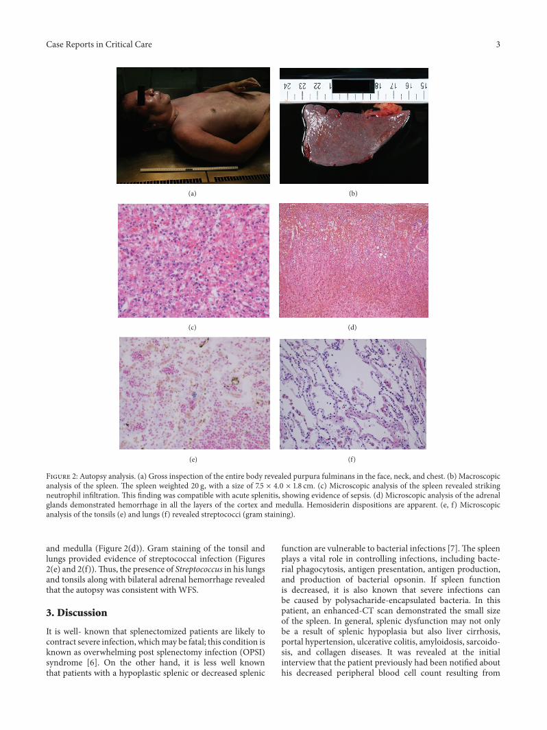
Figure 2: Autopsy analysis. (a) Gross inspection of the entire body revealed purpura fulminans in the face, neck, and chest. (b) Macroscopicanalysis of the spleen. The spleen weighted 20 g, with a size of 7.5 × 4.0 × 1.8 cm. (c) Microscopic analysis of the spleen revealed strikingneutrophil infiltration. This finding was compatible with acute splenitis, showing evidence of sepsis. (d) Microscopic analysis of the adrenalglands demonstrated hemorrhage in all the layers of the cortex and medulla. Hemosiderin dispositions are apparent. (e, f) Microscopicanalysis of the tonsils (e) and lungs (f) revealed streptococci (gram staining).
and medulla (Figure 2(d)). Gram staining of the tonsil andlungs provided evidence of streptococcal infection (Figures2(e) and 2(f)).Thus, the presence of Streptococcus in his lungsand tonsils along with bilateral adrenal hemorrhage revealedthat the autopsy was consistent with WFS.
3. Discussion
It is well- known that splenectomized patients are likely tocontract severe infection, whichmay be fatal; this condition isknown as overwhelming post splenectomy infection (OPSI)syndrome [6]. On the other hand, it is less well knownthat patients with a hypoplastic splenic or decreased splenic
function are vulnerable to bacterial infections [7].The spleenplays a vital role in controlling infections, including bacte-rial phagocytosis, antigen presentation, antigen production,and production of bacterial opsonin. If spleen functionis decreased, it is also known that severe infections canbe caused by polysacharide-encapsulated bacteria. In thispatient, an enhanced-CT scan demonstrated the small sizeof the spleen. In general, splenic dysfunction may not onlybe a result of splenic hypoplasia but also liver cirrhosis,portal hypertension, ulcerative colitis, amyloidosis, sarcoido-sis, and collagen diseases. It was revealed at the initialinterview that the patient previously had been notified abouthis decreased peripheral blood cell count resulting from

4 Case Reports in Critical Care
splenic hypoplasia, but no preventative measures had beentaken.
Howell-Jolly bodies are seen along with a markedlydecreased splenic function. Some reports show that countingHowell-Jolly bodies are a simple and useful measure forassessing splenic function [8] and, in our case, the sampleof peripheral blood cells shows the inclusion of Howell-Jolly bodies. Based on this finding along with the evidenceprovided by the enhanced-CT scan revealing a hypoplasticspleen, the patient was considered to have a decreased splenicfunction.
Purpura fulminans is a critical syndrome, involvingintravascular thrombosis and hemorrhagic infarction of theskin, which rapidly progresses, and is accompanied by vas-cular collapse or DIC [9]. It is characterized by small vesselthrombosis, leading to tissue necrosis, and is associatedwith ahigh mortality rate of approximately 43% [10]. The conditionsometimes requires aggressive surgical treatment, includinglimb amputation, to prevent further dissemination of toxinsor to save patients afflicted by purpura fulminans [10]. In ourcase, with the rapidlyworsening condition, surgical treatmentwas not feasible.
WFS is an emergency condition, characterized by fever,cyanosis, bruises, and/or shock [1]. Even if it is treated,patients with WFS usually die within 24 h after presentationof the syndrome [2]. On autopsy, our case was compatiblewith the diagnosis of WFS. It is challenging to properlydiagnose WFS during the treatment of patients with septicshock and it is supposed that many patients with WFS passaway when left undiagnosed.
Hence, till late, WFS has been considered a post-mortemdiagnosis; however, with the development of imagingmodal-ities, including ultrasonography and CT scan, imaging-baseddiagnosis of adrenal hemorrhage is possible. In general,non-traumatic adrenal hemorrhage revealed by CT scancharacteristically presents as being round or oval, with thestranding of periadrenal fat [11]. In our case, the findings ofadrenal hemorrhage were not recognized in the settings ofthe emergency department. In retrospect, it is apparent thatthe left side adrenal gland was round and accompanied bythe stranding of periadrenal fat (Figure 1(b)). The final diag-nosis of WFS was made possible by autopsy, with extensivebleeding necrosis of the bilateral adrenal glands. Consideringthis condition of WFS, acute adrenal insufficiency would beexpected and, therefore, the described extensive therapeuticstrategywas ineffective in this case. In general, if acute adrenalinsufficiency can be diagnosed quickly in ICU settings, earlyadministration of steroidal drugs may, in fact, be life-saving.When treating patients with severe infection diseases, it isimportant to unravel any existing adrenal insufficiency withthe help of imaging modalities, especially CT scan.
4. Conclusion
We reported a rare case of Waterhouse-Friderichsen syn-drome, presenting with purpura fulminans, resulting from aninvasive pneumococcal infection. In the emergency depart-ment, it is important to be aware of adrenal insufficiency
as well as impaired splenic function whenever patients withsepsis are admitted.
Consent
The family of the deceased gave written informed consent forpublication of this case report and all accompanying images.A copy of the consent form is available for review by theEditor-in-Chief.
Conflict of Interests
The authors declare that they have no competing interests.
Authors’ Contributions
Kazumasa Emori, Nobuhiro Takeuchi and Junichi Sonedatreated the patient. Kazumasa Emori and Nobuhiro Takeuchiwrote the manuscript and Nobuhiro Takeuchi revised andedited the manuscript. All authors read and approved thefinal manuscript.
Acknowledgment
The authors thank Himiko Kodaira, Yuichiro Koma, andHiroshi Yokosaki of Kobe University and Kaori Mori andKumiko Kawabuchi of Department of Laboratory Medicine,Kobe Tokusyukai Hospital. They also thank David Richardfor helping them review the paper.
References
[1] R. Waterhouse, “A case of suprarenal apoplexy,”The Lancet, vol.177, no. 4566, pp. 577–578, 1911.
[2] Y. Hata, T. Chiba, M. Ohtani, S. Ishizawa, and N. Nishida,“An autopsy case of pneumococcal Waterhouse-Friderichsensyndromewith possible functional asplenia/hyposplenia,” Inter-national Journal of Clinical and Experimental Pathology, vol. 8,no. 6, pp. 7518–7525, 2015.
[3] C. Vincentelli, E. G. Molina, and M. J. Robinson, “Fatalpneumococcal Waterhouse-Friderichsen syndrome in a vacci-nated adult with congenital asplenia,” The American Journal ofEmergency Medicine, vol. 27, no. 6, pp. 751.e3–751.e5, 2009.
[4] R. Kanthan, T. Moyana, and J. Nyssen, “Asplenia as a cause ofsudden unexpected death in childhood,” American Journal ofForensic Medicine and Pathology, vol. 20, no. 1, pp. 57–59, 1999.
[5] M. D. Grant, H. I. Horowitz, V. Lorian, and H. R. Brodman,“Waterhouse-Friderichsen syndrome induced by pneumococ-cemic shock,”The Journal of the American Medical Association,vol. 212, no. 8, pp. 1373–1374, 1999.
[6] P. D. Sinwar, “Overwhelming post splenectomy infectionsyndrome—review study,” International Journal of Surgery, vol.12, no. 12, pp. 1314–1316, 2014.
[7] K. Hansen and D. B. Singer, “Asplenic-hyposplenic overwhelm-ing sepsis: postsplenectomy sepsis revisited,” Pediatric andDevelopmental Pathology, vol. 4, no. 2, pp. 105–121, 2001.
[8] G. R. Corazza, L. Ginaldi, and G. Zoli, “Howell-Jolly bodycounting as a measure of splenic function. A reassessment,”Clinical and LaboratoryHaematology, vol. 12, no. 3, pp. 269–275,1990.

Case Reports in Critical Care 5
[9] O. Guelloit, “Note sur trois cas de purpura infectieux foudroy-ant,” in Union Medicale et Scientifique du Nord-Est, vol. 8, pp.25–29, 1884.
[10] B. J. Childers and B. Cobanov, “Acute infectious purpurafulminans: a 15-year retrospective review of 28 consecutivecases,”The American Surgeon, vol. 69, no. 1, pp. 86–90, 2003.
[11] A. Kawashima, C. M. Sandler, R. D. Ernst et al., “Imaging ofnontraumatic hemorrhage of the adrenal gland,” Radiographics,vol. 19, no. 4, pp. 949–963, 1999.

Submit your manuscripts athttp://www.hindawi.com
Stem CellsInternational
Hindawi Publishing Corporationhttp://www.hindawi.com Volume 2014
Hindawi Publishing Corporationhttp://www.hindawi.com Volume 2014
MEDIATORSINFLAMMATION
of
Hindawi Publishing Corporationhttp://www.hindawi.com Volume 2014
Behavioural Neurology
EndocrinologyInternational Journal of
Hindawi Publishing Corporationhttp://www.hindawi.com Volume 2014
Hindawi Publishing Corporationhttp://www.hindawi.com Volume 2014
Disease Markers
Hindawi Publishing Corporationhttp://www.hindawi.com Volume 2014
BioMed Research International
OncologyJournal of
Hindawi Publishing Corporationhttp://www.hindawi.com Volume 2014
Hindawi Publishing Corporationhttp://www.hindawi.com Volume 2014
Oxidative Medicine and Cellular Longevity
Hindawi Publishing Corporationhttp://www.hindawi.com Volume 2014
PPAR Research
The Scientific World JournalHindawi Publishing Corporation http://www.hindawi.com Volume 2014
Immunology ResearchHindawi Publishing Corporationhttp://www.hindawi.com Volume 2014
Journal of
ObesityJournal of
Hindawi Publishing Corporationhttp://www.hindawi.com Volume 2014
Hindawi Publishing Corporationhttp://www.hindawi.com Volume 2014
Computational and Mathematical Methods in Medicine
OphthalmologyJournal of
Hindawi Publishing Corporationhttp://www.hindawi.com Volume 2014
Diabetes ResearchJournal of
Hindawi Publishing Corporationhttp://www.hindawi.com Volume 2014
Hindawi Publishing Corporationhttp://www.hindawi.com Volume 2014
Research and TreatmentAIDS
Hindawi Publishing Corporationhttp://www.hindawi.com Volume 2014
Gastroenterology Research and Practice
Hindawi Publishing Corporationhttp://www.hindawi.com Volume 2014
Parkinson’s Disease
Evidence-Based Complementary and Alternative Medicine
Volume 2014Hindawi Publishing Corporationhttp://www.hindawi.com