case report a case of recurrent pneumothorax … pneumothorax. case report the patient was a...
TRANSCRIPT

CaseReport
Introduction
Birt-Hogg-Dubé syndrome is an autosomal dominant genetic disorder caused by mutations in the folliculin (FLCN) gene. It is characterized by a triad of skin tumors, renal tumors, and multiple pulmonary cysts. This syn-drome was first reported as a genetic skin disorder by Birt, Hogg, and Dubé.1) Subsequently, renal tumors and multiple pulmonary cysts were also considered to be part of the clinical picture.2,3) Herein, we report a case of
A Case of Recurrent Pneumothorax Associated with Birt-Hogg-Dubé Syndrome Treated with Bilateral Simultaneous Surgery and Total Pleural Covering
Kyoshiro Takegahara, MD,1,2 Naoyuki Yoshino, MD, PhD,1 and Jitsuo Usuda, MD, PhD2
Birt-Hogg-Dubé syndrome is an autosomal dominant genetic disorder characterized by a triad of skin tumors, renal tumors, and multiple pulmonary cysts. Our patient was a 40-year-old man with a history of recurrent bilateral pneumothorax and a family history of pneumothorax. The patient visited our department with chest pain and was diagnosed with left pneumothorax based on a chest X-ray. Thoracic computed tomography (CT) showed multiple cysts in both lungs. We performed thoracoscopic bilateral bullectomy with curative intent. Intraoperative observation showed numerous cysts in the lung apex, interlobular region, and mediastinum. We resected the cysts that we suspected to be respon-sible for the symptoms and ligated the lesions, and then performed total pleural covering. After surgery, genetic testing was performed. The result enabled us to diagnose Birt-Hogg-Dubé syndrome in this patient. Although the patient has developed neither recurrent pneu-mothorax nor any renal tumors, to date, long-term monitoring is necessary.
Keywords: Birt-Hogg-Dubé syndrome, pneumothorax, renal cell carcinoma, pleural covering
1Department of Thoracic Surgery, Nippon Medical School Tama Nagayama Hospital, Tama, Tokyo, Japan2Department of Thoracic Surgery, Nippon Medical School Hospital, Tokyo, Japan
Received: December 18, 2016; Accepted: February 17, 2017Corresponding author: Kyoshiro Takegahara, MD. Department of Thoracic Surgery, Nippon Medical School Tama Nagayama Hospital, 1-7-1 Nagayama, Tama, Tokyo 206-8512, JapanEmail: [email protected]©2017 The Editorial Committee of Annals of Thoracic and Cardiovascular Surgery. All rights reserved.
Birt-Hogg-Dubé syndrome diagnosed by the presence of recurrent pneumothorax.
Case Report
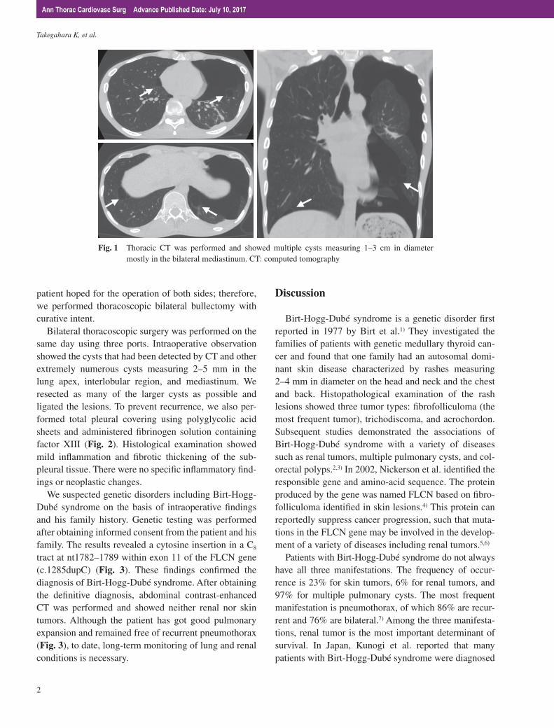
The patient was a 40-year-old man with a history of recurrent pneumothorax (three times in the right and two times in the left lung). The first pneumothorax had occurred at the age of 22 years. His mother and maternal grandmother also had histories of recurrent pneumotho-rax. The patient visited our department with a chief com-plaint of chest pain and left pneumothorax was diagnosed based on a chest X-ray. After placement of a thoracos-tomy tube, thoracic computed tomography (CT) was performed and showed multiple cysts measuring 1–3 cm in diameter mostly in the bilateral mediastinum (Fig. 1). His air leak persisted after thoracostomy tube placement. The patient had bilateral asynchronous pneumothorax. Although the patient visited our department with left pneumothorax this time, the patient had a history of three times pneumothorax in the right lung. In addition, the
Ann Thorac Cardiovasc Surg Advance Published Date: July 10, 2017
doi: 10.5761/atcs.cr.16-00295
1
Takegahara K, et al.
patient hoped for the operation of both sides; therefore, we performed thoracoscopic bilateral bullectomy with curative intent.
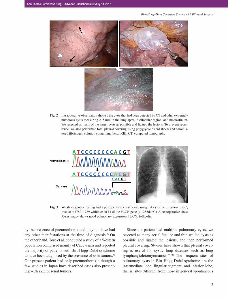
Bilateral thoracoscopic surgery was performed on the same day using three ports. Intraoperative observation showed the cysts that had been detected by CT and other extremely numerous cysts measuring 2–5 mm in the lung apex, interlobular region, and mediastinum. We resected as many of the larger cysts as possible and ligated the lesions. To prevent recurrence, we also per-formed total pleural covering using polyglycolic acid sheets and administered fibrinogen solution containing factor XIII (Fig. 2). Histological examination showed mild inflammation and fibrotic thickening of the sub-pleural tissue. There were no specific inflammatory find-ings or neoplastic changes.
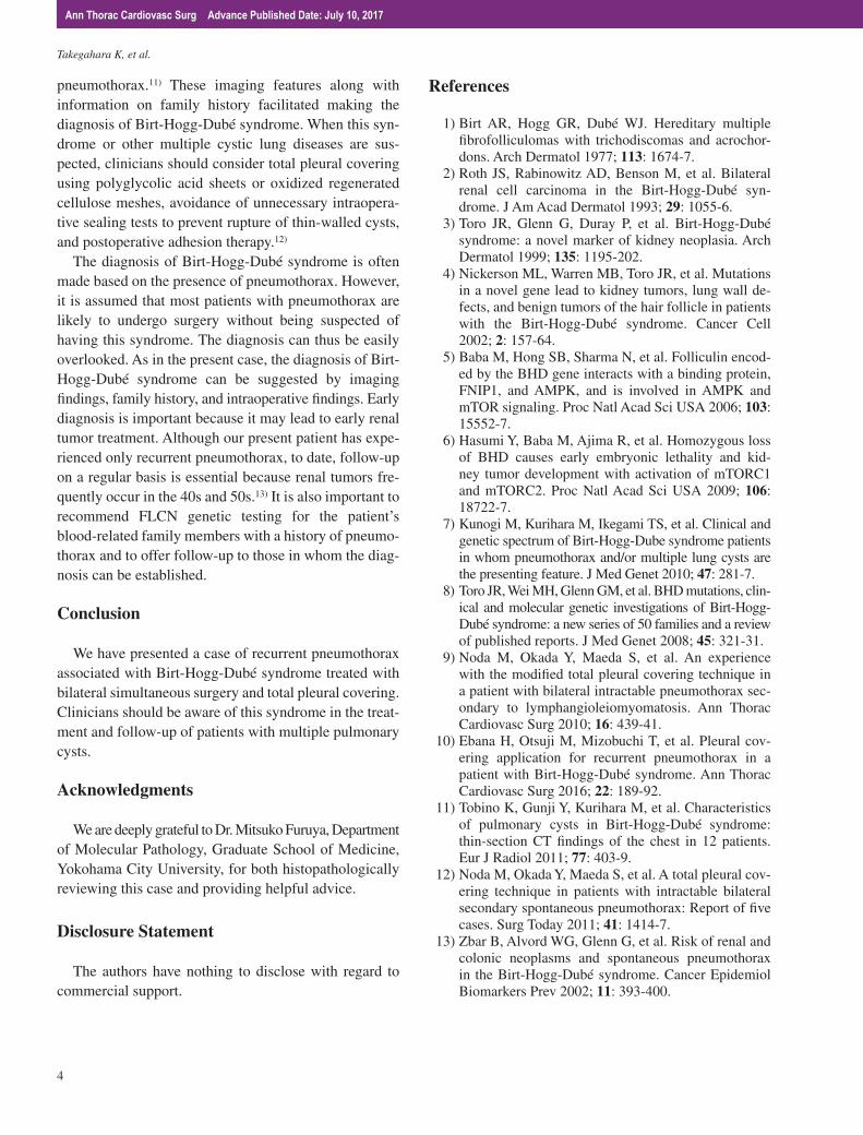
We suspected genetic disorders including Birt-Hogg-Dubé syndrome on the basis of intraoperative findings and his family history. Genetic testing was performed after obtaining informed consent from the patient and his family. The results revealed a cytosine insertion in a C8
tract at nt1782–1789 within exon 11 of the FLCN gene (c.1285dupC) (Fig. 3). These findings confirmed the diagnosis of Birt-Hogg-Dubé syndrome. After obtaining the definitive diagnosis, abdominal contrast-enhanced CT was performed and showed neither renal nor skin tumors. Although the patient has got good pulmonary expansion and remained free of recurrent pneumothorax (Fig. 3), to date, long-term monitoring of lung and renal conditions is necessary.
Discussion
Birt-Hogg-Dubé syndrome is a genetic disorder first reported in 1977 by Birt et al.1) They investigated the families of patients with genetic medullary thyroid can-cer and found that one family had an autosomal domi-nant skin disease characterized by rashes measuring 2–4 mm in diameter on the head and neck and the chest and back. Histopathological examination of the rash lesions showed three tumor types: fibrofolliculoma (the most frequent tumor), trichodiscoma, and acrochordon. Subsequent studies demonstrated the associations of Birt-Hogg-Dubé syndrome with a variety of diseases such as renal tumors, multiple pulmonary cysts, and col-orectal polyps.2,3) In 2002, Nickerson et al. identified the responsible gene and amino-acid sequence. The protein produced by the gene was named FLCN based on fibro-folliculoma identified in skin lesions.4) This protein can reportedly suppress cancer progression, such that muta-tions in the FLCN gene may be involved in the develop-ment of a variety of diseases including renal tumors.5,6)
Patients with Birt-Hogg-Dubé syndrome do not always have all three manifestations. The frequency of occur-rence is 23% for skin tumors, 6% for renal tumors, and 97% for multiple pulmonary cysts. The most frequent manifestation is pneumothorax, of which 86% are recur-rent and 76% are bilateral.7) Among the three manifesta-tions, renal tumor is the most important determinant of survival. In Japan, Kunogi et al. reported that many patients with Birt-Hogg-Dubé syndrome were diagnosed
Fig. 1 Thoracic CT was performed and showed multiple cysts measuring 1–3 cm in diameter mostly in the bilateral mediastinum. CT: computed tomography
2
Ann Thorac Cardiovasc Surg Advance Published Date: July 10, 2017
Birt–Hogg–Dubé Syndrome Treated with Bilateral Surgery
by the presence of pneumothorax and may not have had any other manifestations at the time of diagnosis.7) On the other hand, Toro et al. conducted a study of a Western population comprised mainly of Caucasians and reported the majority of patients with Birt-Hogg-Dubé syndrome to have been diagnosed by the presence of skin tumors.8) Our present patient had only pneumothorax although a few studies in Japan have described cases also present-ing with skin or renal tumors.
Since the patient had multiple pulmonary cysts, we resected as many aerial fistulae and thin-walled cysts as possible and ligated the lesions, and then performed pleural covering. Studies have shown that pleural cover-ing is useful for cystic lung diseases such as lung lymphangioleiomyomatosis.9,10) The frequent sites of pulmonary cysts in Birt-Hogg-Dubé syndrome are the intermediate lobe, lingular segment, and inferior lobe, that is, sites different from those in general spontaneous
Fig. 3 We show genetic testing and a postoperative chest X-ray image. A cytosine insertion in a C8 tract at nt1782–1789 within exon 11 of the FLCN gene (c.1285dupC). A postoperative chest X-ray image shows good pulmonary expansion. FLCN: folliculin
Fig. 2 Intraoperative observation showed the cysts that had been detected by CT and other extremely numerous cysts measuring 2–5 mm in the lung apex, interlobular region, and mediastinum. We resected as many of the larger cysts as possible and ligated the lesions. To prevent recur-rence, we also performed total pleural covering using polyglycolic acid sheets and adminis-tered fibrinogen solution containing factor XIII. CT: computed tomography
Ann Thorac Cardiovasc Surg Advance Published Date: July 10, 2017
3
Takegahara K, et al.
pneumothorax.11) These imaging features along with information on family history facilitated making the diagnosis of Birt-Hogg-Dubé syndrome. When this syn-drome or other multiple cystic lung diseases are sus-pected, clinicians should consider total pleural covering using polyglycolic acid sheets or oxidized regenerated cellulose meshes, avoidance of unnecessary intraopera-tive sealing tests to prevent rupture of thin-walled cysts, and postoperative adhesion therapy.12)
The diagnosis of Birt-Hogg-Dubé syndrome is often made based on the presence of pneumothorax. However, it is assumed that most patients with pneumothorax are likely to undergo surgery without being suspected of having this syndrome. The diagnosis can thus be easily overlooked. As in the present case, the diagnosis of Birt-Hogg-Dubé syndrome can be suggested by imaging findings, family history, and intraoperative findings. Early diagnosis is important because it may lead to early renal tumor treatment. Although our present patient has expe-rienced only recurrent pneumothorax, to date, follow-up on a regular basis is essential because renal tumors fre-quently occur in the 40s and 50s.13) It is also important to recommend FLCN genetic testing for the patient’s blood-related family members with a history of pneumo-thorax and to offer follow-up to those in whom the diag-nosis can be established.
Conclusion
We have presented a case of recurrent pneumothorax associated with Birt-Hogg-Dubé syndrome treated with bilateral simultaneous surgery and total pleural covering. Clinicians should be aware of this syndrome in the treat-ment and follow-up of patients with multiple pulmonary cysts.
Acknowledgments
We are deeply grateful to Dr. Mitsuko Furuya, Department of Molecular Pathology, Graduate School of Medicine, Yokohama City University, for both histopathologically reviewing this case and providing helpful advice.
Disclosure Statement
The authors have nothing to disclose with regard to commercial support.
References
1) Birt AR, Hogg GR, Dubé WJ. Hereditary multiple fibrofolliculomas with trichodiscomas and acrochor-dons. Arch Dermatol 1977; 113: 1674-7.
2) Roth JS, Rabinowitz AD, Benson M, et al. Bilateral renal cell carcinoma in the Birt-Hogg-Dubé syn-drome. J Am Acad Dermatol 1993; 29: 1055-6.
3) Toro JR, Glenn G, Duray P, et al. Birt-Hogg-Dubé syndrome: a novel marker of kidney neoplasia. Arch Dermatol 1999; 135: 1195-202.
4) Nickerson ML, Warren MB, Toro JR, et al. Mutations in a novel gene lead to kidney tumors, lung wall de-fects, and benign tumors of the hair follicle in patients with the Birt-Hogg-Dubé syndrome. Cancer Cell 2002; 2: 157-64.
5) Baba M, Hong SB, Sharma N, et al. Folliculin encod-ed by the BHD gene interacts with a binding protein, FNIP1, and AMPK, and is involved in AMPK and mTOR signaling. Proc Natl Acad Sci USA 2006; 103: 15552-7.
6) Hasumi Y, Baba M, Ajima R, et al. Homozygous loss of BHD causes early embryonic lethality and kid-ney tumor development with activation of mTORC1 and mTORC2. Proc Natl Acad Sci USA 2009; 106: 18722-7.
7) Kunogi M, Kurihara M, Ikegami TS, et al. Clinical and genetic spectrum of Birt-Hogg-Dube syndrome patients in whom pneumothorax and/or multiple lung cysts are the presenting feature. J Med Genet 2010; 47: 281-7.
8) Toro JR, Wei MH, Glenn GM, et al. BHD mutations, clin-ical and molecular genetic investigations of Birt-Hogg-Dubé syndrome: a new series of 50 families and a review of published reports. J Med Genet 2008; 45: 321-31.
9) Noda M, Okada Y, Maeda S, et al. An experience with the modified total pleural covering technique in a patient with bilateral intractable pneumothorax sec-ondary to lymphangioleiomyomatosis. Ann Thorac Cardiovasc Surg 2010; 16: 439-41.
10) Ebana H, Otsuji M, Mizobuchi T, et al. Pleural cov-ering application for recurrent pneumothorax in a patient with Birt-Hogg-Dubé syndrome. Ann Thorac Cardiovasc Surg 2016; 22: 189-92.
11) Tobino K, Gunji Y, Kurihara M, et al. Characteristics of pulmonary cysts in Birt-Hogg-Dubé syndrome: thin-section CT findings of the chest in 12 patients. Eur J Radiol 2011; 77: 403-9.
12) Noda M, Okada Y, Maeda S, et al. A total pleural cov-ering technique in patients with intractable bilateral secondary spontaneous pneumothorax: Report of five cases. Surg Today 2011; 41: 1414-7.
13) Zbar B, Alvord WG, Glenn G, et al. Risk of renal and colonic neoplasms and spontaneous pneumothorax in the Birt-Hogg-Dubé syndrome. Cancer Epidemiol Biomarkers Prev 2002; 11: 393-400.
4
Ann Thorac Cardiovasc Surg Advance Published Date: July 10, 2017