case 2 - right optic nerve head drusen...
TRANSCRIPT

Case 2 - Right Optic Nerve Head Drusen (ONHD) A 41 year old female was referred by her optometrist for a workup for unilateral optic disc drusen, OCT, and visual field changes. The patient was otherwise fit and well and her previous ocular history was unremarkable.
Right Eye Left Eye Unaided Vision 6/6-2 6/6 Intraocular Pressures 14 mmHg 17 mmHg Slit lamp examination Normal Normal Ishihara colour plates 14/14 14/14 Pupil No afferent defect No afferent defect Gonioscopy Normal Normal
Figure 1. Colour photograph of optic nerves Question 1: Comment on the optic nerve appearance of each eye. The left optic nerve head is of normal appearance. It has a CDR of approximately 0.25 and a healthy neuroretinal rim, with well-defined margins. The right optic disc is seen to have blurred margins, pallor, and no discernable optic cup. Multilobular yellowish-white nodules can be appreciated on close inspection, particularly at the periphery of the disc.

Figure 2. HVF 30-2 Right eye

Figure 3. HVF 30-2 Left eye

Figure 4. Retinal nerve fiber layer OCT scan of right and left eye

Figure 5. Ganglion cell complex scans of right and left eye

Figure 6. Macular thickness of right and left eye

Question 2: Comment on the visual field and OCT results for each eye HVF 30-2 Right eye: false negative errors of 46% make the field less reliable. Enlargement of the blind spot with superior and inferior nerve fiber bundle defects are seen. Left HVF 30-2 is reliable (2 fixation losses and 6% false positive errors). The results are within normal limits. OCT RNFL right eye is seen to be thinned globally with thickening of the neuroretinal rim. The classic RNFL ‘double hump’ (RNFL thickest superiorly and inferiorly) pattern is absent. OCT GCL right eye seen to be thinned in all sectors with thinning also present to a moderate degree in the left eye, predominantly in the nasal sectors. OCT macular thickness scan of the right eye demonstrates generalized thinning. The left eye is seen to have a normal macular thickness. Question 3: Given the blurred disc margin of the right optic nerve, comment on the features that indicate a diagnosis of a space occupying lesion is less likely There is unilateral blurring of right disc margins. Normal disc margins left optic nerve Ishihara full and normal both eyes RAPD present Humphery visual fields 30-2: Right eye demonstrates enlargement of the blind spot with superior and inferior nerve fiber bundle defects, however this is not a typical neurological defect. The left visual field is normal. Question 4: Based on the information available please describe additional tests that could be undertaken to confirm the diagnosis for this patient.
• Optic nerve head autofluorescence can elicit superficial buried drusen • B-Scan Ultrasonography to assess for hyperechogenicity of the optic
nerve head at the junction of the retina and the optic nerve. • EDI OCT of the optic disc • CT orbits with thin slices through the optic nerve head
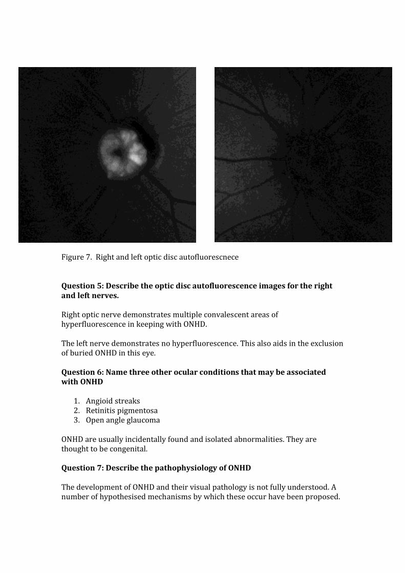
Figure 7. Right and left optic disc autofluorescnece Question 5: Describe the optic disc autofluorescence images for the right and left nerves. Right optic nerve demonstrates multiple convalescent areas of hyperfluorescence in keeping with ONHD. The left nerve demonstrates no hyperfluorescence. This also aids in the exclusion of buried ONHD in this eye. Question 6: Name three other ocular conditions that may be associated with ONHD
1. Angioid streaks 2. Retinitis pigmentosa 3. Open angle glaucoma
ONHD are usually incidentally found and isolated abnormalities. They are thought to be congenital. Question 7: Describe the pathophysiology of ONHD The development of ONHD and their visual pathology is not fully understood. A number of hypothesised mechanisms by which these occur have been proposed.

ONHD are abnormal accumulations of calcified mitochondrial deposits in the optic nerve head with histological composition of calcium and phosphorus crystals. Several mechanisms have been proposed for the formation of ONHD. One accepted theory hypothesises that a small scleral canal may result in axonal stress secondary to the physical limits of large groups of axons passing through a small space which may cause axonal degeneration and ONHD formation. A second theory of ONHD formation is that they are secondary to abnormal optic disc vasculature resulting in ischaemia and impaired axonal metabolism. There is a reported association between ONHD and reduced central retinal artery blood flow. Axonal distress as a mechanism of ONHD formation has been theorized to be related to calcium metabolism. Tso et al put forward the theory that abnormal axonal metabolism leads to the deposition of calcium crystals in mitochondria with subsequent axonal disruption and extrusion of mitochondria into the extracellular space. Birnbaum et al proposed to include papilloedema as a possible cause of acquired disc drusen as optic disc oedema as a result of raised intracranial pressure is known to cause axoplasmic stasis secondary to mechanical obstruction of the axons and/or ischaemia. Their study concluded that there was a higher prevalence of ONHD with resolved papilloedema which suggested a non-coincidental relationship. Question 8: Describe the mechanism of visual field defect development secondary to optic disc drusen Enlarging, non expansile, calcified bodies increase the mechanical stress placed on structures within the scleral canal. This may lead to impairment of blood flow through the optic nerve head thus predisposing to vascular complications. ONHD have higher incidence of vascular complications such as peripapillary haemorrhages, retinal artery occlusions, central retinal vein occlusions, choroidal neovascularization, and anterior ischaemic optic neuropathy. Slowly progressive visual field loss is most likely secondary to mechanical forces of the ONHD resulting in axonal dysfunction and gradual axonal degeneration. ONHD with associated visual field defects progress at a rate of approximately 1.6% per year on Goldmann perimetry.9 Most commonly described visual field defects include enlargement of blind spot, arcuate scotoma and peripheral defects. In a retrospective analysis by Grippo et al of 103 eyes those with visible drusen were found to have significantly more visual field loss compared to those with buried drusen.
Question 9: Describe the relationship between ONHD and ocular hypertension

Ocular hypertension appears to worsen the field loss in ONHD. Grippo et al found that patients with ONHD who also had ocular hypertension had a higher prevalence of visual field loss compared to normotensive eyes with ONHD. This was independent of age, sex, and the drusen visibility. Question 10: Discuss treatment options for ONHD and the evidence for such treatments. No treatment has been proven to alter the clinical course of ONHD. Radial optic neurotomy has been attempted with variable success.
Theoretical thoughts on treatment include lowering of IOP. Eyes with ocular hypertension and ONHD are seen to have a higher prevalence of visual field loss in comparison to normotensive eyes with ONHD. References And Recommended Reading:
1. Kapur R, Pulido JS, Abraham JL, Sharma M, Buerk B, Edward DP. Histologic findings after surgical excision of optic nerve head drusen. Retina 2008;28:143–146.
2. Tso M. Pathology and pathogenesis of drusen of the optic nerve head. Ophthalmology 1981;88:1066–1080.
3. Friedman AH, Henkind P, Gartner S. Drusen of the optic disc. A histopathological study. Trans Ophthalmol Soc UK 1975;95:4–9.
4. Floyd M. Measurement of the scleral canal using optical coherence tomography in patients with optic nerve drusen. Am J Ophthalmol 2005;139:664–669.
5. Auw-haedrich C, Staubach F, Witschel H. Optic disk drusen. Surv Ophthalmol 2002;47:515–532.
6. Abegão Pinto L, Vandewalle E, Marques-Neves C, Stalmans I. Visual field loss in optic disc drusen patients correlates with central retinal artery blood velocity patterns. Acta Ophthalmologica 2014: 92(4):e286-91.
7. Tso MO. Pathology and pathogenesis of drusen of the optic nerve head. Ophthalmology. 1981;88(10):1066-1080.
8. Birnbaum FA, Johnson GM, Johnson LN, et al. Increased prevalence of optic disc drusen after papilloedema from idiopathic intracranial hypertension: on the possible formation of optic disc drusen. Neuro-ophthalmology 2016;40(4):171-180.
9. Lee A, Zimmerman MB. The rate of visual field loss in optic nerve head drusen. American Journal of Ophthalmology. 2005;139(6):1062-1066.
10. Grippo T, Shihadeh W, Schargus M, et al. Optic nerve head drusen and visual field loss in normotensive and hypertensive eyes. J Glaucoma. 2008;17(2):100-104.
11. Nentwich M, Remy M, Haritoglou C, Kampik A. Radial optic neurotomy to treat patients with visual field defects associated with optic nerve drusen. Retina. 2011;31(3):612-615.

Case 2 Test Question 1: Describe the histological composition of ONHD
a. Calcium crystals and phosphorus crystals b. Calcium crystals c. Phosphorus crystals and iron d. Calcium crystals and iron e. Iron
Question 2: Optic nerve head drusen may be associated with all the following conditions except?
a. Angioid streaks b. Retinitis pigmentosa c. pen angle glaucoma d. Usher syndrome e. Ocular hypertension
Question 3: Visual defects secondary to optic nerve head drusen may include all of the following except: 1. enlargement of the blind spot 2. central scotoma 3. peripheral defects 4. arcuate scotoma
a. 3 and 4 b. 2 and 4 c. 1 and 2 d. 1 and 3 e. 1
Question 4: Optic nerve head drusen have increased incidences of the following vascular complications except?
a. peripapillary haemorrhages b. retinal artery occlusions c. anterior ischaemic optic neuropathy d. posterior ischaemic optic neuropathy e. central retinal vein occlusions
Question 5: In patients with ONHD the central retinal artery (CRA) blood flow velocities are lower in comparison to patients without ONHD? True or False? Question 6: ONHD are found in the:
a. Prelaminar portion of the optic nerve head b. Lamina cribrosa region of the optic nerve head c. Retrolaminar region of the optic nerve head

d. Surface nerve fibre layer of the optic nerve head e. Retrolaminar portion of the optic nerve head
Question 7: Prevalence of ONHD is most consistent with which of the following values?
a. <2% b. 3% c. 3-4% d. 4-5% e. 5-6%
Question 8: Diagnosis of ONHD most commonly occurs by which of the following means?
a. Routine consult with optometrist or ophthalmologist with detection of irregular ONH margins
b. Incidental visual field defect finding on routine optometrist visit c. Patients presenting with symptomatically reduced vision d. Reduction in colour vision e. RAPD
Question 9: ONHD may be associated with which of the following comorbidities? 1. Slow peripheral vision loss 2. Central retinal artery occlusions 3. Anterior ischaemic optic neuropathy 4. Choroidal neovascularization 5. Central retinal vein occlusion
a. 1 and 2 b. 1,3, and 5 c. 1,2, and 3 d. 1,2,3, and 5 e. all of the above
Question 10: In patients with ONHD the CRA blood flow velocity is seen to correlate with the extent of visual field defects. True or False? Question 11: Signs that have been seen on fundus examination in patients with ONHD include: 1. dilated veins 2. abnormal early branching of arteries 3. arterial tortuosity 4. arterial vascular loops
a. 2 b. 1 and 2 c. 1 - 3 d. 1 and 3

e. 1 - 4 Question 12: Which of the following are accepted theories and mechanisms for the formation of ONHD? 1. Axonal distress secondary to small scleral canal 2. Abnormal vasculature of the optic disc causing ischaemia and impaired axonal metabolism 3. Altered axonal metabolism results in calcium deposition in mitochondria which are then extruded from degenerating nerve fibres creating a nidus for progressive drusen formation 4. ONHD may be a sequelae of papilloedema
a. 1 and 2 b. 3 and 4 c. 1 and 3 d. 1, 2, and 3 e. 1, 2, 3, and 4
Question 13: The rate of visual field loss per year for patients with ONHD is closest to which of the following?
a. <0.5% per year b. 1-2% per year c. 3% per year d. 4-5% per year e. 5% per year
Question 14: Techniques for diagnosis of ONHD include: 1. B-scan ultrasound 2. Autofluorescence 3. CT scan 4. Humphrey visual field 30-2 5. Humphrey visual field 24-2
a. 1 and 2 b. 1, 2, and 3 c. 1, 2, and 4 d. 1, 2, and 5 e. 1, 2, 4, and 5
Question 15: Dilation of the retinal veins is thought to occur as a result of mechanical compression from the drusen on the vasculature surrounding the ONH. True or False? Question 16: Optic nerve head drusen: 1. are usually bilateral 2. are never unilateral 3. have no established sex predilection

4. ONHD and ocular hypertension is seen more frequently than ONHD and normal IOP
a. 1 b. 1, 3, and 4 c. 1 and 3 d. 2 and 3 e. 3 and 4
Question 17: Which of the following is correct with regard to ONHD? 1. The highest rate of occurrence of visual field defects is in patients with superficial drusen 2. Visual field loss is more frequent in the lower nasal quadrant 3. Generalised constriction of the visual field is not associated with ONHD 4. In most cases visual acuity is well preserved
a. 1 and 2 b. 1, 2, and 3 c. 2, 3, and 4 d. 2 and 4 e. 1, 2, and 4
Question 18: Which of the following is true for ONHD and ocular hypertension?
a. Do not occur together simultaneously b. ONHD (both visible and buried) with concomitant ocular hypertension
does not increase the likelihood of visual field loss c. Only those patients with visible ONHD and ocular hypertension have an
increased likelihood of visual field loss d. Only those patients with buried ONHD and ocular hypertension have an
increased likelihood of visual field loss e. Patients with ONHD and ocular hypertension should be monitored closely
with appropriate treatment of antiglaucomatous medication to prevent further visual field loss progression
Question 19: Which of the following is true for ONHD treatment: 1. There is currently no effective treatment for optic nerve head drusen 2. Patients with ONHD should be followed regularly for the onset of ocular hypertension 3. Visual field defects in eyes with ONHD and are normotensive are uncommon 4. Visual field defects in eyes with ONHD and coexisting ocular hypertension are more common than those eyes with ONHD and normotension
a. 1 and 2 b. 1, 2, and 3 c. 1, 2, 3 and 4 d. 1, 2, and 4 e. 2, 3, and 4

Question 20: Computed tomography scan can demonstrate calcification of the optic nerve head. True or False? Question 21: Identify the correct statement(s): 1. Most patients with ONHD are asymptomatic 2. Reported frequency of asymptomatic patients with of ONHD have visual field defects range from 24% to 87% 3. Patients with ONHD may experience significant visual impairment to the point of legal blindness 4. There is no known treatment for visual field loss associated with ONHD
a. 1 b. 1 and 2 c. 2, 3, and 4 d. 1, 2, and 4 e. 1, 2, 3, and 4
Question 22: Identify the correct statement:
a. Clinically ONHD appear as globular bodies embedded in the optic nerve head
b. In children ONHD are usually exposed c. ONHD become exposed on the disc surface with advancing age thought
secondary to vascular compromise d. ONHD become exposed on the disc surface due to increasing intraocular
pressure thought to be secondary to enlargement by continuous calcium apposition
e. Answers a and b are both correct Question 23: Prevalence of visual field loss in patients' with ONHD: 1. Is dependent upon whether the ONHD are buried or not buried 2. Is dependent upon whether the patient has concomitant ocular hypertension 3. Is independent of the sex of the patient 4. Is dependent upon the age of the patient
a. 1 and 3 b. 1 and 4 c. 2 and 3 d. 1, 2, and 3 e. 1, 2, 3, and 4
Question 24: Identify the correct statement: 1. The racial predilection for ONHD is unknown 2. A recent study has found a higher than expected prevalence of ONHD with resolved papilloedema suggesting a non-coincedental relationship 3. ONHD with resolved paipilloedema is not associated with a worse visual outcome compared with ONHD alone

4. ONHD with resolved papilloedema were seen to associated with worse visual field outcomes compared to ONHD alone
a. 1 and 2 b. 1, 2, and 3 c. 2 and 3 d. 2, 3, and 4 e. 1, 2, 3, and 4
Question 25: B-scan will demonstrated hypoechoic optic nerve heads if drusen are present. True or False?