cardiomyopathy in childhood
TRANSCRIPT

SympoSium: CardiovaSCular mediCine
Cardiomyopathy in childhoodphilip roberts
michael Burch
AbstractCardiomyopathies are a heterogeneous group of disorders of both ge-
netic and non-genetic aetiology. Their importance lies in the fact that
although the mortality for structural congenital heart disease has been
reduced from around 40% to 5% in the past 40 years there has been
little change in the prognosis for paediatric patients with cardiomyopa-
thies. around one-third of children with a cardiomyopathy will die or
require transplantation. advances in genetics over the past decade have
resulted in the recognition that a large proportion of cardiomyopathies
are genetic in origin. management of these patients is best in centres
offering cardiology, genetic and metabolic input. This article aims to
briefly review the broad categories of cardiomyopathies encountered in
childhood.
Keywords cardiomyopathy; childhood
Introduction
The true incidence of cardiomyopathy in childhood remains unclear for several reasons: there is no universally accepted defi-nition or classification of cardiomyopathies and epidemiological studies performed to date have variable inclusion criteria. Fur-thermore, there is likely to be significant geographical variation reflecting both environmental and genetic factors.
Although rare, the importance of cardiomyopathies lies in the fact that around one-third of cardiomyopathies will progress to death or require cardiac transplantation. Cardiomyopathies are the most common reason for cardiac transplant in childhood.1 Although major advances have been made in the management of childhood heart disease, there has been only minimal change in outcome over the past 30 years for those with cardiomyopathies. Cardiomyopathy may be the first manifestation of both an inborn error of metabolism or neuromuscular disorder with attendant genetic implications.
Initial attempts at definition and classification were made in 1980 and 1995 by the World Health Organization and International
Philip Roberts MRCP is at the Department of Paediatric Cardiology,
Cardiothoracic Transplant and Heart Failure, Great Ormond Street
Hospital for Children, London, UK.
Michael Burch FRCP is at the Department of Paediatric Cardiology,
Cardiothoracic Transplant and Heart Failure, Great Ormond Street
Hospital for Children, London, UK.
paediaTriCS and CHild HealTH 19:1
Society and Federation of Cardiology task force. Originally car-diomyopathies were defined as primary myocardial disorders of unknown cause. Where the aetiology of the heart muscle dis-ease was known they were classified as secondary or specific cardiomyopathies.2 Genetic advances of the past decade have resulted in more recent attempts by the American Heart Asso-ciation (AHA) 2006 and European Society of Cardiology 2007 to offer revised classifications with the principle difference between the two being the inclusion of the ‘channelopathies’ by the AHA as cardiomyopathies.3,4
When faced with a symptomatic patient with evidence of car-diac muscle disease on electrocardiography (ECG), chest x-ray and echocardiogram, the question of how best to proceed on the patient journey from diagnosis to therapeutic intervention is in our opinion more pragmatically addressed by aligning with the European Society of Cardiology position statement, remember-ing that this has been principally drawn up by an adult working group.
Several important differentials for ventricular myocardial dysfunction are traditionally not classified as cardiomyopathies. However, these will also be included as, in some cases, such rate related cardiomyopathy and endocrine disorders, treatment can result in complete recovery and, therefore, it is particularly important to identify these cases.
Definition
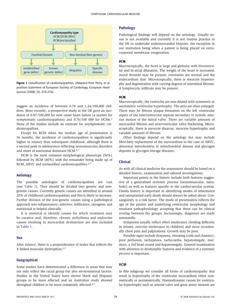
A cardiomyopathy is a myocardial disorder in which the heart muscle is structurally and functionally abnormal. Traditionally, the absence of haemodynamically significant structural heart disease is included and adult definitions would also include the absence of coronary heart disease, systemic and pulmonary hypertension and valvular heart disease significant enough to cause the observed myocardial dysfunction. Clearly it is possible for combinations of the above to coexist.The type of cardiomyopathy is grouped according to ventricular morphology and function into: • dilated cardiomyopathy (DCM) • hypertrophic cardiomyopathy (HCM) • restrictive cardiomyopathy (RCM) • arrhythmogenic right ventricular cardiomyopathy (ARVC) • unclassified.
Each morphological phenotype is then sub-classified into familial/genetic and non-familial/non-genetic. The genetic group can then be further grouped into known genetic types and unidentified defects. The non-genetic group is classified into idio-pathic or specific disease subtypes.
As with many conditions, there can be overlap between the morphological groups and indeed during the evolution and natu-ral history of any cardiomyopathic process progression from one morphology to another. The ventricular morphological classifica-tion and consequent pathophysiology, however, determine both mode of presentation and what medical management strategies are likely to be required (Figure 1).
Incidence
The true incidence of childhood cardiomyopathy is unknown. Studies from Australia, Finland and two regions in North America
15 © 2008 published by elsevier ltd.
SympoSium: CardiovaSCular mediCine
suggest an incidence of between 0.74 and 1.24/100,000 chil-dren. More recently, a prospective study in the UK gave an inci-dence of 0.87/100,000 for new onset heart failure (a marker for symptomatic cardiomyopathies) and 0.76/100 000 for DCMs.5 None of the studies include an estimate for asymptomatic car-diomyopathies.
Except for RCM when the median age of presentation is 36 months, the incidence of cardiomyopathies is significantly higher in infancy than subsequent childhood, although there is a second peak in adolescence reflecting neuromuscular disorders and onset of autosomal dominant HCM.6,7
DCM is the most common morphological phenotype (50%) followed by HCM (40%) with the remainder being made up of RCM, ARVC and unclassified cardiomyopathies.6,7
Aetiology
The possible aetiologies of cardiomyopathies are vast (see Table 1). They should be divided into genetic and non-genetic causes. Currently genetic causes are identified in around 20% of childhood cardiomyopathies and this is likely to increase. Further division of the non-genetic causes using a pathological approach into inflammatory, infective, infiltrative, iatrogenic and nutritional is helpful clinically.
It is essential to identify causes for which treatment may be curative and, therefore, chronic arrhythmia and endocrine causes resulting in myocardial dysfunction are also included in Table 1.
Sex
After infancy, there is a preponderance of males that reflects the X-linked muscular dystrophies.6,7
Geographical
Some studies have demonstrated a difference in areas that may not only reflect the racial group but also environmental factors. Studies in the United States have shown black and Hispanic groups to be more affected and an Australian study showed aboriginal children to be more commonly affected.6,7
Cardiomyopathy typeHCM/DCM/ARVC
RCM/unclassified
Unidentified
gene defect
Known
genetic defectIdiopathic
Specific
disease
Non-familial/Non-geneticFamilial/Genetic
Figure 1 Classification of cardiomyopathies. (adapted from perry, et al.
position statement of european Society of Cardiology, european Heart
Journal (2008) 29, 270-276).
paediaTriCS and CHild HealTH 19:1 1
Pathology
Pathological findings will depend on the aetiology. Usually tis-sue is not available and currently it is not routine practice in the UK to undertake endomyocardial biopsies. the exception in our institution being when a patient is being placed on extra- corporeal membrane oxygenation.
DCMMacroscopically, the heart is large and globular with biventricu-lar and bi-atrial dilatation. The weight of the heart is increased, mural thrombi may be present, coronaries are normal and the endocardium thin. Microscopically, there is myocyte hypertro-phy and degeneration with varying degrees of interstitial fibrosis. A lymphocytic infiltrate may be present.
HCMMacroscopically, the ventricles are non-dilated with symmetric or asymmetric ventricular hypertrophy. The atria are often enlarged. There may be fibrous plaque formation on the left ventricular aspect of the interventricular septum secondary to systolic ante-rior motion of the mitral valve. There are variable amounts of myocardial fibrosis and atrioventricular valve thickening. Micro-scopically, there is myocyte disarray, myocyte hypertrophy and variable amounts of fibrosis.
Other findings depend on the aetiology but may include fibro-fatty replacement of the myocardium in the case of ARVC, abnormal mitochondria in mitochondrial disease and glycogen deposits in glycogen storage diseases.
Clinical
As with all clinical medicine the assessment should be based on a detailed history, examination and tailored investigations.
Important points in the history include both features sugges-tive of a generalised systemic process (neuromuscular, meta-bolic) as well as features specific to the cardiovascular system. Family history is important in identifying modes of inheritance and unexplained early death should always be asked about. Con-sanguinity is a risk factor. The mode of presentation reflects the age of the patient and underlying ventricular morphology and resultant pathophysiology accepting that there can be clinical overlap between the groups. Increasingly, diagnoses are made antenatally.
Symptoms usually reflect effort intolerance (feeding difficulty in infants, exercise intolerance in children) and more occasion-ally chest pain and palpitations. Growth may be poor.
Possible signs include dyspnoea, sweating (cold and clammy), poor perfusion, tachypnoea, tachycardia, hepatomegaly, mur-murs, a 3rd heart sound and hepatomegaly. General examination with attention to dysmorphic features and evidence of a systemic process is important.
HCM
In this subgroup we consider all forms of cardiomyopathy that result in hypertrophy of the ventricular myocardium either sym-metrically or asymmetrically. Haemodynamic causes for ventricu-lar hypertrophy such as arterial valve and great artery stenosis are
6 © 2008 published by elsevier ltd.
SympoSium: CardiovaSCular mediCine
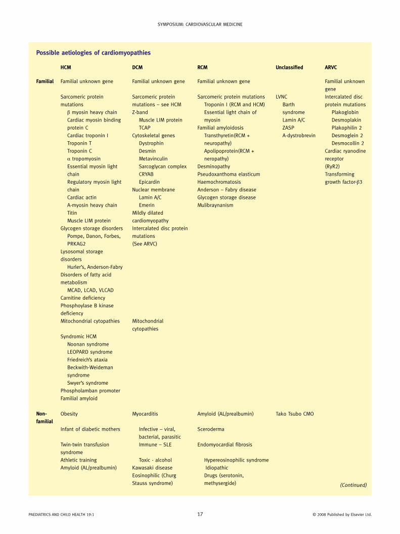
Possible aetiologies of cardiomyopathies
HCM DCM RCM Unclassified ARVC
Familial Familial unknown gene Familial unknown gene Familial unknown gene Familial unknown
gene
Sarcomeric protein
mutations
β myosin heavy chain
Cardiac myosin binding
protein C
Cardiac troponin i
Troponin T
Troponin C
α tropomyosin
essential myosin light
chain
regulatory myosin light
chain
Cardiac actin
a-myosin heavy chain
Titin
muscle lim protein
Sarcomeric protein
mutations – see HCm
Z-band
muscle lim protein
TCap
Cytoskeletal genes
dystrophin
desmin
metavinculin
Sarcoglycan complex
CryaB
epicardin
nuclear membrane
lamin a/C
emerin
mildly dilated
cardiomyopathy
Sarcomeric protein mutations
Troponin i (rCm and HCm)
essential light chain of
myosin
Familial amyloidosis
Transthyretin(rCm +
neuropathy)
apolipoprotein(rCm +
neropathy)
desminopathy
pseudoxanthoma elasticum
Haemochromatosis
anderson – Fabry disease
Glycogen storage disease
mulibraynanism
lvnC
Barth
syndrome
lamin a/C
ZaSp
a-dystrobrevin
intercalated disc
protein mutations
plakoglobin
desmoplakin
plakophilin 2
desmoglein 2
desmocollin 2
Cardiac ryanodine
receptor
(ryr2)
Transforming
growth factor-β3
Glycogen storage disorders
pompe, danon, Forbes,
prKaG2
intercalated disc protein
mutations
(See arvC)
lysosomal storage
disorders
Hurler’s, anderson-Fabry
disorders of fatty acid
metabolism
mCad, lCad, vlCad
Carnitine deficiency
phosphoylase B kinase
deficiency
mitochondrial cytopathies mitochondrial
cytopathies
Syndromic HCm
noonan syndrome
leopard syndrome
Friedreich’s ataxia
Beckwith-Weideman
syndrome
Swyer’s syndrome
phospholamban promoter
Familial amyloid
Non-
familialobesity myocarditis amyloid (al/prealbumin) Tako Tsubo Cmo
infant of diabetic mothers infective – viral,
bacterial, parasitic
Sceroderma
Twin-twin transfusion
syndrome
immune – Sle endomyocardial fibrosis
athletic training Toxic - alcohol Hypereosinophilic syndrome
amyloid (al/prealbumin) Kawasaki disease idiopathic
eosinophilic (Churg
Stauss syndrome)
drugs (serotonin,
methysergide) (Continued)
paediaTriCS and CHild HealTH 19:1 17 © 2008 published by elsevier ltd.

SympoSium: CardiovaSCular mediCine
HCM DCM RCM Unclassified ARVC
drugs - anthracyclines Carcinoid heart disease
pregnancy metastatic cancers
endocrine – vitamin d
deficiency
radiation
nutritional – thiamine,
carnitine, selenium,
drugs - anthracyclines
rate-related
cardiomyopathy
adapted with permission from perry et al. eur Heart J 2008; 29: 270-276. HCm, hypertrophic cardiomyopathy; dCm, dilated cardiomyopathy; rCm, restrictive car-diomyopathy; arvC, arrhythmogenic right ventricular cardiomyopathy; lim, TCap, CryaB, mCad, medium chain acyl-Coa; lCad, long chain acyl-Coa; vlCad, very long chain acyl-Coa; Sle, systemic lupus erythematosis; lvnC, left ventricular non-compaction; ZaSp, Cmo, cardiomyopathy.
Table 1
Possible aetiologies of cardiomyopathies (continued)
excluded. In infancy, HCM is most commonly associated with a syndromal diagnosis or inborn error of metabolism as opposed to later when it is most commonly inherited as an autosomal domi-nant condition with variable expression. HCM is the most common cause of sudden cardiac death in young adults (see Figure 2).
The ventricular hypertrophy usually results in supernor-mal ventricular contraction although this can be variable and depends on the stage of the disease process. Dynamic obstruc-tion of ventricular outflow tracts (both muscular and systolic anterior motion of the mitral valve) and diastolic dysfunction with poor myocardial relaxation can develop. Understanding how a hypertrophied ventricle may behave helps to predict both what symptoms and signs may be anticipated and also what treatment strategies may help. The following are examples of the more common causes of HCM in childhood.
Noonan syndromeThis is relatively common (1:1000–2500 live births) and the most common cause of HCM in children <4 years of age.8 Features include: hypertelorism; down-slanted palpable fissures; low set posteriorly rotated ears with thickened helices; ptosis; deeply grooved philtrum; micrognathia; low posterior hairline; excess neck skin; cryptorchidism in males; superior pectus carina-tum and inferior excavatum; cubitus valgus; clinodactyly; pig-mented naevi; lymphatic dysplasia; and intestinal and pulmonary lymphangiectasia. Antenatal diagnosis is suspected if there is a HCM and pleural effusions. Two-thirds of patients have a cardiac defect most commonly pulmonary valvar stenosis or HCM. The HCM is often severe in the neonatal period but once through this most children do well although the HCM remains significant. Most cases are sporadic, 30% are inherited as an autosomal dominant
paediaTriCS and CHild HealTH 19:1 18 © 2008 published by elsevier ltd.
echocardiograms showing a hypertrophic cardiomyopathy in the long axis a and short axis b of the heart, respectively. The measurements
indicate gross thickening of the left ventricular myocardium.
Figure 2
SympoSium: CardiovaSCular mediCine
condition. LEOPARD syndrome is very similar but with marked pigmented naevi.
Beckwith-Wiedemann syndromeThis is typified by the combination of macrosglossia, exom-phalos and visceromegaly. Other features include neonatal hypoglycaemia secondary to pancreatic cell hyperplasia, trans-verse linear ear lobule creases, hemihypertrophy and malignancy (Wilms, adrenal, hepatic and rhabdomyosarcomas). The con-dition is rare (1:14,000 births) and usually sporadic, although autosomal dominant inheritance is described with variable pen-etrance.9 A variety of structural abnormalities involving chromo-some 11 have been demonstrated.
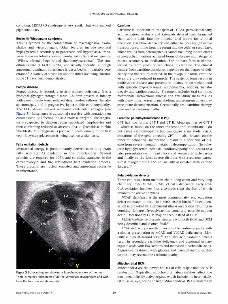
Pompe diseasePompe disease is secondary to acid maltase deficiency. It is a lysosmal glycogen storage disease. Children present in infancy with poor muscle tone, reduced deep tendon reflexes, hepato-splenomegaly and a progressive hypertrophic cardiomyopathy. The ECG shows marked increased ventricular voltages (see Figure 3). Inheritance is autosomal recessive with mutations on chromosome 17 affecting the acid maltase enzyme. The diagno-sis is suspected by demonstrating vacuolated lymphocytes and then confirming reduced or absent alpha1,4 glucosidase in skin fibroblasts. The prognosis is poor with death usually in the first year. Enzyme replacement is being used on a trial basis.
Fatty oxidation defectsMyocardial energy is predominantly derived from long chain fatty acid (LCFA) oxidation in the mitochondria. Several proteins are required for LCFA and carnitine transport in the cardiomyocyte and the subsequent beta oxidation process. These proteins are nuclear encoded and autosomal recessive in inheritance.
Figure 3 echocardiogram showing a four-chamber view of the heart.
There is marked thickening of all the ventricular myocardium and with
time the function will deteriorate.
paediaTriCS and CHild HealTH 19:1 1
CarnitineCarnitine is important in transport of LCFAs, peroxisomal fatty acid oxidation products and ketoacids derived from branched chain amino acids into the mitochondrial matrix for terminal oxidation. Carnitine deficiency can either be primary (defective transport of carnitine from the serum into the cells) or secondary, which results from heterogeneous causes including inborn errors of metabolism, various acquired forms of disease and iatrogenic causes secondary to medication. The primary form is charac-terised by more profound reductions in carnitine. The clinical picture from carnitine deficiency depends on the degree of defi-ciency and the tissues affected. In the myopathic form, carnitine levels are only reduced in muscle. The systemic form results in multisystem disease and presents in infancy or early childhood with episodic hypoglycaemia, ammonaemia, acidosis, hepato-megaly and cardiomyopathy. Treatment includes oral carnitine, bicarbonate, intravenous glucose and anti-failure measures. As with many inborn errors of metabolism, undercurrent illness may precipitate decompensation. Occasionally oral carnitine therapy reverses the cardiomyopathy.
Carnitine palmitoyltransferase (CPT)CPT has two forms: CPT I and CT II. Abnormalities of CPT I – which is found on the inner mitochondrial membrane – do not cause cardiomyopathy but can cause a metabolic crisis. Mutations of the gene encoding CPT II – also located on the inner mitochondrial membrane – result in a spectrum of dis-ease from severe neonatal metabolic decompensation (hypoke-totic hypoglycaemia, acidosis, cardiomyopathy and death) to a later presentation with heart block and ventricular tachycardia and finally to the least severe disorder with recurrent parox-ysmal myoglobinuria and not usually associated with cardiac disease.10
Beta oxidation defectsThese can result from medium chain, long chain and very long chain acyl-CoA (MCAD, LCAD, VLCAD) deficiency. Fatty acyl-CoA oxidation involves four enzymatic steps the first of which involves the above enzymes.
MCAD deficiency is the most common fatty acid oxidation defect estimated to occur in 1:6000–10,000 births.10 Decompen-sation is provoked by intercurrent illness and fasting resulting in vomiting, lethargy, hypoglycaemia, coma and possible sudden death. Occasionally DCM may be seen instead of HCM.
VLCAD deficiency presents similarly with both HCM and DCM being described and is often fatal.11
LCAD deficiency – results in an infantile cardiomyopathy with a similar presentation to MCAD and VLCAD deficiencies. Mor-tality is high at around 50%.12 The fatty acid oxidation defects result in secondary carnitine deficiency and abnormal urinary organic acids with low ketones and increased dicarboxylic acids. Aggressive treatment with glucose and haemodynamic cardiac support may reverse the cardiomyopathy.
Mitochondrial HCMMitochondria are the power houses of cells responsible for ATP production. Typically, mitochondrial abnormalities affect the most metabolically active organs, which include the heart, skele-tal muscles, eye, brain and liver. Mitochondrial DNA is maternally
9 © 2008 published by elsevier ltd.

SympoSium: CardiovaSCular mediCine
inherited and encodes the vital respiratory chain enzymes. How-ever, nuclear encoded proteins are also imported into the mito-chondria so mitochondrial dysfunction can arise from either route. Each cell contains several mitochondria and each mito-chondrion contains several copies of the circular mitochondrial molecule, which in turn has multiple copies encoding ribosomal and transfer RNA. It is this combination that results in a mix of normal and mutant mitochondrial protein and enzyme systems the proportions of which determine the degree to which an organ is affected and the clinical phenotype.13 Cardiac disease is most commonly present with respiratory chain defects.14 Resulting cardiac disease includes HCM, DCM and a mixed hypertrophic-dilated phenotype. The latter should always raise the suspicion of a mitochondrial abnormality when present in an infant.15
In addition to standard treatment strategies, coenzyme Q, car-nitine and vitamins have been tried but typically do not alter the clinical course.15
The diagnosis of a mitochondrial problem should be based on the clinical picture in conjunction with a muscle biopsy show-ing red ragged muscle fibres and mitochondrial respiratory chain analysis of the biopsy tissue.
Autosomal dominant familial HCMThis is secondary to mutations encoding the sarcomeric proteins. Typically the HCM does not develop until the onset of the puber-tal growth spurt and, even then, when an individual has a known mutation, the HCM may only become evident in adult life.
Maternal diabetesMaternal diabetes is a cause of a self-limiting HCM reflecting foe-tal hyperinsulinism secondary to poor maternal glucose control. The myocardial hypertrophy will resolve with time.
Twin-to-twin transfusion syndrome (TTTS)This occurs in approximately 15% of monochorionic twin preg-nancies and results in ventricular hypertrophy of the recipient’s myocardium with the right ventricle being more affected than the left. Significant right ventricular outflow tract obstruction may develop in association with the ventricular hypertrophy. Treat-ment of TTTS remains controversial but in severe cases where laser therapy has been used to treat the placental arteriovenous anasto-moses, regression of the hypertrophy has been documented.16
DCM
In this condition the left ventricle is dilated with the right ven-tricle variably involved (see Figure 3). Typically, ventricular sys-tolic function is reduced and from this the symptoms and signs resulting from pulmonary venous congestion, systemic venous congestion and a low cardiac output state can be predicted. Investigations required are similar to those for HCM but with the addition of viral studies (culture, serology, immune fluorescence, polymerase chain reaction).
The following represent some of the more common causes of DCM.
Myocarditis or inflammatory cardiomyopathyMyocarditis or inflammatory cardiomyopathy may be responsible for up to 40% of DCMs. The most common aetiological agents
paediaTriCS and CHild HealTH 19:1 20
include adenoviruses and enteroviruses (Coxsackie group). Rarer causes include other infections including bacteria, parasitic such as Chagas disease and connective tissue disorders such as systemic lupus erythematosis.
Familial DCMThis occurs in up to 30% of cases. This is usually autosomal dominant but autosomal recessive, X-linked and mitochondrial inheritance are all possible.
Barth syndromeThis is an X-linked cardiac and skeletal myopathy arising from a mutation affecting the tafazzin protein family. Classically, there is a neutropenia and DCM, although left ventricular non-com-paction is noted in some series.7,15 Myocyte hypertrophy and fibrosis is present and mitochondrial abnormalities are noted on electron microscopy of the heart and skeletal muscle. Other findings include hypercholesterolaemia, lactic acidosis, hypogly-caemia and respiratory chain abnormalities. A cardiomyopathy and neutropenia in a male are diagnostic. Urine analysis shows 3-methylglutaconic aciduria. The gene has been mapped to Xp28. Interestingly mutations result in a spectrum of clinical disorders ranging from classic Barth syndrome to DCM to left ventricular non-compaction.15
X-linked DCMX-linked DCM includes those cardiomyopathies that are found in association with the muscular dystrophies, such as Duchenne and Becker. There is also an X-linked DCM that presents in male teenagers, which follows a rapidly progressive course and has been mapped to Xp21.
Vitamin D deficiencyVitamin D deficiency resulting in hyperparathyroidism and hypo-calcaemia is a cause of a severe DCM associated with significant mortality that occurs in breastfed infants of dark-skinned popu-lations where there is a combination of inadequate exposure to sunlight (usually a combination of clothing and northern climate) and inadequate dietary intake. The condition is curable with vita-min D supplementation but recovery of ventricular function on average takes 1 year.17
Rate-related DCM
These arise from any prolonged tachycardia whether supra-ventricular (more commonly) or ventricular in origin. Typi-cally, tachyarrhythmias are well tolerated in childhood in the absence of ischaemic coronary vascular disease. However, eventually the myocardium tires and the ventricles progres-sively dilate.
Treating and controlling the arrhythmia results in cure. An ECG and 24-hour tape should be performed in all patients with a newly diagnosed DCM.
Chemotherapy induced DCMChemotherapy induced DCM is a relatively frequent cause of cardiomyopathy in this patient group and usually related to the use of anthracyclines. Echocardiography is now a standard part of chemotherapy protocols using cardiotoxic agents. New
© 2008 published by elsevier ltd.

SympoSium: CardiovaSCular mediCine
onset effort intolerance should always raise the possibility of an anthracycline-induced cardiomyopathy as part of the differential diagnosis in this patient group (Figure 4).
RCM
This is the rarest form of cardiomyopathy in childhood represent-ing in around 3% of cases. RCM typically presents after the first year of life and later in childhood. Usually no cause is identifiable although a familial variety is recognised.
As implied in the name, the pathophysiology arises from an inability of the ventricular myocardium to relax giving the classic two-dimensional echocardiographic picture of small ventricles associated with large dilated atria known as the ‘ice-cream cone’ appearance (see Figure 5). Symptoms result from systemic and pulmonary venous congestion and a low cardiac output. The dif-ferential diagnosis includes a constrictive pericarditis. This form of cardiomyopathy requires careful ongoing monitoring for the development of pulmonary hypertension. Once this develops cardiac transplantation is recommended to avoid irreversible pulmonary vascular damage.
ARVC
ARVC is defined by the presence of right ventricular dysfunc-tion (regional or global) in the presence of histological evidence of fibro-fatty replacement of the right ventricular myocardium and/ECG abnormalities in accordance with published criteria.4 The estimated prevalence is 1:5000. In parts of Europe, ARVC is a well-recognised cause of sudden death in young people.
paediaTriCS and CHild HealTH 19:1 21
Magnetic resonance imaging (MRI) is now commonly used to demonstrate fibro-fatty change in the myocardium and is excel-lent for assessing ventricular function. Autosomal recessive and dominant mutations in genes encoding cell adhesion have been shown to be responsible.
Unclassified cardiomyopathies
The entity of ventricular non-compaction is included in this group. The normal left ventricular endocardium is usually smooth with minimal trabeculation in contrast to the right ventricle, which is heavily trabeculated. Ventricular non-com-paction is the term given to the echocardiographic finding of abnormal trabeculation of the ventricles with deep sinusoids and recesses typically within the left ventricle (Figure 6). It is felt to represent an embryological arrest and can occur in isola-tion or with structural congenital heart disease. The resultant pathophysiology is variable but usually results from a combina-tion of dilated and hypertrophic physiology. Echocardiographic diagnosis is made when the endocardial layer is twice the thick-ness of the outer epicardium. Barth syndrome can result in this appearance.
Investigations
These should be divided into those that confirm a diagnosis of a cardiomyopathy and those that help identify the underlying aeti-ology. Given the huge number of possible causes a large number of investigations will often need to be taken and commonly a specific aetiological diagnosis is not made.
dilated cardiomyopathy. a a four-chamber echocardiogram showing a hugely dilated left ventricle that is squashing the right ventricle. b is a
short axis view. The myocardium is thinned.
Figure 4
© 2008 published by elsevier ltd.

SympoSium: CardiovaSCular mediCine
Establishing a diagnosis • Chest x-ray • ECG • Echocardiography.
Figure 6 echocardiogram showing the left heart structures and left
ventricular non-compaction. The apex of the left ventricle is normally
smooth but in this case heavily trabeculated.
Figure 5 Four-chamber echocardiogram showing bi-atrial enlargement
also known as the ‘ice-cream cone’ appearance.
paediaTriCS and CHild HealTH 19:1 2
MRI will probably play a larger role in the future. Endomyo-cardial biopsy may be useful but is not commonly performed in the UK.
Establishing aetiologySuggested investigations are listed in Table 2. This list is not comprehensive but serves as a starting point.
All patients should be seen by a paediatric cardiologist but it is helpful and saves time for the initial investigations to be made as several have a significant delay in turnaround time.
Management
Dilated cardiomyopathyDiuretics, angiotensin-converting enzyme inhibitors and beta blockade with a non-selective action are the mainstay of ther-apy. Digoxin has a role with its positive inotropic and negatively chronotropic effect.
Hypertrophic cardiomyopathySelective beta blockade is the mainstay of therapy in young chil-dren. Infants often may need diuretics in the early stages when there may be a difficult balance between the need for an adequate preload to fill stiff hypertrophied ventricles and pulmonary and systemic venous congestion. Anti-arrhythmics and anti-angina therapy may be required.
In older patients, especially where ventricular arrhythmias have been documented and there is a risk of sudden death, auto-matic implantable defibrillators are used.
Surgical myomectomy, in particular where significant left ventricular outflow tract obstruction has developed, as well as transcatheter muscle ablation procedures are recognised forms of therapy in specialised units.
Restrictive cardiomyopathyThe standard strategy is careful observation with ongoing esti-mates of pulmonary artery pressure including cardiac catheteri-sation and early listing for transplantation if pulmonary artery pressures are elevated.
Unclassified cardiomyopathyAs for DCM and HCM.
Inborn error of metabolism - IEMWhen an IEM is suspected or diagnosed on initial screening, discussion with and referral to a specialist paediatric metabolic unit is appropriate. This allows for the consideration of special-ist diets (restrictions and supplementations), ‘chaperone’ therapy and enzyme replacement strategies either by direct replacement or using bone marrow transplantation.
Cardiac transplantationIn the event of medical therapy failing, the final option for treat-ment is cardiac transplantation with or without a mechanical bridge. It is important to remember that cardiac transplantation is not a cure and comes with well-known complications aris-ing from chronic immunosuppressive therapy. However, when significantly symptomatic on maximal medical therapy, the improved quality of life offered by cardiac transplantation can
2 © 2008 published by elsevier ltd.

SympoSium: CardiovaSCular mediCine
Suggested Investigations
Investigation Indications & comments
echocardiogram For ventricular morphology and to exclude structural heart disease especially
anomalous left coronary in dCm
Consider screening of family members
electrocardiogram voltage criteria for chamber enlargement
arrhythmia, giant complexes & short pr interval in pompe
any degree of conduction disturbance possible in iem. low voltages and ST
segment changes in acute myocarditis
24 hour tape arrhythmia (aetiology and secondary complication of cardiomyopathy)
Chest x-ray Heart size
Full blood count neutropaenia with Barth syndrome & organic acidaemias. megaloblastic
anaemia in B1 & B12 deficiency
erythrocyte sedimentation rate & C reactive protein Consider if inflammatory disorder suspected
vacuolated lymphocytes lysosomal storage disorder (mucoploysaccharidoses, mucolipidoses, pompe)
only if patient <2 year of age
urea, creatinine, na, K Baseline pre treatment
Glucose infant of diabetic mother. Hypoglycaemia common in fatty acid oxidation
defects, glycogen storage disorders & mitochondrial cytopathies
Liver function testsinborn error of metabolism and baseline pre treatment
Brain natriuretic peptide more useful for monitoring progress
Creatinine kinase elevated in muscular dystrophies and with myocardial insult
lactate & ammonia raised in mitochondrial disorders, fat oxidation defects, organic acidaemias.
also raised in low cardiac output states
Serum & ionised calcium vitamin d deficiency
parathyroid hormone Breast fed infants in dark skinned families if Ca low
vitamin d levels do only if Ca low and pTH raised, breast-fed infants in dark skinned mothers
micronutrients eg selenium very rare, only if in area of endemic deficiency or patient on parenteral
nutrition or severely malnourished
Thyroid function tests Free T4, thyroid stimulating hormone
Thiamine if megaloblastic anaemia
Carnitine very low in carnitine transporter defect. may be secondarily low in multiple
other iem
acyl carnitine profile (Guthrie card) abnormal in fat oxidation disorders
Transferrin electrophoresis abnormal in congenital disorders of glycosylation
plasma amino acids inborn error of metabolism
antinuclear antibodies (ana) Screen for connective tissue disorders especially systemic lupus erythematosis
anti dna antibodies
rheumatoid factor
viral studies including igG, igm, pCr, npa for rapid
immunofluorescence (respiratory viruses)
liase with local virology laboratory – for enteroviruses including coxsackie
group, adenovirus, parvovirus, echovirus
Consider vZv, eBv
Serum for acute and convalescent titres
urine glycosaminoglycans raised in mucoploysacchardoses
urine oligosaccharides mucolipidoses
urine organic acids abnormal profile in organic acidaemias & mitochondrial disorders. dicarboxylic
aciduria in fat oxidation defects. methylglutaconate raised in Barth syndrome
Second line investigationsSkeletal muscle biopsy, endomyocardial biopsy,
mitochondrial respiratory chain analysis, skin fibroblasts
These investigations are dictated by the clinical scenario and used for
histological diagnosis as well as genetic and enzyme analysis
The above table is by no means comprehensive and serves only as a guide. acknowledgement: dr m Cleary for her guidance on metabolic investigations.dCm, dilated cardiomyopathy; pCr, polymerase chain reaction; npa, nasopharyngeal aspirate; vZv, varicella-zoster virus; eBv, ebstein-Barr virus.
Table 2
paediaTriCS and CHild HealTH 19:1 23 © 2008 published by elsevier ltd.

SympoSium: CardiovaSCular mediCine
be dramatic with increasing graft longevity and a current mean patient survival of 15 years. ◆
REFERENCES
1 Boucek mm, Faro a, novick rJ, Bennett le, Keck Bm, Hosenpud Jd.
The registry of the international Society for Heart and lung
Transplantation: Fourth official pediatric report – 2000.
J Heart Lung Transplant 2001; 20: 39–52.
2 richardson p, mcKenna W, Briston m, et al. report of the 1995
World Health organization/international Society and Federation
of Cardiology Task Force on the definition and Classification of
Cardiomyopathies. Circulation 1996; 93: 841–842.
3 maron BJ, Towbin Ja, Thiene G, et al. Contemporary definitions
and classification of the cardiomyopathies: an american Heart
association Scientific Statement from the Council on Clinical
Cardiology, Heart Failure and Transplantation Committee; Quality
of Care and outcomes research and Functional Genomics and
Translational Biology interdisciplinary Working Groups; and Council
on epidemiology and prevention. Circulation 2006; 113: 1807–1816.
4 elliott p, andersson B, arbustini e, et al. Classification of the
cardiomyopathies: a position statement from the european Society
of Cardiology Working Group on myocardial and pericardial
diseases. Eur Heart J 2008; 29: 270–276.
5 andrews re, Fenton mJ, ridout da, Burch m. new-onset heart failure
due to heart muscle disease in childhood: a prospective study in
the united kingdom and ireland. Circulation 2008; 117: 79–84.
6 lipshultz Se, Sleeper la, Towbin Ja, et al. The incidence of pediatric
cardiomyopathy in two regions of the united States. N Engl J Med
2003; 348: 1647–1655.
7 nugent aW, daubeney pe, Chondros p, et al. The epidemiology of
childhood cardiomyopathy in australia. N Engl J Med 2003; 348:
1639–1646.
8 allanson Je. noonan syndrome. J Med Genet 1987; 24: 9–13.
9 Best lG, Hoekstra re. Wiedemann-Beckwith syndrome: autosomal-
dominant inheritance in a family. Am J Med Genet 1981; 9: 291–299.
10 demaugre F, Bonnefont Jp, Colonna m, Cepanec C, leroux Jp,
Saudubray Jm. infantile form of carnitine palmitoyltransferase ii
deficiency with hepatomuscular symptoms and sudden death.
paediaTriCS and CHild HealTH 19:1 24
physiopathological approach to carnitine palmitoyltransferase ii
deficiencies. J Clin Invest 1991; 87: 859–864.
11 Strauss aW, powell CK, Hale de, et al. molecular basis of human
mitochondrial very-long-chain acyl-Coa dehydrogenase deficiency
causing cardiomyopathy and sudden death in childhood. Proc Natl
Acad Sci U S A 1995; 92: 10496–10500.
12 Wanders rJ, duran m, ijlst l, et al. Sudden infant death and long-
chain 3-hydroxyacyl-Coa dehydrogenase. Lancet 1989; 2: 52–53.
13 lightowlers rn, Chinnery pF, Turnbull dm, Howell n. mammalian
mitochondrial genetics: heredity, heteroplasmy and disease. Trends
Genet 1997; 13: 450–455.
14 mariotti C, Tiranti v, Carrara F, dallapiccola B, didonato S, Zeviani m.
defective respiratory capacity and mitochondrial protein synthesis
in transformant cybrids harboring the trna(leu(uur)) mutation
associated with maternally inherited myopathy and cardiomyopathy.
J Clin Invest 1994; 93: 1102–1107.
15 Towbin Ja, lipshultz Se. Genetics of neonatal cardiomyopathy. Curr
Opin Cardiol 1999; 14: 250–262.
16 manning n. The influence of twinning on cardiac development. Early
Hum Dev 2008; 84: 173–179.
17 maiya S, Sullivan i, allgrove J, et al. Hypocalcaemia and vitamin d
deficiency: an important, but preventable, cause of life-threatening
infant heart failure. Heart 2008; 94: 581–584.
Practice points
• Cardiomyopathies are a heterogeneous group of disorders
arising from both genetic and non-genetic causes
• ventricular morphological classification provides the best idea
as to how to manage individual patients
• Cardiomyopathies remain an important cause of death,
sudden death and cardiac transplantation in both children
and adults
• management is best carried out by paediatric cardiologists
with access to genetic and metabolic teams but shared care
remains an important and necessary resource
© 2008 published by elsevier ltd.