cardiogenic shock: mechanical management · cardiogenic shock: mechanical management kathleen tong,...
TRANSCRIPT

6/14/2016
1
Cardiogenic Shock: Mechanical Management
Kathleen Tong, MDAssistant Clinical Professor
Director, Heart Failure ProgramMedical Director, Ventricular Assist Device Program
UC Davis Medical Center
Disclosures
• Speakers’ Bureau
– Novartis
• May discuss investigational products or off-label use
Objectives
• Review etiology and pathophysiology of shock; specifically differentiating cardiogenic shock from other types
• Describe mechanical treatment options for cardiogenic shock
• Cases of cardiogenic shock treated with mechanical support

6/14/2016
2
What is Shock?
• Circulatory shock - a condition where there is low perfusion to the tissues causing organ damage
– Definition does not specify the cause of low perfusion
Shock Pathophysiology
Something
Low Perfusion
• Metabolic acidosis• Endothelial dysfunction• Cellular injury• Cell death
Shock Pathophysiology
Something
Low Perfusion
• Metabolic acidosis• Endothelial dysfunction• Cellular injury• Cell death
PressorsInotropes
Fluids
BicarbonateCalcium

6/14/2016
3
Shock Pathophysiology
Something
Low Perfusion
• Metabolic acidosis• Endothelial dysfunction• Cellular injury• Cell death
PressorsInotropes
Fluids
BicarbonateCalcium
Types of Shock
• Hypovolemic
• Distributive
– Septic
– Neurogenic
• Cardiogenic
• Other
– Acute RV failure, tamponade
Common Features
• Acute presentation– Ill-appearing patient
– Rapid deterioration
• Hypotension is common though the absence of hypotension should not rule out shock
• End organ damage (rising creatinine, elevated liver enzymes, altered mentation)
• Poor perfusion anaerobic metabolism lactic acidosis

6/14/2016
4
Distinguishing Types of Shock
Shock Patient Identified
Assess Volume Status-history-physical exam-labs (i.e CBC, chemistry, BNP-chest X-Ray
-Bleeding, decreased pointake, diarrhea, vomiting-dry mucous membranes-normal BNP-normal CXR
Treatment-Correct the underlying reason for volume loss -IV fluids, blood product transfusion
Distinguishing Types of Shock
Identify Shock Patient
Pure Hypovolemic Shock
Other Shock
Distributive Shock-Triggered by infection, CNS injury, endocrine derangement, anaphylaxis-High cardiac output-Excess of inflammatory agents (cytokines)
Cardiogenic Shock-Commonly a history of heart failure or cardiac disease-Severe decompensated heart failure
Distinguishing Types of Shock
Identify Shock Patient
Pure Hypovolemic Shock
Other Shock
Distributive Shock-Triggered by infection, CNS injury, endocrinologicderangement, anaphylaxis-High cardiac output-Excess of inflammatory agents (cytokines)
Cardiogenic Shock-Commonly a history of heart failure or cardiac disease-Severe decompensated heart failure – pump failure

6/14/2016
5
Septic vs. Cardiogenic Shock
Septic History/Physical• Infectious symptoms
– Site specific infection symptoms (abscess, cellulitis, urine symptoms, productive cough)
– “Pink”, warm to touch, neck veins flat or not elevated
– SIRS (systemic inflammatory response syndrome)criteria• T >38°C (100.4°F) or <36°C
(96.8°F) • HR >90• RR > 20 breaths/minute• Leukocytosis – WBC
>12K/microliter or bands present
Cardiogenic History/Physical• Congestive symptoms
– Orthopnea, edema, weight gain, increased abdominal girth
• Hypotension, tachycardia, narrow pulse pressure
• Elevated jugular venous pulse, hepatojugular reflux
• Cool to touch, mottled extremities, poor cap refill
• Thready pulses• S3• Crackles/dullness at lung bases
Septic vs. Cardiogenic Shock
Septic Lab/Studies
• Leukocytosis
• Elevated procalcitonin
• Positive cultures
• Elevated AST/ALT
• Normal BNP
• CXR normal or ARDS
Cardiogenic Lab/Studies
• Elevated BNP
• Mildly elevated troponin
• Hyponatremia
• CXR with pulmonary congestion, pleural effusions
Echocardiography
• Echo with hyperdynamic findings
• Echo of HFrEF

6/14/2016
6
Evolution of Cardiogenic Shock
Renin-Angiotensin System ↑ ADH/Vasopressin ↑ Aldosterone ↑
Sympathetic nervous system ↑Contractility ↑
Heart Rate ↑
Systemic Blood Pressure ↑
INSULT
Cardiac Output ↓BP ↓
Evolution of Cardiogenic Shock
Ventricular Mass↑•Myocyte hypertrophy•Dilation•Increased wall stress
Ventricular End Diastolic Volume↑
Renin-Angiotensin System ↑ ADH/Vasopressin ↑ Aldosterone ↑
Sympathetic nervous system ↑Contractility ↑
Heart Rate ↑
Increase in intracardiacpressures
↑ SVR Cardiac Output ↓+
+

6/14/2016
7
Diagnostic Pitfalls
• Patients do not always “read the book”• If hypovolemic shock is not improving despite
adequate volume resuscitation, reconsider assessment• SIRS criteria are not specific
– Cardiogenic shock patients frequently trigger the SIRS criteria protocol in my institution
• Patients can have more than one kind of shock– Infection may trigger cardiogenic shock in a stable heart
failure patient– Advanced cardiogenic shock can look like distributive
shock
• Physical exam can be misleading
Pulmonary Artery CatheterHemodynamics: Pressures measurements
-Right atrial -Right ventricular-Pulmonary artery-Pulmonary capillary wedge
Mixed venous blood sampling-Fick cardiac output (CO)
Temperature sensor-Thermodilution CO
Derived Calculations:
Systemic Vascular Resistance (SVR)Pulmonary Vascular Resistance (PVR)Transpulmonary gradient (TPG)
Measurements Related to Shock
• Volume assessment– Right atrial pressure (RAP)
– Pulmonary capillary wedge pressure (PCWP)
• Cardiac output/Cardiac Index
• Vasoconstriction or vasodilation– Systemic blood pressure, mean arterial pressure
(MAP)
– Systemic vascular resistance (SVR)• SVR = (MAP – RAP)/CO

6/14/2016
8
Hemodynamics of Shock
RA PCWP Cardiac Index SVR
Normal Range (2-6) (6-12) (2.5-4) (800-1200)
Hypovolemic Low Low Normal to high
Normal to high
Distributive Low to normal
Low to normal
High Low
Cardiogenic(left heart)
High High Low High
Cardiogenic(isolated right heart)
High Low to normal
Low Normal to high
Approach to Cardiogenic Shock
• Assess and confirm clinical picture
• History and physical congruent with typical cardiogenic shock Initiate treatment
• Central line and arterial line
• Foley catheter to monitor urine output
• History and physical equivocal Consider right heart catheterization
– If low CI, consider retaining the catheter
Treat the Hemodynamics
• Low cardiac output IV Inotropes
– Dobutamine, Milrinone, lower doses of dopamine
• High SVR Vasodilators
– Nitroglycerin, nitroprusside, hydralazine, ACE inhibitors/angiotensin receptor blockers
• High filling pressures Diuretics, mechanical volume removal
6/14/2016
9
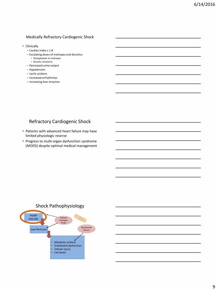
Medically Refractory Cardiogenic Shock
• Clinically– Cardiac Index ≤ 1.8
– Escalating doses of inotropes and diuretics• Tachyphylaxis to inotropes
• Diuretic resistance
– Decreased urine output
– Hypotension
– Lactic acidosis
– Increased arrhythmias
– Increasing liver enzymes
Refractory Cardiogenic Shock
• Patients with advanced heart failure may have limited physiologic reserve
• Progress to multi-organ dysfunction syndrome (MODS) despite optimal medical management
Shock Pathophysiology
PUMP FAILURE
Low Perfusion
• Metabolic acidosis• Endothelial dysfunction• Cellular injury• Cell death
PressorsInotropes
Fluids
BicarbonateCalcium

6/14/2016
10
Helping the Pump
• Many devices available and being developed to augment or replace circulation
– Percutaneous devices
– External pumps
• Peripheral cannulation
• Central cannulation
– Durable implantable pumps
• Left ventricular assist devices
• Total artificial heart
Treatments for acute
shock
Treatments for
relatively stable
patients
Intra-Aortic Balloon Pump (IABP)
• Gated to the cardiac cycle
• Inflates in diastole
• Deflates in systole
– Lowers afterload
IABP
• Easily placed in a cardiac catheterization lab– Low profile – 8 F sheath
• Commonly used for heart failure after acute MI• Can be used for cardiogenic shock without new
ischemic injury– Very modest augmentation of CO– Lowers SVR– Particularly useful if severe MR
• Do not use if significant aortic insufficiency• Tachyarrhythmias limit effect
6/14/2016
11
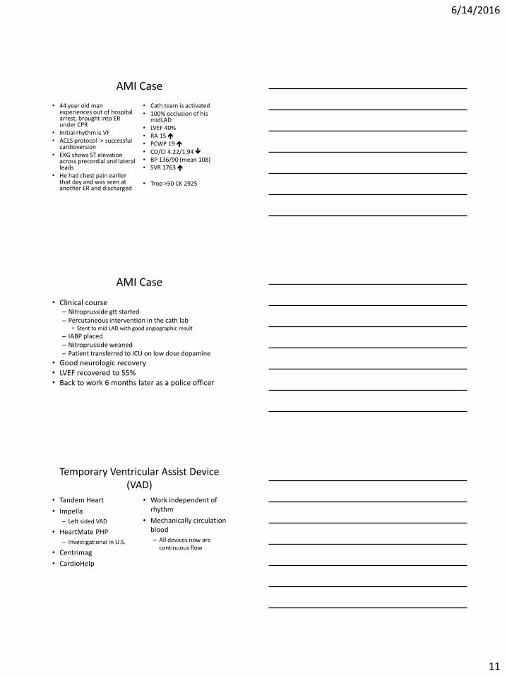
AMI Case
• 44 year old man experiences out of hospital arrest, brought into ER under CPR
• Initial rhythm is VF• ACLS protocol -> successful
cardioversion• EKG shows ST elevation
across precordial and lateral leads
• He had chest pain earlier that day and was seen at another ER and discharged
• Cath team is activated• 100% occlusion of his
midLAD• LVEF 40%• RA 15 • PCWP 19 • CO/CI 4.22/1.94 • BP 136/90 (mean 108)• SVR 1763
• Trop >50 CK 2925
AMI Case
• Clinical course– Nitroprusside gtt started– Percutaneous intervention in the cath lab
• Stent to mid LAD with good angiographic result
– IABP placed– Nitroprusside weaned– Patient transferred to ICU on low dose dopamine
• Good neurologic recovery• LVEF recovered to 55%• Back to work 6 months later as a police officer
Temporary Ventricular Assist Device (VAD)
• Tandem Heart
• Impella
– Left sided VAD
• HeartMate PHP
– Investigational in U.S.
• Centrimag
• CardioHelp
• Work independent of rhythm
• Mechanically circulation blood
– All devices now are continuous flow
6/14/2016
12
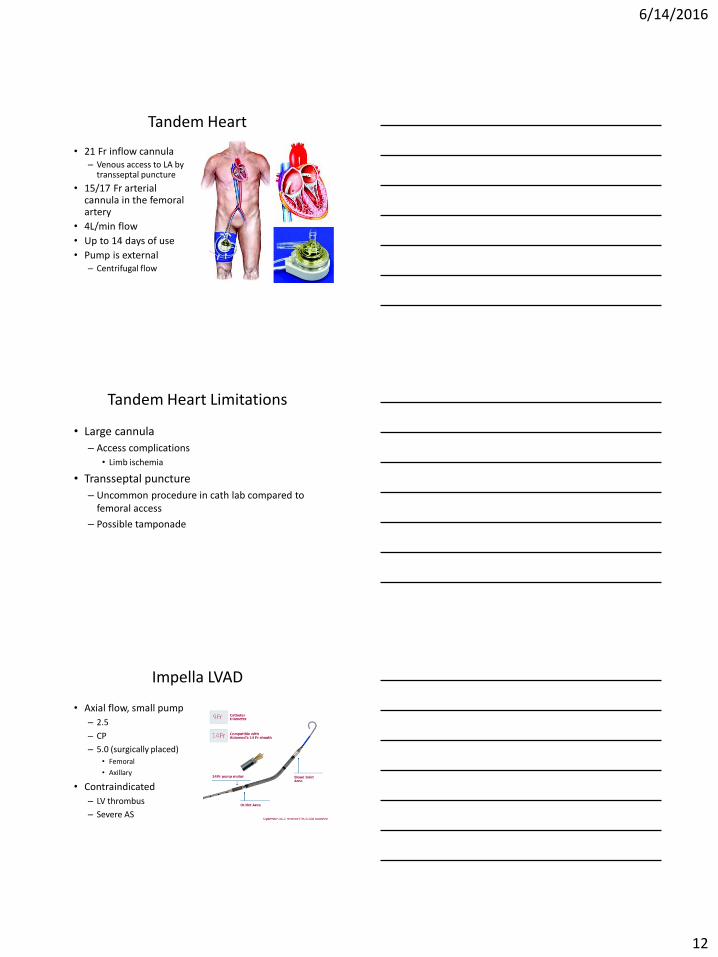
Tandem Heart
• 21 Fr inflow cannula– Venous access to LA by
transseptal puncture
• 15/17 Fr arterial cannula in the femoral artery
• 4L/min flow
• Up to 14 days of use
• Pump is external– Centrifugal flow
Tandem Heart Limitations
• Large cannula
– Access complications
• Limb ischemia
• Transseptal puncture
– Uncommon procedure in cath lab compared to femoral access
– Possible tamponade
Impella LVAD
• Axial flow, small pump
– 2.5
– CP
– 5.0 (surgically placed)• Femoral
• Axillary
• Contraindicated
– LV thrombus
– Severe AS
6/14/2016
13
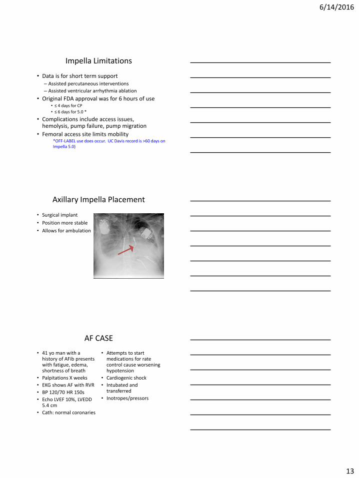
Impella Limitations
• Data is for short term support– Assisted percutaneous interventions
– Assisted ventricular arrhythmia ablation
• Original FDA approval was for 6 hours of use• ≤ 4 days for CP
• ≤ 6 days for 5.0 *
• Complications include access issues, hemolysis, pump failure, pump migration
• Femoral access site limits mobility*OFF-LABEL use does occur. UC Davis record is >60 days on Impella 5.0)
Axillary Impella Placement
• Surgical implant
• Position more stable
• Allows for ambulation
AF CASE
• 41 yo man with a history of AFib presents with fatigue, edema, shortness of breath
• Palpitations X weeks
• EKG shows AF with RVR
• BP 120/70 HR 150s
• Echo LVEF 10%, LVEDD 5.4 cm
• Cath: normal coronaries
• Attempts to start medications for rate control cause worsening hypotension
• Cardiogenic shock
• Intubated and transferred
• Inotropes/pressors

6/14/2016
14
AF CASE
• Right Heart Catheterization (Dopamine, Norepinephrine)– RA 27
– PCWP 41
– Systemic BP 117/88 (MAP 74)
– CO/CI 4.13/1.8
– SVR 910 dynes
• Diagnosis: Cardiogenic shock due to suspected tachycardia mediated cardiomyopathy
AF Case
• Clinical Course
– Impella CP placed via femoral artery
– Pressors/inotropes weaned
– Attempts at cardioversion were unsuccessful
– Impella removed
– AV node ablation
– Biventricular pacemaker implanted
– Repeat echocardiogram showed LVEF improve to 35%
– Patient discharged
Impella RP
• Similar technology to Impella LVAD
• Placed via venous access
• Pumps blood from RV to PA
6/14/2016
15
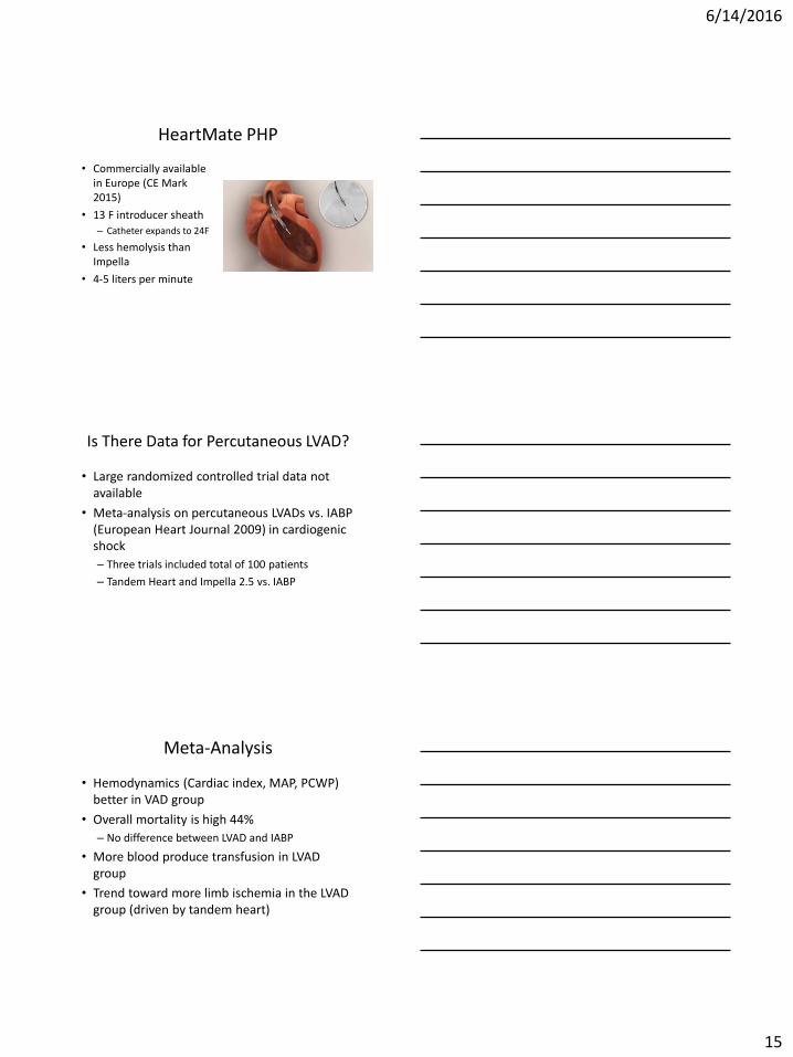
HeartMate PHP
• Commercially available in Europe (CE Mark 2015)
• 13 F introducer sheath
– Catheter expands to 24F
• Less hemolysis than Impella
• 4-5 liters per minute
Is There Data for Percutaneous LVAD?
• Large randomized controlled trial data not available
• Meta-analysis on percutaneous LVADs vs. IABP (European Heart Journal 2009) in cardiogenic shock
– Three trials included total of 100 patients
– Tandem Heart and Impella 2.5 vs. IABP
Meta-Analysis
• Hemodynamics (Cardiac index, MAP, PCWP) better in VAD group
• Overall mortality is high 44%
– No difference between LVAD and IABP
• More blood produce transfusion in LVAD group
• Trend toward more limb ischemia in the LVAD group (driven by tandem heart)

6/14/2016
16
VA ECMO - CARDIOHELP
• Small pump and oxygenator
– Up to 7L/flow
• Peripheral cannulation
– Large bore cannula in vein and artery
• Provides biventricular support
• Portable
VA ECMO CASE
• 35 yo man with known non-ischemic dilated cardiomyopathy– Leaves hospital where he
was was being supported on inotropes
– Flies to Sacramento and presents to ER in severe heart failure
• Echo LVEF 10% LVEDD 8.6 cm, RV dilated and hypokinetic
• HR 120 BP 112/100
• Edematous, cool to touch
• Inotrope started
• PEA arrest while central line is being placed
• AST 4114
• Creatinine 1.4->2.1
• Lactic acid 5.8
VA ECMO CASE
• Code blue called• Surgical consult
– VA ECMO placed
• Hemodynamics stabilized• Dialysis to treat acute tubular necrosis• Advanced heart failure consult team deems that
he is not a psychosocial candidate for advanced heart failure therapies (active substance abuse, no family support)
• Care withdrawn after 7 days
6/14/2016
17
Data for VA ECMO in Cardiogenic Shock
• Series 1: Post-operative cardiogenic shock– 60% weaned from ECMO
– 23% discharged to home
• Series 2: All cardiogenic shock– 40% discharged to home
• Series 3: All cardiogenic shock, single center– 50% discharged to home
• Series 4: Refractory cardiac arrest, single center– 25% discharged home
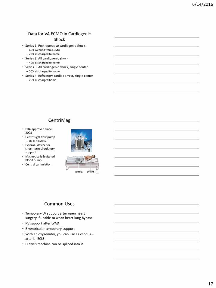
CentriMag
• FDA approved since 2008
• Centrifugal flow pump– Up to 10L/flow
• External device for short-term circulatory support
• Magnetically levitated blood pump
• Central cannulation
Common Uses
• Temporary LV support after open heart surgery if unable to wean heart-lung bypass
• RV support after LVAD
• Biventricular temporary support
• With an oxygenator, you can use as venous –arterial ECLS
• Dialysis machine can be spliced into it
6/14/2016
18
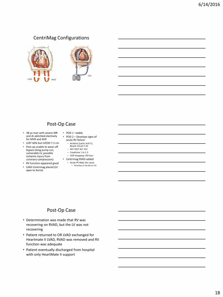
CentriMag Configurations
Post-Op Case
• 38 yo man with severe MR and AI admitted electively for MVR and AVR
• LVEF 60% but LVEDD 7.2 cm
• Post-op unable to wean off bypass (long pump run, vulnerable LV, possible ischemic injury from coronary compression)
• RV function appeared good
• LVAD Centrimag placed (LV apex to Aorta)
• POD 1 –stable
• POD 2 – Develops signs of acute RV failure– Acidosis (Lactic acid 11,
Bicarb 15) pH 7.23
– AST 2027 ALT 214
– Creatinine 1 to 1.3
– UOP dropping <30 hour
• Centrimag RVAD added– Acute PE likely the cause
• Thrombus in the RA on TEE
Post-Op Case
• Determination was made that RV was recovering on RVAD, but the LV was not recovering
• Patient returned to OR LVAD exchanged for Heartmate II LVAD, RVAD was removed and RV function was adequate
• Patient eventually discharged from hospital with only HeartMate II support

6/14/2016
19
What to use when?
High Risk PCI
High RiskVT ablat.
RV Failureonly
LV Failureonly
BiVFailure
Post-opCT
IABP ✔ ✔
ImpellaLVAD
✔ ✔ ✔
Tandem LVAD
✔ ✔
VA ECMO ✔ ✔ ✔ ✔
CentriMag ✔ ✔ ✔ ✔
Principles of Patient Selection
1. Confirm that it is cardiogenic shock– Low cardiac output– Complex patient – try to elucidate which organ derangements
are from heart failure and which are from existing co-morbidities that will not be corrected from improved cardiac output
2. Decide which ventricles need support (LV or RV or both?)3. Discuss your exit strategy
– Temporary support is temporary– Where do you go next? Recovery, durable VAD, transplant
4. Counsel family on the timeline– What will happen if recovery doesn’t occur
Summary
• Shock is an acute condition with high morbidity and mortality if untreated
• Recognizing the underlying reasons for the shock is critical to synthesizing a treatmentplant
• Acute cardiogenic shock can be treated with medical therapy, but medically refractory cases can be treated with mechanical circulatory assist with improvement in mortality
• Mechanical circulatory assist devices are ideally implanted with cardiothoracic surgery and advanced heart failure cardiologist input