carcinogenesis bioassay of zearalenone (cas no
TRANSCRIPT

NATIONAL TOXICOLOGY PROGRAM Technical Report Series No. 235
CARClNOGENESlS BIOASSAY OF
ZEARALENONE (CAS NO. 17924-92-4)
IN F344/N RATS A N D B6C3F1 MICE (FEED STUDY)
U.S. DEPARTMENT OF HEALTH AND HUMAN SERVICES Public Health Service
National Institutes of Health

N A T I O N A L TOXICOLOGY P R O G R A M The National Toxicology Program (NTP), established in 1978, develops and evaluates scientific information about potentially toxic and hazardous chemicals. This knowledge can be used for protecting the health of the American people and for the primary prevention of chemically induced disease. By bringing together the relevant programs, staff, and resources from the U.S. Public Health Service, DHHS, the National Toxicology Program has centralized and strengthened activities relating to toxicology research, testing and test development/validation efforts, and the dissemi- nation of toxicological information to the public and scientific communi- ties and to the research and regulatory agencies. The NTP is comprised of four charter DHHS agencies: the National Cancer Institute, National Institutes of Health; the National Institute of Environmental Health Sciences, National Institutes of Health; the National Center for Toxicological Research, Food and Drug Administra- tion; and the National Institute for Occupational Safety and Health, Centers for Disease Control. In July 1981. the Carcinogenesis Bioassay Testing Program, NCI, was transferred to the NIEHS.

NTP TECHNICAL REPORT ON THE
CARCINOGENESIS BIOASSAY OF
ZEARALENONE
(CAS NO. 17924-92-4)
IN F344/N RATS AND B6C3F1 MICE (FEED STUDY)
NATIONAL TOXICOLOGY PROGRAM Box 12233
Research Triangle Park North Carolina 27709
and Bethesda, Maryland 20205
October 1982
NTP-81 -54 NIH Publication No. 83-1791
U.S. DEPARTMENT OF HEALTH AND HUMAN SERVICES Public Health Service
National Institutes of Health

NOTE T O THE READER
This is one in a series of experiments designed to determine whether selected chemicals produce cancer in animals. Chemicals selected for testing in the NTP carcinogenesis bioassay program are chosen primarily on the bases of human exposure, level of production, and chemical structure. Selection per se is not an indicator of a chemical’s carcinogenic potential. Negative results, in which the test animals d o not have a greater incidence of cancer than control animals, d o not necessarily mean that a test chemical is not a carcinogen, inasmuch as the experiments are conducted under a limited set of conditions. Positive results demonstrate that a test chemical is carcinogenic for animals under the conditions of the test and indicate that exposure to the chemical is a potential hazard to humans. The determination of the risk to humans from chemicals found to be carcinogenic in animals requires a wider analysis which extends beyond the purview of this study.
This study was initiated by the National Cancer Institute’s Carcinogenesis Testing Program, now part of the National Institute of Environmental Health Sciences, National Toxicology Program.
Comments and questions about the National Toxicology Program Technical Reports on Carcino-genesis Bioassays should be directed to the National Toxicology Program, located at Room A-306, Landow Building, Bethesda, M D 20205 (301-496-1 152) or at Research Triangle Park, North Carolina 27709 (919-541-3991).
Although every effort is made to prepare the Technical Reports as accurately as possible, mistakes may occur. Readers are requested to communicate any mistakes to the Deputy Director, NTP (P.O. Box 12233, Research Triangle Park, NC 27709), so that corrective action may be taken. Further, anyone who is aware of related ongoing or published studies not mentioned in this report is encouraged to make this information known to the NTP.
These NTP Technical Reports are available for sale from the National Technical Information Ser-vice, U.S. Department of Commerce, 5285 Port Royal Road, Springfield, VA 22161 (702-487-4650).
Single copies of this carcinogenesis bioassay technical report are available without charge (and while supplies last) from the NTP Public Information Office, National Toxicology Program, P.O. Box 12233, Research Triangle Park, NC 27709.
Zearalenone 2

TABLE OF CONTENTS
Abstract .......................................................................... 7 Contributors ...................................................................... 8 Reviewers ......................................................................... 10 Summary of Peer Review Comments .................................................. 1 1
I . Introduction ................................................................... 13 II . Materials and Methods .......................................................... 17
Chemical Analysis .............................................................. 18 Prechronic Studies .............................................................. 18
Single-Dose Study ............................................................ 18 Fourteen-Day Study .......................................................... 18 Thirteen-Week Study .......................................................... 18
Chronicstudy .................................................................. 19 Clinical Examinations and Pathology ............................................ 19 Data Recording and Statistical Methods ......................................... 20
I I I . Results ........................................................................ 23 Rats .......................................................................... 24
Prechronic Studies ............................................................ 24 Single-Dose Study .......................................................... 24 Fourteen-Day Study ........................................................ 24 Thirteen-Week Study ........................................................ 24
Chronic Study ............................................................... 26 Body Weights and Clinical Signs .............................................. 26 Zearalenone Intake ......................................................... 26 Survival. , ................................................................. 27 Pathology and Statistical Analyses of Results ................................... 28
Mice .......................................................................... 34 Prechronic Studies ............................................................ 34
Single-Dose Study .......................................................... 34 Fourteen-Day Study ........................................................ 34 Thirteen-Week Study ........................................................ 35
Chronic Study ............................................................... 37 Body Weights and Clinical Signs .............................................. 37 Zearalenone Intake ......................................................... 37 Survival. , ................................................................. 38 Pathology and Statistical Analyses of Results ................................... 39
IV . Discussion and Conclusions ..................................................... 47 Rat Study ................................................................... 48 Mouse Study ................................................................ 49
V . References.................................................................... 51
Table 1 Experimental Design and Materials and Methods . , ....................... 21
Table 2
Table 3
Table 4
Table 5
TABLES
Dosage. Survival. and Mean Body Weights of Rats Fed Diets Containing Zearalenone for 14 Days ..................................... 24 Dosage. Survival. Mean Body Weights. and Feed Consumption of Rats Fed Diets Containing Zearalenone for 13 Weeks ........................... 25 Incidence of Compound-Related .Histopathologic Effects in the 13-Week Study of Zearalenone in R a t s . , ......................................... 25
Incidences of Selected Nonneoplastic Lesions in Rats Administered Zearalenone in the Chronic Study ....................................... 28
3 Zearalenone

Table 6
Table 7
Table 8
Table 9
Table 10
Table 11 Table 12
Figure 1 Figure 2
Figure 3
Figure 4
Figure 5
Figure 6 Figure 7
Figure 8
Figure 9
Figure 10
Appendix A
Table AI
Table A2
Table A3
Table A4
Appendix B
Table B1
Table B2
Analysis of Primary Tumors in Male Rats ................................ 29
Analysis of Primary Tumors in Female Rats .............................. 32
Dosage. Survival. and Mean Body Weights of Mice Fed Diets Containing Zearalenone for 14 Days ..................................... 34
Dosage. Survival. Mean Body Weights. and Feed Consumption of Mice Fed Diets Containing Zearalenone for 13 Weeks ........................... 35
Incidence of Compound-Related Histopathologic Effects in the 13-Week Study of Zearalenone in Mice .................................. 36
Analysis of Primary Tumors in Male Mice . . .,............................ 40 Analysis of Primary Tumors in Female Mice .............................. 43
FIGURES
Growth Curves fur Rats Fed Diets Containing Zearalenone . . . . . . . . . . . . . . . . .26
Survival Cunes for Rats Fed Diets Containing Zearalenone . . . . . . . . . . . . . . . . . 27
Growth Curves for Mice Fed Diets Containing Zearalenone . . . . . . . . . . . . . . . . . 37
Survival Curves for Mice Fed Diets Containing Zearalenone . . . . . . . . . . . . . . . .38 Infrared Absorption Spectrum of Zearalenone (Lot 3-75; Batch 01) . . . . . . . . . . .129
Infrared Absorption Spectrum of Zearalenone (Lot 3-75; Batch 02) . . . . . . . . . . .130
Infrared Absorption Spectrum of Zearalenone (Lot 51079; Batch 03) . . . . . . . . .131
Nuclear Magnetic Resonance Spectrum of Zearalenone (Lot 3-75; Batch 01) . . .133
Nuclear Magnetic Resonance Spectrum of Zearalenone (Lot 3-75; Batch 02) . . .135
Nuclear Magnetic Resonance Spectrum of Zearalenone (Lot 5 1079; Batch 03) ............................................................ 136
APPENDIXES
Summary of the Incidence of Neoplasms in Rats Fed Diets Containing Zearalenone ................................................ 55
Summary of the Incidence of Neoplasms in Male Rats Fed Diets Containing Zearalenone ................................................ 56
Summary of the Incidence of Neoplasms in Female Rats Fed Diets Containing Zearalenone ................................................ 60
Individual Animal Tumor Pathology of Male Rats in the 2-Year Study ofzearalenone ................................................. 64
Individual Animal Tumor Pathology of Female Rats in the 2-Year Study ofzearalenone ................................................. 70
Summary of the Incidence of Neoplasms in Mice Fed Diets Containing Zearalenone ............................................ .. . . 77
Summary of the Incidence of Neoplasms in Male Mice Fed Diets Containing Zearalenone ................................................ 78
Summary of the Incidence of Neoplasms in Female Mice Fed Diets Containing Zearalenone ................................................ 82
Zearalenone 4

Table B3
Table B4
Appendix C
Table C l
Table C2
Appendix D
Table DI
Table D2
Appendix E Appendix F
Appendix G
Table GI Appendix H
Table H1
Table H2
Appendix I
Table J2
Appendix K Table K1
Table K2
Table K3
Table I 1
Table 12
Appendix J
Table J 1
Individual Animal Tumor Pathology of Male Mice in the 2-Year Study ofzearalenone ................................................. 86
Individual Animal Tumor Pathology of Female Mice in the 2-Year Study ofzearalenone ................................................. 92 Summary of the Incidence of Nonneoplastic Lesions in Rats Fed Diets Containing Zearalenone ................................................ 99 Summary of the Incidence of Nonneoplastic Lesions in Male Rats Fed Diets Containing Zearalenone ................................................ 100
Summary of the Incidence of Nonneoplastic Lesions in Female Rats Fed Diets Containing Zearalenone ................................................ 107
Summary of the Incidence of Nonneoplastic Lesions in Mice Fed Diets Containing Zearalenone ................................................115 Summary of the Incidence of Nonneoplastic Lesions in Male Mice Fed Diets Containing Zearalenone ................................................ 116
Summary of the Incidence of Nonneoplastic Lesions in Female Mice Fed Diets Containing Zearalenone ................................................ 120
Analysis of Zearalenone (Midwest Research Institute) ..................... i25
Analysis of Formulated Diets for Stability of Zearalenone (Midwest Research Institute) ........................................... 139
Analysis of Formulated Diets for Concentrations of Zearalenone (Southern Research Institute) .......................................... 141 Analysis of Formulated Diets ........................................... 144
Cumulative Mean Body Weight Change of Rats and Mice Fed Diets Containing Zearalenone ................................................ 145
Cumulative Mean Body Weight Change (Relative to Controls) of Rats Fed Diets Containing Zearalenone., ..................................... 146
Cumulative Mean Body Weight Changes (Relative to Controls) of Mice Fed Diets Containing Zearalenone ....................................... 146
Feed Consumption by Rats and Mice Receiving Zearalenone in the Chronic Study .................................................. 147 Feed Consumption by Rats Receiving Zearalenone in the Chronic Study . . . . . . 148
Feed Consumption by Mice Receiving Zearalenone in the Chronic Study . . . . . . 149
Compound Consumption by Rats and Mice Fed Diets Containing Zearalenone . . . . . . . . . . . . . . . . . . . . . . . . . . . . . . . . . . . . . . . . . . . . . . . . . . . . . . . . . . 151
Compound Consumption by Rats Fed Diets Containing Zearalenone . . . . . . . . . 152
Compound Consumption by Mice Fed Diets Containing Zearalenone . . . . . . . . . 152 Historical Incidence of Tumors in Control B6C3F1 Mice1 . . . . . . . . . . . . . . . . . .153
Historical Incidence of Liver Tumors in Untreated Control Female B6C3F1 Mice ........................................................ 154
Historical Incidence of Pituitary Tumors in Untreated Control Male B6C3F1 Mice ................................................... 154
Historical Incidence of Pituitary Tumors in Untreated Control Female B6C3F1 Mice ................................................. 155
5 Zearalenone

Zearalenone 6

CARCINOGENESIS BIOASSAY OF
ZEARALENONE
H 0
0
TRANS-ZEARALENONE CAS NO. 17924-92-4
C18H2205 Mol. wt. 318.36
ABSTRACT A carcinogenesis bioassay of zearalenone, an estrogenic mycotoxin, was conducted by feeding diets
containing 25 or 50 ppm zearalenone to groups of 50 F344/ N rats of each sex and 50 or 100 ppm to groups of 50 B6C3F1 mice of each sex for 103 weeks. Groups of50 rats and50 mice ofeach sex served as controls. Estimates based on food consumption data indicate that the low- and high-dose rats received daily doses of about 1 and 2 mg, respectively, of zearalenone per kg body weight. Low-dose and high-dose mice received estimated daily doses of about 7-10 and 14-20 mg, respectively, of zearalenone per kg body weight.
Survival of dosed and control rats of each sex was comparable. Mean body weight gains of dosed rats of each sex were lower than those of the corresponding controls; depression in mean body weight gain was dose related. Final body weights of dosed rats were<9% lower than those ofcontrol rats. The average daily feed consumption by dosed rats of each sex was 91%-102% that of the controls.
Inflammation of the prostate, testicular atrophy, and hepatocellular cytoplasmic vacuolization (male rats), and nephrosis (male and female rats) were compound-related. Retinopathy and cataracts occurred in low- and high-dose male rats and in low-dose female rats, and were associated with closeness to fluorescent light. N o compound-related, increased tumor incidences were observed in rats in the chronic study.
Survival of dosed and control mice of each sex was comparable. Mean body weight gains of high-dose male and low-dose female mice were lower than those of the controls. Terminal body weights of dosed mice were <8% below those of control mice. The average daily feed consumption by dosed mice of each sex was 97%-99% that of the controls.
Myelofibrosis in the bone marrow, uterine fibrosis, and cystic ducts in the mammary gland were related to administration of zearalenone in female mice. The incidence of hepatocellular adenomas in female mice was dose related (PS0.003),and the incidence of these tumors in high-dose female mice was significantly higher (P10.006) than those in the controls (control, 0/50; low-dose, 2/49; high- dose, 7/49). Pituitary adenomas occurred with statistically significant positive trends (P50.022 for males and P50.001for females). The incidences of these tumors in highdose mice were significantly increased relative to controls (PS0.032 for males: 0 /40 ,4 /45 ,6 /44; and P50.003for females: 3/46, 2/43, 13/42).
Under the conditions of this bioassay, zearalenone was not carcinogenic for F344/N rats of either sex. Zearalenone should be considered carcinogenic in B6C3F1 mice, as evidenced by the increased proportion of male and female mice with pituitary adenomas and by the increased proportion of female mice with hepatocellular adenomas.
7 Zearalenone

CONTRIBUTORS
The bioassay of zearalenone was conducted at Southern Research Institute under a subcontract to Tracor Jitco, Inc., the prime contractor for the Carcinogenesis Testing Program. The 13-week study was begun in June 1977 and completed in September 1977. The chronic study was begun in February 1978 and completed in February 1980.
Principal Contributors at Southern Research Institute 2000 Ninth Avenue South
Birmingham, Alabama 35255 (Conducted bioassay and evaluated tissues)
J . David Prejean, Ph.D. Ruby H. James, B.S. Principal Investigator Chemist
Daniel R. Farnell, D.V.M., Ph.D. Joan B. Belzer Pathologist Animal Care Supervisor
Herschel1 D. Giles, D.V.M., Ph.D. Pathologist
Principal Contributors at Tracor Jitco 1776 East Jefferson Street
Rockville, Maryland 20852 (Prepared Preliminary Summary Report)
Lorne A. Campbell, Ph.D. Marion S. Levy, M.A. Acting Director, Bioassay Technical Editor
Edward T. Cremmins, M.A. Stephen S. Olin, Ph.D. Technical Edi tor P r o g r a m Associate Director
Carolyn E. Dean, B.S. Michael A. Stedham, D.V.M. Production Editor Pathologist
Thomas P. Griffin, D.V.M. William D. Theriault, Ph.D. Laboratory Operations Coordinator Reports Manager
Abigail C. Jacobs, Ph.D. Joseph E. Tomaszewski, Ph.D. Bioscience Writer Chemist
James R. Joiner, Ph.D. John W. Warner, M.S. Statistician Statistician
Zearalenone 8

Principal Contributors at the National Toxicology Program National Institute of Environmental Health Sciences
Research Triangle Park North Carolina 27709
and Bethesda, Maryland 20205
(Evaluated experiment, interpreted the results, and reported the findings)
Dexter S. Goldman, Ph.D. Joseph K . Haseman, Ph.D. Chemical Manager James E. Huff, Ph.D.
Rajendra S. Chhabra, Ph.D. Charles W. Jameson, Ph.D. Michael P. Dieter, Ph.D. E.E. McConnell, D.V.M. J . Fielding Douglas, Ph.D. John A. Moore, D.V.M. Charles K . Grieshaber, Ph.D. Raymond W. Tennant, Ph.D. Larry G. Hart, Ph.D.
Quality assurance of slides and review of tumor diagnoses were conducted at Experimental Pathol- ogy Laboratories, P.O. Box 474, Herndon, VA 22070 by Larry J . Ackerman, D.V.M.
The pathology report from Southern Research Institute, the report from Experimental Pathology Laboratories, and selected slides were evaluated in November 1980 by the National Toxicology Program Pathology Working Group which consisted of:
E.E. McConnell, D.V.M. Michael A. Stedham, D.V.M. National Toxicology Program Tracor Jitco
Gerd Reznick, D.V.M., Ph.D.* Jerrold M. Ward, D.V.M., Ph.D.* National Toxicology Program National Toxicology Program
The chemicals used in this bioassay of zearalenone were analyzed by the Midwest Research Institute, 425 Volker Blvd., Kansas City, Missouri 641 10; reanalysis of the bulk chemical and analyses of dosed feed mixtures were performed by Southern Research Institute.
*Current Address: Laboratory of Comparative Carcinogenesis National Cancer Institute Frederick Cancer Research Center Frederick, Maryland 21701
9 Zearalenone

REVIEWERS
National Toxicology Program Board of Scientific Counselor’s Technical Reports Review Subcommittee
Margaret Hitchcock, Ph.D. (Chairperson) John B. Pierce Foundation Laboratory New Haven, Connecticut
Curtis Harper, Ph.D. Alice Whittemore, Ph.D.* Associate Professor of Pharmacology Stanford University School of Medicine University of North Carolina Palo Alto, California Chapel Hill, North Carolina
A d H o c Subcommittee Panel of Experts
Norman Breslow, Ph.D. Robert A. Scala, Ph.D. University of Washington Exxon Corporation Seattle, W as hington East Millstone, New Jersey
Robert M. Elashoff, Ph.D. Bernard Schwetz, Ph.D. (Principal Reviewer) University of California Toxicology Research Laboratory
at Los Angeles Dow Chemical U.S,A. Jonsson Comprehensive Cancer Center M idland, Michigan Los Angeles, California
Joseph Highland, Ph.D. James Swenberg, Ph.D. Environmental Defense Fund Chief of Pathology Washington, D.C. Chemical Industry Institute of Toxicology
Research Triangle Park, North Carolina
J . Michael Holland, Ph.D. (Principal Reviewer) Stan D. Vesselinovitch, Ph.D. Department of Biology Departments of Radiology and Pathology Oak Ridge National Laboratory University of Chicago Oak Ridge, Tennessee Chicago, Illinois
Frank Mirer, Ph.D. Mary Vore, Ph.D. International Union, University of Kentucky
United Auto Workers College of Medicine Detroit, Michigan Lexington, Kentucky
*Unable to attend December 16, 1981 meeting
Zearalenone 10

SUMMARY OF PEER REVIEW COMMENTS ON THE BIOASSAY OF ZEARALENONE
On December 16, 1981, this report underwent peer review by the National Toxicology Program Board of Scientific Counselors’ Technical Reports Review Subcommittee and associated Panel of Experts. The review meeting began at 9:OO a.m. in Conference Room A, Landow Building, 7910 Woodmont Avenue, Bethesda, Maryland.
Dr. Schwetz, a primary reviewer for the report on the bioassay of zearalenone, agreed with the conclusion that zearalenone was not carcinogenic for rats and suggested that the increase in male and female B6C3F1 mice with pituitary adenomas and in females with hepatocellular adenomas should be considered as indirect evidence of carcinogenicity since there were no observed carcinomas in those organs. He suggested that some mention should be made in the discussion about the relationship between exposure to fluorescent lights and the occurrence of retinopathies and cataracts in rats.
As a second principal reviewer, Dr. Holland pointed out that testicular interstitial tumors in the rat were decreased by zearalenone administration and that potentially serious ophthalmic lesions asso- ciated with the compound were induced. He indicated that this latter finding may be one of the more important results to come from the study and should be given moreattention. He opined that there was no documentation that this effect was due to fluorescent lights in the animal room.
A discussion followed pertaining to the mechanism(s) by which tumors may be induced in animals exposed to hormonally active chemicals such as zearalenone, which has estrogenic activity. There was some disagreement on the wording of the conclusions based on increased incidences of pituitary and hepatocellular adenomas in mice.
Dr. Schwetz moved that the bioassay report on zearalenone be accepted with revisions as indicated. Dr. Vore seconded the motion and the report was approved by the Peer Review Panel (nine affirmative votes with two abstentions: Dr. Highland and Dr. Mirer).
I I Zearalenone

Zearalenone 12

I. INTRODUCTION
13 Zearalenone

I. INTRODUCTION
OH
H 0
TRANS- ZEARALENONE
CAS NO. 17924-92-4 C18H2205 Mol. Wt. 318.36
Trans-zearalenone -(3,4,5,6,9,10-hexahydro-14,I6-dihydroxy-3-methyl-1H-2-benzoxacyclo-tetradecin-1,7 (8H)-dione)-is a non-steroid es- trogenic mycotoxin produced by numerous species of Fusarium, but especially F. roseurn (also known as F. graminearum). Under conditions of high moisture F. roseurn colonizes in maize, bar- ley, oats, and wheat and also invades stored corn (Mirocha et al., 1974). (See the reviews by Chris- tensen, 1979, and by Ciegler, 1975, for a full dis- cussion of the history, production of fungal spe- cies, chemistry, and physiological effects of zearalenone and its derivatives).
Mycotoxicosis and swine estrogenism have been associated for over 50 years. Stob et al., ( 1962) isolated an anabolic uterotophic compound from corn infected with F. graminearum (the imperfect stage of Gibberella zeae); this material was later identified as zearalenone by Urry et al., (1977). Zearalenone is also known as F-2 toxin and as a resorcylic acid lactone (Christensen et al., 1965; Mirocha et al., 1968).
Outbreaks of this Fusarium syndrome have been reported in the United States as well as in Europe (Marasas et al., 1979a); outbreaks are associated with above-average rainfall and infes- tation of stored corn with Fusarium (Mirocha and Christensen, 1974).
The estrogenic syndrome in field and experi- mental animals is characterized by a number of reproductive organ changes that are probably mediated through the endocrine system. Inges- tion of moldy feed by pigs resulted in fetal death, infertility, reduced litter size, abortion, and other
related problems (Nelson et al., 1966; Radnai, 1974). Chang et. al. (1979) reported that the feed- ing of pure zearalenone to healthy, multiparous sows during the pre-estrus or gestation period resulted in these reproductive deficiencies.
Mirocha et al.,( 1968) reported that cattle fed hay with a high (14 mg/ kg) content of zearale- none showed reduced fertility. In experimental animals (young female pigs), as little as 1 mg of zearalenone per day for 8 days induced vulvar tumefaction (Mirocha and Christensen, 1974). At higher doses of zearalenone, young female pigs developed enlarged vulvae and mammary glands; prolapse of the vagina was commonly seen (Mirocha and Christensen, 1974). These effects were reversible upon cessation of zearalenone administration. Histological effects reported included uterine wall edema and epithelial meta- plasia in the cervix and vagina (Kurtz et al., 1969).
The histological changes found in immature female pigs are the same when the gilt is adminis-tered pure zearalenone, Fusarium infested feed, or estradiol (Kurtz et al., 1969). Zearalenone inhibited the production of follicle stimulating hormone (FSH), depressed the maturation of ovarian follicles, and showed a luteotrophic activ- ity which extended the lifespan of corpora lutea and resulted in pseudopregnancy (Chang et al., 1979). Young male pigs administered zearale- none underwent a feminizing effect with testicu- lar atrophy, mammary gland enlargement, and preputial gland swelling (Mirocha and Chris- tensen, 1974).
Zearalenone 14

I. INTRODUCTION
Zearalenone may be teratogenic in pigs, but in most reports of stillbirth, fetal resorption, or undersize litters, moldy feed rather than zearale- none specifically was considered to be the causa- tive factor (reviewed by Mirocha and Chris- tensen, 1974). Studies using zearalenone as a teratogenic agent in swine were carried out by Chang et al.( 1979). Infertility, pseudopregnancy, reduced litter size, fetal malformation, and prob- able fetal resorption were noted in this study, but there was no clear evidence of teratogenicity. In multigeneration studies of the teratogenicity of zearalenone in rats, Bailey et al. (1976) were unable to find teratogenic effects at daily intakes of 0.1-10 mg zearalenone per kilogram of body weight. Ruddick et al. (1976) administered zea- ralenone at levels of 0.075-10 mg/ kg to pregnant rats; fetal skeletal anomalies were found in the offspring of rats given oral doses of zearalenone above 1 mg/ kg body weight.
The known uterotrophic action of zearalenone is, on a cellular level, translated into cellular pro- liferation and increased rate of synthesis of target cell protein, DNA, and RNA (Ueno and Yaga- saki, 1975; Ueno et al., 1974). Zearalenone and its reduced derivative, zeranol, compete with estradiol for unfilled nuclear estrogen receptor sites in a human breast cancer cell line (Martin et al., 1978), in rat mammary glands (Boyd and Wittliff, 1978), in the immature rat uterus (Kat- zenellenbogen et al., 1979), in bovine liver (Inge- rowski and Stan, 1979), and in the calf uterus (Kiang et al., 1978). The filled cytoplasmic estro- gen receptors are translocated to the nucleus, where subsequent biochemical events are influ- enced. In the immature mouse uterus, zeranol was the strongest competitor for cytoplasmic estradiol receptor sites, stronger than either zea- ralenone or diethylstilbestrol (DES) (Greenman et al., 1979). In their physiological, cellular, and subcellular actions, zearalenone and zeranol mimic both estradiol, the natural female mam- malian circulating hormone, and DES, the syn- thetic anabolic non-steroid estrogen. The physio- logical and carcinogenic properties of DES were reviewed in IARC publications in 1974 and 1979.
Zearalenone is produced by large-scale deep fermentation. (See Hidy et al., 1977, for a review of the commercial processes.) The recovered zea- ralenone is converted via a 4H reduction to its reduced (3S,7R) derivative zeranol, commercially known as Ralgro@*. Zeranol was approved by the FDA in 1969 for use as a growth promoter in cattle and sheep (Federal Register, 1969). Feedlot cattle and sheep receive ear implants of zeranol;
growth, carcass grade, and feed conversion are improved (Brown, 1970; Sharp and Dyer, 1971; Borger et al., 1973). Implantation is discontinued 40-65 days prior to slaughter for sheep and 65 days prior to slaughter for cattle to reach the FDA’s zero-residue (less than 20 ppb) require- ment for tissue content of zeranol (US CFR, 1973; US CFR, 1974). Zeranol and zearalenone show similar physiological activities in all sys-tems tested.
Zearalenone, when administered to whole animals, is excreted either unchanged or as zeara- lenol (2H reduction) (Ueno and Tashiro, 1981). The excretory products can be either the intact molecule or the glucuronides (Mirocha et al., 1981). Rat liver homogenates also catalyze both the conjugation and the reduction of zearalenone (Kiessling and Pettersson, 1978). While the re- versible reduction of zearalenone to zearalenol can be catalyzed by a bacterial beta-hydroxy ste- roid dehydrogenase (Kiessling and Pettersson, 1978), there is no evidence for or against this route in mammalian tissue.
There has been no direct evidence that zea- ralenone, or its metabolic products, are carcino- gens. Schoental(1978; 1979) has pointed out that mqcotoxins may be responsible for unexplained variations in background tumor rates in labora- tory animals. Marasas et al. (1979b) reported that in the African Republic of Transkei there is a correlation between geographical distribution of esophageal cancer and levels of Fusarium infestation of corn, the main dietary staple.
Livestock feeds contaminated by mycotoxin- producing microorganisms may present a health ha7ard to man, because the toxins can pass into milk or edible tissues (Schoental, 1975). When 7earalenone was administered to lactating sows, it was metabolized and appeared in the milk as 7earalenol (Palyusik et al., 1980). Suckling female piglets fed exclusively on this milk developed red and swollen vulvae, the typical sign of zearale- none mycotoxicosis.
Although zearalenone and zearalenol were posititt in the rec- assay in Baciffussubrilis M45 (Ueno and Kubota, 1976), zearalenone is reported to be not mutagenic for Salmonella typhimurium TA 1535, TA 1537. TA 1538, TA 98, and TA
*International Minerals and Chemical Corpora- tion, 133 l South First Street, Terra Haute, Indi- ana 47808.
15 Zearalenone

I . INTRODUCTION
100, with or without activation by rat liver mi- crosomes (Kuczuk et al., 1978; Wehner et al., 1978; NTP Technical Bulletin, 1980).
Zearalenone was tested by the Bioassay Pro- gram because of the potential for chance expo- sure of the general population to Fusarium-infested foods or grains, the use of a chemically
reduced (but metabolically unrelated) form, zeranol, a s a cattle or lamb growth promoter, and because it is a non-steroidal anabolic estro- genic compound showing physiological and sub-cellular activities similar to those of DES, a non-steroidal anabolic estrogen for which there is sufficient evidence of carcinogenicity in ani- mals and in humans (IARC, 1979).
Zearalenone 16

II. MATERIALS AND METHODS
CHEMICAL ANALYSIS
PRECHRONIC STUDIES SingleDose Study Fourteen-Day Study Thirteen-Week Study
CHRONIC STUDY Clinical Examinations and Pathology Data Recording and Statistical Methods
Zearalenone17

II . MATERIALS AND METHODS: CHEMICAL ANALYSIS
CHEMICAL ANALYSIS
The trans-zearalenone (CAS No. 17924-92-4) used in this bioassay was obtained from Com- mercial Solvents Corp., IMC Chemical Group, Inc. (Terre Haute, IN), in three batches. Lot No. 3-75 (Batch 01) was used for the single-dose and 14-day studies; Lot No. 3-75 (Batch 02) was used for the 13-week studies and for the first 19 months of the chronic studies; and Lot No. 51079 (Batch 03) was used for the remaining 5 months of the chronic studies. The third batch was a homogeneous mixture of two lots obtained from the manufacturers (Lot Nos. 626973 and 626990).
Results of analyses of the three batches of zea- ralenone at Midwest Research Institute (MRI) were consistent with the literature values (Appen- dix E). A trace impurity in each of three batches was detected by thin-layer chromatography; and a minor impurity with an area 0.04% that of the major peak was detected before the major peak by high-pressure liquid chromatography only in Lot No. 51079 (Appendix E).
Southern Research Institute (SoRI) reanalyzed the bulk chemical, which was stored in the dark at 5OC, versus a reference sample, which was stored at -2OoC, periodically throughout the entire study. Reanalysis included infrared spectroscopy
and gas-liquid chromatography (3% OV-l column after derivatization with a Tri-Sil/ BSA reagent). The results of these analyses indicated no change in the chemical throughout the study.
M R1 determined that chemical/feed mixtures were stable at temperatures up to 45OC for 2 weeks (Appendix F) and thus required no special storage. (See Table 1 for storage conditions of formulated diets a t SoRI.) The GLC procedure (Appendix G, Table G1) utilized by SoRI during the subchronic and the first year of the chronic studies for the analyses of formulated diets was based upon extraction of the dosed feed, cleanup of the extraction on a florasil column, conversion of zearalenone to the trimethylsilyl derivative, and analysis of the derivatized zearalenone by gas chromatography. This procedure proved troublesome in that it produced equivocal results due to reaction of the silylating reagent with feed components. Subsequently, a more reliable pro-cedure was developed based on the HPLC method used for chemical reanalysis. The new method was used during the second year of the chronic study.
Bulk zearalenone was stored in the dark at 5°C.
PRECHRONIC STUDIES
Single-Dose Study Groups of five male and five female F344/N
rats were administered single doses of 250 to 4,000 mg/ kg zearalenone in corn oil by gavage. Groups of five male and five female B6C3F1 mice received doses of 125 to 2,000 mg1 kg by the same route. Animals were killed on day 16. Details of animal maintenance and examination are provided in Table 1.
Fourteen-Day Study Groups of five male and five female F344/N
rats and groups of five male and five female B6C3F1 mice were fed diets containing 6.000, 12.500, 25,000, 50,000, or 100,000 ppm zearale- none for 14 days (Table I ) . Animals were observed twice daily for mortality and were weighed on days I and 15. Necropsies were performed on all animals on days 16-2 I .
Thirteen-Week Study Subchronic studies were conducted to evaluate
the cumulative toxicity of zearalenone and to determine the concentrations to be used in the chronic study.
Groups of 9 or I O F344; N rats of each sex and groups of I O B6C3F1 mice of each sex were fed diets containing 0, 30, 100, 300, 1,000, or 3,000 ppm zearalenone for 13 weeks (Table 1) . Dosed feed. control diets, and water were available ad libitum.
Animals were checked for mortality and signs of morbidity twice daily. Those animals that were judged moribund were killed and necropsied. Each animal was given a clinical examination weekly, including palpation for tissue masses or swelling. Body weight and feed consumption data were collected weekly.
Zearalenone 18

II . MATERIALS AND METHODS: CHEMICAL ANALYSIS
At the end of the 13-week study, survivors were killed with carbon dioxide. Necropsies were performed on all animals found dead during the study, unless precluded in whole or in part by autolysis or cannibalization, and on animals that survived to the end of the study. Thus the number of animals from which particular organs or tissues were examined microscopically varies and does not necessarily represent the number of animals that were placed on study in each group. The following specimens were examined for con- trol and high-dose groups: gross lesions, tissue masses, abnormal lymph nodes, skin, mandibu- lar lymph nodes, mammary gland, salivary gland, thigh muscle, bone marrow, bone (rib), thymus,
trachea, lungs and bronchi, heart, thyroid, para- thyroid, esophagus, stomach, duodenum, jeju- num, ileum, colon, mesenteric lymph nodes, liver, gallbladder (mice), pancreas, spleen, kid- neys, adrenals, urinary bladder, seminal vesicles/- prostate/ testes or ovaries] uterus, brain, and pituitary.
Tissues were preserved in 10% neutral buffered formalin, embedded in paraffin, sectioned, and stained with hematoxylin and eosin.
Histopathologic examination for all other dosed groups was limited to the pituitary, adrenals, uri- nary bladder, bone, bone marrow, testes or ovar-ies, prostate or uterus, and seminal vesicles.
CHRONIC STUDY
Groups of 50 rats of each sex were fed diets containing 0, 25, or 50 ppm zearalenone for 103 weeks, and groups of 50 mice of each sex were fed diets containing 0, 50, or 100 ppm zearale- none for the same period of time. Control and dosed groups were of the same strain, sex, and age range and from the same source and ship- ment. All animals shared the same room, and all aspects of animal care and maintenance were similar (Table 1). No other chemicals were on test in that room.
The mixing schedule and storage conditions were based on the results of a stability study. Sample diets formulated with 1,400 or 100,000 ppm zearalenone and stored for 2 weeks at - 2 O O
to 45OC were found to be stable (Appendix F). The concentrations of zearalenone were deter- mined in blindly selected batches of formulated diets (Appendix G).
Clinical Examinations and Pathology Morbidity and mortality checks were made
twice daily. Body weights, feed consumption by cage, and clinical signs were recorded monthly. The mean body weight of each group was calcu- lated by dividing the total weight of all surviving animals in the group by the number of surviving animals in the group. The average feed con- sumption per animal was calculated by dividing the total feed consumption measured for all cages by the number of surviving animals in the group. Moribund animals and animals that sur- vived to the end of the bioassay were killed using carbon dioxide and necropsied.
Examinations for grossly visible lesions were performed on major tissues or organs. Tissues were preserved in 10% neutral buffered formalin, embedded in paraffin,. sectioned, and stained with hematoxylin and eosin. The following were examined microscopically: tissue masses, ab- normal lymph nodes, skin, mandibular lymph nodes, mammary gland, salivary gland, thigh muscle, sciatic nerve, bone marrow, femur, thy- mus, trachea, lungs and bronchi, heart, thyroid, parathyroid, esophagus, stomach, duodenum, jejunum, ileum, colon, mesenteric lymph nodes, liver, gallbladder (mice), pancreas, spleen, kid- neys, adrenals, urinary bladder, seminal vesicles/- prostate/ testes or ovaries/ uterus, pituitary, brain, and any grossly abnormal tissues or organs. Spe- cial staining techniques were used as necessary.
Necropsies were performed on all animals found dead and on those killed at the end of the study, unless precluded in whole or in part by autolysis or cannibalization. Thus, the number of animals from which particular organs or tissues were examined microscopically varies and is not necessarily equal to the number of animals that were placed on study in each group.
The histopathologic examination for the rat study was performed by Dr. Farnell and for the mouse study by Dr. Giles. The slides, individual animal lifetime records, gross and microscopic necropsy results, animal tumor pathology descrip- tion, and tumor pathology summary tables were sent to a second pathologist for quality review. This review included reconciliation of the tumor
19 Zearalenone

II. MATERIALS AND METHODS: CHRONIC STUDY
pathology summary tables with the individual animal records, verification of tissue and slide count, as well as a review of the quality of a percentage of the slides. The second pathologist, in addition, reviewed all tumor diagnoses and all tissues in which there was an increased incidence of tumors. Any discrepancies between the reports of the original pathologist and the reviewing pathologist as well as all tumor target tissue (liver, pituitary) slides were submitted to and evaluated by the NTP Pathology Working Group as described by Ward et al. (1978). The classifica- tion of neoplastic nodules was done according to the recommendations of Squire and Levitt (1975) and the National Academy of Sciences (1980). The diagnoses represent a consensus of contract- ing pathologists and the NTP Pathology Work- ing Group.
Data Recording and Statistical Methods Data on this experiment were recorded in the
Carcinogenesis Bioassay Data System (Linhart et al., 1974). The data elements include descriptive information on the chemicals, animals, experi- mental design, clinical observations, survival, body weight, and individual pathologic results, as recommended by the International Union Against Cancer (Berenblum, 1969).
Probabilities of survival were estimated by the product-limit procedure of Kaplan and Meier (1958) and are presented in this report in the form of graphs. Animals were statistically cen- sored as of the time that they died of other than natural causes or were found to be missing; animals dying from natural causes were not sta- tistically censored. Statistical analyses for a pos- sible dose-related effect on survival used the method of Cox (1972) for testing two groups for equality and Tarone’s (1975) extensions of Cox’s methods for testing for a dose-related trend.
The incidence of neoplastic or nonneoplastic lesions has been given as the ratio of the number of animals bearing such lesions at a specific ana- tomic site to the number of animals in which that site was examined. In most instances, the denom- inators included only those animals for which that site was examined histologically. However, when macroscopic examination was required to detect lesions (e.g., skin or mammary tumors) prior to histologic sampling, or when lesions could have appeared at multiple sites (e.g., lym- phomas), the denominators consist of the numbers of animals necropsied.
For the statistical analysis of tumor incidence data, two different methods of adjusting for intercurrent mortality were employed. Each used the classical methods for combining contingency tables developed by Mantel and Haenszel(l959). Tests of significance included pairwise compari- sons of high- and low-dosed groups with controls and tests for overall dose-response trends.
The first method of analysis assumed that all tumors of a given type observed in animals dying before the end of the study were “fatal”; i.e., they either directly or indirectly caused the death of the animal. According to this approach, the pro- portions of tumor-bearing animals in the dosed and control groups were compared at each point in time at which an animal died with a tumor of interest. The denominators of these proportions were the total number of animals at risk in each group. These results, including the data from animals killed at the end of the study, were then combined by the Mantel-Haenszel methods to obtain an overall P-value. This method of adjust-ing for intercurrent mortality is the life table method of Cox (1972) and of Tarone (1975).
The second method of analysis assumed that all tumors of a given type observed in animals dying before the end of the study were “inciden- tal”; i.e., they were merely observed at autopsy in animals dying of an unrelated cause. According to this approach, the proportions of animals found to have tumors in dosed and control groups were compared in each of the five time intervals: 0-52 weeks, 53-78 weeks, 79-92 weeks, week 93 to the week before terminal kill, and the terminal kill period. The denominators of these proportions were the number of animals actually autopsied during the time interval. The individ- ual time interval comparisons were then com- bined by the previously described methods to obtain a single overall result. (See Pet0 et al., 1980, for the computational details of both methods.)
In addition to these tests, one other set of sta-tistical analyses was carried out and reported in the tables analyzing primary tumors: the Fisher exact test for pairwise comparisons and the Cochran-Armitage linear trend test for dose- response trends (Armitage, 1971; Gart et al., 1979). These tests were based on the overall pro- portion of tumor-bearing animals. All reported P values are one-sided.
Zearalenone 20

For studies in which there is little effect of the three methods, the final interpretation of the compound administration on survival, the results data will depend on the extent to which the of the three alternative analyses will generally be tumor under consideration is regarded as being similar. When differing results are obtained by the cause of death.
TABLE 1. EXPERIMENTAL DESIGN AND MATERIALS AND METHODS
Experimental Design
Size of Test Groups
Doses
Duration of Dosing
Type and Frequency of Observation
Necropsy and Histologic Examination
Animals and Animal Maintenance
Species
Animal Source
Time Held Before Start of Test
Age When Placed on Study
Method of Animal Distribution
Feed
Bedding
Water
S inglcDwe
5 males and 5 females of each species
Rats: 250, 500. 1.000, 2,000, 01
4,000 mg/ kg zearalenone in corn oil by gavage Mice: 125, 250. 500, 1.000. or 2,000 mg/ kg zearalenone in corn oil by gavage
Single dose; killed on day 16
Observed twice daily for mortality
N o necropsy or histologic examination
F344/NRats; B6C3F1 Mice
Frederick Cancer Research Center (Frederick, MD)
Rats. 8 days: mice. 7 days
6 weeks
Animals assigned to cages according to table of random numbers; cages assigned to test group according to new table of random numbers
Wayne Lab Elox@ meal, Allied Mills, Inc. (Chicago. I L ) Available ad libirum
Beta Chips@. Northeastern Products Corp. (Warrensburg, NY); replaced biweekly
Water bottles
14-Day Study
5 males and 5 females of each species
Rats and Mice: 6.000. 12,500. 25,000, 50,000, or 100,ooO ppm zearalenone in feed
14 days; rats killed on days 16-17; mice killed on days 16-21
Same as Single-Dose Study
All animals necropsied; no histologic examination
Same as Single-Dose Study
Same as Single-Dose Study
8 days
5 weeks
Same as Single-Dose Study
Same as Single-Dose Study
Same as Single-Dose Study
Tap water via automatic system. Edstrom Industries (Waterford, W11
13-Week Study
9 or IO male rats; IO female rats; I O male and I O female mice
Rats and Mice: 0, 30, 100, 300, 1,000, or 3,000 ppm zearalenone in feed
13 weeks; killed on days 92-96
Observed twice daily; weighed once per week
AI1 animals necropsied. Complete histologic exam on controls and highdose; pituitary. adrenal, urinary bladder, bone, bone marrow, testes or ovaries, prostate or uterus and seminal vesicles examined on all other animals.
Same as Single-Dose Study
Same as Single-Dose Study
7 days
5 weeks
Same as Single-Dose Study
Same as Single-Dose Study
Same as Single-Dose Study
Same as 14-Day Study
2-Year Study
K) males and 50 females of each species
Rats: 0, 25.. or 50 ppm zearalenone in feed Mice: 0, 50, or 100 ppm zearalenone in feed
Start dates were Feb. 7, 1978, for rats and Feb 14. 1978. for mice. Rats killed on days 729-744; mice on days 735-756.
Observed twice daily; weighed every 4-5 weeks
All animals necropsied; complete histologic exam on all animals
Same as Single-Dose Study
Harlan Industries, Inc (Indianapolis, IN)
12 days
Rats: 5 weeks Mice: 7 weeks
Same as Single-Dose Study
Same as Single-Dose Study
Beta Chips@ and Sawdust, PWI, Inc. (Lowville, NY)
Same as 14-Day Study
Zearalenone21

TABLE I. EXPERIMENTAL DESIGN AND MATERIALS AND METHODS (Continued)
Single-Dose I4-Dny Study 13-Week Study 2-Year Study
Cages
Animals per Cage
Cage Filters
Animal Room Environment
Other Chemicals on Test in Same Room
Chemical Vehicle and Chemical Feed Mixture Preparation
Maximum Storage Time
Storage Conditions
Rats and Mice: Stainless steel, Hahn Roofing and Sheet Metal Co. (Birmingham. AL)
5
Remay polyester. Dupont lf2024 Snow Filtration Co. (Cincinnati, OH): replaced every 2 weeks
Rat5 and Mice: temperature 23" t 3°C; uncontrolled humidity
D-Mannitol. stannous chloride. ziram. propyl gallate
Weighed amount of zearalenone mixed with corn oil
Rats: Stainless steel. Hahn Roofing and Sheet Metal Co. (Birmingham, AL) Mice: Polycarbonate. Lab Products. Inc. (Garfield. NJ)
5
Same as Single-Dose Study
Rats: temperature 23" f 3°C: uncontrolled humidity; 9 hours fluorescent light; air changed 15 times per hour Mice: temperature 23" f 3°C; uncontrolled humidity: 12 hours fluorescent light; room air changed I5 times per hour
Rats: D-mannitol, ziram, propyl gallate Mice: propyl gallate
Weighed amount of zearalenone mixed with small amount of ground feed; premix and additional meal mixed in Pat terson-Kelp Twin-Shell V-Blender (equipped with an intensifier bar) for 5 minutes
14 days
Stored in sealed plastic containers a t room temperature
Polycarbonate. Lab Products, Inc. (Garfield, NJ); changed twice per week
5
Same as Single-Dose Study
Temperature 23O f 3OC; uncontrolled humidity; 12 hours fluorescent light; room air changed 15 times per hour
None
Same as 14-Day Study
14 days first 4 weeks; 7 days thereafter
Same as 14-Day Study
Same as 13-Week Study
5
Same as Single-Dose Study
Same as 13-Week Study
None
Same as 14-Day Study
14 days
Stored in plastic bags at 5°C the first week, and at 21° - 23OC the second week
Zearalenone 22

III. RESULTS
RATS
PRECHRONIC STUDIES Single-Dose Study
Fourteen-Day Study Thirteen-W eek Study
CHRONIC STUDY
Body Weights and Clinical Signs
Zearalenone Intake
Survival
Pathology and Statistical Analyses of Results
MICE
PRECHRONIC STUDIES
Single-Dose Study
Fourteen-Day Study
Thirteen-W eek Study
CHRONIC STUDY Body Weights and Clinical Signs
Zearalenone Intake
Survival
Pathology and Statistical Analyses of Results
23 Zearalenone

III . RESULTS: RATS-PRECHRONIC STUDIES
PRECHRONIC STUDIES
Single-Dose Study All rats lived to the end of the 15-day observa-
tion period. Rats of each sex receiving 2,000 or 4,000 mg/ kg zearalenone were slightly inactive after dosing, but they returned to normal by day 2.
Fourteen-Day Study All animals survived to the end of the dosing
period. A generalized, dose-related decrease in mean body weight gain was observed (Table 2). Male rats fed 100,000 ppm zearalenone lost weight. Ruffled, wet fur and diarrhea were seen in 3 i 5 or 415 rats in each group receiving 12,500 to 100,000 ppm. Distention of the uterine horns was observed in 4 / 5 or 5 / 5 females in each dosed group.
Thirteen-Week Study No compound-related deaths occurred.
Weight gain was depressed by more than 17% in
rats of either sex receiving 100 ppm or more 7earalenone, but feed consumption by dosed and control groups was comparable (Table 3).
Atrophy of the seminal vesicles and testes and fibromuscular hyperplasia of the prostate occurred in 90%- 100% of the male rats fed 1,000 or 3,000 ppm zearalenone (Table 4). Ductular hyperplasia of the mammary gland occurred in 6/10 males and lOil0 females fed 3,000 ppm. Chromophobe hyperplasia of the pituitary occurred in 6/ I O males and 7 /10 females fed 3,000 ppm, 2 / 9 males and 21 I O females fed 1,000 ppm, and 1/ 10 females fed 100 ppm; this lesion was not seen in controls. Osteopetrosis was seen in most dosed groups; it was observed in 90%- 100% of the females fed 100 ppm or more and in 90%- 100% of the males fed 1,000or 3,000 ppm.
Because of the depression of weight gain, endocrine system involvement, and osteopetro- sis at higher dose levels, doses of 25 and 50 ppm zearalenone in feed were selected for both male and female rats in the chronic study.
TABLE 2. DOSAGE, SURVIVAL, AND MEAN BODY WEIGHTS OF RATS FED DIETS CONTAINING ZEARALENONE FOR 14 DAYS
Mean Body Weight (grams) Dose Survival (ppm) (4 Initial Final Change
Males 6.000 5 5 80.4 i2. I I 95.2 * 1.07 +14.8 i 1.59
12.500 5 5 82.8 i3.87 101.2 i2.35 t 18.4i4.26 25.000 5 5 85.6 i4.31 95.6 i2.66 +10.0 i 5.32
50.000 5 5 80.8 i 1.56 80.6 i 1.81 ~ 0.2 i 1.66 IOO.OOO 5 5 82.4 i 5.48 76.4 i2.77 6.0 i 3.36
Females 6.000 5 5 67 6 i2 86 91 6 5 2 0 6 t24 0 .f I 79
12.500 5 5 67 8 i2 56 86 4 i2 01 + 1 8 6 + I 2 9 25.000 5 5 7 3 0 ~ 3I I 86 8 i 1 93 +I3 8 i I 69 50.000 5 5 61 6 i I63 77 6 i 1 96 t160i I67
I00.000 5 5 60 8 i 2 06 65 4i4 01 t 4 6 i 2 0 9
(a) Number sur\i\ing number per group
Zearalenone 24

TABLE 3. DOSAGE, SURVIVAL, MEAN BODY WEIGHTS, AND FEED CONSUMPTION OF RATS FED DIETS CONTAINING ZEARALENONE FOR 13 WEEKS
Weight Change Average Mean Body Weight (grams) Relative to Daily Feed
Dose Control (c) Consumption (ppm) Survival (a) Initial Final (b) Change (percent) (grams)
Males
0 101 I O 88.7 f 2.88 325.5 f 7.14 t236.8 f8.00 19.9 30 919 (d) 87.0 I 4.38 304.4 f 2.87 +214.4f4.51 - 9.5 18.8
100 I O / I O 81.6 f 5.25 254.0 i2.65 + 172.4 f4.54 -27.2 18.8 300 I O / I O 80.5 f 3.44 201.6 I 3.69 t121.1 I 2.57 -48.9 18.6
1,000 9 i 9 (4 77.1 I 2.39 179.3 k 1.78 t102.2 f 2.59 -56.8 19.5 3,000 I O / 10 86.6 f 5.46 1 7 3 . 6 i 1.90 + 87.0 f 5.42 -63.3 19.1
Females
0 101 I O 79.2 f 2.70 191.7 f 1.47 + I 12.5 & 2.36 13.2 30 I O / I O 78.5 i3.43 187.7 i2.09 t109.2 t 3.92 - 2.9 12.4
IO0 101 I O 77.8 I 2.38 170.2 f 2.60 + 92.4 f 3.80 - I 7.9 12.7 300 I O , I O 80.1 k 3.40 158.1 i 3.34 + 78.0 I 3.41 -30.7 14.2
1,000 101 I O 82.6 f 2.79 149.1 f 1.91 + 66.5 f 3.34 -40.9 13.8 3,000 10 I O 77.2 f 2.51 145.6 f 3.00 + 68.4 i2.62 -39.2 14.4
(a) Number surviving/ number initially in the group. (b) Mean body weight of survivors of the group f standard error of the mean. (c) Weight change of the dosed group relative to that of the controls
Weight Change (Dosed Group) Weight Change (Control Group) X I 00
Weight Change (Control Group) (d) One female rat was found in this group of males and was killed.
TABLE 4. INCIDE4CE OF COMPOLJYD-RELATED HISTOPATHOLOGIC EFFECTS IN THE 13-WEEK STUDY OF ZEARALENONE IN RATS
Mammary Seminal Pituitary Gland L terus Prostate Vesicles Testes Bone
Dose Hyperplasia Adenoma Ductular Fndometrial Fibromuscular Atrophy Atrophy Aspermalo- Osteopetrosis (ppm) Hyperplasia Hyperplasia Hyperplasia genesis
Males
~0 0 I O 0 I O 0 IO 0 I O 0 9 0 I O 0 IO 0 I O 10 0 9 0 9 0 9 0 9 1 9 0 9 0 9 0 9
~100 0 I O 0 I O 0 I O 0 10 7 I O 0 I O 0 10 0 I O 7 0 0 0 I O 0 I O 0 I O - 7 I O I O I O 7 I O h IO 0 I O
I 000 1 9 0 10 0 10 9 9 9 9 9 9 9 9 9 9~
3 000 h 10 0 I O 6 I O - 9 I O 9 IO I O 10 I O I O I O I O
Females
0 0 I O 0 10 0 I O 0 I O - - - 0 10 70 0 10 0 10 0 I O 0 I O - - 5 I O~ -
100 I I O 0 I O 0 I O I I O ._ - - - I O 10 700 0 10 0 I O 0 10 2 10 - - -. - 9 I O
loo0 2 10 0 I O 0 10 8 I O - - ._ IO 10 3 000 7 I O 0 I O I O I O I O I O - - - IO 10
25 Zearalenone

III. RESULTS: RATS-CHRONIC STUDY
CHRONIC STUDY
Body Weights and Clinical Signs Mean body weight gains of dosed males
(throughout the study) and dosed females (dur- ing the second year of the study) were lower than those of the controls (Figure 1 and Appendix H , Table HI ) . The decrement in weight gain was dose related. The average daily feed consump- tion per rat by low- and high-dose rats was 102% (16.61 16.3) and 91% (14.8/16.3) that of the con- trols for males and 96% (10.81 11.3) and 98% (1 I . 11 11.3) for females (Appendix I , Table 1I ) . No compound-related clinical signs were observed.
Zearalenone Intake
The average daily intake of zearalenone by male and female rats at various intervals during the 104-week study is shown in Table J1 (Appendix J) . The low-dose animals (male and female) received about 1 mg of zearalenone per kg body weight per day during the major portion of the study; high-dose animals received about twice this daily amount. These daily intake amounts should be considered as useful approxi- mations that are dependent on the accuracy of the measurement of feed consumption.
o o o o 0 0 0 0 0 0 0 0 0 0 8 A A
-i? c 3w-x u 5 R Pb
BL zw-$ e
I MALE RATS
0 CONTROL
0 LOWDOSE
A HIGH DOSE
TIME ON STUDY IWEEKSI
- Ii?
FEMALE RATS
0 CONTROL
0 LOWDOSE I
Figure 1 . Growth Cunres for Rats Fed Diets Containing Zearalenone
Zearalenone 26

III. RESULTS: RATS-CHRONIC STUDY
Survival the high-dose group lived to the termination period of the study at 104-106 weeks. In female Estimates of the probabilities of survival of rats, 401 50 (80%) of the controls, 38/50(76%) of male and female rats fed diets containing zear- the low-dose, and 41/50 (82%) of the high-dose alenone at the concentrations of this bioassay, group lived to the termination period of the together with those of the control groups, are study at 104-106 weeks. The survival incidence shown by the Kaplan and Meier curves in Figure of control male rats includes one animal that 2. No significant differences in survival were died a natural death during the termination observed between any groups of male or any period; this animal is listed in Appendix Table groups of females. AI as dying a natural death.
In male rats, 39/50 (78%) of the controls, 38/50 (76%) of the low-dose, and37/50(74%) of
1 w
0 80
0.80
0 7 0 1 I I I I I I
O CONTROL
0 LOWDOSE
0 100 2 0 prHIGHDOSEi
I
I 1 I
I
0 15 30 45 60 75 90 105 120
T I M E ON STUDY (WEEKS1
1 00
om
0 80
0.70 a 2
5 o.60
FEMALE RATS
0 CONTROL
0 LOWDOSE
0 10 A H I G H D O S E
OW 2 TIME O N STUDY (WEEKS1 O 0O 0
Figure 2. Sunrival Curves for Rats Fed Diets Containing Zearalenona
27 Zearalenone

III. RESULTS: RATS-CHRONIC STUDY
Pathology and Statistical Analyses of Liver: Cytoplasmic vacuolization was found in increased incidence in dosed male rats. Results
Histopathologic findings on neoplasms in rats are summarized in Appendix A, Tables A l and A2; Tables A3 and A4 give the survival and tumor status for each individual animal in the male rat and female rat studies, respectively. Tables 6 and 7 contain the statistical analyses of those primary tumors that occurred with an inci- dence of at least 5% in one of the three groups.
Findings on nonneoplastic lesions are sum- marized in Table 5 and Appendix C , Tables Cl and C2.
Kidnej': Nephrosis, typical of nephropathy seen in aging F344/N rats, was observed in increased incidence in dosed rats of either sex.
Eve: Retinopathy and cataracts were observed in increased incidence in low- and high-dose male rats (Table C l ) and in low-dose female rats (Table C2).
Prostate: Inflammation was observed in in-creased incidence in dosed male rats.
Testis: Compound-related atrophy was seen in male rats; incidence: control, 1/50 (2%); low-dose, 26/50 (52%); high-dose, 17/50 (34%).Inter-stitial-cell tumors occurred witti a statistically significant negative trend (PzO.042, incidental tumor test; incidence: control, 45/50, 90%; low- dose, 43/50, 86%; high-dose, 39/50, 78%).
Pituitary: Low-dose male rats showed a signif-icant (P<0.05) increase in pituitary adenomas; however, there was no significant dose-related trend (Table 5). When pituitary adenomas and carcinomas were combined for analysis, none of the tests indicated a significant change in tumor rate (Table 5).
TABLE 5. INCIDENCES OF SELECTED NONNEOPLASTIC LESIONS IN RATS ADMINISTERED ZEARALENONE IN THE CHRONIC STUDY
Kidney: Nephrosis Males Females
Eye: Retinopathy Males Females
Eye: Cataracts Males Females
Liver: Cytoplasmic Vacuolization Males
Prostate: Inflammation
Testis: Atrophy
Low High Controls Dose Dose
43/50 (86%) 24/50 (48%)
4 I I50 (82%) 26/50 (52%)
27/50 (54%) 8 /50 (16%)
0150 (0%) 6/ 50 ( 12%) 8/50 (16%)
151'49 (31%) 31/50 (62%) 22,150 (44%)
1I 50 (2%) 26/50 (52%) 17/50 (34%)
Zearalenone 28

TABLE 6. ANALYSIS OF PRIMARY TUMORS IN MALE RATS (a)
Control
Lung: Alveolar/Bronchiolar Adenoma or Carcinoma Tumor Rates
Overall (b) 3/49 (6%) Adjusted (c) 7.0% Terminal (d) 1 I39 (3%)
Statistical Tests (e) Life Table P=0.246N Incidental Tumor Test P=0.202N Cochran-Armitage Trend,
Fisher Exact Tests P=0.234N
Hematopoietic System: Undifferentiated Leukemia Tumor Rates
Overall (6) 9/ 50 ( 1 8%) Adjusted (c) 21.7% Terminal (d) 7/39 (18%)
Statistical Tests (e) Life Table P=0.362N Incidental Tumor Test P=0.262N Cochran-Armitage Trend,
Fisher Exact Tests P=0.330N
Hematopoietic System: Lymphoma or Leukemia Tumor Rates
Overall (b) 9/50 (18%) Adjusted (c) 21.7% Terminal (d) 7/39 (18%)
Statistical Tests (e) Life Table P=O .47 7N Incidental Tumor Test P=O. 376N Cochran-Armitage Trend,
Fisher Exact Tests P=0.444N
Pituitary: Adenoma Tumor Rates
Overall (b) 5/46 ( I 1%) Adjusted (c) 13.3% Terminal (d) 4/36 ( 1 1%)
Statistical Tests (e) Life Table P=0.192 Incidental Tumor Test P=0.266 Cochran-Armitage Trend,
Fisher Exact Tests P=0.236
Pituitary: Adenoma or Carcinoma Tumor Rates
Overall (6) 6/46 (13%) Adjusted (c) 15.1% Terminal (d) 4/36 ( I 1%)
Statistical Tests (e) Life Table P=0.199 Incidental Tumor Test P=0.290 Cochran-Armitage Trend,
Fisher Exact Tests P=0.246
Low Dose
2/50 (4%;) 4.9% 1 /38 (3%)
P=O.5 13N P=0.474N
P=0.490N
4/50 (8%) 9.6% 21 38 (5%)
P=O. 135N P=O. I20N
P=O.1 17N
5/50 (10%) 12.1% 3/38 (8%)
P=0,215N P=O. 198N
P=O. l94K
13 49 (27%) 30.3% 8/37 (2295)
P=0.042 P=0.048
P=0.045
13149 (27%) 30.3% 8/37 (22%)
P=0.075 P=0.095
P=0.082
High Dose
1 I47 (2%) 2.8% 1/36 (3%)
P=O .3 35 N P=0.286N
P=0.324N
7 \ 5 0 (14%) 15.4% 2/37 (5%)
P=0.429N P=0.3ION
P=0.393N
8/50 (16%) 17.8% 3,'37 (8%)
P=0.534N P=0.420N
P=O.500N
9 / 5 0 (18%) 22.7% 7/37 (19%)
P=0.204 P=0.246
P=0.243
10'50 (20%) 24.6% 7137 (19%)
P=0.216 P=0.290
P=0.262
29 Zearalenone

TABLE 6. ANALYSIS OF PRIMARY TUMORS IN MALE RATS (a) (Continued)
Control Low Dose
High Dose
Adrenal: Pheochromocytoma Tumor Rates
Overall (b) 5 50 (10%) 51 50 (10%) Adjusted (c) 11.9% 12.7% Terminal (d) 2 38 (5%) 4 37 (11%)
Statistical Tests (e) Life Table P=0.535 P=0.6I O P=0.601 Incidental Tumor Test P=0.530h’ P=0.626N P=0.6 15N Cochran-Armitage Trend,
Fisher Exact Tests P=0.566 P=0.630 P=0.630 Adrenal: AI1 Pheochromocytomas Tumor Rates
Overall (b) Adjusted (0 Terminal (d)
Statistical Tests (e)
7 50(14%) 16 8% 5 39(13%)
5 50 (10%) I I .9% 2 38 (5%)
Life Table P=O 523 P=0.575 Incidental Tumor Test P=O 51 IN P=0.576N Cochran-Armitage Trend.
Fisher Exact Tests P=O 559 P=0.380N P=0.613
Thyroid: C-cell Adenoma Tumor Rates
Overall (b) Adjusted (c)
3 49 (6%) 7.2%
7 50 (14%) 17.9%
6 50(12%) 15.4%
Terminal (d) I 39 (3%) 6 38 (16%) 5137 (14%) Statistical Tests (e)
Life Table P=O. I89 P=O. I53 P=0.224 Incidental Tumor Test P=0.24I P.0.146 P.0.290 Cochran-Armitage Trend,
Fisher Exact Tests P=0.219 P=O. 167 P=0.254
Thyroid: C-cell Adenoma or Carcinoma Tumor Rates
Overall lb) Adjusted (c) Terminal (d)
3 49 (6%) 7 2% 1 39 (3%)
8 50 (16%) 19.69 6 38 (16%)
8’50(16%) 20.1% 6 37 (16%:)
Life Table Statisticdl Tests (e)
P=O081 P=O.100 P.0.093 Incidental Tumor Test P=O 118 P=O.I02 P=O. 136 Cochran-Armitage Trend,
Fisher Exact Tests P=O 095 P=O. 106 P=O. 106
Pancreatic Islets: Islet-Cell Adenoma or Carcinoma Tumor Rates
Overall (b) Adjusted (c) Terminal (d)
3 49 (6%) J 4% 2 39 (5%)
3 50(6%) 7.9% 3 38 (8%)
Statistical Tests (e) Life Table P=O 114N P=0.648 P=O.I36N Incidental Tumor Test P=O 098N P=0.652 P=O. 105N Cochran-Armitage Trend.
Fisher Exact Tests P=O 098N P=0.651N P=O.I 17h’
Zearalenone 30

TABLE 6. ANALYSIS OF PRIMARY TUMORS IN MALE RATS (a) (Continued)
Low High Control Dose Dose
Mammary Gland: Fibroadenoma Tumor Rates
Overall (b) 1/50(2%) 3/50 (6%) Adjusted (c) 2.6% 6.5% Terminal (d) 1 I39 (3%) 0/38 (0%)
Statistical Tests (e) Life Table P=0.383 P=0.300 P=0.482 Incidental Tumor Test P=0.45I P=0.390 P=0.482 Cochran-Armitage Trend,
Fisher Exact Tests P=0.400 P=0.309 P=O.SOO Testis: All Interstitial-Cell Tumors Tumor Rates
Overall (b) 45/50 (90%) 431 50 (86%) 39/50 (78%) Adjusted (r) 97.8% 91.7% 86.4% Terminal (d) 38/39 (97%) 34/38 (89%) 31 137 (849%)
Statistical Tests (e) Life Table P=0.231 N Incidental Tumor Test P=0.042N Cochran-Armitage Trend,
Fisher Exact Tests P=0.064N P=0.380N P=0.086N
(a) Dosed groups received doses of 25 or 50 ppm of zearalenone in the diet. (b) Number of tumor bearing animals/ number of animals examined at the site. (r) Kaplan-Meier estimated lifetime tumor incidence after adjusting for intercurrent mortality. (d) Observed tumor incidence at terminal kill. (e) Beneath the control incidence are the P values associated with the trend test. Beneath the dosed group
incidence are the P values corresponding to pairwise comparisons between that dosed group and the controls. The life table analysis regards tumors in animals dying prior to terminal kill as being (directly or indirectly) the cause of death. The incidental tumor test regards these lesions as nonfatal. The Cochran- Armitage and Fisher exact tests compare directly the overall incidence rates. A negative trend is indicated by (N).
31 Zearalenone

TABLE 7. ANALYSIS OF PRIMARY TUMORS IN FEMALE RATS (a)
Control
Hematopoietic System: Undifferentiated Leukemia Tumor Rates
Overall (b) 7 50 (14%) Adjusted (c) 16.7% Terminal (d) 6 40( l5%)
Statistical Tests (e) Life Table P=O 075N Incidental Tumor Test P=0.096N Cochran-Armitage Trend,
Fisher Exact Tests P=0.073N
Hematopoietic System: Lymphoma or Leukemia Tumor Rates
Overall (b) 8/50 (16%) Adjusted (c) 19.2% Terminal (d) 7 40(18%)
Statistical Tests (e) Life Table P=0.086N Incidental Tumor Test P=O.I 14N Cochran-Armitage Trend,
Fisher Exact Tests P=0.084N
Pituitary: Adenoma Tumor Rates
Overall (b) 13 49 (27%) Adjusted (c) 28.5% Terminal (d) 8 40 (20%)
Statistical Tests (e) Life Table P=0.446 Incidental Tumor Test P=0.388 Cochran-Armitage Trend.
Fisher Exact Tests P=0.456
Pituitary: Adenoma or Carcinoma Tumor Rates
Overall (b) 14149 (29%) Adjusted (c) 30.0% Terminal (d) 8 40 (20%)
Statistical Tests (e) Life Table P=0.525 Incidental Tumor Test P=0.467 Cochran-Armitage Trend.
Fisher Exact Tests P=0.544
Thyroid: C-cell Adenoma Tumor Rates
Overall (b) 5 49 (10%) Adjusted (c) 12.5% Terminal (d) 5 '40 ( I 3%)
Statistical Tests (e) Life Table P=0.437N Incidental Tumor Test P=0.463N Cochran-Armitage Trend.
Fisher Exact Tests P=0.434N
Low Dose
7 50(14%) I6 3% 4 38 (11%:)
P=O 583 P=O 566
P=O 613
7 50 (149) I6 3% 4 38 (114)
P=O 529N P=O 5507%
P=O SOON
15 50 (30%) 33 2% 9 38 (24%)
P=O 382 P=O 460
P=O437
16 50 (32%) 34 8% 9 38 (24%)
P=O 385 P=O 475
P=O 440
6 50 (12%) I4 7% 4 38 ( I l n , )
P=O 470 P=O 513
P=O 514
High Dose
2 ' 50 (4%) 4 9% 2 41 (5%)
P=O 078N P=0.090N
P=O 080N
3 50 (6%) 6 9% 2 41 (5%)
P=O 099N P=O 123N
P=O loois
14 49 (29%) 29 9% 8 40 (20%)
P=O 488 P=O 479
P=O 500
14 49 (29%) 29 9 5 8 40 (20%)
P=O 561 P=O 560
P=O 588
4 49 (8%;) 9 5% 3 40 ( 7 q )
P=O 500N P=O520N
P=O 500K
Zearalenone 32

TABLE 7. ANALYSIS OF PRIMARY TUMORS IN FEMALE RATS (a) (Continued)
Low High Control Dose Dose
Thyroid: C-cell Adenoma or Carcinoma Tumor Rates
Overall (b) 5149 (10%) 7/50 (14%) Adjusted (c) 12.5% 17.2% Terminal (d) 5/40 (13%) 5/38 (13%)
Statistical Tests (e) Life Table P=0.439 P=0.350 P=0.500 Incidental Tumor Test P=0.405 P=0.388 P=0.468 Cochran-Armitage Trend,
Fisher Exact Tests P=0.439 P=0.394 P=0.500
Mammary Gland: Fibroadenoma Tumor Rates
Overall (b) Si50 (18%) 14'50 (28%) 9/50 (18%) Adjusted (c) 20.9% 34.7% 20.9% Terminal (d) 7/40 (18%) 12/38 (32%) 7141 (17%)
Statistical Tests (e) Life Table P=0.530N P=O. 148 P=0.587N Incidental Tumor Test P=0.5 13 P=O. 152 P=0.546 Cochran-Armitage Trend,
Fisher Exact Tests P=0.548 P=O.171 P=0.602
Uterus: Endometrial Stromal Polyp Tumor Rates
Overall (b) 4 '50 (8%;) 8/49 (16%) 5/49 (10%) Adjusted (c) 10.0% 20.6% 12.1% Terminal (d) 4140 (10%) 7\37 (19%) 4/40 (10%)
Statistical Tests (e) Life Table P=0.437 P=O. I46 P=O. 500 Incidental Tumor Test P=0.410 P=O. I38 P=0.485 Cochran-Armitage Trend,
Fisher Exact Tests P=0.424 P=O.I68 P=0.487
(a) Dosed groups received doses of 25 or 50 ppm of zearalenone in the diet. (b) Number of tumor bearing animals/ number of animals examined at the site. (c) Kaplan-Meier estimated lifetime tumor incidence after adjusting for intercurrent mortality. (d) Observed tumor incidence at terminal kill. (e) Beneath the control incidence are the P values associated with the trend test. Beneath the dosed group
incidence are the P values corresponding to pairwise comparisons between that dosed group and the controls. The life table analysis regards tumors in animals dying prior to terminal kill as being (directly or indirectly) the cause of death. The incidental tumor test regards these lesions as nonfatal. The Cochran- Armitage and Fisher exact tests compare directly the overall incidence rates. A negative trend is indicated by (N).
33 Zearalenone

III. RESULTS: MICE-PRECHRONIC STUDIES
PRECHRONIC STUDIES
Single-Dose Study One male mouse administered 1,000 mg/kg
zearalenone died due to gavage error. All other mice lived to the end of the observation period.
Fourteen-Day Study All animals survived to the end of the dosing
period. A generalized, dose-related decrease in mean body weight gain was observed (Table 8).
Two of five male mice fed 100,000ppm and415 and 3/ 5 female mice fed 50,000 or 100,000ppm, respectively, lost weight. (No controls were used in this study.)
Distention of the uterine horns was observed in 2 5 to 415 female mice in each dosed group. All dosed mice had small thymuses. Distention of the urinary bladder was seen in 215 and 315 male mice receiving 12,500 or 25,000 ppm, respectively.
TABLE 8. DOSAGE, SURVIVAL, AND MEAN BODY WEIGHTS OF MICE FED DIETS CONTAINING ZEARALENONE FOR 14 DAYS
Mean Body Weight (grams) Dose (Ppm) Survival (a) Initial Final Change (6)
Males
6.000 5 5 20.4 5 1.21 21.0 i 1.14 +0.6k 0.24 12,500 25.000
5 5 5 5
19.85 0.66 20.4 i0.75
20.8 i0.66 2 1.2 L 0.58
+ l . O i O . O O +0.8L 0.49
50,000 5 5 18.4 i0.75 19.2 i0.86 +0.8i0.20 I00.000 5 5 20.0 L 0.63 19.6i 1.03 -0.4 i0.5 I
Females
6.000 5 5 17.8i0.58 18.6 i0.60 +0.8k 0.20 12,500 25.000
5 5 5 5
16.8L 0.37 16.8i0.37 17.2 i0.37
18.8i0.37 +2.0i0.32 +0.4 L 0.40
50.000 5 '5 18.0 i0.63 17.4 L 0.40 -0.6 5 0.8I 100.000 5 5 17.6i0.40 16.8i0.37 -0.8 i0.20
(a) Number surkiking number per group. (b) Weight change of the group k standard error of the mean
Zearalenone 34

III. RESULTS: MICE-PRECHRONIC STUDIES
Thirteen-Week Study males and 5 / 10females fed 300 ppm, 4/ I O males Two of the I O female mice fed 3,000 ppm died. and 2/ I O females fed 100 ppm, and O / 10 male
Weight gain was depressed by 14% or more in and O / 10 female controls. Myelofibrosis of the male mice fed 300 ppm or more (Table 9). Feed bone marrow was seen in I O / 10 males fed 3,000 consumption by dosed and control mice was ppm or 1,000 ppm, 6/8 females fed 3,000 ppm, comparable. and 3 / 10 females fed 1,000 ppm. Endometrial
hyperplasia of the uterus was seen in 1 / 10 toAtrophy of the seminal vesicles and cytoplas- I O / I O female mice in all dosed groups, but the
mic vacuolization of the adrenal were found in incidence was not dose related. I O / I O or 9 / 10 male mice fed 1,000or 3,000 ppm 7earalenone, and squamous metaplasia of the Because of the severity of the lesions observed prostate was seen in 8/10 males fed 3,000 ppm in mice at doses of 300 ppm or more, doses of 50 (Table IO). Osteopetrosis was observed in I O / 10 and 100 ppm zearalenone in feed were selected males and 6 /8 females fed 3,000 ppm, I O / I O for mice in the chronic study. males and 5 / I O females fed 1,000 ppm, 7 / I O
TABLE 9. DOSAGE, SURVIVAL, MEAN BODY WEIGHTS, AND FEED CONSUMPTION OF MICE FED DIETS CONTAINING ZEARALENONE FOR 13 WEEKS
Weight Change Average Mean Body Weight (grams) Relative to Daily Feed
Dose Control (c) Consumption (ppm) Survival (a) Initial Final (b) Change (percent) (grams)
Males
0 I O / I O 19.0 k0.39 3 1.2 ~ 0 . 4 9 +12.2 M.20 7.7 30 IO/ I O 19.6 k0.34 3 I .4 i0.54 + I I .8 20.36 - 3.3 7.4
100 I O / I O 19.6 M.37 32.2 k0.65 +12.6 M . 6 7 + 3.3 7.9 300 IO/I O 19.2 M.51 29.6 k0.64 +10.4 k0.70 -14.8 7.9
1,000 I O / I O 18.9 H . 5 5 28.3 k0.82 + 9.4 k0.40 -23.0 8. I 3,000 I O / I O 19.6 M . 4 3 26.5 k0.78 + 6..9 k0.55 4 3 . 4 8. I
Females
0 I O / I O 16.6 20.56 24.8 k0.20 + 8.2 20.63 8.0 30 I O / I O 16.2 k0.47 24.8 20.36 + 8.6 20.54 + 4.9 7.7 IO0 I O / 10 16.6 20.31 24.8 20.33 + 8.2 k0.36 0.0 7.8 300 I O / I O 16.0 i0.37 24.3 20.50 + 8.3 20.30 + 1.2 7.3
1.OOo I O / I O 15.5 M.60 23.6 k0.65 + 8.1 k0.60 - 1.2 6.7 3,OOo 8/ I O 15.9 M.50 23.8 k0.53 + 7.9 k0.68 - 3.7 7.0
(a) Number surviving/ number initially in the group. (b) Mean weight change of the survivors of the group k standard error of the mean. (c) Weight change of the dosed group relative to that of the controls
Weight Change (Dosed Group) - Weight Change (Control Group) x 100
Weight Change (Control Group)
35 Zearalenone

--
TABLE 10. INCIDENCE OF COMPOUND-RELATED HISTOPATHOLOGIC EFFECTS IN THE 13-WEEK STUDY O F ZEARALENONE IN MICE
Adrenal (Zona Seminal Urinary
Reticularis) Uterus Vagina Prostate Vesicles Testes Bladder Bone Bone Marrow ~ ~ - _ _ _ ~ Dose Cytoplasmic Endometrial Hyperkera- Squamous Atrophy Atrophy Aspermato- Fibrous Osteopc Myelo- Neutrophilic (ppm) Vacuolization Hyperplasia tosis Metaplasia genesis hyperplasia troris fibrosis hyperplasia
Males
0 30
100 300
I.ooo 3.ooo
Females
0 0 I O 0 I O 0 I O - .~ - - 0 I O 0 I O 0 I O 0 I O 30 0 I O I O I O 0 I O - - - - 0 IO 0 I O 01 I O 0 I O
100 0 I O 7 10 0 10 - - 0 I O 2 I O 01 I O 0 I O- ~
~300 h I O I I O 0 I O - - - 0 10 5 IO 0 I O O i I O - ~I.W0 4 I O h I O 0 I O - - 0 I O 5 I O 3 10 2 , IO
3.ooo 6 8 7 x 4 8 - - - - 1 8 6 8 6 8 1 8
Zearalenone 36

III. RESULTS: MICE-CHRONIC STUDY
CHRONIC STUDY
Body Weights and Clinical Signs No significant reduction in body weight gain
was apparent in the male mice during the second half of the chronic study. Low-dose female mice showed a reduced rate of weight gain, but this was not evident in the high-dose female mice (Figure 3 and Appendix H, Table H2). The aver- age daily feed consumption per mouse in the low- and high-dose groups was 99% (6.81 6.9) and 97% (6.7/6.9) that of the controls for males and 97% (6.7/6.9) for both dosed groups of females (Appendix I , Table 12). No other compound- related clinical signs were observed.
1
0 0 10 20 2n 4b 50 fa 70
Zearalenone Intake The average daily intake of zearalenone by
male and female mice at various intervals during the 104-week study is shown in Table J2 (Appendix J). The low-dose animals (male and female) received about 7-10 mg of zearalenone per kg body weight per day during the major portion of the study; high-dose animals received about twice this daily amount. These daily intake amounts should be considered as useful approx- imations that are dependent on the accuracy of the measurement of feed consumption.
MALE MICE
0 CONTROL
0 LOWOOSE
0 HlGH DOSE
FEMALE MICE
0 CONTROL
0 LOWDOSE
A HiGHDOSE
I en 90 Vm 110
TIME O N STUDY IWEEKSI
Figure 3. Growth Cuwes for Mice Fed Diets Containing Zearalenone
37 Zearalenone

III. RESULTS: MICE-CHRONIC STUDY
Survival Estimates of the probabilities of survival of
male and female mice fed diets containing zea- ralenone at the concentrations of this bioassay, together with those of the control group, are shown by the Kaplan and Meier curves in Figure 4. No significant differences in survival were observed between any groups of either sex.
In male mice, 42/50 (84%’) of the controls, 40/ 50 (80%) of the low-dose, and 441 50 (88%) of the high-dose group lived to the termination period of the study at 105-108 weeks. In female mice, 37/50 (74%) of the controls, 37/50 (74%) of the low-dose, and 32/50 (64%) of the high-dose
group lived to the termination period of the study at 105-108 weeks. The I O male control mice listed in Appendix B, Table B1, animal disposition summary as “scheduled sacrifice” have been included in the termination period. These ani- mals were killed at week 105. The eight female control mice listed as “scheduled sacrifice” in Appendix B, Table B2, animal disposition sum- mary have also been included in the termination period; these mice were killed at weeks 105 (six animals) and 106 (two animals). In addition, one male and one female mouse in the low-dose groups died natural deaths during the termina- tion period; for statistical purposes, these animals were considered to be killed at termination.
0 BO
0 80
2 0 7 0
E O B 0
:050
9 o m
E 030
>
VI
t
a
-
0 20
0 10
OW 0 15 30 45 80 76
T I M E ON STUDY IWEEKSI
80 105 110
IL,” 050..
t-j 0 4 0 - -
L 0 0 3 0 -
FEMALE M I C E
0 2 0 - 0 CONTROL
0 L O W W S E
0 10.
0 00 t p “‘GnDosE~
0 1s 30 15 SO 75 90 10s 120
Figure 4. Survival Curves for Mice Fed Diets Containing Zearalenone
Zearalenone 38

III. RESULTS: MICE-CHRONIC STUDY
Pathology and Statistical Analyses of Results
Histopathologic findings on neoplasms occur- ring in mice are summarized in Appendix B, Tables B1 and B2; Tables B3 and B4 give the survival and tumor status for each individual animal in the male and female mouse studies, respectively. Findings on nonneoplastic lesions are summarized in Appendix D, Tables D1 and D2. Tables 11 and 12 contain thestatisticalanal- yses of those primary tumors that occurred with an incidence of a t least 5% in one of the three groups.
Bone Marrow: Myelofibrosis was observed at increased incidences and with increased severity in dosed female mice (control, 8/49, 16%; low- dose, 32/48, 67%; high-dose 34/49, 69%).
Uterus: Fibrosis in the uterus/ myometrium was observed at increased incidences in dosed female mice (control, 1/50, 2%; low-dose, 5/49, 10%; high-dose, 17/48, 35%).
Mammary Gland:Dosed females had increased incidences of cystic ducts (control, O / 50,096; low-dose, 3/49,6%; high-dose, 15/49,31%). Adenocar- cinomas were observed at increased incidences in dosed female mice (control, 1 / 50, 2%; low-dose, 3/49, 6%; high-dose, 3/49, 6%); these increases were not statistically different from controls. The carcinoma in the control animal was type B; in the dosed females, four tumors were type C, one was adenosquamous, and one was type A.
Liver: Hepatocellular adenomas occurred in female mice with a statistically significant
( P 5 0.003) positive trend. The incidences in the high-dose group were statistically significant (P50.006) in individual comparisons with the control group ( O j S O , 2/49, 7/49). The occur- rence of liver tumors in dosed male mice was not statistically significant.
Compared with hepatocytes elsewhere in the liver, those in lesions diagnosed as hepatocellular adenomas were often larger, had cytoplasm that was more vacuolated, and often had cytoplasm with a different tinctorial quality. Hepatocellular carcinomas generally had a more disorderly arrangement of hepatocytes than did hepatocel- lular adenomas, and there was usually evidence of invasive growth into adjoining hepatic tissue. The arrangement of hepatocytes into trabeculae or islands of neoplastic hepatocytes was a criter- ion for diagnosing lesions as hepatocellular car- cinomas. No toxic hepatic lesions were found.
Piruitary: Adenomas occurred in both male and female mice with statistically significant posi- tive trends (P10.022 for males and P10.001 for females). The increased incidences of adenomas were statistically significant in high-dose males (PS0.032) and high-dose females (P10.003.); see Tables 1 1 and 12 for incidence rates.
Pituitary adenomas were usually characterized by a nodular, solid proliferation of pleomorphic epithelial cells which was frequently associated with blood-filled vascular spaces and hemosid- erin deposition. The diagnosis of pituitary car- cinoma was made when one or more of the fol- lowing were observed: unusually large growths with bizarrely shaped epithelial cells, an increased mitotic index, or invasion into the brain.
39 Zearalenone

TABLE 1 1 . ANALYSIS OF PRIMARY TUMORS IN MALE MICE (a)
Lung: Alveolar/Bronchiolar Adenoma Tumor Rates
Overall (b) Adjusted (c) Terminal (d)
Statistical Tests (e) Life Table Incidental Tumor Test Cochran-Armitage Trend,
Fisher Exact Tests
Lung: Alveolar/Bronchiolar Carcinoma Tumor Rates
Overall (b) Adjusted (c) Terminal (d)
Statistical Tests (e) Life Table Incidental Tumor Test Cochran-Armitage Trend,
Fisher Exact Tests
Control
7/50 (14%) 16.7% 7/42 (17%)
P=0.479 P=0.479
P=0.442
4150 (8%) 9.2% 4/42 (7%)
P=0.403N P=0.454N
P=0.42I N
Lung: Alveolar/Bronchiolar Adenoma or Carcinoma Tumor Rates
Overall (b) 1 1 / 50 (22%) Adjusted (c) 25.5% Terminal (d) ~ 24 (24%)
Statistical Tests (e) Life Table P=0.505N Incidental Tumor Test P=0.538 Cochran-Armitage Trend,
Fisher Exact Tests P=0.549 Hematopoietic System: Malignant Lymphoma, Lymphocytic Type Tumor Rates
Overall (b) 1150 (2%) Adjusted (c) 2.4% Terminal (d) I /42 (2%;)
Statistical Tests (e) Life Table P=O. 167 Incidental Tumor Test P=0.206 Cochran-Armitage Trend,
Fisher Exact Tests P=O. I47 Hematopoietic System: Malignant Lymphoma, Mixed Type Tumor Rates
Overall (b) 5/50 (10%) Adjusted (c) 11.1% Terminal (d) 2i42 (5%')
Statistical Tests (e) Life Table P=O. l47N Incidental Tumor Test P=0.200N Cochran-Armitage Trend,
Fisher Exact Tests P=O. 147N
Low Dose
5/49 (10%) 12.8% 5/39 (13%)
P=0.431N P=0.43I N
P=0.394N
3/49 (6%) 7.3% 2/39 (5%)
P=OS1 1 N
P=0.323N
4/50 (8%) 10.0% 4/40 (10%)
P=O. 165 P=O.I65
P=O.181
2 '50 (4%) 4.9% I Il40 (3%)
P=0.2 18N
High Dose
8/50 (16%) 18.2% 8/44 (18%)
P=0.539 P=0.539
P=0.500
3/50 (6%) 6.8% 3/44 (7%)
P=0.478N P=0.5 17N
P=0.500N
1 1/50 (22%) 25.0% I I I44 (25%)
P=O S49N P=O S73N
P=0.595
4/50 (8%) 8.4% 2/44 (5%)
P=O. I99 P=0.3 I9
P=O.181
2150 (4%) 4.5% 2/44 (5%)
P=0.2I I N P=0.290N
P=0.2 18N
Zearalenone 40

TABLE 11 . ANALYSIS OF PRIMARY TUMORS IN MALE MICE (a) (Continued)
Low High Control Dose Dose
Hematopoietic System: Malignant Lymphoma, All Types Tumor Rates
Overall (&) 6/50 (12%) 7/50 (14%) 6 50 (12%) Adjusted (c) 13.3% 16.3% 12.7% Terminal (d)
Statistical Tests (e) 3/42 (7%) 5 40 (13Cn,) 4'44 (9%)
Life Table P=0.53I N P=0.451 P=0.59I N Incidental Tumor Test P=0.503 P=0.423 P=0.576N Cochran-Armitage Trend,
Fisher Exact Tests P=0.559 P=0.500 P=0.620 Liver: Hepatocellular Adenoma Tumor Rates
Overall (b) Adjusted (c)
4'50 (8%) 9.5%
3 '50 (6%) 7.3%
7 49 (14%) 15.9%
Terminal (d) 4/42 (10%) 2140(5%) 7144 (16%) Statistical Tests (e)
L.ife Table P=0.218 P=0.532N P=0.288 Incidental Tumor Test P=0.191 P=0.540N P=0.288 Cochran-Armitage Trend,
Fisher Exact Tests P=O. 187 P=0.500N P=0.25I
Liver: Hepatocellular Carcinoma Tumor Rates
Overall (b) Ad,iusted (c)
I5/ 50 (30%') 33.0%
19/50 (38%) 44.0%
7/49 (14%) 15 9%
Terminal (d) 12/42 (29%) 16 40 (40%) 7'44 (16%) Statistical Tests (e)
Life Table P=0.04I N P=0.2 I7 P=0.040N Incidental Tumor Test P=0.07I N P=0.237 P=O. 104% Cochran-Armitage Trend,
Fisher Exact Tests P=O.O52N P=O 263 P=O050N
Liver: Hepatocellular Adenoma or Carcinoma Tumor Rates
Overall (b) Adjusted (c)
19/50 (38%) 41.9%
22/50 (44%) 49.9%
14/49 (29%) 3I .8%
Terminal (d) Statistical Tests 2(e)
16/42 (38%) 18/40 (45%) 14/44 (32%)
Life Table P=0.147N P=0.277 P=O. 162N Incidental Tumor Test P=0.237N P=0.295 P=0.308N Cochran-Armitage Trend,
Fisher Exact Tests P=O. 196N P=0.342 P=0.2 17N
Pituitary: Adenoma Tumor Rates
Overall (b) Adjusted (c)
0140 (0%) 0.0%
4/45 (9%)9.9%
6/44 (14%) 13.8%
Terminal (d) Statistical Tests (e)
0134 (0%) 2/37 ( 5 % ) 4\40 (10%)
Life Table P=0.022 P=0.065 P=0.026 Incidental Tumor Test P=O.OlO P=0.046 P=0.032 Cochran-Armitage Trend,
Fisher Exact Tests P=0.017 P=0.074 P=O.O I7
41 Zearalenone

TABLE 11. ANALYSIS OF PRIMARY TUMORS IN MALE MICE (a) (Continued)
Low High Control Dose Dose
Pituitary: Adenoma or Carcinoma Tumor Rates
Overall (b) 0/40 (0%) 5/45 ( 1 1%) 6/44 (14%) Adjusted (c) 0.0% 1 I .7% 13.8% Terminal (d) 0134 (0%) 2/37 (5%) 4/40 (10%)
Statistical Tests (e) Life Table P=0.027 P=0.036 P=0.026 Incidental Tumor Test P=0.006 P=0.025 P=0.032 Cochran-Armitage Trend,
Fisher Exact Tests P=0.022 P=0.037 P=0.017
Adrenal: Cortical Adenoma Tumor Rates
Overall (6) 2/50 (4%) I / 50 (2%) 4/49 (8%) Adjusted (cj 4.8% 2.1% 9.0% Terminal (d) 2/42 (5%) 0/40 (0%) 3/43 (7%)
Statistical Tests (e) Life Table P=0.250 P=0.5I I N P=0.348 Incidental Tumor Test P=0.255 P=0.367N P=0.309 Cochran-Armitage Trend,
Fisher Exact Tests P=0.232 P=0.500N P=0.329
Harderian Gland: Adenoma Tumor Rates
Overall (b) 6/50 (12%) l0/50 (20%) IO/ 50 (20%) Adjusted (c) 14.3% 23.6% 22.1% Terminal (d) 6/42 (14%) 8/40 (20%) 9/44 (20%)
Statistical Tests (e) Life Table P=0.209 P=O. 179 P=0.236 Incidental Tumor Test P=O. 199 P=0.2 18 P=0.215 Cochran-Armitage Trend,
Fisher Exact Tests P=O.178 P=0.207 P=0.207
(a) Dosed groups received doses of 50 or 100 ppm of zearalenone in the diet. (b) Number of tumor bearing animals/number of animals examined at the site. (c) Kaplan-Meier estimated lifetime tumor incidence after adjusting for intercurrent mortality. (d) Observed tumor incidence at terminal kill. (e) Beneath the control incidence are the P-values associated with the trend test. Beneath the dosed group
incidence are the P-values corresponding to pairwise comparisons between that dosed group and the controls. The life table analysis regards tumors in animals dying prior to terminal kill as being (directly or indirectly) the cause of death. The incidental tumor test regards these lesions as non-fatal. The Cochran- Armitage and Fisher exact tests compare directly the overall incidence rates. A negative trend is indicated by (N).
Zearalenone 42

TABLE 12. ANALYSIS OF PRIMARY TUMORS IN FEMALE MICE (a)
Control
Lung: Alveolar/Bronchiolar Adenoma Tumor Rates
Overall (b) 2/48 (4%) Adjusted (c) 5.6% Terminal (d) 2/36 (6%)
Statistical Tests (e) Life Table P=O.450N Incidental Tumor Test P=0.343N Cochran-Armitage Trend,
Fisher Exact Tests P=0.398N
Lung: Alveolar/Bronchiolar Adenoma or Carcinoma Tumor Rates
Overall (b) 3/48 (6%) Adjusted (c) 7.6% Terminal (d) 2/36 (6%)
Statistical Tests (e) Life Table P 4 . 299N Incidental Tumor Test P=0.209N Cochran-Armitage Trend,
Fisher Exact Tests P=0.245N
Hematopoietic System: Malignant Lymphoma, Lymphocytic Type Tumor Rates
Overall (b) 3/50 (6%) Adjusted (c) 8.1% Terminal (d) 3/37 (8%)
Statistical Tests (e) Life Table P=0.5 I3 Incidental Tumor Test P=0.524 Cochran-Armitage Trend,
Fisher Exact Tests P=0.578
Hematopoietic System: Malignant Lymphoma, Histiocytic Type Tumor Rates
Overall (b) 2/50 (4%) Adjusted (c) 5.3% Terminal (d) 1/37 (3%)
Statistical Tests (e) Life Table P=0.339 Incidental Tumor Test P=0.358 Cochran-Armitage Trend,
Fisher Exact Tests P=0.384
Hematopoietic System: Malignant Lymphoma, Mixed Type Tumor Rates
Overall (b) I O / 50 (20%) Adjusted (c) 24.4% Terminal (d) 7/37 (19%)
Statistical Tests (e) Life Table P=0.429 Incidental Tumor Test P=0.529N Cochran-Armitage Trend,
Fisher Exact Tests P=O. 534
Low Dose
4/48 (8%) 10.0% 3/37 (8%)
P=0.347 P=0.427
P=0.339
4/48 (8%) 10.0% 3137 (8%)
P4.504 P=0.57I
P=0.500
2/49 (4%) 5.4% 2/37 (5%)
P=0.500N P=0.500N
P=0.510N
0/49 (0%) 0.0% Oi37 (0%)
P=0.243N P=0.264N
P=0.253N
6/49 (12%) 15.6% 5137 (14%)
P=0.210N P=0.220N
P=0.220N
High Dose
1/49 (2%) 3.1% 1/32 (3%)
P=0.541N P=0.54I N
P=0.492N
1/49 (2%) 3.1% 1I32 (3%)
P=0.363N P=0.348N
P=0.30IN
3/49 (6%) 9.0% 2/32 (6%)
P=0.58I P=0.597
P=0.65I
3/49 (6%) 7.2% Oi32 (0%)
PS.432 P=0.458
P=0.490
10/49 (20%) 25.7% 5i 32 ( 16%)
P=0.462 P=0.547N
P=0.579
43 Zearalenone

TABLE 12. ANALYSIS OF PRIMARY TUMORS IN FEMALE MICE (a) (Continued)
Hematopoietic System: Malignant Lymphoma, Tumor Rates
Overall (b) Adjusted (c) Terminal (d)
Statistical Tests (e) Life Table Incidental Tumor Test Cochran-Armitage Trend,
Fisher Exact Tests
Liver: Hepatocellular Adenoma Tumor Rates
Overall (b) Adjusted (c) Terminal (d)
Statistical Tests (e) Life Table Incidental Tumor Test Cochran-Armitage Trend,
Fisher Exact Tests
Liver: Hepatocellular Carcinoma Tumor Rates
Overall (b) Adjusted (c) Terminal (d)
Statistical Tests (e) Life Table Incidental Tumor Test Cochran-Armitage Trend,
Fisher Exact Tests
Liver: Hepatocellular Adenoma or Carcinoma Tumor Rates
Overall (b) Adjusted (c) Terminal (d)
Statistical Tests (e) Life Table Incidental Tumor Test Cochran-Armitage Trend,
Fisher Exact Tests
Pituitary: Adenoma Tumor Rates
Overall (b) Adjusted (c) Terminal (d)
Statistical Tests (e) Life Table Incidental Tumor Test Cochran-Armitage Trend,
Fisher Exact Tests
Control
A11 Types
151 50 (30%) 36.2% I 1 / 37 (30%)
P=0.307 P=0.428
P=0.431
0150 (0%) 0.0% 0,'37 (0%)
P=0.002 P=0.002
P=0.003
3 I50 (6%) 8.1% 3 37 (8%)
P=0.487 P=0.564
P=0.561
31 50 (6%) 8.1% 3/37 (8%)
P=O.O I4 P=0.020
P=0.026
3'46 (7%) 8.6% 3 35 (9%)
P<O.OOI P<O.OOI
P=O.OOl
Low Dose
9/49 (18%) 22.5% 7/37 (19%)
P=O. 129N P=O. l38N
P=O. I32N
2/49 (4%) 4.9% 1/37 (3%)
P=0.235 P=0.215
P=0.242
6/49 (12%;) 14.0% 2/ 37 (5%)
P=0.253 P=O. I94
P=0.233
7/49 (14%) 16.4% 3/37 (8%)
P=O.I69 P=O. I24
P=O. 151
2/43 (5%) 5.5% 1 ' 33 (3%)
P=0.517N P=0.558N
P=0.532N
High Dose
16/49 (33%) 38.0% 71 32 (22%)
P=0.327 P=0.490
P=0.473
7/49 (14%) 21 .O% 6/32 (19%)
P=0.005 P=0.006
P=0.006
3/49 (6%) 8.3% 2/32 (6%)
P=0.602 P=0.636N
P=0.65I
10/49 (20%) 28.7% 8/32 (25%)
P=0.019 P=0.029
P=0.033
13/42 (31%) 40.9% 10'28 (36%)
P=0.002 P=0.002
P=0.003
Zearalenone 44

TABLE 12. ANALYSIS OF PRIMARY TUMORS IN FEMALE MICE (a) (Continued)
Low High Control Dose Dose
Pituitary: Adenoma or Carcinoma Tumor Rates
Overall (b) Adjusted (c) Terminal (d)
Statistical Tests (e) Life Table Incidental Tumor Test Cochran-Armitage Trend,
Fisher Exact Tests
Mammary Gland: Adenocarcinoma Tumor Rates
Overall (b) Adjusted (c) Terminal (d)
Statistical Tests (e) Life Table Incidental Tumor Test Cochran-Armitage Trend,
Fisher Exact Tests
Harderian Gland: Adenoma Tumor Rates
Overall (b) Adjusted (c) Terminal (d)
Statistical Tests (e) Life Table Incidental Tumor Test Cochran-Armitage Trend,
Fisher Exact Tests
3/46 (7%) 8.6%
2/43 (5%) 5.5%
15/42 (36%) 44.4%
3/35 (9%) 1/33 (3%) 10i28 (3%)
P<O.OOl P=OS17N P<O.OOl P<O.OOl P=0.558N P<O.OOI
P<O.OOl P=0.532N P=O.OOl
1/50 (2%) 2.7%
3/49 (6%) 7.1%
3 i 49 (6%) 7.4%
1 I37 (3%) 1I37 (3%) 1 / 32 (3%)
P=O ,209 P=0.416
P=0.31 1 P=0.322
P=O .27 5 P=0.493
P=0.233 P=0.30I P=0.301
1i50 (2%) 2.7%
0149 (0%) 0.0%
3/49 (6%) 9.4%
1 I37 (3%) 0137 (0%) 31 32 (99%)
P=O.144 P=O. 500N P=0.254 P=O.144 P=0.500N P=0.254
P=O. I73 P=0.505N P=0.301
Dosed groups received doses of 50 or 100 ppm of zearalenone in the diet. Number of tumor bearing animals/ number of animals examined at the site. Kaplan-Meier estimated lifetime tumor incidence after adjusting for intercurrent mortality. Observed tumor incidence at terminal kill. Beneath the control incidence are the P-values associated with the trend test. Beneath the dosed group incidence are the P-values corresponding to pairwise comparisons between that dosed group and the controls. The life table analysis regards tumors in animals dying prior to terminal kill as being (directly or indirectly) the cause of death. The incidental tumor test regards these lesions as non-fatal. The Cochran- Armitage and Fisher exact tests compare directly the overall incidence rates. A negative trend is indicated by (N).
45 Zearalenone

Zearalenone 46

IV. DISCUSSION AND CONCLUSIONS
47 Zearalenone

IV. DISCUSSION AND CONCLUSIONS
Rat Study Zearalenone and zearalenol have been shown
to bind to the uterine cytoplasmic estrogen recep- tors of Holtzman and Sprague-Dawley rats and to elicit the translocation of the cytosol-receptor complex to the nucleus (Katzenellenbogen et al., 1979; Kiang et al., 1978). Zearalenone has also been found to bind to the mammary gland estro- gen receptors of Sprague-Dawley rats (Boyd and Wittliff, 1978). The physiological consequences of these subcellular events were shown clearly in the subchronic tests. In the 14-day study, disten- tion of the uterine horns occurred in 4 / 5 or 515 F344/ N rats fed diets containing 6,000-100,000 ppm zearalenone. Endometrial hyperplasia of the uterus was observed in21 I O , 8/ I O , and 101' I O female rats administered 300, 1,000. or 3,000 ppm for 13 weeks; at the 3,000-ppm dose, ductu- lar gland hyperplasia of the mammary gland was found in 6 ' I O males and 10:'lO females.
Effects on the pituitar). prostate, testes, and bone in rats may also be linked to the binding of Tearalenone or its metabolite to steroid receptors. Chromophobe hyperplasia of the pituitary occurred in 1 ' 10 females fed 100 ppm, 21 10 females and 2i9 males fed 1.000 ppm, and 6/ 10 males and 71 10 females fed 3,000 ppm zearale- none for 13 weeks. Other hyperplastic effects on the pituitary have also been observed in rats administered other estrogenic compounds. Increased incidences of pituitary tumors have been found in rats and mice in other studies after they received implantations of cholesterol pellets containing diethylstilbesterol (Andervont et al., 1960; DeNicola et al., 1978) and after subcu- taneous injections of estradiol benzoate (Gard- ner, 1941) or diethylstilbesterol dipropionate (Jacobi et al., 1975). In this N C I / N T P chronic study an increased incidence of pituitary tumors occurred in B6C3F1 mice but not in F344/N rats.
lmplants of zearalenone induce squamous metaplasia and hq perplasia of calf prostate glands (Rothenbacher et al., 1975) as well as testicular atrophy (Ralston. 1978). In the present 13-week stud), fibromuscular hyperplasia of the prostate was seen in 9 , 9 and 9: I O male rats fed diets con- taining 1.000 or 3.000 ppm. In the chronic study, inflammation of the prostate occurred in 3 I 50 and 22/50 male rats administered 25 or 50 ppm (control incidence, 15 49).
Atrophy of the seminal vesicles occurred in 90%-l00% of the male rats fed 1.000 or 3,000 ppm zearalenone for 13 weeks; atrophy of the testes was found at increased incidences relative
to controls in males administered 25 or 50 ppm for 103 weeks. Aspermatogenesis was found in 60%-100% of the animals receiving 300-3,000 ppm in the 13-week study. Interstitial-cell tumors of the testes occurred with a statistically signifi- cant negative trend in rats fed 25 or 50 ppm for 103 weeks (P=0.042 in the incidental tumor test; control 45/50, 90%: low-dose, 43/50, 86%; high-dose 39/50, 78%). The decreased incidence of tes- ticular interstitial-cell tumors is probably related to testicular atrophy.
Osteopetrosis occurred with a dose-related incidence in female rats administered diets con- taining 30-3,000 ppm zearalenone for 13 weeks. Incidences of 90%-100% were found in groups of females administered 100 ppm or more and in males administered 1,000 or 3,000 ppm. Female rats were considerably more sensitive to this chemically-induced effect ( 5 ; I O a t 30 ppm) than were males. Yet, osteopetrosis was not observed in either male or female rats in the chronic study. Bailey et al. (1976) reported that ingestion of more than 0.1 mg zearalenone per kg per day increased the number and extent of distribution of medullary trabeculae in the femurs of male and female rats in a multigeneration study of the toxic effects of zearalenone. The absence of this response in the current study is unexplainable.
Studies on the metabolic fate of 3Wzearalenone in Wistar rats have shown that radioactivity accumulates in both the liver and kidney (Ueno et al., 1977). Cytoplasmic vacuolization of the liver was observed at increased incidences in male rats fed diets containing 25 or 50 ppm zearale- none in the chronic study. The vacuolization, however, was minimal and was focal in most instances. Nephrosis. typical of that seen in aging F344/ N rats, was observed a t increased inci- dences in males and females fed 25 or 50 ppm zearalenone (control males, 30i 50, 60%; low-dose males, 43/50,86%; high-dose males,41/50, 82%; control females, I O / 50, 20%; low-dose females, 24/ 50, 48%; high-dose females, 26/ 50, 52%).
Clinically, opacity or cloudiness of the lens was noted at increased incidences in male rats fed diets containing 25 or 50 ppm zearalenone and in female rats fed 25 ppm. The increased incidences were also associated with animals in the uppermost rows of cages and with proximity to fluorescent light. Inasmuch as the eye was not one of the organs sampled routinely, only those eyes with clinical or gross abnormalities were examined microscopically. Almost all rats in which opacity or cloudiness of the lens was
Zearalenone 48

IV. DISCUSSION AND CONCLUSIONS
observed were also found to have cataractous or shrunken lenses and retinal degeneration or atrophy. The relationship between cataracts, the test chemical, light intensity, and retinal degen- eration (which was usually severe in the present study) is not known.
Lenticular problems associated with expo- sure of test or control animals to fluorescent light have been noted in several Bioassay Pro- gram studies a t this same laboratory (NTP 1982a; N T P 1982b; NTP 1982~) .
For male and female rats there were no compound-related increases in any tumor type.
Mouse Study Effects similar to those found in rats were also
observed in mice. Distention of the uterine horns occurred in 2/ 5 to 4/ 5 females receiving 6,000- 100,000ppm zearalenone for 14 days. Fibrosis of the uterus/ myometrium was seen at increased incidences in mice administered 50 or 100 ppm for 103 weeks. The number of females with cystic ducts in the mammary glands was increased in the dosed groups in the chronic study.
Atrophy of the seminal vesicles was seen in all male mice fed diets containing 1,000 or 3,000 ppm zearalenone in the 13-week subchronic study and vacuolization of the adrenal glands was observed in 90% of the male mice fed diets con- taining the same doses of the test chemical; squamous metaplasia of the prostate occurred in 8 / 10 males administered the 3,000-ppm dose.
Osteopetrosis was observed in I O / 10 males and 6 /8 females fed diets containing 3,000 ppm zearalenone for 13 weeks, in 10/10 males and 5 / 10 females administered 1,000 ppm, in 7/ 10 males and 5 / I O females receiving 300 ppm, and in 4,1 10 males and 2 / I O females administered 100 ppm. Osteopetrosis, noted at most dose levels in the 13-week study, was not seen in the chronic study. Certain minimal bony changes were noted in the femurs of the chronic study high-dose female mice; these changes were characterized by increased fibrous osteodystrophy. Sass and Mon- tali ( 1980) recently reported that the spontaneous myelofibrosis found in B6C3F1 mice resem-bles the estrogen-induced myelofibrosis described by other investigators (Seaman et al., 1979; Sokoloff et al., 1967; Silberberg and Silberberg, 1970). In the current experiment, myelofibrosis of the bone marrow occurred in I O / I O males fed diets containing 3,000 or 1,000 ppm for 13 weeks and in 6 /8 females fed 3,000 ppm and 3/10 females fed 1,000 ppm; this effect occurred with increased incidence and severity (compared with
the controls) for females receiving 50 or 100ppm zearalenone for 103 weeks.
Administration of zearalenone to mice in the present study was associated with increased inci- dences of neoplastic lesions of the liver and of the pituitary.
The incidence of hepatocellular adenomas was dose-related in female mice administered diets containing 0,50, or 100 ppm zearalenone for 103 weeks (P10.003) ; the incidences in females receiving the highest dose were significantly (P50.006) higher than those in the controls. The incidence of hepatocellular adenomas for females administered 50 or 100 ppm zearalenone was 2149 (4%) and 7/49 (14%), respectively. The inci- dence in the control group was 0 / 5 0 (O%), com-pared with an average rate of 14/498 (2.8%) for control female B6C3F1 mice at this laboratory. The historical incidence of liver tumors in untreated control female B6C3F1 mice in all Bioassay Program laboratories is summarized in Appendix K, Table K I . The incidence of hepatocellular carcinomas (3 150, 6/49, 3/49) was similar for the control and dosed groups.
Pituitary adenomas occurred with statistically significant positive trends in mice administered 50 or 100 ppm zearalenone for 103 weeks (PC0.022 for males and P50.001 for females). The increased incidences of pituitary adenomas in mice receiving the higher dose were each statis- tically significant (P50.032 in males, P50.003 in females). These increases are considered to be due to the dietary administration of zearale- none. In addition to the pituitary adenomas, two other high-dose female mice had pituitary carcinoma. The incidence of pituitary adenomas and carcinomas (of all types) in untreated con- trol B6C3F1 mice at this laboratory is 0/399 for males and 21 I428 (4.9%) for females(individua1 group incidences ranged from 0% to 6 /48 , 12.5%)(Appendix K, Tables K2 and K3). Pitui- tary tumors have also been found at increased incidences in various strains of rats and mice, including F344 males and C57BL males, admin- istered other estrogenic compounds such as diethylstilbesterol (Andervont et al., 1960), estradiol benzoate (Gardner, 1941), or diethyl-stilbesterol dipropionate (Jacobi et al., 1975).
The body weight gain reduction anticipated from the 13-week study was not duplicated throughout the chronic study. Based on weight gain data only, it appears that the mice could have tolerated higher doses of zearalenone. However, based on histological findings in both male and female mice, it appears likely that the
49 Zearalenone

IV. DISCUSSION AND CONCLUSIONS
levels used in the chronic study approached a maximum tolerated dose.
As can be observed from the analysis of the data given in Tables 1 1 and 12, the significance of the dose-related increase in hepatocellular adenomas in female mice is not markedly affected when adenomas or carcinomas are con- sidered together. Hepatocellular adenomas are considered to be part of the process of hepatic carcinogenesis (Stewart et al., 1980) which is induced by hepatocarcinogens but not by non- carcinogens. National and international agen- cies (IARC, 1980; IRLG, 1979) have stated that few, if any, chemicals produce only benign tumors in any species; agents that significantly increase the incidence of benign tumors are today viewed with a s much suspicion as they would have been had the induced tumors been diagnosed as malignant. In general, existing evi- dence supports the progression, in many tissues, of adenomas to carcinomas and, accordingly, a chemical that induces adenomas represents a potential carcinogenic risk to humans (IARC, 1980).
Despite the similarities between zearalenone and DES in their physiological responses and subcellular actions, there appear to be significant differences in tumor target tissues. Following oral doses of DES, Sprague-Dawley rats deve- loped mammary fibroadenomas, pituitary tu-mors (not specified as adenomas or carcinomas),
hepatomas, and a hemangio-endothelioma (Gib- son et al., 1967). Such tumors were not seen in the F344/N rats in this study. Various strains of mice receiving oral doses of DES (gavage or feed) developed mammary tumors, uterine horn adenocarcinomas, cervical adenocarcinomas, and osteosarcomas (reviewed in IARC, 1979). The B6C3F1 mice used in this study did not develop tumors in these locations. It is not pres- ently clear if these target tissue differences are due to strain, to the chemical tested (despite phy- siologically similar mechanisms), or to both.
Administration of zearalenone was related to the occurrence of inflammation of the prostate, testicular atrophy, and cytoplasmic vacuolization of the liver in male F344/N rats and to nephrosis in rats of either sex. In female B6C3F1 mice, zearalenone increased the incidence of bone marrow myelofibrosis, uterine fibrosis, and mammary cystic ducts.
Conclusions: Under the conditions of this bio- assay, zearalenone was not carcinogenic f o r F344/ N rats of either sex. Zearalenone should be considered carcinogenic in B6 C3Fl mice, as evi- denced b,\, the increased proportion of' male and ,female mice uiith pituitarjs adenomas and b.\i the increased proportion of,female mice M'ith hepa- tocellular adenomas.
Zearalenone 50

V. REFERENCES
Zearalenone51

V. REFERENCES
Andervont, H.; Shimkin, M.; Canter, H., Sus- ceptibility of seven inbred strains and the FI hybrids to estrogen-induced testicular tumors and occurrence of spontaneous testicular tumors in strain BALB/c mice. J . Natl. Cancer Inst. 25: 1069; 1960.
Armitage, P., Statistical methods in medical research. New York: John Wiley & Sons, Inc.; 197 11362-365.
Bailey, D.; Cox, G.; Morgareidge, K.; Taylor, J., Acute and subacute toxicity of zearalenone in the rat. Toxicol. Appl. Pharmacol. 37: 144; 1976.
Berenblum, I., ed., Carcinogenicity testing: a report of the panel on carcinogenicity of the Cancer Research Commission of UICC. Geneva: International Union Against Cancer, Vol. 2; 1969. Borger, M.; Wilson, L.; Sink, J.; Ziegler, J.; Davis, S., Zeranol and dietary protein level effects on live performance, carcass merit, certain endocrine factors and blood metabolite levels of steers. J. Animal Sci., 36:706-711; 1973.
Boyd, P.: Wittliff, J., Mechanism of Fusarium mycotoxin action in mammary gland. J . Toxicol. Environ. Health. 4:1-8; 1978.
Brown, R., An anabolic agent for ruminants. J. Amer. Vet. Med. Assoc. 157:1537-1539; 1970. Chang, K.: Kurtz, H.; Mirocha, C., Effects of the mycotoxin zearalenone on swine reproduction. Amer. J . Vet. Res. 40:1260-1267; 1979.
Christensen, C., In: W. Shimeda (ed.), Confer- ence on mycotoxins in animal feeds and grains related to animal health, Food and Drug Admin- istration, Rockville, M D , June, 1979. Available through the National Technical Information Service, Springfield, VA 22161 as PB-300 300; FDA/BVM-79/ 139.
Christensen, C.: Nelson, G.; Mirocha, C., Effect on the white rat uterus of a toxic substance iso- lated from Fusarium. Appl. Microbiol. 13:653- 659; 1965.
Ciegler, A,, Mycotoxins: occurrence, chemistry, biological activity. Lloydia. 38:21-35; 1975.
Cox, D., Regression models and life tables. J . R. Stat. SOC. B34:187-220; 1972.
DeNicola, A,; von Lawzewitsch, I . ; Kaplan, S.; Libertun, C., Biochemical and ultrastructural studies on estrogen in estrogen-induced pituitary tumors in F344 rats. J . Nat. Cancer Inst. 61:753: 1978.
Federal Register, 34: 18243; 1969.
Gardner, W . , Effect of estrogen on the incidence of mammary and pituitary tumors in hybrid mice. Cancer Res. 1:345; 1941.
Gart, J.; Chu, K.; Tarone, R., Statistical issues in interpretation of chronic bioassay tests for carci- nogenicity. J . Natl. Cancer Inst. 62:957; 1979. Gibson, J.; Newborne, J.; Kuhn, W.; and Elsea, J., Comparative chronic toxicity of three oral estrogens in rats. Toxicol. Appl. Pharmacol. 11:489-510; 1967.
Greenman, D.; Mehta, R.; Wittliff, J., Nuclear interaction of Fusarium mycotoxin with estra- diol binding sites in the mouse uterus. J . Toxicol. Environ. Health 5:593-598: 1979.
Hidy, P.; Baldwin, R.; Greasham, R.; Keith, C.; McMullen, J., Zearalenone and some deriva-tives: production and biological activities. Adv. Appl. Microbiol. 22:59-82; 1977.
IARC, Diethylstilboestrol and diethylstilboestrol dipropionate. IARC monographs on the evalua- tion of the carcinogenic risk of chemicals to humans: sex hormones ( I I ) ; Vol. 21. World Health Organization, Lyons, France, 1979: 173- 213. IARC, Diethylstilboestrol (Stilboestrol), IARC monographs on the carcinogenic risk of chem- icals to man: sex hormones. Vol. 6. World Health Organization, Lyons, France, 1974:55-76.
IARC, Long term and short term screening assays for carcinogens: a critical appraisal. IARC Monographs, Supplement 2. World Health Organization, Geneva, 1980:69. IRLG, Scientific basis for identification of poten- tial carcinogens and estimation of risks. J . Natl. Cancer Inst. 63:242-268; 1979. Ingerowski, G . ;Stan, H., In vitro metabolism of the anabolic drug zeranol. J . Environ. Pathol. Toxicol. 2: 1 173-1 183; 1979. Jacobi, J.; Lloyd, H.; Meares, J., Induction of pituitary tumors in male rats by a single dose of estrogen. Horm. Metab. Res. 7:228-230; 1975.
Kaplan, E.; Meier, P., Nonparametric estimation from incomplete observations. J. Amer. Stat. ASSOC.53:457-48 1; 1958. Katzenellenbogen, B.; Katzenellenbogen, J . ; MoKdecai, D., Zearalenone: characterization of the estrogenic potencies and receptor interactions of a series of fungal B-resorcylic acid lactones. Endocrinology 105:33-40; 1979.
Zearalenone 52

V. REFERENCES
Kiang, D.; Kennedy, B.; Pathre, S.;Mirocha, C . , Binding characterization of zearalenone analogs to estrogen receptors. Cancer Res. 38:3611-3615; 1978.
Kiessling, K.; Pettersson, H., Metabolism of zea- ralenone in rat liver. Acta Pharmacol. Toxicol. 43:285-290; 1978.
Kuczuk, M.; Benson, P.; Heath, H.; Hayes, A., Evaluation of the mutagenic potential of myco-toxins using Salmonella typhimurium and Sac-charomyces cerevisiae. Mutat. Res. 53: 11-20; 1978. Kurtz, H.; Nairn, M.; Nelson, G.; Christensen, C.; Mirocha, C., Histologic changes in the genital tracts of swine fed estrogenic mycotoxin. Am. J. Vet Res. 30:551; 1969.
Linhart, M.; Cooper, J.; Martin, R.; Page, N.; Peters, J., Carcinogenesis bioassay data system. Comp. Biomed. Res. 7:230-248; 1974. Mantel, N.; Haenszel, W., Statistical aspects of the analysis of data from retrospective studies of disease. J. Natl. Cancer Inst. 22:719-748; 1959.
Marasas, W.; Leistner, L.; Hofmann, G.; Eck-ardt, C., Occurrence of toxigenic strains of Fusa-rium in maize and barley in Germany. Eur. J . Appl. Microbiol. 7:289-305; 1979a.
Marasas, W.; van Rensburg, S.; Mirocha, C., Incidence of Fusarium species and the mycotox- ins, deoxynivalenol and zearalenone, in corn produced in esophageal cancer areas in Transkei. J . Agric. Food Chem. 27:1108-1112; 1979b. Martin, P.; Horowitz, K.; Ryan, D.; McGuire, W., Phytoestrogen interaction with estrogen reception in human breast cancer cells. Endocri- nology 103:1860-1867; 1978.
Mirocha, C.; Christensen, C., Oestrogenic myco- toxins synthesized by Fusarium. In: Purchase, I . , ed., Mycotoxins. Amsterdam: Elsevier Press, 1974: 129-147.
Mirocha, C.; Christensen, C.; Nelson, G., Estro-genic metabolite produced by Fusarium grami- nearum in stored corn. Appl. Microbiol. 15:497- 503; 1967.
Mirocha, C.; Harrison, J.; Nichols, A.; McClin- tock, M., Detection of a fungal estrogen (F-2) in hay associated with infertility in dairy cattle. Appl. Microbiol. 16:797-798; 1968. Mirocha, C.;Schauerhamer, B.; Pathre, S. , Iso-lation, detection, and quantitation of zearale- none in maize and barley. J. Assoc. Off. Anal. Chem. 57:1104-1110; 1974.
Mirocha, C.; Pathre, S . ; Robison, T., Compara- tive metabolism of zearalenone and transmission into milk. Food Cosmet. Toxicol. 19:25-30; 1981. National Academy of Sciences. Histologic typing of liver tumors of the rat. J . Natl. Cancer Inst. 64: 179; 1980. Nelson, G.; Christensen, C.; Mirocha, C., Feeds, fungi and animal health. Minn. Sci. 23:12-13; 1966. NTP, Technical Bulletin, Aug. 1980. NTP, National Toxicology Program, NTP Tech- nical report on the carcinogenesis bioassay of allyl isothiocyanate, NTP TR 234, Department of Health and Human Services, Research Trian- gle Park, North Carolina, 1982a. NTP, National Toxicology Program, NTP Tech- nical report on the carcinogenesis bioassay of D-mannitol, N T P TR 236, Department of Health and Human Services, Research Triangle Park, North Carolina, 1982b. NTP, National Toxicology Program, NTP Tech- nical report on the carcinogenesis bioassay of stannous chloride, NTP T R 23 1 , Department of Health and Human Services, Research Triangle Park, North Carolina, 1982~. Palyusik, M.; Harrach, B.; Mirocha, C.J.; Pathre, S.V., Transmission of zearalenone and zeara-lenol into porcine milk. Acta Vet. Acad. Sci. Hung. 28:2 17-222; 1980.
Peto, R.; Pike, M.; Day, N.: Gray, R.; Lee, P.; Parish, S.; Peto, J.; Richard, S.;Wahrendorf, J . , Guidelines for simple, sensitive, significant tests for carcinogenic effects in long-term animal experiments. International Agency for Research Against Cancer. Monographs on the long-term and short-term screening assays for carcinogens: a critical appraisal. Geneva: World Health Organ- ization. Supplement 2; l980:3 1 1. Radnai, J., Reproductive consequences of feed-ing Fusarium infested corn in two pig farms. Magy Allatoru Lapja. 29:539-543; 1974. Ralston, A.. Effect of ieralenol on weaning weight of male calves. J . Animal Sci. 47:1203- 1206; 1978. Rothenbacher, H.; Wiggins, J.; Wilson. L., Path-ological changes in endocrine glands and certain other tissues of lambs implanted with the syn- thetic growth promotant zeranol. Amer. J. Vet. Res. 36:1313-1316; 1975.
\
Ruddick, J.; Scott, P.; Harwig, J . , Teratological evaluation of zearalenone administered orally to the rat. Bull. Environ. Contam. Toxicol. 15:678- 68 1 ; 1976.
Zearalenone53

V. REFERENCES
Sass, B.: Montali. R . . Spontaneous fibro-osseous lesions in aging female mice. Lab Animal Sci. 30:907-909; 1980. Schoental, R.. Mycotoxicoses “by proxy.” Inter- nat. J . Environ. Studies 8:171-175; 1975. Schoental. R.. Mycotoxins in food and the varia- tions in tumor incidence among laboratory rodents.. h’utr. Cancer l:13-14: 1978. Schoental. R., The role of Fusarium mycotoxins in the aetiology of tumours of the digestive tract and of certain other organs of man and animals. Front. Gastrointest. Res. 4: 17-24; 1979. Seaman, W.; Gindhart, T.; Greenspan, J.; Black-man, M.; Talal. N., Natural killer cells, bone, and the bone marrow: studies in estrogen-treated mice and in congenitally osteopetrotic (mi/ mi) mice. J . Immunology 122:254 1 : 1979. Sharp, G.; Dyer, I . . Effect of zearalenol on the performance and carcass composition of grow- ing-finishing ruminants. J . Animal Sci. 33:865; 1971. Silberberg, M.: Silberberg. R.. Age-linked modi- fication of the effect of estrogen on joints and cortical bone of female mice. Gerontology 16:201-211; 1970. Sokoloff, L.: Varney, D.: Scott, J . , Sex hor- mones. bone changes, and osteoarthritis in DBA 2Jn mice. Arth. Rheum. 8:1027-1038; 1967. Squire, R . ; Levitt. M. , Report of a workshop on classification of specific hepatocellular lesions in rats. Cancer Res. 35:3214: 1975. Stewart, H.; Williams, G.; Keyssen, C.; Lom- bard, L.: Montali. R.. Histologic typing of liver tumors of the rat. J. Natl. Cancer Inst. 64:180- 206: 1980. Stob. M.; Baldwin. R.: Tuite, J . ; Andrews, F.; Cillette. K . . Isolation of a n anabolic, uterotro- phic compound from corn infected with Gibber-ella zeae. Nature 196:1318; 1962.
Tarone, R., Tests for trend in life table analysis. Biometrika 62:679-682; 1975. Ueno, Y.; Ayaki, S.; Sato. N.; Ito, T., Fate and mode of action of 7earalenone. Ann. Nutr. Alim. 3 I :935-948; 1977. Ueno, Y.; Kubota, K . , DNA-attacking ability of carcinogenic mycotoxins in recombination - defi-cient mutant cells of Bacillus subtilis. Cancer Res. 36:445-45 1 : 1976. Ueno, Y.; Shimada, N.; Yagasaki, S.; Enomoto, M ., Toxicological approaches to the metabolites of Fusaria. VII . Effects of iearalenone on the uteri of rats and mice. Chem. Pharm. Bull. (Tokyo) 2:22:2830-2835; 1974. Ueno. Y.: Tashiro, F., a-Zearalenol, a ma.jor hepatic metabolite in rats of iearalenone, an estrogenic mycotoxin of Fusarium species. J . Biochem. 89:563-571; 1981. Ueno. Y.; Yagasaki, S.,Toxicological approaches to the metabolites of Fusaria. X . Accelerating effect of zearalenone on RNA and protein syn- thesis in the uterus of ovariectomized mice. Jap. J . Exp. Med. 45:199-206; 1975. US CFR, United States Code of Federal Regula- tions, 21 C F R 135b.12, 1973. US CFR, United States Code of Federal Regula- tions, 21 CFR 1358.64, 1974. Urry . W.: Wehrmeister. H.: Hodge. E.: Hidy, P., Tetrahedron Letters, 27:3109-31 14: 1966. Ann. Nutr. Alim. 31:935-948; 1977. Ward. J.; Goodman, D.: Griesemer, R.: Hard- isty, J . ; Schueler, R.; Squire, R.; Strandberg, J., Quality assurance for pathology in rodent carci- nogenesis tests. J . Environ. Pathol. Toxicol. 21371-378; 1978. Wehner, F.; Marasas. W.; Thiel. P.. Lack of mutagenicit? to Salmonella tjsphimuriurn of some Fusarium mycotoxins. Appl. Environ. M icrob. 351659-662; 1978.
Zearalenone 54

APPENDIX A
SZJMMARY OF THE INCIDENCE OF NEOPLASMS IN RATS FED DIETS CONTAINING ZEARALENONE
55 Zearalenone

TABLE A l .
SUMMARY OF THE INCIDENCE OF NEOPLASMS IN MALE RATS FED DIETS CONTAINING 2EAR AL E NO NE
CONTROL LOW DOSE HIGH DOSE . . . . . . . . . . . . . . . . . . . . . . . . . . . . . . . . . . . . . . . . . . . . . . . . . . . . . . . . . . . . . . . . . . . . . . . . . . . . . . . . A N I M A L S I N I T I A L L Y I N STUDY 5 0 50 5 0 A t i I MA L S N E C R 0P SIE D 5 0 50 5 0 AN I f l A L S EX A M I N ED H ISTOP ATHO L O G I C A L L Y 5 0 50 50 . . . . . . . . . . . . . . . . . . . . . . . . . . . . . . . . . . . . . . . . . . . . . . . . . . . . . . . . . . . . . . . . . . . . . . . . . . . . . . . . INTEGUMENTARY SYSTEM
* M E D I A S T I N U M (50) (50) (50) F I B R O U S H I S T I O C Y T O M A , M A L I G N A N T 1 ( 2 % )
* S K I N ( 5 0 )P A P I L L O M A , NOS 1 (2%)SQUAMOUS C E L L P A P I L L O M A 1 ( 2 % )B A S A L - C E L L TUMOR SEBACEOUS ADENOMA 1 ( 2 % )ADENOSQUAMOUS CARCINOMA 1 ( 2 % )
*SUBCUT T I S S U E (50) (50) ( 5 0 ) F I B R O M A 2 ( 4 % )FIBROSARCOMA 1 ( 2 % ) 1 ( 2 % )L I P O M A 1 ( 2 % ). . . . . . . . . . . . . . . . . . . . . . . . . . . . . . . . . . . . . . . . . . . . . . . . . . . . . . . . . . . . . . . . . . . . . . . . . . . . . . . .
S E S P I R A T O R Y SYSTEM
#HOSE ( 5 0 ) ( 5 0 ) ( 5 0 ) SQUAMOUS C E L L P A P I L L O M A 1 ( 2 % )
#TRACHEAL GLAND ( 4 6 ) ADENOCARCINOMA, NOS
H E M A T O P O I E T I C SYSTEM
D I G E S T I V E SYSTEM
# S A L I V A R Y GLAND (50) SARCOMA, NOS
# L I V E R (50) (50) (50)
# N U M B E R OF A N I M A L S W I T H T I S S U E E X A M I N E D M I C R O S C O P I C A L L Y X N U M B E R OF A N I M A L S N E C R O P S I E D
Zearalenone 56

U R I N A R Y SYSTEM
# K 1 DNEY (50) (50) ( 5 0 )SARCOMA, NOS 1 ( 2 % ) NEPH R O B L AS T OMA 1 ( 2 % )
E N D O C R I N E SYSTEM
# P I T U I T A R Y ( 4 6 ) ( 4 9 ) ( 5 0 ) -NEOPLASM, NOS 1 ( 2 % )
CARCINOMA,NOS 1 ( 2 % ) 1 ( 2 % ) ADENOMA, NOS 5 ( 1 1 % ) 13 ( 2 7 % ) 9 (18%)
#ADRENAL (50) ( 5 0 ) ( 5 0 )Pti EO CH R OM 0CYT OMA 5 ( 1 0 % ) 5 ( 1 0 % ) 5 ( 1 0 % )PHEOCHROMOCYTOMA, M A L I G N A N T 2 ( 4 % ) 2 ( 4 % )
# T H Y R O I D ( 4 9 ) ( 5 0 ) (50 1 F O L L I C U L A R - C E L L ADENOMA 1 ( 2 % )C - C E L L ADENOMA 3 ( 6 % ) 7 ( 1 4 % ) 6 ( 1 2 % )C-CELL CARCINOMA 1 ( 2 % ) 2 ( 4 % )
# P A N C R E A T I C I S L E T S ( 4 9 ) ( 5 0 ) (50)I S L E T - C E L L ADENOMA 2 (4%) 2 ( 4 % ) I S L ET-CEL L CARCINOMA 1 ( 2 % ) 1 ( 2 % ) . . . . . . . . . . . . . . . . . . . . . . . . . . . . . . . . . . . . . . . . . . . . . . . . . . . . . . . . . . . . . . . . . . . . . . . . . . . . . . . .
R E P R O D U C T I V E SYSTEM
MMAFIMA R Y GL A N D (50) (50) ( 5 0 1 1 ( 2 % )
1 ( 2 % )1 ( 2 % )1 ( 2 % ) 3 ( 6 % ) 2 ( 4 % )
H P R E P U T I A L GLAND ( 5 0 ) (50) ( 5 0 ) C A R C I N O M A t N O S 1 ( 2 % ) SQUAHOUS C E L L ~. ( 2 % ) CARCINOMA 1 ADENOMA, NOS- 1 ( 2 % ) 2 ( 4 % ) 1 ( 2 % )
#PROSTATE ( 4 9 ) ( 5 0 ) ( 4 9 )ADENOCARCINOMAt NOS 1 ( 2 % )
# T E S T I S ( 5 0 ) ( 5 0 ) (50)I N T E R S T I T I A L - C E L L TUMOR 4 5 ( 9 0 % ) 4 2 ( 8 4 % ) 3 9 ( 7 8 % )I N T E R S T I T I A L - C E L L TUMOR, M A L I G N A 1 ( 2 % )
HSCROTUM (50)SQUAMOUS C E L L P A P I L L O M A
# N U M B E R OF A N I M A L S W I T H T I S S U E E . X A M I N E D M I C R O S C O P I C A L L Y 3 N U M B E R O F A N I M A L S N E C R O P S I E D
5 1 Zearalenone

TABLE A l , MALE RATS: NEOPLASMS (CONTINUED)
NERVOUS SYSTEM
# B R A I N ( 5 0 ) ( 5 0 ) ( 5 0 )ASTROCYTOMA 1 ( 2 % )
# P A L L I U M ( 5 0 ) ( 5 0 ) ( 5 0 )GLIOMA, NOS 1 ( 2 % )
MUSCULOSKELETAL SYSTEM
N O N E . . . . . . . . . . . . . . . . . . . . . . . . . . . . . . . . . . . . . . . . . . . . . . . . . . . . . . . . . . . . . . . . . . . . . . . . . . . . . . . .
. . . . . . . . . . . . . . . . . . . . . . . . . . . . . . . . . . . . . . . . . . . . . . . . . . . . . . . . . . . . . . . . . . . . . . . . . . . . . . . .
BODY C A V I T I E S
* M E D I A S T I N U M SQUAMOUS C E L L CARCINOMA, METASTA
( 5 0 ) 1 ( 2 % )
( 5 0 ) ( 5 0 )
*ABDOMINAL C A V I T Y MESOTHELIOMA B E N I G N
( 5 0 1 1 ( 2 % )
( 5 0 ) ( 5 0 )
NMES ENT ERY MESOTHELIOMA B E N I G N
( 5 0 )
% T U N I C A V A G I N A L I S MESOTHELIOMA B E N I G N
( 5 0 )1 ( 2 % )
( 5 0 ) ( 5 0 )
A L L OTHER SYSTEMS
* M U L T I P L E ORGANS ( 5 0 ) ( 5 0 ) ( 5 0 )ALVEOLAR/BRONCHIOLAR CA, METASTA 1 (2%) 1 ( 2 % )PHEOCHROMOCYTOMA, M E T A S T A T I C 1 ( 2 % )MESOTHELIOMA, NOS 1 ( 2 % ) 1 ( 2 % )
T H I G H CARCINOMA,NOS 1
X NUMBER OF A N I M A L S W I T H T I S S U E E X A M I N E D M I C R O S C O P I C A L L Y * NUMBER OF A N I M A L S N E C R O P S I E D
Zearalenone 58

CONTROL LOW DOSE HIGH DOSE ................................................................................ A N I M A L D I S P O S I T I O N SUMMARY
A N I M A L S I N I T I A L L Y I N STUDY 5 0 5 0 5 0 NATURAL D E A T H a 6 4 3 M O RIBUND- SA C R I FIc E 6 8 1 0 SCHEDULED S A C R I F I C E 6 A C C I D E N T A L L Y K I L L E D TERMINAL SACRIFICE 32 3 8 37 A N I M A L M I S S I N G
TUMOR SUMi lARY
TOTAL A N I f l A L S W I T H P R I M A R Y TUMORS% 50 5 0 4 8 T O T A L P R I M A R Y TUMORS 102 93 8 7
TOTAL A N I M A L S W I T H B E N I G N TUMORS 47 47 46 T O T A L B E N I G N TUMORS 7 3 80 67
TOTAL A N I M A L S W I T H M A L I G N A N T TUMORS 2 1 10 15 T O T A L M A L I G N A N T TUMORS 2 6 12 18
2 1 T0T A L S EC0N D A RY T UP1 0 RS 2
TOTAL A N I M A L S W I T H SECONDARY TUMORS# 2 2 1
TOTAL A N I M A L S W I T H TUMORS U N C E R T A I N - B E N I G N OR M A L I G N A N T 3 1 2
T O T A L U N C E R T A I N TUMORS 3 1 2
TOTAL A N I M A L S W I T H TUMORS U N C E R T A I N - P R I M A R Y OR M E T A S T A T I C
T O T A L U N C E R T A I N TUMORS
* P R I M A R Y TUMORS: A L L TUMORS EXCEPT SECONDARY TUMORS # SECONDARY TUMORS: M E T A S T A T I C TUMORS OR TUMORS I N V A S I V E I N T O AN ADJACENT ORGAN ___-____________________________________- _
59 Zearalenone

TABLE A2.
SUMMARY OF THE INCIDENCE OF NEOPLASMS IN FEMALE RATS FED DIETS CONTAINING ZEARALENONE
CONTROL LOW DOSE HIGH DOSE - - - - - - - - - - - - - - - - - - - -____________________- - - - - - - - - - - - - - - - - - - - - - - - , - - - - - - - - - - - - - - - -
A N I M A L S I N I T I A L L Y I N STUDY 5 0 5 0 5 0 A N I M A L S N E C R O P S I E D 50 5 0 5 0 A H I M A L S EXAPl INED H I S T O P A T H O L O G I C A L L Y 50 50 50 . . . . . . . . . . . . . . . . . . . . . . . . . . . . . . . . . . . . . . . . . . . . . . . . . . . . . . . . . . . . . . . . . . . . . . . . . . . . . . . . INTEGUMENTARY SYSTEM
% S K I N ( 5 0 ) ( 5 0 ) ( 5 0 )SQUAMOUS C E L L P A P I L L O M A 1 ( 2 % )KERATOACANTHOMA 1 ( 2 % )
XSUBCUT T I S S U E ( 5 0 ) ( 5 0 ) ( 5 0 )CARCINONA 9 NOS 1 ( 2 % )SARCOMA> NOS 2 ( 4 % )FIBROMA 2 ( 4 % ). . . . . . . . . . . . . . . . . . . . . . . . . . . . . . . . . . . . . . . . . . . . . . . . . . . . . . . . . . . . . . . . . . . . . . . . . . . . . . . .
R E S P I R A T O R Y SYSTEM
#TRACHEA C-CELL CARCINOMA, I N V A S I V E
(42) ( 4 6 ) ( 4 7 )1 (2%)
#LUNG. .-- . ALVEOLAR/BRONCHIOLAR ADENOMA ALVEOLAR/BRONCHIOLAR CARCINOMA SARCOMA, NOS, M E T A S T A T I C
( 4 8 ). -
1 ( 2 % ) ( 5 0 )
2 ( 4 % )
1 (2%)
( 5 0 ) 1 ( 2 % )1 ( 2 % )
H E M A T O P O I E T I C SYSTEM
* M U L T I P L E ORGANS ( 5 0 ) ( 5 0 ) ( 5 0 )PlAL I G . LYilPHOMA , H I S T I O C Y T I C T Y P E 1 ( 2 % )U N D I F F E R E N T I A T E D L E U K E M I A 7 ( 1 4 % ) 7 ( 1 4 % ) 2 ( 4 % )
C I R C U L A T O R Y SYSTEM
# NUMBER OF A N I M A L S W I T H T I S S U E E X A M I N E D M I C R O S C O P I C A L L Y * NUMBER OF A N I M A L S N E C R O P S I E D
Zearalenone 60

D I G E S T I V E SYSTEM
# L I V E R N E O P L A S T I C NODULE HEPATOCELLULAR CARCINOMA
(50) ( 5 0 ) 1 ( 2 % )
(50)1 ( 2 % ) 1 ( 2 % )
U R I N A R Y SYSTEM
ENDOCRINE SYSTEM
# P I T U I T A R Y CARCINOMA,NOS ADENOMA, NOS
( 4 9 ) 1 ( 2 % )
13 ( 2 7 % )
(50) 1 ( 2 % )
15 ( 3 0 % )
( 4 9 )
14 ( 2 9 % )
#ADRENAL C O R T I C A L ADENOMA PHEOCHROMOCYTOMA
(50) (50)
1 (2%)
(50)1 ( 2 % )
#THYROID. . ..- - -ADENOMA, NOS F O L L I C U L A R - C E L L ADENOMA C-CELL ADENOMA C-CELL CARCINOMA
1 5
(2%)( 1 0 % ) 6
1 ( 1 2 % )( 2 % )
4 2
( 8 % )( 4 % )
#PARATHYRO ADENOMA
I D , NOS
( 4 3 ) ( 4 5 ) ( 4 4 ) 1 ( 2 % )
REPRODUCTIVE SYSTEM
%MAMMARY GLAND ADENOCARCINOMA, NOS CYSTADENOMA, NOS
(50)1 ( 2 % )
( 5 0 )2 ( 4 X )
(50)1 ( 2 % )1 ( 2 % )
# NUMBER OF A N I M A L S W I T H T I S S U E E X A M I N E D M I C R O S C O P I C A L L Y % NUMBER OF A N I M A L S N E C R O P S I E D
61 Zearalenone

TABLE A2. FEMALE RATS: NEOPLASMS (CONTINUED)
X P R E P U T I A L GLAND C A R C I N O M A t N O S ADENOPlAt NOS
X V A G I N A ( 5 0 ) ( 5 0 ) GRANULAR-CELL TUMORt B E N I G N 1 ( 2 % )
S P E C I A L SENSE ORGANS
% E Y E L I D (50) ( 5 0 ) (501 FIBROSARCOMA 1 ( 2 % )
BODY C A V I T I E S
XTHORAX ( 5 0 ) ( 5 0 ) ( 5 0 ) FIBROMA 1 ( 2 % )
X M E D I A S T I N U M (501 ( 5 0 ) (50) ADEGOCARCINOMA, NOS 1 ( 2 % )
XABDOMINAL WALL ( 5 0 ) ( 5 0 ) (50) F I B R O M A 1 ( 2 % )
XMESENTERY ( 5 0 ) ( 5 0 ) (50) L I P O M A 1 ( 2 % )
# NUMBER O F A N I M A L S W I T H T I S S U E E X A M I N E D M I C R O S C O P I C A L L Y * NUMBER OF A N I M A L S N E C R O P S I E D
Zearalenone 62

TABLE A2. FEMALE RATS: NEOPLASMS (CONTINUED) . . . . . . . . . . . . . . . . . . . . . . . . . . . . . . . . . . . . . . . . . . . . . . . . . . . . . . . . . . . . . . . . . . . . . . . . . . . . . . . .
A N I M A L S I N I T I A L L Y I N STUDY NATURAL DEATH@ Pi0RI BUG D - S A CRIFIC E SCHEDULED S A C R I F I C E A C C I D E N T A L L Y K I L L E D T ERM INA L S A C RIFIC E A N I M A L M I S S I N G
TOTAL A N I M A L S W I T H P R I M A R Y TUMORSX TOTAL P R I M A R Y TUMORS
TOTAL A N I M A L S W I T H B E N I G N TUMORS TOTAL B E N I G N TUMORS
TOTAL A N I M A L S W I T H M A L I G N A N T TUMORS TOTAL M A L I G N A N T TUMORS
TOTAL A N I M A L S W I T H SECONDARY TUMORS# T 0 T A L S EC0N DA RY T U N O RS
TOTAL A N I M A L S W I T H TUMORS U N C E R T A I N -B E N I G N OR M A L I G N A N T
TOTAL U N C E R T A I N TUMORS
TOTAL A N I M A L S W I T H TUMORS U N C E R T A I N -P R I M A R Y OR M E T A S T A T I C
TOTAL U N C E R T A I N TUMORS
50 2 8 6
3 4
32 52
29 3 9
13 13
50
12
38
4 0 6 7
3 5 4 9
15 17
1 1
1 1
50
9
41
3 3 4 9
28 3 9
9 9
1 2
1 1
63 Zearalenone
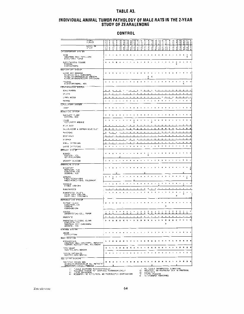
TABLE A3.
INDIVIDUAL ANIMAL TUMOR PATHOLOGY OF MALE RATS IN THE 2-YEAR STUDY OF ZEARALENONE
L U N G S A N D B R O N C H I I + + + * + + + t + + t t + + + + + t + + t - t t t S Q U A M O U S C E L L C A R C I N O M A I A L V E O L A R / B R O N C H I O L A R ADENONA x x A L V E G L A R I B R O N C N I O L A R C A R C I N O M A X
T R A C H E A . . . . . . . . . . . . . . . . . . . . . . . . . A D E N O C A R C I N O M A , N O S X
H E N A T G P O I E T I C S Y S T E M
B O N E MARROW - 1 , + i i ++ + + + + + + + + + + + + + + + + + + + - S P L E E N . . . . . . . . . . . . . . . . . . . . . . . . . L Y N P H N O D E S . . . . . . . . . . . . . . . . . . . . . . . . .
T n m u s + + + t i - + + + + - t - + + + + + + + + - + + -
C I R C U L A T O R Y S Y S T E N
H E A R T + + + + + + + t + + + t ' + + + + t + + + - t t t
D I G E S T I V E S Y S T E N
S A L I V A R Y G L A N D . . . . . . . . . . . . . . . . . . . . . . . . .
S A R C O N A . N O S
L I V E R . . . . . . . . . . . . . . . . . . . . . . . . . . N E O P L A S T I C N O D U L E X X
B I L E D U C T . . . . . . . . . . . . . . . . . . . . . . . . .
G A L L B L A D D E R 6 COMMON B I L E DUCT . N H N N N N N N N N N N H N N N N N N N N N N N N .
P A N C R E A S + + + + + + + + + i + + + * + + + + + t + - i + t
+ - - i i - * - + - + + t i - t - + + + + - t + +E S O P H A G U S
S T O n A C H . . . . . . . . . . . . . . . . . . . . . . . . .
S M A L L I N T E S T I N E + + t + + + + + + + + + + + + + + + + + t - t + t
L A R G E I N T E S T I N E * + + + + + + + + + + + t + + + + t + + + - t i t
URINARY S Y S T E M
K I D N E Y l + + + + + + + + + + + + + + + t t t t * t * t t+ I
P I T U I T A R Y N E O P L A S M , N O S C A R C I N O N A , N O S A D E N G N A , NOS X X X1A D R E N A L I + + ; + + + + + t + + + + + + + + + + + t t + t + PHEOCHROMOCYTOMA X
T H Y R O I D + * + + + + + + + + + + + + + + t + + + + - t + t l C - C E L L ADENOMA X
P A R A T H Y R O I D . . . . . . . . . . . . . . . . . . . . . . . . .
P A N C R C A T I C I S L E T S . . . . . . . . . . . . . . . . . . . . . . . . . I S L E l - C E L L ADENOMA I S L E T - C E L L C A R C I N O N A
R E P R O D U C T I V E S Y S T E N I M A N M P R Y G L A N D . . . . . . . . . . . . . . . . . . . . . . . . . +I CARC1NOMA.NOS
F I B R O M A XI F I B R O A D E H O M A
T E S T I S I ; ; + + + + + + + + + + + + + + + + + + + + t * tX I N T E R S T I T I A L - C E L L TUMOR X X X x x x x x x x X x x x x X x x
P R O S T A T E . . . . . . . . . . . . . . . . . . . . . . . . .
P R E P U T I A L / C L I T G R A L G L A N D I N N N H N N N N N N N N N H N N N N N N N N N N ~ C A R C I N O N A . N O 5 SQUANOUS C E L L C A R C I N O M A X ADENOMA, NOS !
B R A I N / + + + + + + + + + + + t + + + + + + + + + + + + + I A S T R O C Y T O M A
._B O D Y C A V I T I E S
N E D I A S T I N U N ~ N N N N N N N N N N N N N N N N N N N N N N N N N SQUAMOUS C E L L C A R C I N O N A , M E T A S T A T I F I B R O U S H I S T I O C Y T O M A , M A L I G N A H T
P E R I T O N E U M N N N N N N N N N N N N N N N N N H N N N N N N N M E S O T H E L I O P I A B E N I G N
. . . . . . . . . . . . . . . . . . . . . . . . I T U N I C A V A G I N A L 1 5 N E S O T H E L I O P l A B E N I G N
A L L O T H E R S Y S T E N S
m u i T i P t E O R G A N S nos H N N N N N N N H N N N N N N N N N N N N N N N N A L V E O L A R I B R O N C H I O L A R C A , M E T A S T A T I U N D I F F E R E N T I A T E D L E U K E M I A X X X
+ : T I S S U E E X A M I N E D M I C R O S C O P I C A L L Y : H0 T I S S U E I N F O R M A T I O N S U B M I T T E D - : R E Q U I R E D T I S S U E NOT E X A M I N E D M I C R O S C O P I C A L L Y C : N E C R O P S Y , N O H I S T O L O G Y D U E TO P R O T O C O L x : ~ U M O RI H C I D E N C E A : A U T O L Y S I S H : NECROPSY, NO A U T O L Y S i S , NO f l i C R O S C O P I C E X A F I I N I T I O N p1' A N I P i A L n I S S I N G
B : NO N E C R O P S Y P E R F O R M E D
Zearalenone 64

TABLE A3. MALE RATS: TUMOR PATHOLOGY (CONTINUED) CONTROL
S K I N I + N + + N N + + + + + + t + + + i + + + + + + + tj 5 0 x j S Q U A M O U S C E L L P A P I L L O M A X B A S A L - C E L L TUMOR X
S U B C U T A N E O U S T I S S U E + N i + N N * + + + + + + t + + + + + + + + + + F I B R O ' I A X F I B R O S A R C O M A i X +I 5 0 ;
L U N G S A N D B R O N C H I / + + ~ + i + + i + + + + + + + + + + + + + + t + SQUArtOUS C E L L C A R C I N O M A X A l V E O L A R / B R @ ~ C H I O L A R ADENOMA X I 3 1 A L V E O L A R / B R O N C H I O L A R C A R C I N O V A X
T R A C H E A . . . . . . . . . . . . . . . . . . . . . . . . . . . 4 9 1 A O E N O C A R C I N C M A I NOS I I I
H E M A T O P O I E T I C S Y S T E M I I
B O N E MARROW i , l t t , t ~ t i + + ~ + * t + - + t 4 9~ ~ + ~ ~
S P L E E N i + + + , + + + i + + + + + + + + i + + + + + 50
t y w n NODES t + i + t t i i i + i i ' t ' i i i + t t i i t i 5 0 , .
i w m u s ~ + - + - - + - + + + + + + + + + - + - - + + - + + / 37 1
C I R C U L A T O R Y S Y S T E M I
O I G E S T I V E S Y S T t M
S A L I V A R Y G L A N D SARCOMA, N O S
L I V E R N E O P L A S T I C N O D U L E
B I L E D U C T I + + *+ + + + + + + + + + + i i + + + + + + + + +5 0 1
G A L L B L A D D E R 8 COMMON B I L E D U C T 15 0 X . PA NC R E A 5 + + + + * + + + + + + + + + + * + + + * + * + + + 4 9 .
E S O P H A G U S . . . . . . . . . . . . . . . . . . . . . . . . . 3 9 .
S T O M A C H J + + , + + i + + + + i + + + + t t + + + t i t + 5 0 . S N A L L I N T E S T I N E + * + + + + + + + + * + + + + * + + + + + * + + + 49
L A R G E I N T E S T I N E ~ + + + , + + + + * + + + + t + + + + + * + + t + t4 9 1 I
U R I N A R Y SYSTEPI
SARCOMA, N O S I j l N E P H R O B L A S T O M A
U R I N A R Y B L A D D E R + + + + + * + + + + * + + + + + + + + + + + + + + l48 I I
E N D O C R I N E S Y S l E M t
A D S E N A L . . . . . . . . . . . . . . . . . . . . . . . . . . . 50 I P H E O C d R D n D C Y T O n A X X X I 5 I P H E O C H R O M O C Y T C ~ A . M A L I G N A N T 1 X
T H Y R O I D I + + + i + + + + + + + + + + + + + + + + + + + + + I 4 9 C - C E L L ADENOMA X J
I P A R A T H Y R O I D . . . . . . . . . . . . . . . . . . . . . . . . . . 4 :
I P A N C R C A T I C I S L E T S i + + + + + + + + + + + + + + + + + + + + + t + t t iX 4 9 12 1
I S L E T - C E L L ADENOMA I S L E I - C E L L C A R C I N O M A I I I !
C A R C I N O M A , N O S X I I F I E R O N A 1 1 ' F I E R O A O E H O M A X
I T E S l I S i + + + i + + + + + + + + + + + + t + + + + + + + l5 0
I N T E R S T I T I A L - C E L L TUMOR . x x x x x x x x x x x x x x x x x x x x x x x X I 4 5 .
P R O S T A T E . . . . . . . . . . . . . . . . . . . . . . . . . . 4 9
P R E P U T I A L I C L I T O R A L G L A N D I N N N n N N N N N n H H N H N N N N H N N N H H H I 5 0 x 1 C A R C I H O N A r N O S I I SQUAMOUS C E L L C A R C I N O M A I A D E N O N A , H05 X
N E R \ ' O U S S Y S T E M
B R A I N A S T R O C Y T O M A 1 1 1
B O D Y C A V I T I E S I
F I B R O U S H I S T I O C Y T O M A , V A L I G N A N T X
P E R I T O H E U l N N H N N N N N N H N N ~ N N H N H N H N N N N N 50" M E S O T H E L I O V A B E N I G N X 1
T UNICA V A G IIiA L I S + + i + t , + + + * + + + i + + + + + + + + + + + 5 0 x 1 V E S O T H E L I O I I A B E N I G N X I I
A L L O T P E R S Y S T E M S I
+ : TISSUE E X A V I H E D V I C R O S C O P I C A L L Y : NO T I S S U E I N F O R V A T I O N S U B M I T T E D - : R E Q U I R E D TISSUE N O T E X A M I H E D MICROSCOPICALLY C : N E C R O P S Y , NO H I S T O L O G Y DUE 1 0 P R O T O C O L X : IUNDR I N C I D E N C E A : A U I O L Y S I S H ; N E C R O P S Y , N O A U T O L Y S I S , NO N I C R O S C O P I C E X A V I H A T I O N M; A N I N A L PIISSING
B : NO N E C R O P S Y P E R F O R N E D
65 Zearalenone

TABLE A3.
INDIVIDUAL ANIMAL TUMOR PATHOLOGY OF MALE RATS I N THE 2-YEAR STUDY OF ZEARALENONE
LOW DOSE a N i r i A t I 0 1 0 D 0 1 D I 0 1 D I D I D I D I D I 0 1 0 0 0 D I 0 1 D I D I D I D I 0 1 D I 0 1 D NUMBER I O 1 D I D I D I 0 1 D I 0 1 D I 1 1 I I I I 1 1 I I I I I I I I I I I I 21 21 21 2 1 2 1 21D I
~
I 1 1 Z l 31 41 51 6 1 7 1 8 1 9 1 0 1 I I 2 1 31 4 1 5 1 6 1 7 1 E l 9 1 0 1 I I 2 1 3 1 41 L WEEKS O N S T U D Y
51 5 1 5 1 51 2. I N T E G U M E N T A R Y S Y S T E M
S K I N I + + + + + + + + + + + H + + + + + t i + [+ + + i i s Q u a n o u s C E L L PAPILLOMA
R E S P I R A T O R Y S Y S T E M I LUNGS a t n BRONCHI j + . . . . . . . . . . . . . . . . . . . . . . . . .
C A R C I N O M A . N O S , M E T A S T A T I C 1 X A L V E O L A R / B R O N C H I O L A R ADENOMA I X I A L V E O L A R / B R O N C H I O L A R C A S C I N O M A 4 X
T R A C H E A + + + + + + - i - * * + + + + + + + + * + - + * +
B O N E NARROW . . . . . . . . . . . . . . . . . . . . . . .
S P L E E N +:I + + i + + + + + + + + + + + + + + + +i i + + : LYMPH N O D E S . . . . . . . . . . . . . . . . . . . . . . . . .
T H Y N U S . . . . . . . . . . . . . . . . . . . . . . . . . .
C I R C U L A T O R Y S Y S T E M
H E A R T . . . . . . . . . . . . . . . . . . . . . . . . . .
D I G E S T I V E S Y S T E M
S A L I V A R Y G L A N D . . . . . . . . . . . . . . . . . . . . . . . . .
L I V E R . . . . . . . . . . . . . . . . . . . . . . . . . B I L E D U C T . . . . . . . . . . . . . . . . . . . . . . . . . G A L L B L A D D E R & COMMON B I L E D U C T H N H H H + ~ H ~ N N N H H N H N H ~ N H N N N H
I P A N C R E A S . . . . . . . . . . . . . . . . . . . . . . . . . E S O P H A G U S . . . . . . . . . . . . . . . . . . . . . . . . . S T O N A C H + * + + + + + + + + + + + + + + i t + + + * + + +
SMALL I N T E S T I N E t - + + + + + + + i i + + + + * + + + + + + + + +
L A R G E I N T E S T I N E . . . . . . . . . . . . . . . . . . . . . . . . . . . A D E N O N A T O U S P O L Y P , NOS X
U R I N A R Y S Y S T E M I . . . . . . . . . . . . . . . . . . . . . . . . .K I D N E Y
U R I N A R Y B L A D D E R I + * * + + + + + + + + + + + + + + + + + + + + + + l
E N D O C R I N E S Y S T E M
P I T u I T A R v ADENOMA, NOS x x x x Xi
A D R E N A L . . . . . . . . . . . . . . . . . . . . . . . . . . PHEOCHROMOCYTOMA X Xj X
T H Y R O I O j ; + + + + + + + + + + i + + + + + + + + + + + + + iI C - C E L L ADENOMA C - C E L L C A R C I N O M A I
P A R A T H Y R O I D . . . . . . . . . . . . . . . . . . . . . . . . . .
P A N C R E A T I C I S L E T S / + + + + + + + + + + + + + + + i + + + + + + + t t ~ I S L E T - C E L L ADENOMA I X I I S L E T - C E L L C A R C I N O M A ! I
I R E P R O D U C T I V E S Y S T E M I
MAMMARY G L A N D I i + + + + + t i+ + + + + + + + N + + + N + + + + l F I B R O A D E N O M A
T E S T I S . . . . . . . . . . . . . . . . . . . . . . . . . . I N T E R S T I T I A L - C E L L TUMOR I X X X x x x x x x x x x x x x x x x x x x x x I N l E R S T I T I A L - C E L L TUMOR, N A L I G Y A Nk + + Y + + + + + + + + + * + + + + t + l
P R O S T A T E
P R E P U T I A L l C L I T O R A L G L A N D ~ H N ~ N H H N N N H H H N N N N H H N N N N H N H x x ~ ADENOMA, N O S
I N E R V O U S S Y S T E M
Z Y M B A L ' S O L A N D / H N H N N N H N H + N + H N N N N N N N N N N ~ ~ / SQUAMOUS C E L L C A R C I N O M A 1 I
8 N E S E H T E R Y ~ N N N H N N N N N H N N NH N H N N N N N N N N N l
M E S O T H E L I O N A B E N I G N I I
A L L OTHER S Y S T E M S 1 M U L T I P L E ORGANS NOS / H N ~ N N ~ N N N N H N N H N N N N N N N N N N N /
A L V E O L A R / B R O N C H I O L A R C A , M E T A S T A T M E S O T H E L I O M A , NOS I M A L I G . L Y M P H O M A , I Y M P H O C Y T I C T Y P E 1 U N D I F F E R E N T I A T E D L E U K E M I A x x
T H I G H NOS i I c a R c I N o m a , N o s X
S C R O T U M NOS souamous C E L L ? a P I t t o n a X I
i: T I S S U E E X A M I N E D N I C R O S C O P I C P L L Y : NO T I S S U E I N F O R N A T I O N S U B N I T T E D - : R E Q U I R E D T I S S U E H O T E X l i M I N E D M I C R O S C O P I C A L L Y c : N E C R O P S Y , NO H I S T O L O G Y D U E T O P R O T O C D X : T U M O R I N C I D E N C E A : A U T O L Y S I S N : N E C R O P S Y , NO A U T O L Y S I S , NO M I C R O S C O P I C E X A I I I N A T I O N M : A N I M A L N I S S I N G
B: NO N E C R O P S Y P E R F O R M E D
Zearalenone 66
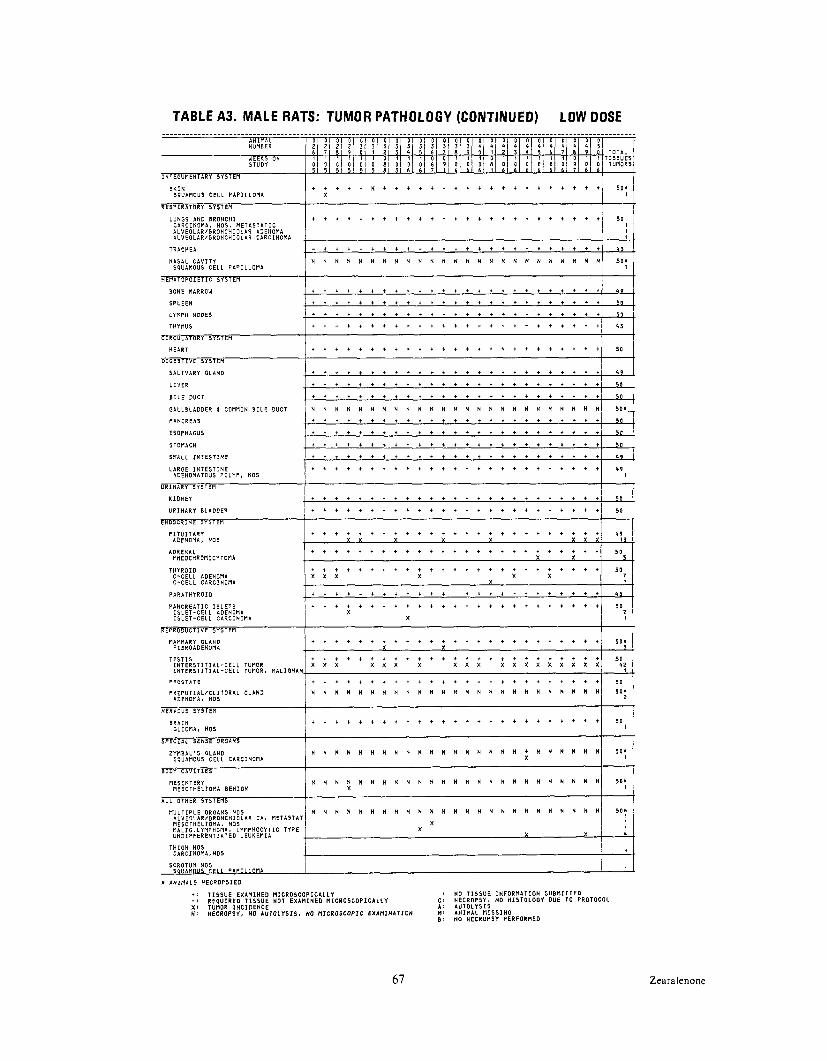
TABLE A3. MALE RATS: TUMOR PATHOLOGY (CONTINUED) LOW DOSE
I + ' + i N t + t + + t t t t t t t t + t t t t t S O XS K I N S Q U A M O U S C E L L P A P I L L O M A X I
L U N G S A N D + + + + + + + + + t + + + + t + + + + + + + + t + lB R O N C H I 5 0 1 C A R C I N O M A , NOS, N E T A S T A T I C I A L V E O L A R I B R O N C H I O L A R ADENOMA A L V E O L A R I B R O N C H I O L A R C A R C I N O N A
T R A C H E A . . . . . . . . . . . . . . . . . . . . . . . . . $ 3
N A S A L C A V I T Y N N N N N N N N N N N N N N N N N N N N N N N N N SQUAMOUS C E L L P A P I L L O N A 5 0 : i
H E M A T O P O I E T I C S Y S T E N
B O N E MARROW t l l t , t + t ~ l t ~ + ~ t , l ~ t + ~4 9 , ~ t +
S P L E E N t + + t + + * ' + l + i + + t + i + + + + + + + + 5 0
L Y f i P n NODES t + + + + + + t + + + + + t + * + + t + + + + + + so THYMUS + t - + t ~ t + ~ + l + + i - + - t - * + $3+ ~ ~ ~
C I R C U L A T O R Y S Y S T E M
H E A R T + + + + + + * + + + + + + + + + + + + + + + + t + l15 0
O I O E S T I V E S Y S T E n t + + + + + + + + + + + i + + + + * + t + t + t + t l 5 9 .S A L I V A R Y G L A N O
. . . . . . . . . . . . . . . . . . . . . . . . . 5 0 L I V E R
. . . . . . . . . . . . . . . . . . . . . . . . . 5 0 B I L E DUCT
G A L L B L A D D E R 6 COMNON B I L E D U C T N N N N N N N N N N N N N N N N N N N N N N N N N SO*.{
P A N C R E A S . . . . . . . . . . . . . . . . . . . . . . . . . 5 0
E S O P H A G U S . . . . . . . . . . . . . . . . . . . . . . . . . s o
S T O N A C H + * + + + + + + + + + + + + + + + + + * + + + + + Is o S M A L L I N T E S T I N E + + . * + + + t + + i + * + + + + + + + + + * + + +4 9
I I L A R G E I N T E S T I N E 4 9 ' + + + + + + t + + + + * + + + + * + t + + + + t + l
A D E N O N A T G U S P O L Y P , NOS I I
K I D N E Y . . . . . . . . . . . . . . . . . . . . . . . . . 5 0
U R I N A R Y B L A D D E R . . . . . . . . . . . . . . . . . . . . . . . . . . 50 I
E N D O C R I N E S Y S T E N I P I T U I T A R Y . . . . . . . . . . . . . . . . . . . . . . . . . . 4 9
ADENOHA, NOS
. . . . . . . . . . . . . . . . . . . . . . . . . 5 0 A D R E N A L PNEOCHROMOCYTONA X X 5
T 1+ + + + + + + + + + t + + + + t + t t t t t t + t i5 0T H Y R O I D C - C E L L ADENOMA / x x x X I ! I C - C E L L C A R C I N O N A i ++ + + - + + + + + + + - + + + + - + t + t + t t l 4 3P A R A T H Y R O I D
P A N C R E A T I C I S L E T S . . . . . . . . . . . . . . . . . . . . . . . . . 50: 1I S L E T - C E L L ADENONA I X + /
I S L E T - C E L L C A R C I N O M A
MAMNARY G L A N D . . . . . . . . . . . . . . . . . . . . . . . . . 5 0 " F I B R O A O E N O N A X X 3 .
. . . . . . . . . . . . . . . . . . . . . . . . . 50 I N T E R S T I T I A L - C E L L TUMOR x x x x x x x X X X X X X X X X X X X 4 2 I N T E R S T I T I A L - C E L L TUMOR, M A L I G N P N . I .
T E S T I S
P R O S T A T E I l l l + ~ , ~ l + ' t , t + , + l t l t t t *
P R E P U T I A L I C L I T O R A L G L A N D ~ N N N N N N N N N N N N N N N N N N N N N N N N 5 0 s ADENOMA, NOS +I
N E R V O U S S Y S T E M
+ + + t + t + + + + t + t t + t + t + t t i t + tsoB R A I N I IG L I O M A , N O S
S P E C I A L S E N S E OROANS
Z Y M B A L ' S G L A N D I N N N N N H N N N H N N N N N N N N t N N N N N N s o n SQUAMOUS C E L L C A R C I N O M A
I BODY C A V I T I E S
M E S E N T E R Y N N N N N n N N N N N N H N n N N N N N N N N H N / 5 0 1 ' M E S O T H E L I O M A B E N I G N
A L L O T H E R S Y S T E N S I CA. nETA57AT / N N N N N N N N N N N N N N N N N N N N N N N N N
P I U L T I P L E ORGANS NOS A L V E O L A R / B R O N C H I O L A R M E S O T H E L I O M A . NOS M A L I G . L Y P l P H O M A , L Y M P H O C Y T I C T Y P E U N D I F F E R E N T I A T E D L E U K E M I A
T H I G H NOS C A R C I H O M A , N O S
S C R O T U N N O S S Q U A M O U S C E L L P l P I L L O P l A I L
A N I M A L S N E C R O P S I E D
t i T I S S U E E X A M I N E D M I C R O S C O P I C A L L Y I NO T I S S U E I N F O R M A T I O H S U B M I T T E D -: R E Q U I R E D T I S S U E H O T E X A M I N E D M I C R O S C O P I C A L L Y c: N E C R O P S Y , NO H I S T O L O G Y D U E TO P R O T O C O I X : T U N O R l N C I D E N C E A : A U T O L Y S I S H: NECROPSY, no i u io t ys rs . NO NICROSCOPIC EXAMINATION M: ANIMAL HISSING
B : NO N E C R O P S Y P E R F O R N E D
67 Zearalenone
X

TABLE A3.
INDIVIDUAL ANIMAL TUMOR PATHOLOGY OF MALE RATS IN THE 2-YEAR
S K I N P A P I L L O M A , NOS S E B A C E O U S ADENOMA ADENOSQUAMOUS C A R C I N O M A
S U B C U T A N E O U S T I S S U E F I B R O S A R C O M A L I P O M A
L U N G S A N D B R O N C H I A L V E O L A R / B R O N C H I O L A R C A R C I N O M A
S P L E E N
L Y M P H N O D E S
S A L I V A R Y G L A N D
L I V E R N E O P L A S T I C N O D U L E H E P A T O C E L L U L A R C A R C I N O M A
8 I L E D U C T
G A L L B L A D D E R i COPIMON B I L E D U C T
P A N C R E A S
E S O P H A G U S
STOMACH
S M A L L I N T E S T I N E
ADENOMA, NOS
A D R E N A L PHEOCHRONOCYTOMA PHEOCHROMOCYTOMA. M A L I G N A N T
T H Y R O I D F O L L I C U L A R - C E L L ADENOMA C - C E L L ADENONA C - C E L L C A R C I N O M A
P A R A T H Y R O I D
R E P R O D U C T I V E S Y S T E R
MAMMARY G L A N D AOENOMA, NOS F I B R O A D E N O M A
T E S T I S I N T E R S T I T I A L - C E L L TUMOR
P R O S T A T E A D E N O C A R C I N O H A . N O S
P R E P U T I A L I C L I T O R A L G L A N D A D E N D H A . NOS
@ R V D U S S Y S I E P I
B R A I N
A L L O T H E R S Y S l E M S
M U L T I P L E ORGANS N O S PHEOCHROMOCYTONA. M E T A S T A T I C M E S O T H E L I O N A . N D S M A L I G N A N T L Y R P H O B A , M I X E D T Y P E U N D I F F E R E N T I A T E D L E U K E M I A
STUDY OF ZEARALENONE
HIGH DOSE
+ + + + + + + + t * + + + + t t t t t t t t + + + X
X
I + + + t t + + + + t t t * + + t t + + * t + + +:I
+ + + + + + + + + * * t + * + + t * * * t t + + t
X x x x x + + + + + + * * + + t + * * + * + + + * + t t + +
X x x
~ + + + + t + + + t + t * * t + + + t * * t * * * * X
X X L.
+ + + + - + + + - t t t t - + + t t t - t t t - +
+ * + + + + t + + t * * t t t t + + t + t t t
X X
+ + + + + + + + + + t * t t + + + t + * + t t t t x x x x x x x x x x x x x x x x x x x x
+ - + + + + + + * + + + * t + + + + + * t t t t t
N N N N N N N N N N N N N N N N N N N N N N U N N X
+ t + + + + + + + + + t t + + + + + * + + * t t t
N N N N N N N N N
x x x i i T I S S U E E X A N I N E D M I C R O S C O P I C A L L Y -: R E Q U I R E D T I S S U E NOT E X A M I N E D M I C R O S C O P I C A L L Y X : TUMOR I N C I D E N C E N : N E C R O P S Y , NO A U T O L Y S I S , NO M I C R O S C O P I C E X A M I N A T I O N
N N N N N N N N N N N N U U N N
X
x x : NO T I S S U E I N F O R M A T I O N S U B M I T T E D
C : N E C R O P S Y , NO H I S T O L O G Y D U E 10 P R O l O C O l A : A U T O L Y S I S M : A N I M A L M I S S I N G I): NO N E C R O P S Y P E R F O R M E D
Zearalenone 68

TABLE A3, MALE RATS: TUMOR PATHOLOGY (CONTINUED) HIGH DOSE
S K I N + t + t + + + + + + + + t + t + t t t t t t t t i 5 0 x 1 P A P I L L O M A , NOS I 1 S E B A C E O U S ADENOMA X 1 1 ADENOSPUAMOUS C A R C I N O M A
I S U B C U T A N E O U S T I S S U E . . . . . . . . . . . . . . . . . . . . . . . . .
F I B R O S A R C O M A X I i p n m I * I 1 -- I -. .
RESPI~? L U N G S A N 0 B R O N C H I l + + + + + + + + + + + + + + + + + + t t + t + + t47
A L V E O L A R / B R O N C H I O L A R C A R C I N O M A . T R A C H E A + + + + + + + + + + + + + t + + i t - + i t + + t 4 6
P I
B O N E MARROW . . . . . . . . . . . . . . . . . . . . . . . . . 5 0 1
S P L E E N + + + + + + + + + + + + + + + + + t + t t + t + 5 0
L Y M P H N O D E S . . . . . . . . . . . . . . . . . . . . . . . . . 5 0
T H Y M U S . . . . . . . . . . . . . . . . . . . . . . . . . 4 3 -.t p IH E A R T + + + + + + + + + + + + + t * + + l t t t t l + t l 50
FD I G E S T I V E S Y S T E M 1 S A L I V A R Y G L A N D + + + + + + + + + + + + + + + + t t + t + + + t + 50 I
I+ + + + + t + + + + + + + t + t + + t t + t t + t l5 0 1L I V E R
N E O P L A S T I C N O D U L E H E P A T O C E L L U L A R C A R C I N O M A X
B I L E D U C T . . . . . . . . . . . . . . . . . . . . . . . . . 5 0
G L L L B L A D D E R I COMMON B I L E D U C T N N N N N N H N N N N H N N N N N N N n N N n N N 5 0 s T
P A N C R E A S . . . . . . . . . . . . . . . . . . . . . . . . . 50
E S O P H A G U S . . . . . . . . . . . . . . . . . . . . . . . . . 4 9
S T O M A C H . . . . . . . . . . . . . . . . . . . . . . . . . 5 0
S M A L L I N T E S T I N E . . . . . . . . . . . . . . . . . . . . . . . . . 4 9
L A R G E I N T E S T I N E
U R I N A R Y S Y S T E M
K I D N E Y I
+ t + + + + + + + + + + + + + + + + i + + + t t + 50
U R I N A R Y B L A D D E R + + + + + + + + + + + + + 1 + t + 1 + t + t t + t 4 9 1
E N D O C R I N E S Y S T E M I P I T U I T A R Y / + + + + + + + + + + t + + + + + t + + + + + t + + li50
C A R C I N O M A , N O S X I I ADENOMA. N O S X x x X
A D R E N A L + + + * + * + + * + + + + + + + + + t + + + t t PHEOCHROMOCYTOMA X PHEOCHROMOCYTOMA, M A L I G N A N T
T H Y R O I D . . . . . . . . . . . . . . . . . . . . . . . . 50 F O L L I C U L A R - C E L L ADENOMA C - C E L L ADENOMA X X C - C E L L C A R C I N O M A
I P A R A T H Y R O I D . . . . . . . . . . . . . . . . . . . . . . . . . . 4 3
I R E P R O D U C T I V E S Y S T E M
MAMMARY G L A N D I + t + + + + + + t + + + t + + + + + + + + + + + +II 50"I ADENOMA, NOS F I B R O A D E N O M A
I 5 o iT E S T I S l t , + , + + + , + + t + t + + t + t + + + t + t t !
I N T E R S T I T I A L - C E L L TUPIOR I X X X X X Y x x x x x x x x x x x x X I 3 9 1 IP R O S T A T E + + t + + + + * + t + . + * + + + + + + + + + + t l
A D E N O C A R C I H O M A , N O S X I 49,
AOENOEIA, N N N N N N N N N N N N N IP R E P U T I A L I C L I T O R A LNOS G L A N D N N N N N N N N N N N N '
R E R V O U SS Y S T E M
B R A I N + + + + + + + + + + + + + + + + t + + t + i t t t 50
1 -.
IM U L T I P L E ORGANS NOS N N N N N N N N N N N N N N N N N N N N N N N N N ~ PHEOCHROPIOCYTDMA, M E T A S T A T I C M E S O T H E L I O M A , N O S P l A L I G N A N T LYMPHOMA, M I X E D T Y P E X U N D I F F E R E N T I A T E D L E U K E M I A X
69 Zearalenone

TABLE A4.
INDIVIDUAL ANIMAL TUMOR PATHOLOGY OF FEMALE RATS IN THE 2-YEAR STUDY OF ZEARALENONE
CONTROL
S K I N + + + + + + N + + + N + + N + + t + N N t t t t N K E R A T O A C A N T H O M A X
R E S P I R A T O R Y S Y S T E M
L U N G S A N D B R O N C H I . . . . . . . . . . . . . . . . . . . . . . . . . A L V E O L A R l B R O N C H I D L A R ADENOMA _.
T R A C H E A . . . . . . . . . . . . . . . . . . . . . . . . .
H E M A T O P O I E T I C S Y S T E N
B O N E MARROW . . . . . . . . . . . . . . . . . . . . . . . . . P l A L I G . L Y M P H O M A , H I S T I O C Y T I C T Y P E . X
S P L E E N + + + + + + + + + + + + + + + + + + + t + t t t t ~
L Y N P H N O D E S + + + + + + , + + + + + + + + + + + + * + + t t t ~
T H Y M U S . . . . . . . . . . . . . . . . . . . . . . . . . R R C U L A T O R Y S Y S T E M
H E A R T . . . . . . . . . . . . . . . . . . . . . . . . . . D I G E S T I V E S Y S T E M
S A L I V A R Y G L A N D + + + + + + + + t * * + + + + + + + t t t t t t t ,
+ + + + + + + * + + + + + + + + + t + t + t t t + ~L I V E R
+ + + + t t + t + t + + + + t + t t t t t t t t t ,B I L E D U C T
G A L L B L A D D E R L COMMON B I L E D U C T N N N N N N ~ N N N N N H N H NN N N N N N N n n . P A N C R E A S + + * + + + + + + + + + t + + + t t + t i t t t t
. . . . . . . . . . . . . . . . . . . . . . . . . E S O P H A G U S
S T O M A C H + + + + + t + + + + t + t t t t t t t i + + t t i l S A R C O N A , NOS X
+ + + + + + + * - + + t + + + + + + + + + * + t tS M A L L I N T E S T I N E
. . . . . . . . . . . . . . . . . . . . . . . . .L A R G E I N T E S T I N E
U R I N A R Y S Y S T E M
. . . . . . . . . . . . . . . . . . . . . . . . . . K I D N E Y
. . . . . . . . . . . . . . . . . . . . . . . . . U R I N A R Y B L A D D E R
E N D O C R I N E S Y S T E M
P I T U I T A R Y + + + + + + + t t + + + + + t * + + + + + + + t + C A R C I N O M A , N O S ADENOMA, N O S X x x x X X X
A D E N O I I A , NOS F O L L I C U L A R - C E L L ADENOMA X C - C E L L ADENOMA X X
- + + + + - - + + - + t t + + + + + + + * * + + +P A R A T H Y R O I D
P A N C R E A T I C I S L E T S . . . . . . . . . . . . . . . . . . . . . . . . . I S L E T - C E L L ADENOMA X
R E P R O D U C T I V E S Y S T E M
M A M N A R Y G L A N D + + + + + + + + + + + + + + + + + + t N + + + + N A D E N O C A R C I N O M A , N O S X F I B R O A D E N O N A X X x x x X
P R E P U T I A L I C L I T O R A L O L A N D N N N N N N N N N N N N N N N N N N N N N N N N N C A R C I N O M A , N O S X
V A G I N A N N N N N N N N N N N N H N N N N N N N N N N N N G R A N U L A R - C E L L TUMOR, B E N I G N X
. . . . . . . . . . . . . . . . . . . . . . . . . U T E R U S E N D O M E T R I A L S T R O M A L P O L Y P X
t + + + + t + + + * + + t + + + t t t t + t t t tO V A R Y
N E R V O U S S Y S T E M
+ + + + + + + + + + + + + + + + + t * t * t * l *B R A I N
S P E C I A L S E N S E ORGANS
Z Y M B A L ' S O L A N D ~ N N N N N ~ N N N H N N N N N N N N N N N N N N SQUAPIOUS C E L L C A R C I N O M A
B O D Y C A V I T I E S
P E R I T O N E U M N N N N N N N N N N N N N N N N N N N N N N N N N F I B R O M A
M E S E N T E R Y N N N N N N N N N N N N N N N N N N N N N N N N N L I P O M A X
A L L O T H E R S Y S T E M S
M U L T I P L E ORGANS NOS N N N N N N N N N N N N N N N N N N N N N N N N N U N D I F F E R E N T I A T E D L E U K E M I A X x x
: NO T I S S U E I N F O R M A T I O N S U B M I T T E DI : T I S S U E E X A N I N E D M I C R O S C O P I C A L L Y c : N E C R O P S Y , no HISTOLOGY DUE T O P R O T O C O L - : R E W I R E D T I S S U E N O T E X A N I N E D M I C R O S C O P I C A L L Y A : A U T O L Y S I S t i : N E C R O P S Y , no MICROSCOPIC M : ANIMAL mxssInnx : TUNOR I N C I D E N C E
NO A u r o t y s I s , EXAMINATION B : NO N E C R O P S Y P E R F O R M E D
Zearalenone 70

TABLE A4. FEMALE RATS: TUMOR PATHOLOGY (CONTINUED) CONTROL
P IT U 1T A R Y I - + + + + + + + + t + + + + + + t + + + + + * t 4 9 C A R C I N O M A l N 0 5 X 1 ADENOMA. NOS x x X x x L 13 .
. . . . . . . . . . . . . . . . . . . . . . . . . 50
T H Y R O I D + + + + + + * + + + + * + + + + + + + * * * ( * * 4 9 ADENONA, H 0 5 X 1 F O L L I C U L A R - C E L L ADENOMA 1 C - C E L L AOENOMA X X X 5 .
P A R A T H Y R O I D . . . . . . . . . . . . . . . . . . . . . . . . . 4 3 P A N C R E A T I C I S L E T S + + + + + + + + + * * + + t + + t + t + t + + t t 50
I S L E T - C E L L ADENOMA I
R E P R O D U C T I V E S Y S T E M ~
I MAMMARY G L A N D + + t + + + + + + t + + + + + + + + + + N + + + t S O 11A O E N O C A R C I N O N A ~ NOS
F I B R O A D E N O N A ~
P R E P U T I A L I C L I T O R A L G L A H O N N N N N N N H N N N N N N N N H N N N N N N N N C A R C I N O N A , N O S 50:
V A G I N A N N N N N N N N H N N N N N N N N N N N N N N N N 5 0 s G R A N U L A R - C E L L TUMOR, B E U I G N
U T E R U S + * + + + * * + t + * + + + r + + + + + t + + + t 5 0 ENDOVETRIAL STROMAL P O L Y P X X Y 4
OVARY . . . . . . . . . . . . . . . . . . . . . . . . . 5 0 -.
P E R I T O N E U M N N N N N N N N N N N N N N N N N N N W N N N N N 501 F I B R O M A X 1
N E S E N T E R Y N N N N N N N N N N H N N N N N N N N N N N N N N son L I P O N A
M U L T I P L E ORGANS NOS N N N N N H N N N N N N N N N N N N N N N N N H N s o x
t: TISSUE E X A M I N E D M I C R O S C O P I C A L L Y t NO T I S S U E I N F O R M A T I O N S U B M I T T E D C : N E C R O P S Y , N O H I S T O L O G Y D U E TO P R O T O C O L
-: R E Q U I R E D T I S S U E NOT E X A M I N E D P I I C R O S C O P I C A L L I A : A U T O L Y S I SX: TUMOR I N C I D E N C E N: N E C R O P S Y , NO A U T O L Y S I S I NO M I C R O S C O P I C E X A M I N A T l O N M: & N l M A L M I S S I N G
II NO N E C R O P S Y P E R F O R M E D
71 Zearalenone

TABLE A4.
INDIVIDUAL ANIMAL TUMOR PATHOLOGY OF FEMALE RATS IN THE 2-YEAR STUDY OF ZEARALENONE
LOW DOSE
S K I N . . . . . . . . . . . . . . . . . . . . . . . . . SQUAMOUS C E L L P A P I L L O N A
S U B C U T A N E O U S T I S S U E + * * + + + * + + + + + + + + + + + + + + + + + + I C A R C I N O M A , N O S SARCOMA, NOS I F I B R O M A I X X I
L U N O S A N D B R O N C H I . . . . . . . . . . . . . . . . . . . . . . . . . . A L V E O L A R / B R O N C H I O L A R ADENOMA X SARCONA, N O S , N E T A S T A T I C
T R A C H E A i: + + + + + + + + i + + + + I + + + + - + + + t - .
H E M A T O P O I E T I C S Y S T E M I
B O N E MARROW . . . . . . . . . . . . . . . . . . . . . . . . . S P L E E N . . . . . . . . . . . . . . . . . . . . . . . . . L Y M P H N O O E S . . . . . . . . . . . . . . . . . . . . . . . . . THYMUS . . . . . . . . . . . . . . . . . . . . . . . . .
C I R C U L A T O R Y S Y S T E W
H E A R T . . . . . . . . . . . . . . . . . . . . . . . .
L I V E R . . . . . . . . . . . . . . . . . . . . . . . . . N E O P L A S T I C N O D U L E X .
B I L E D U C T . . . . . . . . . . . . . . . . . . . . . . . . . . G A L L B L A O D E R k COMWON B I L E D U C T . N N N N N H N N N H N N N N N N N N N N N N N n N_.
P A N C R E A S . . . . . . . . . . . . . . . . . . . . . . . . . E S O P H A O U S . . . . . . . . . . . . . . . . . . . . . . . . . S T O M A C H . . . . . . . . . . . . . . . . . . . . . . . . . S R A L L I N T E S T I N E . . . . . . . . . . . . . . . . . . . . . . . . .
L A R G E I N T E S T I N E . . . . . . . . . . . . . . . . . . . . . . . . . . U R I N A R Y S Y S T E N
K I D N E Y . . . . . . . . . . . . . . . . . . . . . . . . . U R I N A R Y B L A D D E R . . . . . . . . . . . . . . . . . . . . . . . . .
E N D O C R I N E S Y S T E M
. . . . . . . . . . . . . . . . . . . . . . . . . . X I
x x x x x x x . . . . . . . . . . . . . . . . . . . . . . . . .
X I T H Y R O I D I + + + + + + I + + + + + + + * + + + + + + + + + + I
C - C E L L ADENOWA X X X C - C E L L C A R C I N O M A X
P A R A T H Y R O I D . . . . . . . . . . . . . . . . . . . . . . . . .
P A N C R E A T I C I S L E T S + + + + + ' + 1 + + + + + + * i + + + + + + + + * I I S L E T - C E L L C A R C I N O M A X I
R E P R O D U C T I V E S Y S T E M
WAMMARY G L A N D . . . . . . . . . . . . . . . . . . . . . . . . . A D E N O C A R C I N O M A , N O S X X F I B R C A O E N O M A x x x x x x x x .
U T E R U S . . . . . . . . . . . . . . . . . . . . . . . . . E N D O W E T R I A L S T R O M A L P O L Y P X x x
O V A R Y I t + + + + + + + + + + + + + + + + + + + + + + + + I
N E R V O U S S Y S T E M
B R A I N . . . . . . . . . . . . . . . . . . . . . . . . . S P E C I A L S E N S E ORGANS
E Y E A P P E N D A G E S I N N N N N N N N N N N N N N N N N N N N N N NI F I B R O S A R C O M A ! !
M E D I A S T I N U M / N N N N N N N N N N N N N N N N N N N N H N N N N / A C E N O C A R C I N O M A , NOS ! !
M U L T I P L E ORGANS NOS I N N N N N N N N N N N N N H N N N N N N N N N N N /U N D I F F E R E N T I A T E D L E U K E M I A
+ : T I S S U E E X A N I N E C W I C R O S C O P I C A L L Y : no TISSUE INFORMATION SUBMITTED - : R E Q U I R E D T I S S U E NOT E X A M I N E D M I C R O S C O P I C A L L Y C : N E C R O P S Y , NC H I S T O L O G Y D U E TO P R O T O C O L X : TUMOR I N C I D E N C E A : A U T O L Y S I S N : N E C R O P S Y . NO A U T O L Y S I S , NO W I C R O S C O P I C E X A M I N A T I O N W : A N I M A L M I S S I H O
B : NC N E C R O P S Y P E R F O R M E D
Zearalenone 72

TABLE A4. FEMALE RATS: TUMOR PATHOLOGY (CONTINUED) LOW DOSE
WEEKS ST'JDY
I N T E G U f l E N T A R Y S Y S T E M
S K I N SQUAMOUS C E L L P A P I L L O M A
S U B C U T A N E O U S T I S S U E C A R C I N O M A , N O S S A R C O n A , N O S F I B R O P I A
R E S P I R A T O R Y S Y S T E N
L U N G S A N D B R O N C H I
ON
+ i 1 t t t t + + + + + + H + + + + + + + + + + t /X I
5 0 x 1 I
+ + +
X
+ + + t + + * + + + ~ +
X
+ + , X
+ + + t + t 'I I A L V E O L A R / B R O N C H I O L A R ADENONA SARCOMA. NOS, M E T A S T A T I C
T R A C H E A I
+ - + + + + + + + + + + + - + + + + + + + t t t t l$ 6 I H E f l A T O P O I E T I C S Y S T E M
B O N E MARROU . . . . . . . . . . . . . . . . . . . . . . . . . 5 8 .
S P L E E N . . . . . . . . . . . . . . . . . . . . . . . . . s o L Y M P H N O D E S . . . . . . . . . . . . . . . . . . . . . . . . . 5 0
THYMUS + + + - + + + + t + + + + - + + + + + + + + t + t 4 7 1
C I R C U L A T O R Y S Y S T E M
H E A R T
D I G E S T I V E S Y S T E M
S A L I V A R Y G L A N D
L I V E R N E O P L A S T I C N O D U L E
B I L E D U C T
G A L L B L A D D E R L COMMON B I L E D U C T
P A N C R E A S
E S O P H A G U S
STOMACH
S n A L L I N T E S T I N E
L A R G E I N T E S T I N E
U R I N A R Y S Y S T E M
K I D N E Y
U R I N A R Y B L A D D E R
E N D O C R I N E S Y S T E N
P I T U I T A R Y C A R C I H O N A , N O S ADENOMA, NOS
A D R E N A L PHEOCHROMOCYTOMA
T H Y R O I D C - C E L L ADENOMA C - C E L L C A R C I N O N A
P A R A T H Y R O I D
P A N C R E A T I C I S L E T S I S L E T - C E L L C A R C I N O M A
R E P R O D U C T I V E S Y S T E M
N A N l l A R Y G L A N D A D E N O C A R C I N O M A I NOS F I B R O A D E N I M A
U T E R U S E N D O M E T R I A L S T R C f l A L P O L Y P
O V A R Y
N E R V O U S S Y S T E N
B R A I N
S P E C I A L S E N S E ORGANS
E Y E A P P E N D A G E S F I B R O S A R C O M A
BODY C A V I T I E S
M E D I A S T I N U M A D E N O C A R C I N O M A , N 0 5
A L L O T H E R S Y S T E M S
M U L T I P L E ORGANS N 0 5 U N D IF F ER E-
* A N I M A L S N E C R O P S I E D
1
-
+ + + + + + + + + + , + + + + t + + + + + t + + + l1 5 0 1 X 6 1
I . . . . . . . . . . . . . . . . . . . . . . . . . . 4 5
I+ + + + + + + + + + + + + + + + + + + t t t t i ' l 5 0
! I !
i i+ + + + + + + + + + + + + * I t + I t + + + + + t! s o y ! I L 1
x x x x X
+ + + + * + + + + + * + + + + + - + + t + + i t X X Y X x +I Tal
. . . . . . . . . . . . . . . . . . . . . . . . . 4 9
. . . . . . . . . . . . . . . . . . . . . . . . . SO
N N N N N N N N N
-N N N N N N N N N
X X
+ : T I S S U E E X A M I N E D M I C R O S C O P I C A L L Y - : R E Q U I R E D T I S S U E N O 1 E X A N I N E D N I C R O S C O P I C A L L Y X : TUMOR I N C I D E N C E H : N E C R O P S Y , NO A U T O L Y S I S , H0 M I C R O S C O P I C E X A M I N A l l O N
N N N N N N N N N N N N N N N N / 5 0 x 1 I l l
N N N N N N N N N n N N N N n H I 5 0 x 1 X X
: N O T I S S U E I N F O R M A T I O N S U B M I T T E D c : N E C R O P S Y , N O H I S T O L O G Y D U E T O P R O T C C O l A: A U T O L Y S I S M : A N I M A L I 1 1 5 5 1 N G B : N O N E C R O P S Y P E R F O R N E O
73 Zearalenone

TABLE A4.
INDIVIDUAL ANIMAL TUMOR PATHOLOGY OF FEMALE RATS I N THE 2-YEAR STUDY OF ZEARALENONE
HIGH DOSE
E S O P H A G U S ~ t t t t t * + + + + + * t + * t t t t + + + + t i
S T O M A C H . . . . . . . . . . . . . . . . . . . . . . . . . S M A L L I N T E S T I N E + + + 1 + + + + + + + + + + t * + + + + + + i t .
L A R G E I N T E S T I N E . . . . . . . . . . . . . . . . . . . . . . . . . . \ l R I N A R Y S Y S l E N
K I D N E Y + + + + + + + + + t + + + + + t + + + + + + + t +
U R I N A R Y B L A D D E R + + + - + , * + - t t + + + + t * + + + + + + t t ~
E N D O C R I N E S Y S T E M
P I T U I T A R Y + * * * + + + + + + t * + + + + + + + + + + + + +A O E N O I I A , N O S X X x x x X
A D R E N A L + + t t + + + + + + + + * + + 1 + + + + + + + + + I C O R T I C A L ADENOMA c C - C E L L / + + + + t t + + + + + + * * + - + + + + + + * + +T H Y R O I D ADENOMA X x x C - C E L L C A R C I N O M A 4
P A R A T H Y R O I D + + + * t + + + + + + t + + + - + + + + + t + + - ADENOMA, N O S X
~ t + + + + + t + + + + + + + + t t + t + + + + t t
I
ADENOMA, NOS
U T E R U S * * + + + + + + - * t t * + + + + + + + + + + t t lE H D O I I E T R I A L S T R O N A L P O L Y P X X X
O V A R Y ~ , t , * + t l - . * t t t . l ~ . l t , , , ~ ~
N E R V O U S S Y S T E M
B R A I N + + t + + + + + + + + + + t t + + + + + + + + i +
M U S C U L O S K E L E T A L S Y S T E M I
M U S C L E . . . . . . . . . . . . . . . . . . . . . . . . . RHABDOMYOSARCOMA X
B O D Y C A V I T I E S
P L E U R A F I B R O M A [ N N N N N N N N N N N N N N N N N N N N N N N N NI C - C E L L C A R C I N O M A , N E T A S T A T I C M A L I G . L Y M P H O M A , H I S T I O C Y T I C T Y P E X
t i T I S S U E E X A M I N E D M I C R O S C O P I C A L L Y : NO T I S S U E I N F O R M A T I O N S U B M I T T E D - : R E Q U I R E D T I S S U E NOT E X A M I N E D M I C R O S C O P I C A L L Y C : N E C R O P S Y , NO H I S T O L O G Y D U E TO P R O T O C O L X : TUMOR I N C I D E N C E A : A U T O L Y S I S H : N E C R O P S Y , NO A U T O L Y S I S , NO M I C R O S C O P I C E X A M I N A T I O N M : A N I M A L M I S S I N G
B: NO N E C R O P S Y P E R F O R M E D
~
/
Zearalenone 74

X
LUNGS AND B R O N C H I I + + + + + + + + + + + + + + + + + + + * + + * 5 0 )A L V E O L A R I B R O N C H I O L A R ADENOMA A L V E O L A R l b R O N C H I O L A R C A R C I N O M A X 1
T R P C H E A + + + + + + - - + + + + + + + + I + + + t + + t + 47 C - C E L L C A R C I N O M A . I N V A S I V E X I
pp-. B O N E MARROW + + - - t + + + + , + + + , + + + + + , + * I + * 4 1 .
S P L E E N + t + + + + + + + l t ~ t i + t t t t t + 5 0 .~ ~ ~ ~
L Y M P H N O D E S + + t + + + + + + + t + + + + + + t + t . t * t * 4 9
THYVUS + + + + + + + + - 4 + + + + + - + + - t + + t + + $ b
a R C U L A T O R Y S Y S T E M
HEA'RT + + + + + + + *. + + + + + + + + + + + t t + + I + 50
a I O E S T I V E S Y S T E N
S A L I V A R Y G L A N D t + + + + + + + + + + + + + + + + t + t t + t t + 50
L I V E R [ + + + + + i + + + + + I + + + + f t t t + i t + t 50 N E O P L P S T I C N O D U L E H E P A T O C E L L U L A R C A R C I N O M A 1
B I L E D U C T
O A L L B L A D D E R L COMVON B I L E DUCT
P A N C R E A S
E S O P H A G U S
S T O V P C H
SMALL INTESTINE
L A R G E I N T E S T I N E
O M E T R I A L S T R O M A L P O L Y P
V U L T I P L E ORGANS NOS i n n N N N N N n n N N N N N n n N N N N N n N N N 5 0 1 C - C E L L C A R C I N O V A , M E T A S T A T I C n A L i o . L i m f H o n A , HISTIOCYTIC T Y P E I U N D I F F E R E N T I A T E D L E U K E M I A X I
A N I M A L S N E C R O P S I E D
t: T I S S U E E X A M I N E D M I C R O S C O P I C A L L Y i NO lIS5UE I N F O R O A T I O N S U B O I I T E D - : R E Q U I R E D T I S S U E NOT E X A M I N E D M I C R O S C O P I C A L L Y E8 N E C R O P S Y , N O H I S T O L O G Y DUE T O P R O T O C O l x : T U M O R INCIDENCE A : A U T O L Y S I S H: N E C R O P S Y . NO AUTOLYSIS, H0 M I C R O S C O P I C E X A M I N A T I O N M: MIMA1 P I I S S I N G
b: N O N E C R O P S Y P E R F O R M E D
TABLE A4. FEMALE RATS: TUMOR PATHOLOGY (CONTINUED) HIGH DOSE
75 Zearalenone

Zearalenone 76

APPENDIX B
SUMMARY OF THE INCIDENCE OF NEOPLASMS IN MICE FED DIETS CONTAINING ZEARALENONE
77 Zearalenone

TABLE 61.
SUMMARY OF THE INCIDENCE OF NEOPLASMS IN MALE MICE FED DIETS CONTAINING ZEARALENONE
CONTROL LOW DOSE HIGH DOSE. . . . . . . . . . . . . . . . . . . . . . . . . . . . . . . . . . . . . . . . . . . . . . . . . . . . . . . . . . . . . . . . . . . . . . . . . . . . . . . . A N I M A L S I N I T I A L L Y I N STUDY 5 0 5 0 5 0 A N I M A L S N E C R O P S I E D 5 0 5 0 5 0 A N I M A L S E X A M I N E D H I S T O P A T H O L O G I C A L L Y 5 0 50 5 0 . . . . . . . . . . . . . . . . . . . . . . . . . . . . . . . . . . . . . . . . . . . . . . . . . . . . . . . . . . . . . . . . . . . . . . . . . . . . . . . . I N T E G U M E N T A R Y SYSTEM
X S K I N (50) ( 5 0 ) (50) SQUAMOUS C E L L P A P I L L O M A 1 ( 2 % ) FIBROSARCOMA 1 ( 2 % )
XSUBCUT T I S S U E ( 5 0 ) ( 5 0 ) (50)SARCOMA, NOS 1 ( 2 % ) 1 ( 2 % )FIBROSARCOMA 1 ( 2 % )N E U R I L E M O M A t M A L I G N A N T 1 ( 2 % ). . . . . . . . . . . . . . . . . . . . . . . . . . . . . . . . . . . . . . . . . . . . . . . . . . . . . . . . . . . . . . . . . . . . . . . . . . . . . . . .
R E S P I R A T O R Y SYSTEM
#LUNG (50) ( 4 9 ) (50)H E P A T O C E L L U L A R CARCINOMA, METAST 1 ( 2 % ) 3 ( 6 % )A L V E O L A R / B R O N C H I O L A R ADENOMA 7 ( 1 4 % ) 5 ( 1 0 % ) 8 ( 1 6 % )A L V E O L A R / B R O N C H I O L A R CARCINOMA 4 ( 8 % ) 3 ( 6 % ) 3 ( 6 % ). . . . . . . . . . . . . . . . . . . . . . . . . . . . . . . . . . . . . . . . . . . . . . . . . . . . . . . . . . . . . . . . . . . . . . . . . . . . . . . .
H E M A T O P O I E T I C SYSTEP!
X M U L T I P L E ORGANS ( 5 0 ) ( 5 0 ) ( 5 0 ) MALIG,LYFIPHOMAt L Y M P H O C Y T I C T Y P E 1 ( 2 % ) 1 (2%) 4 -( 8 % )MA L IG , L Y MP H O P l A p H IS T I O CY T IC TY P E 1 ( 2 % )M A L I G N A N T LYElPHOPIA, M I X E D T Y P E 5 ( 1 0 % ) 2 ( 4 ! < ) I (2%)
# S P L E E N ( 5 0 ) M A L I G . L Y M P H O M A t L Y M P H O C Y T I C T Y P E
# P E Y E R ' S P A T C H ( 4 9 ) ( 4 9 )M A L I G N A N T LYMPHOMAt M I X E D T Y P E 1 ( 2 % )
#DUODENUM ( 4 9 ) ( 4 9 ) ( 4 9 ) M A L I G . L Y M P H O M A , L Y M P H O C Y T I C T Y P E 2 (4%)
# NUMBER OF A N I M A L S W I T H T I S S U E E X A M I N E D M I C R O S C O P I C A L L Y X NUFlDER O F A N I M A L S N E C R O P S I E D
Zearalenone 78

TABLE B1. MALE MICE: NEOPLASMS (CONTINUED)
CONTROL LOW DOSE HIGH DOSE . . . . . . . . . . . . . . . . . . . . . . . . . . . . . . . . . . . . . . . . . . . . . . . . . . . . . . . . . . . . . . . . . . . . . . . . . . . . . . . . C I R C U L A T O R Y SYSTEM
* M U L T I P L E ORGANS HEFl ANGI0 SA R CON A
HSUDCUT T I S S U E ti EMA NG10S AR CON A
(50)I ( 2 % )
(50)
# S P L E E N H EFM N G IOMA ti EMA NGI OSARCOMA
# P I T U I T A R Y ( 4 5 ) ( 4 4 ) CARCINOMA,NOS 1 ( 2 % ) ADENOMA 8 NOS 4 ( 9 % ) 6 ( 1 4 % )
UADRENAL ( S O ) (SO) ( 4 9 )C O R T I C A L ADENOMA 2 ( 4 % ) 1 ( 2 % ) 4 ( 8 % )PHEOCHROMOCYTOMA 1 ( 2 % )
# T H Y R O I D ( 5 0 ) ( 4 9 ) ( 4 9 ) FOL L I C UL A R - C E L L ADENOMA 2 ( 4 % ) 1 ( 2 % )
U P A N C R E A T I C I S L E T § ( 5 0 ) ( 4 9 ) ( 4 9 )1 ( 2 % )
# NUMBER O F A N I M A L S W I T H T I S S U E E X A M I N E D M I C R O S C O P I C A L L Y M NUMDER OF A N I M A L S N E C R O P S I E D
79 Zearalenone

TABLE B1. MALE MICE: NEOPLASMS (CONTINUED)
CONTR0L LOW DOSE HIGH DOSE . . . . . . . . . . . . . . . . . . . . . . . . . . . . . . . . . . . . . . . . . . . . . . . . . . . . . . . . . . . . . . . . . . . . . . . . . . . . . . . . R E P R O D U C T I V E SYSTEM
# T E S T I S ( 5 0 ) ( 5 0 ) ( 5 0 )I N T E R S T I T I A L - C E L L TUMOR 1 ( 2 % ). . . . . . . . . . . . . . . . . . . . . . . . . . . . . . . . . . . . . . . . . . . . . . . . . . . . . . . . . . . . . . . . . . . . . . . . . . . . . . . .
NERVOUS SYSTEM
NONE . . . . . . . . . . . . . . . . . . . . . . . . . . . . . . . . . . . . . . . . . . . . . . . . . . . . . . . . . . . . . . . . . . . . . . . . . . . . . . . . S P E C I A L SENSE ORGANS
# H A R D E R I A N GLAND (501 ( 5 0 ) (50)ADENOMA, NOS 6 ( 1 2 % ) 1 0 ( 2 0 % ) 1 0 ( 2 0 % ). . . . . . . . . . . . . . . . . . . . . . . . . . . . . . . . . . . . . . . . . . . . . . . . . . . . . . . . . . . . . . . . . . . . . . . . . . . . . . . .
MUSCULOSKELETAL SYSTEM
NONE . . . . . . . . . . . . . . . . . . . . . . . . . . . . . . . . . . . . . . . . . . . . . . . . . . . . . . . . . . . . . . . . . . . . . . . . . . . . . . . . BODY CAV ITIES
NONE . . . . . . . . . . . . . . . . . . . . . . . . . . . . . . . . . . . . . . . . . . . . . . . . . . . . . . . . . . . . . . . . . . . . . . . . . . . . . . . . A L L OTHER SYSTEMS
% M U L T I P L E ORGANS ( 5 0 ) SARCOMA, NOS
A N I M A L D I S P O S I T I O N SUMMARY
A N I M A L S I N I T I A L L Y I N STUDY 50 50 5 0 NATURAL DEATH@MORIBUND S A C R I F I C E
6 2
4 7
1 4
SCHEDULED S A C R I F I C E 1 0 T E R M I N A L S A C R I F I C E A C C I D E N T A L L Y K 1 L L ED
32 3 9 44 1
A N I M A L M I S S I N G
a INCLUDES A U T O L Y Z E D ANIMALS
# NUMBER OF A N I M A L S W I T H T I S S U E E X A M I N E D M I C R O S C O P I C A L L Y NUMBER OF A N I M A L S N E C R O P S I E D
Zearalenone 80

TUMOR SUMMARY
TOTAL A N I M A L S W I T H P R I M A R Y TUMORSX 37 4 1 38 TOTAL P R I M A R Y TUMORS 5 4 60 5 5
TOTAL A N I M A L S LJ ITH B E N I G N TUMORS 20 2 1 2 9 TOTAL B E N I G N TUMORS 2 4 2 6 37
TOTAL A N I M A L S W I T H M A L I G N A N T TUMORS 2 4 2 9 1 7 TOTAL M A L I G N A N T TUMORS 3 0 3 4 18
TOTAL A N I M A L S W I T H SECONDARY TUMORS# 1 3 T O T A L SECONDARY TUMORS 2 3
T O T A L A N I M A L S W I T H TUMORS U N C E R T A I N - B E N I G N OR M A L I G N A N T
TOTAL U N C E R T A I N TUNORS
T O T A L A N I M A L S W I T H TUMORS U N C E R T A I N - P R I M A R Y 0R PlET AS T A TIC
T O T A L U N C E R T A I N TUMORS
Zearalenone81

TABLE 82.
SUMMARY OF THE INCIDENCE OF NEOPLASMS IN FEMALE MICE FED DIETS CONTAINING ZEARALENONE . . . . . . . . . . . . . . . . . . . . . . . . . . . . . . . . . . . . . . . . . . . . . . . . . . . . . . . . . . . . . . . . . . . . . . . . . . . . . . . .
A N I M A L S I N I T I A L L Y I N STUDY 5 0 so A N I M A L S N E C R O P S I E D 50 4 9 A N I M A L S E X A M I N E D H I S T O P A T H O L O Q I C A L L Y 50 4 9
I N T E G U M E N T A R Y SYSTEM
M S K I N SQUAMOUS C E L L CARCINOMA
( S O )1 (2%)
( 4 9 ) ( 4 9 )
( S O ) ( 4 9 )1 ( 2 % )
( 4 9 )
1 (2%)
#LUNG ( 4 8 ) ( 4 8 ) ( 4 9 )H E P A T O C E L L U L A R CARCINOMA, METAST 1 ( 2 % ) 1 ( 2 % )A L V E O L A R / B R O N C H I O L A R ADENOMA 2 ( 4 % ) 4 ( 8 % ) 1 ( 2 % )A L V E O L A R / B R O N C H I O L A R CARCINOMA 1 ( 2 % )
H E M A T O P O I E T I C SYSTEM
* M U L T I P L E ORGANS ( 5 0 ) ( 4 9 ) ( 4 9 )M A L I G N A N T LYMPHOMA, NOS 1 ( 2 % )MALIG.LYMPHOMA, LYMPHOCYTIC T Y P E 2 ( 4 % ) 2 ( 4 % ) 3 ( 6 % )MALIG.LYMPHOMA, H I S T I O C Y T I C T Y P E 2 ( 4 % ) 3 ( 6 % )M A L I G N A N T LYMPHOMA, M I X E D T Y P E 9 (18%) 5 ( 1 0 % ) 9 (18%)
#SPLEEN ( 5 0 ) ( 4 9 ) ( 4 9 ) MALIG.LYMPHOMA, LYMPHOCYTIC T Y P E 1 ( 2 % )M A L I G N A N T LYMPHOMA, M I X E D TYPE 1 ( 2 % )
#MESENTERIC L . NODE ( 5 0 ) ( 4 8 ) ( 4 9 )M A L I G N A N T LYMPHOMA, M I X E D T Y P E 1 ( 2 % )
# P E Y E R ' S PATCH ( 4 8 ) ( 4 5 ) ( 4 9 )1GNA P
# NUMBER OF A N I M A L S W I T H T I S S U E E X A M I N E D M I C R O S C O P I C A L L Y H NUMBER OF A H I M A L S NECROPSIED
Zearalenone 82

TABLE 82. FEMALE MICE: NEOPLASMS (CONTINUED)
C I R C U L A T O R Y SYSTEM
X M U L T I P L E ORGANS ( 5 0 ) ( 4 9 ) HEMANGIOSARCOMA
# S P L E E N ( 5 0 ) ( 4 9 ) HEMANGIOSARCOMA 1 ( 2 % )
#UTERUS HEMANGIOMA
D I G E S T I V E SYSTEM
# L I V E R ( 5 0 ) (49) ( 4 9 )HEPATOCELLULAR ADENOMA 2 ( 4 % ) 7 ( 1 4 % )HEPATOCELLULAR CARCINOMA 3 ( 6 % ) 6 (12%) 3 ( 6 % )
ENDOCRINE SYSTEM
# P I T U I T A R Y ( 4 3 ) ( 4 2 ) CARCINOMApNOS 2 (5%) ADENOMA, NOS 2 ( 5 % ) 13 ( 3 1 % )
#ADRENAL ( 4 9 ) ( 4 7 )C O R T I C A L ADENOMA 1 ( 2 % ) 1 ( 2 % ) PHEOCHROMOCYTOMA 1 ( 2 % )
# NUMBER OF A N I M A L S W I T H T I S S U E E X A M I N E D M I C R O S C O P I C A L L Y K NUMBER OF A N I M A L S N E C R O P S I E D
83 Zearalenone

TABLE B2. FEMALE MICE: NEOPLASMS (CONTINUED) -------------------------------------------------*------------------------------
#UTERUS ( 5 0 ) ( 4 8 ) ENDOMETRIAL STROMAL POLYP 1 ( 2 % )
S P E C I A L SENSE ORGANS
* H A R D E R I A N GLAND ( 5 0 ) ( 4 9 ) (49)ADENOMA, NOS 1 (2%) 3 (6%). . . . . . . . . . . . . . . . . . . . . . . . . . . . . . . . . . . . . . . . . . . . . . . . . . . . . . . . . . . . . . . . . . . . . . . . . . . . . . . .
MUSCULOSKELETAL SYSTEM
KBONE ( 5 0 ) ( 4 9 ) ( 4 9 )OSTEOSARCOMA 1 (2%). . . . . . . . . . . . . . . . . . . . . . . . . . . . . . . . . . . . . . . . . . . . . . . . . . . . . . . . . . . . . . . . . . . . . . . . . . . . . . . .
BODY C A V I T I E S
NONE. . . . . . . . . . . . . . . . . . . . . . . . . . . . . . . . . . . . . . . . . . . . . . . . . . . . . . . . . . . . . . . . . . . . . . . . . . . . . . . . A L L OTHER SYSTEMS
L E G OSTEOSARCOMA 1 NEURILEMOMA, M A L I G N A N T 1 . . . . . . . . . . . . . . . . . . . . . . . . . . . . . . . . . . . . . . . . . . . . . . . . . . . . . . . . . . . . . . . . . . . . . . . . . . . . . . . .
A N I M A L D I S P O S I T I O N SUMMARY
A N I M A L S I N I T I A L L Y I N STUDY 5 0 5 0 5 0 NATURAL DEATH@ 7 1 0 4 MORIBUND S A C R I F I C E 6 4 14 SCHEDULED S A C R I F I C E a A C C I D E N T A L L Y K I L L E D T E R M I N A L S A C R I F I C E 29 36 32 A N I M A L M I S S I N G
-3 I N C L U D E S AUTOLYZED A N I M A L S
# NUMBER OF A N I M A L S W I T H T I S S U E E X A M I N E D M I C R O S C O P I C A L L Y * NUMBER OF A N I M A L S N E C R O P S I E D
Zearalenone 84

TUMOR SUMMARY
T O T A L A N I M A L S W I T H P R I M A R Y TUMORS% 28 2 5 4 0 T O T A L P R I M A R Y TUMORS 3 4 3 3 5 3
T O T A L A N I M A L S W I T H B E N I G N TUMORS 8 1 3 2 2 T O T A L B E N I G N TUMORS 9 13 25
T O T A L A N I M A L S W I T H M A L I G N A N T TUMORS 2 3 i a 27 T O T A L M A L I G N A N T TUMORS 25 2 0 28
T O T A L A N I M A L S W I T H SECONDARY TUMORS# 1 1 TOTAL SECONDARY TUMORS 1 1
T O T A L A N I M A L S W I T H TUMORS U N C E R T A I N - B E H I G H O R M A L I G N A N T
TOTAL U N C E R T A I N TUMORS
T O T A L A N I M A L S W I T H TUMORS U N C E R T A I N - P R I M A R Y OR M E T A S T A T I C
T O T A L U H C E R T A I N TUMORS
% P R I M A R Y TUMORS: A L L TUMORS EXCEPT SECONDARY TUMORS # SECONDARY TUMORS: M E T A S T A T I C TUMORS O R TUMORS I N V A S I V E I N T O AN ADJACENT ORGAN . . . . . . . . . . . . . . . . . . . . . . . . . . . . . . . . . . . . . . . . . . . . . . . . . . . . . . . . . . . . . . . . . . . . . . . . . . . . . . . .
85 Zearalenone

TABLE 83.
INDIVIDUAL ANIMAL TUMOR PATHOLOGY OF MALE MICE IN THE 2-YEAR STUDY OF ZEARALENONE
CONTROL
I IS K I N + + + + + + + + + + + + + * + + + + + + t * * t t
SQUAMOUS C E L L P A P I L L O M A X
S U B C U T A N E O U SSARCOMA, N O S T I S S U E . . . . . . . . . . . . . . . . . . . . . . . . . . H E M A N G I O S A R C O M A I
R E S P I R A T O R Y S Y S T E M I L U N G S A N D B R O N C H I . . . . . . . . . . . . . . . . . . . . . . . . .
X A L V E O L A R / B R O N C H I O L A RA L V E O L A R / B R O N C H I O L A R
ADENOMA C A R C I N O M A
x x X
X
T R A C H E A I + + + + + + + + + + + + + + + + + + + + + + t t *
H E M A T O P O I E T I C 5 Y 5 T E M
B O N E MARROW + + + + + + + + + + + + + + + + + + + + + * ' * * S P L E E N . . . . . . . . . . . . . . . . . . . . . . . . .
HEF1ANGIOSARCOMA
L Y M P H N O D E S . . . . . . . . . . . . . . . . . . . . . . . . . THYMUS . . . . . . . . . . . . . . . . . . . . . . . . .
C I R C U L A T O R Y S Y S T E M
H E A R T . . . . . . . . . . . . . . . . . . . . . . . . . D I G E S T I V E S Y S T E M
S A L I V A R Y G L A N D + + + + + + + + + + + t + + + + + + + + + t t + *
L I V E R t t t + + + t + + + t t t + + t t + + t t t + + + H E P A T O C E L L U L A R ADENOMA H E P A T O C E L L U L A R C A R C I N O M A 4 1I
B I L E D U C T + t + + + + + + + + + + + t t + t * * t t t t t t
G A L L B L A D D E R & COMMON B I L E D U C T + t t + + + + + + + + + + + + t + + t + +
P A N C R E A S . . . . . . . . . . . . . . . . . . . . . . . . . . H E M A N O I O M A
E S O P H A G U S . . . . . . . . . . . . . . . . . . . . . . . . . S T O M A C H . . . . . . . . . . . . . . . . . . . . . . . . . S M A L L I N T E S T I N E . . . . . . . . . . . . . . . . . . . . . . . . . L A R G E I N T E S T I N E . . . . . . . . . . . . . . . . . . . . . . . . .
F O L L I C U L A R - C E L L ADENOPIA X
P A R A T H Y R O I D . . . . . . . . . . . . . . . . . . . . . . . . . R E P R O D U C T I V E S Y S T E M
MAMMARY G L A N D . + N N N N N N N N N N I H N N N N N N N N N N N .
T E S T I S . . . . . . . . . . . . . . . . . . . . . . . . . I N T E R S T I T I A L - C E L L TUPIDR X
P R O S T A T E . . . . . . . . . . . . . . . . . . . . . . . . . N E R V O U S S Y S T E M
B R A I N . . . . . . . . . . . . . . . . . . . . . . . . .
S P E C I A L S E N S E ORGANS
H A R D E R I A N G L A N D / + + + + + + + + + * + * + + + + + + + + + + + + ADENOMA, NOS X X x x + iI
M U L T I P L ESARCOMA, ORGANS N O S NOS / N N N N N N N N N N N N N N N N N N N N N N N N N
H E M A N G I O S A R C O M A X M A L I G . L Y M P H O M A , L Y M P H O C Y T I C T Y P E M A L I G N A N T L Y M P H O M A . M I X E D T Y P E X X X X h
L A N I M A L S N E C R O P S I E D
+ : T I S S U E E X A M I N E D M I C R O S C O P I C A L L Y : NO T I S S U E I N F O R M A T I O N S U B M I T T E D - : R E Q U I R E D T I S S U E N O T E X A M I N E D M I C R O S C O P I C A L L Y C : N E C R O P S Y , N O H I S T O L O G Y D U E T O P R O T O C O L X : TUMOR I N C I D E N C E A : A U T O L Y S I S N : N E C R O P S Y . NO A U T O L Y S I S , NO M I C R O S C O P I C E X A P I I N A T I O N M: A N I M A L M I S S I N G
B: NO N E C R O P S Y P E R F O R M E D
Zearalenone 86

X
TABLE 83. MALE MICE: TUMOR PATHOLOGY (CONTINUED) CONTROL
S K I N I + + + + + + + + + + + + + + + + + + + + + + + + I 5 0 s1
SQUAMOUS C E L L P A P I L L O M A ____ L 2 SUBCUTANEOIJS T I S S U E + + + + + * + + + + + i + + + i + + t + + + + * I s o xi
SARCONA, NOS X I 1 H E M A H O I O S A R C O M A X ! '
I Ik k L U N G S A N D B R O N C H I . . . . . . . . . . . . . . . . . . . . . . . . .
H E P A T O C E L L U L A R C A R C I N O N A , M E l A S T A l A L V E O L A R l B R O N C H I O L A R AOENOMA I x x X x I 7 A L V E O L A R / B R O N C H I O L A R C A R C I N O M A 4 x X __
T R A C H E A 1 + + * + + + + + + + + t + + * +
~
H E M A T O P O I E l I C 5 Y S T E N -+ B O N E MARROW t : + + + + + + + + + + + + + + + + + + - 1 4 9i ; ; ; ;
IS P L E E N + + + , + + + + + + + * + + + l + + i + ~
H E M A N G I O S A R C O M A
L Y M P H H O D E S
T H Y M U S I + + + + * + + + + + + + + + + + + + A + - + + + + I I
C I R C U L A T O R Y S Y S T E M
H E A R T / + * + + + + t -+ + + + + + + t + l + + + + + + I 50
D I G E S T I V E S Y S T E M
S L L I V A R Y G L A N D I + + + + + + + t + + + + + + +-+ + + + + i + + + + I 50
L I V E S 1 ; ; + + + + + + + + + + + + + + + + + + + + + + i i 5 0 H E P A T O C E L L U L A R AOENOMA X 1 4 H E P A I O C E L L U L A R C A R C I N O N A X X x x x x
H E M A N G I O M A ___._ E S O P H A G U S + + t i t + + . + + + + + + + * i t + + + + i + + Ip_
S T O M A C H . . . . . . . . . . . . . . . . . . . . . . 50-
- + + + + + + + + - + + + - i t - t + - - + I
A D R E N A L
P I T U I T A R Y + + + I 4 0
+ + + + + * + + + + ' + + + + + + i + t + + + r C O R T I C A L r(DEH0MP. x ___-- +P
T H Y R O I D . . . . . . . . . . . . . . . . . . . . . . . . . . 50 F O L L I C U L A R - C E L L ADENOMA X 1 2
, i t + - * - + + - + + + - + + + - + +P A R A T H Y R O I D
R E P R O D U C T I V E 5 Y S T E M
AOENDMA, N O S I X
A N I N A L S N E C R O P S I E D
+ : T I S S U E E X A N I N E D M I C R O S C O P I C A L L Y : NO T I S S U E I N F O R M A T I O H S U B M I T T E D -: R E Q U I R E D T I S S U E N O T E X A M I N E D M I C R O S C O P I C A L L Y c : H E C R O P S Y , NO H I S T O L O G Y D U E T O P R O T O C O L Y : TUMOR I N C I D E N C E A : A U T o L Y S I 5 N : N E C R O P S Y , H0 A U T O L Y S I S , N O M I C R O S C O P I C E X A M I H A T I O N M: A N I M A L P I I S S I N G
B : NO N E C R O P S Y P E R F O R M E D
87 Zearalenone

TABLE B3.
INDIVIDUAL ANIMAL TUMOR PATHOLOGY OF MALE MICE IN THE 2-YEAR STUDY OF ZEARALENONE
LOW DOSE
I S K I N + N + t N + + + + + + N + + + + t t + + + t + + t
F I B R O S A R C O M A
S U B C U T A N E O U S T I S S U E i + N t + ~ + + + + + + N + + + + + + + + + + + + SARCOMA, N O S t i F I B R O S A R C O M A ! !
L U N G S AND B R O N C H I . . . . . . . . . . . . . . . . . . . . . . . . . H E P A T O C E L L U L A R C A R C I N O M A . M E T A S T A I * (A L V E O L A R / B R O N C H I O L A R ADENOMA I X X A L V E O L A R / B R O N C H I O L A R C A R C I N O M A 1 , + + + + + + + + + + +5T R A C H E 4 + + + + + t i +
H E M A T O P O I E T I C S Y S T E M I BONE MARROW . . . . . . . . . . . . . . . . . . . . . . . . .
I IS P L E E N . . . . . . . . . . . . . . . . . . . . . . . . . .
H E M A N G I O M A X H E M A N G I O S A R C O M A X M A L I G . L Y M P H O M A , L Y M P H O C Y T I C T Y P E
L Y M P H N O D E S . . . . . . . . . . . . . . . . . . . . . . . . . THYMUS + - * + - + + - - + + , , + + - + + + - + + + + - ~
C I R C U L A T O R Y S Y S T E M
H E A R T I + + + + + 1 + + + + + + + + + + + + + + + + + + + ~ I
S A L I V A R Y G L A N D . . . . . . . . . . . . . . . . . . . . . . . . .
L I V E R ADENOMA . . . . . . . . . . . . . . . . . . . . x . x . . . . ;'H E P A T O C E L L U L A R ' H E P A T O C E L L U L A R C A R C I N O M A X x x x x X4 x
P A N C R E A S . . . . . . . . . . . . . . . . . . . . . . .
E S O P H A G U S t x + + + + + + + + * + + + + + + + + + + + + + + : S M A L L I N T E S T I N E . . . . . . . . . . . . . . . . . . . . . . . . . . .
M A L I G . L Y M P H O M A , L Y M P H O C Y T I C T Y P E L-L A R G E I N T E S T I N E ~ + + + + * + + + + + + + + + + + + + * + + + * + +
I U R I N A R Y S Y S T E M
K I D N E Y . . . . . . . . . . . . . . . . . . . . . . . . . . . U R I N A R Y B L A D D E R I + + + + + + + + + i t + + + + + + + + + + + ++ I+
I E N D O C R I N E S Y S T E M
ADENOMA, N05
A O R E N A L ADENOMAC O R T I C A L ......................... T H Y R O I D I t + + + * + + + + + + + 1 + + + + + + + + + + + + I
F O L L I C U L A R - C E L L ADENOMA
P A R A T H Y R O I D + + + + - + + - - - + - t - - - t t - + - + - + -
N E R V O U S S Y S T E M
B R A I N 1 , - + + * + + + + + + + + + + + + * * + + + + + t
S P E C I A L S E N S E ORGANS
M U L T I P L E ORGANS NOS / N N ~ U N ~ N ~ N N N N N ~ ~ ~ N N N N N H N N N ~ M A L I G . L Y M P H O M A L Y M P H O C Y T I C T Y P E X M A L I G . L Y M P H O M A : H I S T I O C Y T I C T Y P E I M A L I G N A N T LYMPHOMA. M I X E D T Y P E I X X i
+ : T I S S U E E X A N I N E D M I C R O S C O P I C A L L Y : NO T I S S U E I N F O R M A T I O N S U B M I T T E D -: R E Q U I R E D T I S S U E NOT E X A M I N E D M I C R O S C O P l C A L L Y c : N E C R O P S Y , NO H I S T O L O G Y D U E TO PROTOCOL X : TUMOR I N C I D E N C E A : A U T O L Y S I S N : N E C R O P S Y , NO A U T O L Y S I S , NO M I C R O S C O P I C E X A M I N A T I O N M : A N I M A L M I S S I N G
B : N O N E C R O P S Y P E R F O R M E D
Zearalenone 88

TABLE 83. MALE MICE: TUMOR PATHOLOGY (COWTINUED) LOW DOSE
S K I N I t t + + + + + + + + + + + + + + + + N + + t + + + I 5 o * i F I B R O S A R C O M A X I
I S U B C U T A N E O U S T I S S U E / + + + + + + + + + + + + + + + + + + N + + + + + s i 5 0 x 1
SARCOMA. NOS X I I I F I B R O S A R C O M A I X I I
R E S P I R A T O R Y S Y S T E N I 1 L U N G S A N D B R O N C H I + + + + + + + + + * + + + + - + + * t + t + t + t i 49 i
H E P A T O C E L L U L A R C A R C I N O M A , M E T A S T A i x x 3 1 A L V E O L A R / B R O N C H I O L A R AOEHOMA I X Y X 1 5 A L V E O L A R / B R O N C H I O L A R C A R C I N O M A J X Y X
T R A C H E A ~ + + + + + + + + + i + + + + + + + + + + + t i + + I I5 0
H E P I b T O P O I E T I C S Y S T E M I I
B O N E MARROW . . . . . . . . . . . . . . . . . . . . . . . . . 5 0
S P L E E N . . . . . . . . . . . . . . . . . . . . . . . . . H E M b N G I O M A H E M A N G I O S A R C O M A I 1 1 M A L I G . L Y N P H O M A . L Y P I P H O C Y T I C T Y P E 1 X
D I G E S T I V E S Y S T E M I I I
S A L I V A R Y G L A N D . . . . . . . . . . . . . . . . . . . . . . . . . . 5 0 I
L I V E R I + + + + + + + + + + + + + + + + + + + + + + + + + I 5 0 H E P A T O C E L L U L A R AOENOMA I 3 H E P A T O C E L L U L A R C A R C I N O M A 1<. x x X Y x x
. . . . . . . . . . . . . . . . . . . . . . 5 0B I L E D U C T . . . .
GALLBLADDER L COMMON BILE D U C T n + + N + + t + N + + n t + + + + + + i + + + + H s o x
P A N C R E A S . . . . . . . . . . . . . . . . . . . . . . . . . 4 9
E S O P H A G U S . . . . . . . . . . . . . . . . . . . . . . . . . 5 0
STOMACH . . . . . . . . . . . . . . . . . . . . . . . . . . 50
SMALL I N T E S T I N E - + + + + + + + + + + + + t + + * + * * * t + t * ~4 9 1 M A L I G . L Y M P H O M A , L Y M P H O C Y T I C T Y P E . X X I 2 .
L A R G E I N T E S T I N E . . . . . . . . . . . . . . . . . . . . . . . . . . 50
U R I N A R Y S Y S T E M
K I D N E Y 1 , + + + + + + + + + + + + + + + + + + + + + + + + 5 0 . U R I N A R Y B L A D D E R . . . . . . . . . . . . . . . . . . . . . . . . . . . 5 0 1
I E N D O C R I N E S Y S T E M I
I P I T U I T A R Y 1 , + i + 1 + + + - + + + + + + - + + + + + + + t + I 4 5
C A R C I N O P I A , N O S I I I ADENOMA, NOS
I A D R E N A L . . . . . . . . . . . . . . . . . . . . . . . . . . 5 0 I
C O R T I C A L ADENOMA X I I _
T H Y R O I D . . . . . . . . . . . . . . . . . . . . . . . . . . 4 9 F O L L I C U L A R - C E L L ADENONA X I I .
P A R A T H Y R O I D + + . . ~ . - + + . - - + - + + + + - + - - * - - ~ 2 4 I
P A N C R E A T I C I S L E T S I t + + + + + + + t i t t - i i + + + + + + + + t + l 4 9 I
m D U C T I V E S Y S T E M II I
M A M M R Y G L A N D ~ ~ ~ N N + ~ N N N + N N N N ~ ~ + N5 0 x N N N + + ~
T E S T I S + * + + + + + + + * + + + + + + + t i + + + + t +50
P R O S T A T E 1 , + , + + + + + + + + + + + + + + + + + + + + + + + 5 0 I
N E R V O U S S Y S T E M I i
H A R D E R I A N G L A N D / + + + + + + + + + + + + + + + + + + + + I + + + t i 5 0 1 ADENOMA. NOS x x X I I O I
I A L L O T H E R S Y S T E M S I
M U L T I P L E ORGANS NOS ~ N N N N N N N N N N N N N N N N N N N N N N N N N M A L 1 G . L Y M P H O M A , L Y N P H O C Y T I C T Y P E I M A L I G . L Y M P H O M A . H I S T I O C Y T I C T Y P E I X M A L I G N A N T LYMPHOMA, M I X E D T Y P E I I 2 1
A N I M A L S N E C R O P S I E D
+ : T I S S U E E X A M I N E D M I C R O S C O P I C A L L Y i NO T I S S U E I H F O R M A T I O N 5 U B M I T T E O - : R E Q U I R E D T I S S U E t l P T E X A M I N E D M I C R O S C O P I C A L L Y C : N E C R O P S Y , N O H I S T O L O G Y D U E TO P R O T O C O I X : TUMOR I N C I D E N C E A : A U I O I Y S I S H I H E C R O P S Y , N O A U T O L Y S I S , H0 M I C R O S C O P I C E X A M I N A T I O N M: A N I N b l M I S S I N G
B : no N E C R O P S Y PERFORMED
89 Zearalenone
X

TABLE 83.
INDIVIDUAL ANIMAL TUMOR PATHOLOGY OF MALE MICE IN THE 2-YEAR STUDY OF ZEARALENONE
HIGH DOSE
H E M A T O P O I E T I C S Y S T E M
B O N E MARROW . . . . . . . . . . . . . . . . . . . . . . . . . S P L E E N . . . . . . . . . . . . . . . . . . . . . . . . .
E N D O C R I N E S Y S T E M I
P I T U I T A R Y I + + + , - - , I + + + + x + + + + i ix - + ++ x + + + I ADENONA, N O 5
A D R E N A L + + + + + + + + + + + + + + + + t + + t + t - t
R E P R O D U C T I V E S Y S T E M
MAMMARY G L A N D . ' N N N N 4. T E S T I S + , + * * + + + + + + + + + + + + + * + + + + + + P R O S T A T E . . . . . . . . . . . . . . . . . . . . . . . . . .
M A L I G . L Y M P H O M A . L Y M P H O C Y T I C T Y P E X X
Zearalenone 90

TABLE 83. MALE MICE: TUMOR PATHOLOGY (CONTINUED) HIGH DOSE
/ + + + + + + + + + + + + + + + + + + + + + + + I + s a A L V E O L A R / B R O N C H I O L A R ADENOMA x x x 8
L U N G S A N D B R O N C H I
T R A C H E A . . . . . . . . . . . . . . . . . . . . . . . . . . 6 9
H E M A T O P O I E T I C S Y S T E M
B O N E MARROW . . . . . . . . . . . . . . . . . . . . . . . . . 6 9 ,
S P L E E N ' + + + + + + + , + - * + t + + + ~ t + t i t t i4 9 H E M A N G I O M A -. I
L Y M P H N O D E S . . . . . . . . . . . . . . . . . . . . . . . . . 50
T H Y f l U S + - + + + + + + - + - + + + + I + + t + + t + i t I 4
C I R C U L A T O R Y S Y S T E N
H E A R T ! + + + + + + + + * + + + + t + + + + * + t + t + t 50
DIGESTIVE s y s i m I SALIVARY G L A N D . . . . . . . . . . . . . . . . . . . . . . . . . 5 0
L I V E R + + + + + + + + + + - + + t + t + + t + t + + + + 6 9 H E P A T O C E L L U L A R ADENOMA H E P A T O C E L L U L A R C A R C I N O M A
B I L E D U C T
G A L L B L A D D E R L COMMON B I L E DUCT 6:::lii;iP A N C R E A S + + + E S O P H A G U S ' 1 , ' - + + + * + - + * + + + + + * + + t + + t 4
S T O M A C H + + + + + + + - + + + + + + + + + + t + t
S M A L L I N T E S T I N E 4 9 + + + + + + + * * + - + + ~ + t + + + t t t t t i M A L I G N A N T L Y M P H O N A , M I X E D T Y P E
L A R G E I N T E S T I N E 4 9 1 I * t + + + t + + + + - + + * + + + + + + + + + + + (
U R I N A R Y S Y S T E M
K I D N E Y + + + + + + + + t + + + + + + + + + + + + t + t + 50
U R I N A R Y B L A D D E R + + + + + + + + + + + t + t + t + + t + t + + + + 5 0
ADENOMA, NOS
A O R E N A L . . . . . . . . . . . . . . . . . . . . . . . . . 49 C O R T I C A L ADENOMA X I PHEOCHROMGCYTOMA I
T H Y R O I D + + + + - + + + + + + + + + + + + + + + + t . + + I 9
P A R A T H Y R O I D + + * + - - - - + - l - - + + - + + + + + + + + +3 4 -.REPRODUCTIVE-
N A M M A R Y G L A N D S + +A+ + + + + + + + N X . T E S T I S + + + + + + + + + + + + + + + + + + t + t + i t +50
P R O S T A T E + + + + + + + + + + + + t + + + + + t + t + t + + 50
N E R V O U S S Y S T E M
B R A I N I + + + + + + + + + + t + + + + + + + i + t + t + + 5 0
S P E C I A L S E N S E ORGANS
H A R D E R I A N G L A N D I + + + + + + + + + + t + t + * + + + * + * + t + * 5 0 ADENOMA, N O S x x x 10
A L L O T H E R S Y S T E M S
M U L T I P L E ORGANS NOS ~ N N N N N N N N N N N N N ~ N ~ N N N N ~ N ~ N N H E M A N G I O S A R C O M A M A L I G . L Y M P H O M A I L Y M P H O C Y T I C T Y P E 1 X 4 1
G N A N T L YMPHOMA. M I X E O T Y P E I X
8 A N I M A L S N E C R O P S I E D
+ : T I S S U E E X A M I N E D M I C R O S C O P I C A L L Y : NO T I S S U E I N F O R M A T I O N S U B M I T T E D - : R E Q U I R E D T I S S U E N O 1 E X A M I N E D M I C R O S C O P I C A L L Y c : N E C R O P S Y , N O H I S T O L O G Y Q U E 10 P R O T O C O I X : T U N O R I N C I D E N C E A : A U T O L Y S I S N : N E C R O P S Y , N O A U I O L Y S 1 5 , NO M I C R O S C O P I C E X A N l N A T l O N U: A N I M A L R I S S I N G
B: NO H E C R O P B Y P E R F O R M E D
91 Zearalenone

TABLE 84.
INDIVIDUAL ANIMAL TUMOR PATHOLOGY OF FEMALE MICE IN THE 2-YEAR STUDY OF ZEARALENONE
CONTR01
S K I N / ~ + ~ ~ + + + ~ + ~ + + + + + + + + + + + ~ + ~ SQUAMOUS C E L L C A R C I N O M A X
:I L U N G S A N D B R O N C I I l . . . . . . . . . . . . . . . . . . . . . . . . .
A L V E O L A R l B R O N C H l O L A R ADENOMA X A L Y E O L A R I B R O N C H I O L A R C A R C I N O M A
T R A C H E A + , + + * t * + t + , + + + t + + + + + + t + t t
N-
B O N E MARROW . . . . . . . . . . . . . . . . . . . . . . . . .
S P L E E NH E M A H G I O S A R C O M A ~ + , + + + + + + + + + t * + + + + + * + t t t t tX
P I A L I G . L Y M P H O M A , L Y M P H O C Y T I C T Y P E M A L I O N A N T LYPIPHOMA, M I X E D T Y P E 4
L Y M P H N O D E S . . . . . . . . . . . . . . . . . . . . . . . . . THYMUS + * + * + * + + + + + + * + + + + + + + * + + - +
C I R C U L A T O R Y S Y S T E M
H E A R T . . . . . . . . . . . . . . . . . . . . . . . . . .
BIOESTIVE S Y S T E M
S A L I V A R Y G L A N D . . . . . . . . . . . . . . . . . . . . . . . . .
L I V E R * + + + + + + + + * + + * + + + + + + + + + + + + H E P A T O C E L L U L A R C A R C I H O M A X X
B I L E D U C T . . . . . . . . . . . . . . . . . . . . . . . . . G A L L B L A D D E R L COMMON B I L E D U C T I + 1 I + + t N + + I + + t t + + + t + I + + + + +
. . . . . . . . . . . . . . . . . . . . . . . . . P A N C R E A S
+ + + + + + + + + + t + + + + + + * + * + t + t tE S O P H A G U S
+ + + + + + + + * + + + + + + - + + + + + t + i tS T O M A C H
t - t + * + + + + + + + + + + + + + + + + * + t +S M A L L I N T E S T I N E
L A R G E I N T E S T I N E , + * + * + + * + + + * - + + + + + + + + + + + t l
URINARY SYSTEM
. . . . . . . . . . . . . . . . . . . . . . . . . K I D N E Y
U R I N A R Y B L A D D E R + + * + * + + + + + - + + + + t + * + t + t * t +
E N D O C R I N E S Y S T E M
R E P R O D U C T I V E S Y S T E M
+ + + N + + + + + + + + + + + + + + + + t + t t +MAMMARY G L A N D ADENOCARCINOMA^ N O S X
* + * + + + + + + + + * + + + * + + + + t t t + tU T E R U S SQUAMOUS C E L L C A R C I N O M A X E N D O M E T R I A L S T R O M A L P O L Y P X HEnANGIPP11 X
O V A R Y I + + + + + + + + + + + + + t + - + - - - + t + t + l I
N E R V O U S S Y S T E M
B R A I N * + + + + + + + i + i + + + + + + + t i t + t l1 I
S P E C I A L S E N S E ORGANS
H A R D E R I A N G L A N D N N N N N N N N N N N N N N H N N N N N N N N N N l ADENOMA, N O S X
ALL O T H E R S Y S T E N S
M U L T I P L E ORGANS L Y M P H O C Y T I C ~ N N N N N N N N N N N N ~M A L I G . L Y M P H O M 4 . N O S T Y P E N N N N N N N N N N N N N
M A L I O . L Y M P H O N A , H I S T I O C Y T I C T Y P E M A L I G N A N T L Y H P H O M A , M I X E D T Y P E 4 X X X X x x
L E G NOS I I OSTEOSARCOMA N E U R I L E M O M A . M A L I G N A N T
I : T I S S U E E X A M I N E D M I C R O S C O P I C A L L Y i NO T I S S U E I N F O R P I A T I O N S U B M I T T E D - : R E Q U I R E 0 T I S S U E N O T E X A M I N E D M I C R O S C O P I C A L L Y c : N E C R O P S Y , NO H I S T O L O G Y D U E T O P R O T O C O L X : TtItIUR I N C I D E N C E A : A U T O L Y S I S N : N E C R O P S Y , NO A U T O L Y S I S , NO M I C R O S C O P I C E X A M I N A T I O N M : A N I N A L M I S S I N G
B : NO N E C R O P S Y P E R F O R M E D
Zearalenone 92

TABLE 84. FEMALE MICE: TUMOR PATHOLOGY (CONTINUED) CONTROL
SKIN I + + + + + + + N + t + + + + + i t + N t t + i t + s o x SQUAPlOUS C E L L C P R C I N O M A
I
L U N G S A N D B R O N C H I / ; * + + + + + i t + + + t + + + * + + + , + - + t4 8 A L V E O L A R / B R O N C H I O L A R ADENOMA
T R A C H E A + + + - * + * 1 + * + 1 + i + I + * + t + * - t t 48 -.P
B O N E MARROW . . . . . . . . . . . . . . . . . . . . . . . . . 4 9
S P L E E NH E M A N G I O S A R C O M P / + + + + + * + t + + + + + + * + * + + * + t t t t 50
M A L I O . L Y M P H O M A . L Y M P H O C Y T I C T Y P E X 1
L Y M P H N O D E S . . . . . . . . . . . . . . . . . . . . . . . . . 5 0
THYMUS . . . . . . . . . . . . . . . . . . . . . . . . . . 4 5 -.
C I RCU L
H E A R T . . . . . . . . . . . . . . . . . . . . . . . . . 4 9
O I G E S T I V E S Y S T E M
S A L I V P R Y G L A N D
L I V E R H E P A T O C E L L U L A R C A R C I N O M A
B I L E D U C T
G A L L B L A D D E R L COMMOH B I L E D U C T
P P N C R E A S
E S O P H A G U S
S T O M A C H
S M A L L I N T E S T I N E
L A R G E I N T E S T I N E
U R I N A R Y S Y S T E M
K I D N E Y . . . . . . . . . . . . . . . . . . . . . . . . . 50
U R I N A R Y B L A D D E R + + + + + + * + + + + * + + t + + * + + + t , t + 4 9
E N O O C R I N E S Y S T E M
P I T U I T A R Y . . . . . . . . . . . . . . . . . . . . . . . . . 4 6 ADENOMA, NOS X 3
A D R E N A L * + + + + + + * + * + * + + + + + + + * + + + + + 5 0
T H Y R O I D + + + - + + + - + * + + + + + + t + + * + + - + t1 7
P A R A T H Y R O I D + + - - + - + - - + + * - - + + + + * - + + - + + 36
R E P R O D U C T I V E S Y S T E M
MAMMARY G L A N D I + + + + + + + N + + + N + + + + + N + t + I + t t 5 a x
U T E R U S . . . . . . . . . . . . . . . . . . . . . . . . . 50 5 8 U A M G U S C E L L C A R C I N O M A 1 E N D O M E T R I A L S T R O M A L P O L Y P I
O V P R Y I + - + * + 1 + + - + + + + + + + + + + I + * * 4 4
N E R V O U S S Y S T E M I B R P I N 1 , + + + + + + + + + + t + + + * + + + + + + + t + 5 0
S P E C I P L S E N S E ORGANS
H A R D E R I A N G L A N D N N N N N N N N N N N N N N N N N N N N N N N N N S O * ADENONA, NOS
A L L O l H E R S Y S T E M S
M U L T I P L E ORGANS N O S / N N N N N N N N N N N N N N N N N N N H N N N N N 50% M A L I G . L Y E I P H O M A L Y M P H O C Y T I C T Y P E M A L I G . L Y M P H O M A : H I S T I O C Y T I C T Y P E I X X 2 M A L I G N A N T L I I I I P H O M A , f l I X E D T Y P E X x x 9
L E G N O S i OSTEOSARCOMA I X I
+: T I S S U E E X A M I N E D M I C R O S C O P I C A L L Y f no T I S S U E I N F O R M A T I O H S U B M I T T E D -: R E Q U I R E D T I S S U E N O T E X A M I N E D M I C R O S C O P I C A L L Y C i N E C R O P S Y , NO H I S T O L O G Y D U E T O P R O T O C O L X : TUMOR I N C I D E N C E A 2 A U T O L Y S I S H: N E C R O P S Y , NO A U T O L Y S I S , NO M I C R O S C O P I C E X A M I N A l l O N M: A N I M A L M I S S I N G
B i N O N E C R O P S Y P E R F O R M E D
93 Zearalenone

TABLE 84.
INDIVIDUAL ANIMAL TUMOR PATHOLOGY OF FEMALE MICE I N THE 2-YEAR STUDY OF ZEARALENONE
LOW DOSE
I D , D , " I L , I N T E G U M E N T A R Y S Y S T E M I
S U B C U T A N E O U S T I S S U E i N + + ~ + + n ++ + + n + ~ + t + + t + + + + + + /F I B R O S A R C O M A
I R E S P I R A T O R Y S Y S T E N
T R A C H E A + t t A + t t , , + l l . + + , + * , + , - + + ~
B O N E MARROW + + r A t + r t t r + + + + + + + + t + t t * r *
S P L E E N + + + A + + + + + + + + + + + + + + + + * * t t l
L Y M P H N O D E S + + + A + + + + + + + + + + + + + + + + + + + + + N A L I G N A N T L Y M P H O N A , N I X E D T Y P E . X
THYMUS t ~ ~ A t r + * * , , * - t t t + t ~ ~ + , ~ + +
C I R C U L A T O R Y S Y S T E N
H E A R T / + t t A t t * * * * * * + + + + + + * + + + + + + !
U I b t S I I Y t 5 1 5 1 t m
S A L I V A R Y G L A N D
L I V E R l t t t A t t + + t + + + + * + + * * + + + + + + + H E P A T O C E L L U L A R A D E N O N A X H E P A T O C E L L U L A R C A R C I N O M A X X X Y L.
B I L E D U C T I * + A + + + + + + + * , + r + + + + + + + + + *
G A L L B L A D D E R 8 CONMON B I L E D U C T t ~ , ~ ~ n ~ ~ * n t t * * r r i + + + + + , n ~
P A N C R E A S
E S O P H A G U S
S T O N A C H S Q U A M O U S C E L L P A P I L L O N A
S M A L L I N T E S T I N E
L A R O E I N T E S T I N E
"nlnanr 3 1 1 1 C " I I
K I D N E Y + + + A * + t + + + + + * + t + . * * * + + * + +
U R I N A R Y B L A D D E R / t + + A + + + * * * * * * + t + + + * + + * * * +
E N D O C R I N E S Y S T E M I t
I R E P R O D U C T I V E S Y S T E N
MANMARY G L A N D / i t + A + t + + + + + + + + + + + + + + + + I A D E N O C A R C I N O N A , NOS ADENOSQUAPIOUS C A R C I N O N A
U T E R U S ~ + + + A + t + + + + + + + t * * * t + + + + + + +
O V A R Y t t t A + + + + + + * + * + * * * + + + t + + + + ~ H E N A N O I O M A X
h E R V O U S S Y S T E M
B R A I N ~ + t t A + + t t t * + t * + + * + + + + * t + + + ,
I M U L T I P L E ORGANS N O S I N N N A N N N N N N N N N N N N N N N N N N N N N
M A L I G N A N T LYMPHOMA, N O S I M A L I O . L Y M P H O I I A I L Y M P H O C Y T I C T Y P E I X M A L I G N A N T LYMPHOMA, M I X E D T Y P E I x x
t i T I S S U E E X A M I N E D M I C R O S C O P I C A L L Y : NO T I S S U E I N F O R M A T I O N S U B M I T T E D - : R E Q U I R E D T I S S U E NOT E X A M I N E D N I C R O S C O P I C A L L Y C : N E C R O P S Y , NO H I S T O L O G Y D U E TO P R O T O C O L X : TUMOR I N C I D E N C E A : A U T O L Y S I S N : N E C R O P S Y , NO A U T O L Y S I S , NO M I C R O S C O P I C E X A N I N A T I O N M : A N I M A L M I S 5 1 N G
B : NO N E C R O P S Y P E R F O R M E D
Zearalenone 94

TABLE B4. FEMALE MICE: TUMOR PATHOLOGY (CONTINUED) LOW DOSE
S U B C U T A N E O U S T I S S U E r + N + i t + + + + + + + + + * * * + * t N ~ + t 49. F I B R O S A R C O M A X 1
L U N G S AND B R O N C H I I + + - + + + + + + + + + + + + + + I + + + t + t + 48 H E P A T O C E L L U L A R C A R C I N O M A . M E T A S T A
K I D N E Y
U R I N A R Y B L A D D E R + * + + + - + + + + * + + * + + * * * * + t t + +48
E N D O C R I N E S Y S T E N
P I T U I T A R Y ADENONA, NOS
+ + + + + + - - + + - + + t * + * * + + t t t ' X
4 32 .
A D R E N A L C O R T I C A L ADENOMA PHEOCHROPlOCYTONA
+ ~ + + * + + * * + + t + * + + * t t * * I * * *
X
4 9I I
T H Y R O I D F O L L I C U L A R - C E L L ADENOMA
. . . . . . . . . . . . . . . . . . . . . . .X
. . 9 8 I
P A R A T H Y R O I D + - - + + + + * - * + + + - - - + + - + * + t + + 34
R E P R O D U C T I V E S Y S T E M
MAMNARY G L A N D A D E N O C A R C I N O M A , NOS ADENOSQUAMOUS CARCINOP14
+ + N + + + I + + * + + 1 * X
+ + * + + + + X
+ t 4 9 N 3 I
U T E R U S + + + + + + + + + + + + + + + + + + I + * t i * * 4 9 .
OVARY + + - + + + + + I , , + + 1 + + + * * + t * * * 48H E M A N G I O N A ~.
NERVOUS S Y S T E M
B R A I N + + + + + + + + + , + + * + + + + + * * * + t + + 49
A L L OTHER S Y S T E M S
"111 T l P l_-F " P C b Y 9 N"< N N N N N N N N N N N Ni
M A L I G . L Y M P H D N A , L Y M P H O C Y T I C T Y P E I
N N N N N N N N N N N N N .~ . .6*u
X 2
* A N I M A L S N E C R O P S I E D
+ : T I S S U E E X A M I N E D M I C R O S C O P I C A L L Y I - : R E Q U I R E D T I S S U E N O T E X A M I N E D M I C R O S C O P I C L L L Y ca X: TUMOR I N C I D E N C E A : Ni N E C R O P S Y . NO A U T O L Y S I S , NO M I C R O S C O P I C E X A M I N A T I O N M:
I :
NO TISSUE I N F O R M A T I O N S U B M I T T E D N E C R O P S Y . NO H I S T O L O G Y D U E T O P R O T O C O L A U T O L Y S I S A N I U A L M I S S I N G NO N E C R O P S Y P E R F O R M E D
95 Zearalenone

TABLE B4.
INDIVIDUAL ANIMAL TUMOR PATHOLOGY OF FEMALE MICE IN THE 2-YEAR STUDY OF ZEARALEN ONE
HIGH DOSE
OSTEOSARCOMA
R E S P I R A T O R Y S Y S T E M
L U N G S A N D B R O N C H I / + + A + + + + + + + + + + + + + + + + + + + + + + H E P A T O C E L L U L A R C A R C I N O M A . P I E T A S T A I
H E f l A T O P O I E T I C S Y S r E N
B O N E MARROW ~ ~ ~ + + A + + + + + + + + + + + + + + + i + t + + * +
S P L E E N + * A + + * + + + t t + t + t + + + + + + + + t t l H E M A N G I O S A R C O M A X
L Y M P H N O D E S + + A i + + + + + + + + + + + + + + + + + + + + +
T H Y M U 5 + + A + + + + * + + + + + + , + + + + + + + + + + l
C I R C U L A T O R Y S Y S f E M
I L I V E R + + A + + + + + + + + + + + + + + + + + + t + t + ~
H E P A T O C E L L U L A R ADENOMA X X X X X I H E P A T O C E L L U L A R C A R C I N O M A X X
G A L L B L A D D E R 1 COMMON B I L E D U C T ~ + + A + + + + + + + + + + N + + + + + + + + + + +
P A N C R E A S t t A * , t ~ t + t + * r r + + i + t r ~ ~ ~ * ~
+ + A + + + + + + + + + - + + + + + + + + + + + tE S O P H A G U S
t + A * t * t + l + t + t + + + l + t + , t , t ~S T O M A C H
S M A L L I N T E S T I N E , * A , + t + * , t , t , , , + + t i * ' t ' l t M A L I G N A N T L Y N P H O M A , M I X E D T Y P E .
L A R G E I N T E S T I N E + + A + + t + t + t + + + + i + + + l t + t + + f
U R I N A R Y S Y S T E M
K I D N E Y t , A * t , ~ , , t + t ~ , l t * t i t , ~ , ~ ~
U R I N A R Y B L A D D E R I , t A l t ~ t l ~ i t , t ~ ~ t ' t , ~ , ~ ~ ~ ~
E N D O C R I N E S Y S T E M
P I T U I T A R Y r l A ' * * t , t + + , * i * , , ~ - t ~ t - , - 1C A R C I N O I I A , N O S X A D E N O N A t NOS X X X X X X
A D R E N A L t * A t l + ' , , + ~ t + t , + + + + + + + + ~ t C O R T I C A L ADENOMA
T H Y R O I D , t A * * l * t * * * i - ' , ' , ' * ' , , ~ ~ ~
P A R A T H Y R O I D ~ , * ~ t + + * , - r , r - t * * r i ~ - r i * * * I I
R E P R O D U C T I V E S Y S T E M
MAmMARY G L A N D I r A t , l , * + , , l , + , l ~ + ' + ~ + t ~ ~ l A D E N O C A R C I N O N A . NOS X
U T E R U S , * A t " * * * l , * l * t * , l t * , ~ , ~ t
O V A R Y t + A l * , , ~ l + * * + l ~ t ' + + + + + + t ~
N E R V O U S S Y S T E M
B R A I N ) t . ~ * t , - + * , ~ t ~ * , , , ~ , , , , , ~ ~
S P E C I A L S E N S E ORGANS
H A R D E R I A N O L A N D N N A N N N N N N N N N N N N N N N N N N N N N N ~ AOENOPIA, N O S
M U S C U L O S K E L E T A L S Y S T E M
B O N E N N A N N N N N N N N N N N N N N N N N N N N N N ~ OSTEOSARCOMA X
A L L O T H E R S Y S T E M S 1 M U L T I P L E ORGANS NOS I N N A N N N N N N N N N N N N N N N N N N N N N N
H E M A N G I O S A R C O N A M A L I G . L Y I I P H O f l A , L Y M P H O C Y T I C T Y P E X X X1 M A L I G . L Y M P H O N A t H I S T I O C Y T I C TYPE 1 X X
I : T I S S U E E X A M I N E D M I C R O S C O P I C A L L Y : NO T I S S U E I N F O R M A T I O N S U B M I T T E D - : R E Q U I R E D T I S S U E NOT E X A M I N E D M I C R O S C O P I C A L L Y c ; N E C R O P S Y , NO H I S T O L O G Y D U E TO PROTOCOL X : TUMOR I N C I D E N C E A : A U T D L Y 5 I S N : N E C R O P S Y , H0 A U T O L Y S I S , NO M I C R O S C O P I C E X A M I N A T I O N PI: A N I M A L M I S S I N G
B : N O N E C R O P S Y P E R F O R M E D
Zearalenone 96

TABLE 84. FEMALE MICE: TUMOR PATHOLOGY (CONTINUED) HIGH DOSE
SUBCUTAWEOUS T I S S U E O S T E P S A R C O M A
/ + + + t + + + + + + t * + + 4 + X
* * t 1 + + + + 4 9 " I
H E P A T O C E L L U L A R C A R C I N O M A ,
T R A C H E A
DIE TIC S Y S I E R
B O N E MARROW
S P L E E N
L Y M P H N O D E 5
H E M A H O I O S A R C O M A
lHYMUS
m A T O R Y S Y S T E M
H E A R T
M E T A S T A
+ + + l . * , ' ~ * . ~ + ~ , . + + , * * * + ~l d
* * + * ~ + + * + + * + + + ~ * + ~ + + * ~ t4 9
* + . + + t + * t t t ~ + t ~ t ~ t * t ~ t * *4 9
+ , * , . t * . + , * + * t + * + + * + + * + * * 4 9
I
+ + * * + t - + * + * * * + * * * * t t + + + + +41
* + t t + + + + t + , t + * . t + * + + + t * t 1 49
+
*
t
+
L I V E R H E P A T O C E L L U L A R H E P A T O C E L L U L A R
ADENOMA C A R C I N O M A
* * + , * * * * . X
~ * + .
X
+ * l t X
* * * + ~ * + * 4 9 73
PAWCREAS
E S O P H A G U I
STOMACH
SMALL I N T E S T I N E M A L I G n A H l LYMPHOMA, M I X E D T Y P E X
+ + + * t + * * t t t t ~ + + l + * t t + + + t
C A R C I N O M A , H O S ADENOMA, NOS
C O R T I C A L ADENOMA A D R E N A L
THIRDID
HARDERIAN G L A N D n n n n n n n n N H n n N n n n N n n n n n N N n l 4 9 s ADENOI IA , NOS X I J
RUSCULGSKELETALSYSTEM I I BONE j n n n N n n N N n n N N n n n N n n n n n n n n n l 4 9 i
OSTEOSARCOMA I I ALL OTHER S Y S T E M S I I
M A L I O . L Y t l P I I O N A . L Y M P H O C Y T I C T Y P E M A L l G . L Y P I P H O P I A . H I S T l O C Y T I C T Y P E X 1 3 M A L I G N A N T LYMPHOI IA . M I X E D TYPE X x x X X
I A H I M A L S H E C R O P S I E O
* I T I S S U E E X A M I N E D M I C R O S C O P I C A L L Y 8 no T I S S U E I H F O R M A T I O N SUBMITTED -: R E Q U I R E D T I S S U E NOT E X A M I N E D M I C R O S C O P I C A L L Y C8 N E C R O P S Y , II0 H I S T O L O G Y D U E T O P R O T O C O L x: runon I n c I o E n c E A I AUTOLYSIS N8 H E C R O P S Y . NO A U T O L Y S I S , W0 M I C R O S C O P I C EXAHINATlOW M1 ANIMAL M I S S I H O
II no NECROPSY PERFORMED
97 Zearalenone

Zearalenone 98

APPENDIX C
SUMMARY OF THE INCIDENCE OF NONNEOPLASTIC LESIONS IN RATS FED DIETS CONTAINING ZEARALENONE
99 Zearalenone

TABLE C l ,
SUMMARY OF THE INCIDENCE OF NONNEOPLASTIC LESIONS IN MALE RATS FED DIETS CONTAINING ZEARALENONE
CONTROL LOW DOSE HlaH DOSE. . . . . . . . . . . . . . . . . . . . . . . . . . . . . . . . . . . . . . . . . . . . . . . . . . . . . . . . . . . . . . . . . . . . . . . . . . . . . . . . A N I M A L S I N I T I A L L Y I N STUDY 5 0 5 0 5 0 A H I M A L S N E C R O P S I E D 5 0 5 0 so ANIP lALS E X A M I N E D H I S T O P A T H O L O G I C A L L Y 5 0 SO 5 0. . . . . . . . . . . . . . . . . . . . . . . . . . . . . . . . . . . . . . . . . . . . . . . . . . . . . . . . . . . . . . . . . . . . . . . . . . . . . . . . INTEGUMENTARY SYSTEM
% S K I N ( 5 0 ) ( 5 0 1 ( 5 0 )E P I D E R M A L I N C L U S I O N CYST 1 ( 2 % )ATROPHY, NOS 1 ( 2 % )H Y P E R P L A S I A , FOCAL 1 ( 2 % )S K I N TAG 2 ( 4 % )
HSUBCUT T I S S U E ( 5 0 ) ( 5 0 ) ( 9 0 ) E P I D E R M A L I N C L U S I O N CYST 2 (4%) 1 ( 2 % ) . . . . . . . . . . . . . . . . . . . . . . . . . . . . . . . . . . . . . . . . . . . . . . . . . . . . . . . . . . . . . . . . . . . . . . . . . . . . . . . .
R E S P I R A T O R Y SYSTEM
#LUNG/BRONCHIOLE ( 4 9 ) ( 5 0 ) ( 4 7 )H Y P E R P L A S I A , NOS 3 ( 6 % ) 1 ( 2 % ) 1 ( 2 % )H Y P E R P L A S I A , ADENOMATOUS 1 ( 2 % )
#LUNG ( 4 9 ) ( 5 0 ) (47)IN F L AMMA T I ON IN T ER S TITIA L 1 ( 2 % ) PNEUMONIA, CHRONIC M U R I N E 1 ( 2 % ) H Y P E R P L A S I A , ALVEOLAR E P I T H E L I U M 3 ( 6 % ) 1 ( 2 % ) 1 ( 2 % )
# A L V E O L A R E P I T H E L I U M ( 4 9 ) (50) ( 4 7 1 H Y P E R P L A S I A , ADEHOMATOUS 1 ( 2 % ) . . . . . . . . . . . . . . . . . . . . . . . . . . . . . . . . . . . . . . . . . . . . . . . . . . . . . . . . . . . . . . . . . . . . . . . . . . . . . . . .
H EM A T 0P0IETIC SY ST EM
#BONE MARROW ( 4 9 ) ( 4 9 ) ( 5 0 ) H I S T I O C Y T O S I S 1 ( 2 % ) H Y P O P L A S I A , H E M A T O P O I E T I C 1 ( 2 % )
# S P L E E N ( 5 0 ) ( 5 0 ) (50)I N F L A M M A T I O N , CHRONIC 1 ( 2 % )
X NUMBER O F A N I M A L S W I T H T I S S U E E X A M I N E D M I C R O S C O P I C A L L Y NUMBER OF A N I M A L S N E C R O P S I E D
Zearalenone IO0

TABLE Cl , MALE RATS: NONNEOPLASTIC LESIONS (CONTINUED)
FIBROSIS, FOCAL H E M ATOPOIESIS
#MA
I C H O I D
# M E S E N TERIC L. N O D E HYPERPLASIA, NOS H Y P E RPLASIA, RETICULUM CELL
#LIVER L E U KOCYTOSIS, NOS HEM A T O P 0 IES I S
# K I D N E Y H Y P ERPLASIA, LYMPHOID
C I R C U L A T ORY SYSTEM
# L U N G T H R O MBOSIS, NOS
#HEART I N F LAMMATION, C H R O N I C
# M Y O C A R DIUM IN FL ANI1 AT I ON N O S IN F L Ai 1P1A T I O N, INT ERS T I T I A L IN F L A P!: 1 A T I 0H t CH R0NIC FIBROSIS FIBROSIS, FOCAL FIBROSIS, D I F F U S E
# C A R D I A C V A L V E I N F L AMMATION, F I B R I N O U S
H A R T E R Y INFLAMMATION, FOCAL
1 (2%) 1 (2%)2 (4%)
( 5 0 )
2 (4%)
( 5 0 )
5 ( 1 0 % )
1 ( 2 % )
( 5 0 )1 ( 2 % )2 (4%)1 ( 2 % )
( 5 0 ) ( 5 0 )1 ( 2 % )
( 5 0 )
1 ( 2 % )
( 5 0 ) ( 5 0 )5 ( 1 0 % ) 1 ( 2 % )
( 5 0 )
( 4 9 ) 1 ( 2 % )
( 5 0 ) ( 5 0 )4 ( 8 % )
(49)
18 (37%)1 ( 2 % )
2 (4%)
( 5 0 )2 (4%)
6 ( 1 2 % ) 1 ( 2 % )2 (4%)
( 5 0 )
1 ( 2 % )3 ( 6 % )
6 ( 1 2 % )
( 5 0 )
( 5 0 )1 (2%)
( 5 0 )
Zearalenone101

TABLE Cl . MALE RATS: NONNEOPLASTIC LESIONS (CONTINUED)
C H O L A N G I O F I B R O S I S 1 ( 2 % ) N E C R O S I S , NOS 1 ( 2 % ) C Y T O P L A S M I C V A C U O L I Z A T I O N 6 ( 1 2 % ) 8 ( 1 6 % ) B A S O P H I L I C CYTO CHANGE 1 ( 2 % ) ATROPHY, FOCAL 1 ( 2 % ) H Y P E R P L A S I A , FOCAL 1 ( 2 % ) A N G I E C T A S I S 1 ( 2 % )
# H E P A T I C L O B U L E ( 5 0 ) ( 5 0 ) ( 5 0 ) H Y P E R P L A S I A , NOS 1 ( 2 % )
# L I V E R / C E N T R I L O B U L A R ( 5 0 )ATROPHY, NOS 3 ( 6 % )
# B I L E DUCT ( 5 0 ) H Y P E R P L A S I A , NOS 1 ( 2 % )H Y P E R P L A S I A , FOCAL 1 ( 2 % )
#PANCREAS ( 4 9 ) ( 5 0 ) ( 5 0 )I N F L A M M A T I O N , CHRONIC 9 (18%) 1 ( 2 % )I N F L A M M A T I O N , CHRONIC FOCAL 1 ( 2 % )ATRCPHY, NOS 1 ( 2 % )ATROPHY, FOCAL 2 ( 4 % )
# P A N C R E A T I C A C I N U S ( 4 9 ) ( 5 0 ) ( 5 0 )ATROPHY, NOS 1 ( 2 % )ATROPHY, FOCAL 4 ( 8 % ) 3 (6%)
# G A S T R I C MUCOSA ( 5 0 ) ( 5 0 ) ( 5 0 )IN F 1APlPl A TI0N, N0S 1 ( 2 % ) H Y P E R P L A S I A , NOS 1 ( 2 % )
# G A S T R I C SUBMUCOSA ( 5 0 ) ( 5 0 ) ( 5 0 )I N F L A M M A T I O N , NOS 1 ( 2 % )
# C A R D I A C STOMACH ( 5 0 ) ULCER, NOS I N F L A M M A T I O N A C T I V E CHRONIC H Y P E R P L A S I A , E P I T H E L I A L
U R I N A R Y SYSTEM
# K I D N E Y ( 5 0 ) ( 5 0 ) ( 5 0 ) SONEPHROSIS 1 (3%)
W NUMBER OF A N I M A L S W I T H T I S S U E E X A M I N E D M I C R O S C O P I C A L L Y * NUMBER OF A N I M A L S N E C R O P S I E D
Zearalenone I02

TABLE C l . MALE RATS: NONNEOPLASTIC LESIONS (CONTINUED)
# U R I N A R Y BLADDER ( 5 0 ) ( 4 9 )IN F L A MMA TI0N p HEM0R R H A GIC 1 ( 2 % )
E N D O C R I N E SYSTEM
# P I T U I T A R Y.. . - - - .. . HYPERPLASIA,H Y P E R P L A S I A ,
NOS FOCAL
( 4 6 )..
1 ( 2 % )
( 4 9 )2 (4%)
( 5 0 )3 ( 6 % )1 ( 2 % )
#ADRENAL C Y T O P L A S M I C V A C U O L I Z A T I O N
( 5 0 ) ( 5 0 ) ( 5 0 )1 ( 2 % )
#ADRENAL CORTEX C Y T O P L A S M I C V A C U O L I Z A T I O N C Y T O L O G I C A L T E R A T I O N , NOS H Y P E R P L A S I A , FOCAL
( 5 0 )
1 ( 2 % )
( 5 0 )2 ( 4 % ) 1 ( 2 % )
( 5 0 )1 (2%)
#ADRENAL FOCAL
MEDULLA C E L L U L A R CHANGE
( 5 0 ) ( 5 0 ) ( 5 0 )I ( 2 % )
# T H Y R O I D C Y S T I C F O L L I C L E S DEGENERATION, C Y S T I C H Y P E R P L A S I A , C Y S T I C H Y P E R P L A S I A , C-CELL
( 4 9 )
1 ( 2 % )
( 5 0 )
1 ( 2 % )
7 (1W)
( 5 0 )1 ( 2 % )
2 (4%)3 ( 6 % )
5 ( 1 0 % )
# P A R A T H Y R O I D H Y P E R P L A S I A , NOS
( 4 1 ) ( 4 3 )1 (2%)
( 4 3 )
H Y P E R P L A S I A , NOS # P A N C R E A T I C I S L E T S
1 ( 2 % )( 4 9 ) ( 5 0 ) ( 5 0 )
# NUMBER O F A N I M A L S W I T H T I S S U E E X A M I N E D M I C R O S C O P I C A L L Y H NUMBER OF A N I M A L S N E C R O P S I E D
103 Zearalenone

TABLE C l . MALE RATS: NONNEOPLASTIC LESIONS (CONTINUED)
*MAMMARY GLAND CYST, NOS C Y S T I C DUCTS I N F L A M M A T I O N i CHRONIC HYPE! R P L A S I A i NOS HYP ERPL AS I A i C Y S T I C
* P R E P U T I A L GLAND CYST, NOS C Y S T I C DUCTS I N F L A M M A T I O N , NOS IN F L AMMA T I ON I S U P P U R A T I V E I N F L A M M A T I O N A C T I V E CHRONIC I N F L A M F I A T I O N , GRANULOMATOUS H Y P E R P L A S I A , NOS H Y P E R P L A S I A , C Y S T I C
#PROSTATE CYST, NOSHEM OR RH A G E I N F L A h l M A T I O N . N O S- ... - ..... . .. . - -. . . ..- - INF L A t1MA T I0N i SUP PUR A TIV E I N F L A M f l A T I O N ACUTE AND CHRONIC I N F L AMHAT I O N s CHRONIC I N F L A M M A T I O N ; CHRONIC S U P P U R A T I V F I B R O S I S , D I F F U S E H Y P E R P L A S I A , FOCAL
* S E M I N A L V E S I C L E H Y P E R P L A S I A , NOS
* C O A G U L A T I N G GLAND HEMORRHAGE
X T E S T I S I N F A R C T , NOS CAL C I F I C A 1 I O N I FOCAL ATROPHY, NOS H Y P E R P L A S I A , I N T E R S T I T I A L C E L L
*EPIDIDY M IS ( S O )A i S P E R M A T I C 1 ( 2 % )
X NUMBER OF A N I M A L S W I T H T I S S U E E X A M I N E D M I C R O S C O P I C A L L Y * NUMBER OF A N I M A L S N E C R O P S I E D
Zearalenone 104

TABLE C l . MALE RATS: NONNEOPLASTIC LESIONS (CONTINUED)
NERVOUS SYSTEM
# B R A I N ( 5 0 ) HEMORRHAGE
#HYPOTHALAMUS ( 5 0 ) ( 5 0 ) COMPRESSION 2 (4%) 1 ( 2 % )
S P E C I A L SENSE OROANS
*EYE ( 5 0 ) ( 5 0 ) ( 5 0 )- . HEMORRHAGE I (2%)R E T I N O P A T H Y 1 4 ( 2 8 % ) 27 ( 5 4 % )CATARACT 1 1 ( 2 2 % ) 2 5 ( 5 0 % )
N E Y E / R E T I N A ( 5 0 ) DEFORMITY, NOS 1 ( 2 % )INF L A PlMA T I 0N , C H R 0NIC 1 ( 2 % ) ATROPHY, NOS 2 (4%)
w E Y E / C R Y S T A l L I N E L E N S ( 5 0 ) ( 5 0 ) ( 5 0 ) M I N E R A L I Z A T I O N 1 ( 2 % ) F I B R O S I S I (2%)
BODY C A V I T I E S
M E S E N T E R Y ( 5 0 ) ( 5 0 ) ( 5 0 ) ST E A T 1 T I S 3 ( 6 % ) 3 ( 6 % ) 2 (4%) I N F L A M M A T I O N , CHRONIC 1 ( 2 % ) LIPOGRANYLOMA 1 ( 2 % ) ADHESION, NOS 1 ( 2 % ) N E C R O S I S , F A T 5 ( 1 0 % ) 3 ( 6 % )................................................................................
A L L OTHER SYSTEMS
OMENTUM NECROSIS , F A T 1 2 1
4 NUMBER OF A N I M A L S W I T H T I S S U E E X A M I N E D M I C R O S C O P I C A L L Y NUMBER OF A N I M A L S N E C R O P S I E D
ZearalenoneI05

TABLE C l . MALE RATS: NONNEOPLASTIC LESIONS (CONTINUED) ................................................................................
8 NUMBER OF A N I M A L S W I T H T I S S U E E X A M I N E D M I C R O S C O P I C A L L Y * NUMBER OF A N I M A L S N E C R O P S I E D
Zearalenone 106

TABLE C2.
SUMMARY OF THE INCIDENCE OF NONNEOPLASTIC LESIONS IN FEMALE RATS FED DIETS CONTAINING ZEARALENONE
CONTROL LOW DOSE HIGH DOSE . . . . . . . . . . . . . . . . . . . . . . . . . . . . . . . . . . . . . . . . . . . . . . . . . . . . . . . . . . . . . . . . . . . . . . . . . . . . . . . . A N I M A L S I N I T I A L L Y I N STUDY 50 50 5 0 A N I M A L S N E C R O P S I E D 56 50 5 0 A N I M A L S E X A M I N E D H I S T O P A T H O L O G I C A L L Y 5 0 50 50 . . . . . . . . . . . . . . . . . . . . . . . . . . . . . . . . . . . . . . . . . . . . . . . . . . . . . . . . . . . . . . . . . . . . . . . . . . . . . . . . INTEGUMENTARY SYSTEM
* S K I N ( 5 0 ) ( 5 0 ) ( 5 0 ) ULCER, NOS 1 ( 2 % ) H Y P E R P L A S I A , NOS 1 ( 2 % )
%SUBCUT T I S S U E ( 5 0 ) ( 5 0 1 ( 5 0 )Iti FL A P?l A T I0N 5UP P U R A T IV E 2 ( 4 % )
R E S P I R A T O R Y SYSTEM
#LUNG/BRONCHIOLE ( 4 8 ) ( 5 0 ) ( 5 0 ) H Y P E R P L A S I A , ADENOMATOUS 1 ( 2 % )
#LUNG ( 4 8 ) ( 5 0 ) ( 5 0 ) CONGESTION, NOS 1 ( 2 % ) EDEMA, NOS 1 (2%) H Y P E R P L A S I A , ALVEOLAR E P I T H E L I U M 1 (2%) 1 ( 2 % )
#AL VEOL AR E P I THEL I U M ( 4 8 ) ( 5 0 ) ( 5 0 ) H Y P E R P L A S I A , ADENOMATOUS 2 ( 4 % )
H E M A T O P O I E T I C SYSTEM
#BONE MARROW ( 4 8 ) H Y P E R P L A S I A , NOS
# S P L E E N ( 5 0 ) ( 5 0 ) ( 5 0 1 HEPiOSIDEROSI S 2 ( 4 % ) 3 ( 6 % )ATROPHY, FOCAL 1 ( 2 X )H Y P E R P L A S I A , H E M A T O P O I E T I C 1 ( 2 % )HEMATOP0IE S I S 2 ( 4 % )
# M A N D I B U L A R L . NODE ( 5 0 ) ( 5 0 ) ( 4 9 ) H F M O S I D F R O S I S 1 ( 3 % )
# NUMBER OF A N I M A L S W I T H T I S S U E E X A M I N E D M I C R O S C O P I C A L L Y W NUPlBER O F A N I M A L S N E C R O P S I E D
107 Zearalenone

TABLE C2. FEMALE RATS: NONNEOPLASTIC LESIONS (CONTINUED) . . . . . . . . . . . . . . . . . . . . . . . . . . . . . . . . . . . . . . . . . . . . . . . . . . . . . . . . . . . . . . . . . . . . . . . . . . . . . . . .
# M E D I A S T I N A L L - N O D E H Y P E R P L A S I A , NOS
MFEMUR M Y E L O F I B R O S I S
( 5 0 ) ( 5 0 ) ( 5 0 )1 ( 2 % )
#LUNG H Y P E R P L A S I A , L Y M P H O I D
( 5 0 )2 (4%)
( 5 0 )
# L I V E R L E U K O C Y T O S I S , NOS
( 5 0 )1 ( 2 % )
( 5 0 )5 ( 1 0 % )
( 5 0 )
#THYMUS H Y P E R P L A S I A , E P I T H E L I A L
( 4 3 ) ( 4 7 )1 ( 2 % )
( 4 6 )
. . . . . . . . . . . . . . . . . . . . . . . . . . . . . . . . . . . . . . . . . . . . . . . . . . . . . . . . . . . . . . . . . . . . . . . . . . . . . . . . C I R C U L A T O R Y SYSTEM
N M U L T I P L E ORGANS P E R I A R T E R I T I S
NMEDIA S TIEl UM THROMBUS, MURAL
#HEART I N F L A M M A T I O N ,
# B A S E OF HEART I N F L A M M A T I O N ,
#MYOCARDIUMI N F L A M M A T I O N I N F L A M M A T I O N ,I N F L A M M A T I O N , F I B R O S I S
CHRONIC
FOCAL
NOS M U L T I F O C A L CHRONIC
F I B R O S I S , D I F F U S E
M E S E N T E R I C ARTERY IEl FL Atll.1A T I O N, CHRONIC N E C R O S I S , F I B R I N O I D
# L I V E R THROMBOSIS, NOS
# NUMBER OF A N I M A L S W I T H T I S S U E W NUMBER OF A N I M A L S N E C R O P S I E D
( 5 0 )
( 5 0 )
( 4 9 )
( 5 0 ) ( 5 0 ) 1 ( 2 % )
( 4 9 )
( 4 9 ) ( 5 0 )1 ( 2 % )
1 ( 2 % )1 ( 2 % )
1 ( 2 % )2 (4%)
( 5 0 )1 ( 2 % )1 ( 2 % )
( 5 0 )
( 5 0 ) ( 5 0 )
E X A M I N E D M I C R O S C O P I C A L L Y
( 5 0 )
1 ( 2 % )
( 5 0 )
( 5 0 )1 ( 2 % )
Zearalenone 108
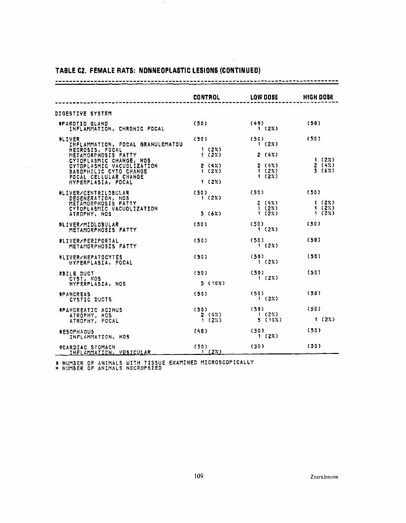
TABLE C2. FEMALE RATS: NONNEOPLASTIC LESIONS (CONTINUED) . . . . . . . . . . . . . . . . . . . . . . . . . . . . . . . . . . . . . . . . . . . . . . . . . . . . . . . . . . . . . . . . . . . . . . . . . . . . . . . .
CONTROL LOW DOSE HIGH DOSE. . . . . . . . . . . . . . . . . . . . . . . . . . . . . . . . . . . . . . . . . . . . . . . . . . . . . . . . . . . . . . . . . . . . . . . . . . . . . . . . D I G E S T I V E SYSTEM
# P A R O T I D GLAND I N F L A M M A T I O N , C H R O N I C FOCAL
( 9 0 )
# L I V E R I N F L A M M A T I O N , FOCAL N E C R O S I S . FOCAL r i E iAiioRPHos I s F A T T Y
GRANULOMATOU ( 5 0 )
1 ( 2 % ) t ( 2 % )
( 5 0 ) 1 ( 2 % )
2 ( 4 % ) C Y T O P L A S M I C CHANGE, NOS C Y T O P L A S M I C V A C U O L I Z A T I O N B A S O P t i I L I C C FOCAL C E L L U L H Y P E R P L A S I A ,
:YTO CHANGE AR CHANGE
FOCAL
2 (4%)1 ( 2 % )
1 ( 2 % )
2 ( 4 % )1 ( 2 % ) 1 ( 2 % )
# L IV E R I C EH T R I L 0BU L A R DEGEN ERA T I0N, N 0S METAMORPHOSIS F A T T Y C Y T O P L A S M I C V A C U O L I Z A T I O N ATROPHY, NOS
( 5 0 )1 ( 2 % )
3 ( 6 % )
( 5 0 )
2 (4%)1 ( 2 % ) 1 ( 2 % )
# L I V E R / M I D L O B U L A RMETAMORPHOSIS F A T T Y
( 5 0 ) ( 5 0 ) 1 ( 2 % )
# L I V E R / P E R I P O R T A LMETAf lORPHOSIS F A T T Y
( 5 0 ) ( 5 0 ) 1 (2%)
# L I V E R / H E P A T O C Y T E SH Y P E R P L A S I A , FOCAL
( 5 0 ) ( 5 0 )1 ( 2 % )
# B I L E DUCT CYST NOS H Y P E R P L A S I A , NOS
( 5 0 )
5 ( 1 0 % )
( 5 0 )1 ( 2 % )
#PANCREAS C Y S T I C DUCTS
( 5 0 ) ( 5 0 )1 ( 2 % )
# P A N C R E A T I C A C I N U S ATROPHY, ATROPHY,
NOS FOCAL
#ESOPHAGUSINFL A MMA T ION , N O S
( 4 8 )
# C A R D I A C STOMACH I N F L A M M A T I O N , V E S I C U L A R
( 5 0 )1 ( 2 % )
# NUMBER OF A N I M A L S W I T H T I S S U E E X A M I N E D M I C R O S C O P I C A L L Y W NUMBER O F A N I M A L S N E C R O P S I E D
( 5 0 )
1 ( 2 % )2 ( 4 % )3 (6%)
( 5 0 )
1 ( 2 % )1 ( 2 % )1 ( 2 % )
( 5 0 )
( 5 0 )
( 5 0 )
( 5 0 )
( 5 0 )
109 Zearalenone

TABLE C2. FEMALE RATS: NONNEOPLASTIC LESIONS (CONTINUED) ................................................................................
................................................................................ CONTROL LOW DOSE HIGH DOSE
U R I N A R Y SYSTEM
# K I D N E Y ( 5 0 ) ( 5 0 ) ( 5 0 )HYDRONEPHROSIS 1 ( 2 % ) N E P H R O S I S , NOS 10 ( 2 0 % ) 2 4 (48%) 26 ( 5 2 % ) N E C R O S I S , NOS 1 ( 2 % )
#K IDN E Y / P E L V I S ( 5 0 ) ( 5 0 ) ( 5 0 )I N F L A M M A T I O N , A C U T E I C H R O N I C 1 (2%)C A L C I F I C A T I O N , NOS 1 ( 2 % )
# U R I N A R Y BLADDER ( 4 9 ) ( 4 8 ) ( 4 8 ) H Y P E R P L A S I A , E P I T H E L I A L 1 (2%) ................................................................................
E N D O C R I N E SYSTEM
# P I T U I T A R Y ( 4 9 ) ( 5 0 ) ( 4 9 )H Y P E R P L A S I A , NOS 6 (12%) 4 (8%) 7 ( 1 4 % )H Y P E R P L A S I A , FOCAL 1 (2%) 1 (2%)
# A N T E R I O R P I T U I T A R Y ( 4 9 ) ( 5 0 ) HEMORRHAGIC CYST A N G I E C T A S I S
#ADRENAL ( 5 0 ) ( 5 0 ) C A L C I F I C A T I O N , FOCAL 1 (2%) C Y T O P L A S M I C V A C U O L I Z A T I O N 2 (4%)
#ADRENAL CORTEX ( 5 0 ) ( 5 0 ) ( 5 0 ) 11EM0 RRH A GIC CYST 1 (2%) C Y T O P L A S M I C V A C U O L I Z A T I O N 1 (2%) 1 ( 2 % )H Y P E R P L A S I A , FOCAL 1 (2%)
#ADRENAL MEDULLA ( 5 0 ) ( 5 0 ) ( 5 0 )HEMORRHAGIC CYST 1 (2%)H Y P E R P L A S I A , NOS 1 ( 2 % )
# T H Y R O I D ( 4 9 ) ( 5 0 ) ( 4 9 )U L T I M O B R A N C H I A L CYST 2 (4%)C Y S T I C F O L L I C L E S 1 ( 2 % )H Y P E R P L A S I A , C Y S T I C 1 ( 2%)H Y P E R P L A S I A , C-CELL 9 (18%) 6 ( 1 2 % ) 7 ( 1 4 % )
# T H Y R O I D P A R A F O L L I C U L ( 4 9 ) ( 5 0 ) ( 4 9 ) RTROPHY, FOCAL 1 (2%)-
I NUMBER OF A N I M A L S W I T H T I S S U E E X A M I N E D M I C R O S C O P I C A L L Y * NUMBER OF A N I M A L S N E C R O P S I E D
Zearalenone 110

TABLE C2. FEMALE RATS: NONNEOPLASTIC LESIONS (CONTINUED) . . . . . . . . . . . . . . . . . . . . . . . . . . . . . . . . . . . . . . . . . . . . . . . . . . . . . . . . . . . . . . . . . . . . . . . . . . . . . . . .
R E P R O D U C T I V E SYSTEM
# M A MM A R Y G L A N D ( 5 0 ) ( 5 0 ) ( 5 0 ) GALACTOCELE 1 ( 2 % ) CYST, N O S 1 0 ( 2 0 % ) 3 4 (68%) t o ( 2 0 % ) C Y S T I C DUCTS 1 8 ( 3 6 % ) 1 8 ( 3 6 % ) ABSCESS, NOS 1 ( 2 % ) IN F L AMMAT I O N , C H R O N I C 1 ( 2 % )
1 ( 2 % )H Y P E R P L A S I A , C Y S T I C 1 ( 2 % ) A D E N O S I S 2 ( 4 % ) 2 ( 4 % )
X MAP1MA RY L 0B U L E ( 5 0 ) ( 5 0 ) ( 5 0 ) H Y P E R P L A S I A , NOS 1 (2%) 1 (2%)
% P R E P U T I A L GLAND ( 5 0 ) ( 5 0 ) ( 5 0 ) C Y S T I C DUCTS 1 ( 2 % ) IN F L ArlMA T ION , NOS 1 ( 2 % ) IN F L A K I A TION, SUP P U R A TIV E 1 ( 2 x 1 2 (4%) H Y P E R P L A S I A , C Y S T I C 2 (4%) 2 (4%) 1 ( 2 % )
# C L I T O R A L GLAND ( 5 0 ) ( 5 0 ) ( 5 0 ) H Y P E R P L A S I A , FOCAL 1 ( 2 % )
#UTERUS (50) ( 4 9 ) ( 4 9 ) HYDROMETRA 1 ( 2 % ) 3 (6%) HEMATOMETRA 1 ( 2 % )
R C E R V I X U T E R I ( 5 0 1 ( 4 9 ) ( 4 9 ) H Y P E R P L A S I A , E P I T H E L I A L 1 ( 2 % )
(50) ( 4 9 ) ( 4 9 )#UTERUS/ENDOMETRIUM HYPERPL A S 1 A , FOCAL 1 ( 2 % ) 1 ( 2 % ) H Y P E R P L A S I A , C Y S T I C 2 ( 4 % ) 5 ( 1 0 % )
'NERVOUS SYSTEM
*CHOROID PLEXUS L A T E R (50) ( 5 0 ) ( 5 0 ) A N G I E C T A S I S 1 ( 2 % )
# NUMBER OF A N I M A L S W I T H T I S S U E E X A M I N E D M I C R O S C O P I C A L L Y X NUMBER OF A N I M A L S N E C R O P S I E D
1 1 1 Zearalenone

TABLE C2. FEMALE RATS: NONNEOPLASTIC LESIONS (CONTINUED)
S P E C I A L SENSE ORGANS
XEYE HEMORRHAGE I N F L Ai l r l AT I O N , CHRONIC R ET INOP A THY CATARACT
% EY E ICORNEA I N F L AMFl AT I O N , FOCAL
% EYE/ R ET IN A ATROPHY, NOS
N E Y E I C R Y S T A L L I N E L E N S M I N E R A L I Z A T I O N
MZYMBAL 'S GLAND H Y P E R P L A S I A , C Y S T I C
3 ( 6 % )4 (8%)
( 5 0 )
( 5 0 )
( 5 0 )
6 ( 1 2 % )1 ( 2 % )
28 ( 5 6 % )26 ( 5 2 % )
( 5 0 )
( 5 0 )3 ( 6 % )
( 5 0 )1 (2%)
( 5 0 )
1 ( 2 % )
8 ( 1 6 % ) 6 ( 1 2 % )
( 5 0 ) 1 ( 2 % )
( 5 0 )1 ( 2 % )
( 5 0 )
( 5 0 ) 1 ( 2 % )
BODY C A V I T I E S
ALL OTHER SYSTEMS
% M U L T I P L E ORGANS ( 5 0 ) ( 5 0 )HEMORRHAGE 1 ( 2 % )
# NUMBER NUMBER
OF A N I M A L S OF A N I M A L S
W I T H T I S S U E N E C R O P S I E D
E X A M I N E D M I C R O S C O P I C A L L Y
( 5 0 )
Zearalenone 112

TABLE C2. FEMALE RATS: NONNEOPLASTIC LESIONS (CONTINUED) ................................................................................
T A I L H Y P E R K E R A T O S I S 1
FOOT H Y P E R K E R A T O S I S 1
OMEHTUM NECROSIS , F A T 2 1
S P E C I A L MORPHOLOGY SUMMARY
H 0 L E S I O N REPORTED 4. . . . . . . . . . . . . . . . . . . . . . . . . . . . . . . . . . . . . . . . . . . . . . . . . . . . . . . . . . . . . . . . . . . . . . . . . . . . . . . . # NUNBER OF A N I M A L S W I T H T I S S U E E X A M I N E D M I C R O S C O P I C A L L Y
NUMBER OF A N I M A L S N E C R O P S I E D
I13 Zearalenone

Zearalenone 114

APPENDIX D
SUMMARY OF THE INCIDENCE OF NONNEOPLASTIC LESIONS IN MICE FED DIETS CONTAINING ZEARALENONE
I I5 Zearalenone

TABLE 01.
SUMMARY OF THE INCIDENCE OF NONNEOPLASTIC LESIONS IN MALE MICE FED DIETS CONTAINING ZEARALENONE
N S K I N ( 5 0 ) (50)IN F L A PIN A T I 0N, A CUT E F0CA L 3 ( 6 % ) IN FL ANMA T ION , Cl lR ON IC 1 ( 2 % ) I N F L A M M A T I O N , C H R O N I C FOCAL 3 ( 6 % ) 2 (4%)
NSUBCUT T I S S U E ( 5 0 ) ( 5 0 ) (50)I N F L A M M A T I O N ACUTE AND C H R O N I C 1 ( 2 % )I N F L A M M A T I O N , CHRONIC FOCAL 1 ( 2 % ). . . . . . . . . . . . . . . . . . . . . . . . . . . . . . . . . . . . . . . . . . . . . . . . . . . . . . . . . . . . . . . . . . . . . . . . . . . . . . . .
R E S P I R A T O R Y SYSTEM
#LUNG ( 5 0 ) ( 4 9 ) (50)CONGESTION, NOS 1 ( 2 X )B R 0N C H0I'N EUMO N IA , FO C A L 14 ( 2 8 % ) 6 (12::) 12 ( 2 4 % )P N E U V O N I A , CHRONIC P l U R I N E 1 ( 2 % ) H Y P E R P L AS IA , A D ENO MAT 0US 1 ( 2 % ) H Y P E R P L A S I A , ALVEOLAR E P I T H E L I U M 1 (Z?!) . . . . . . . . . . . . . . . . . . . . . . . . . . . . . . . . . . . . . . . . . . . . . . . . . . . . . . . . . . . . . . . . . . . . . . . . . . . . . . . .
H E M A T O P O I E T I C SYSTEM
# H A R D E R I A N GLAND ( 5 0 ) ( 5 0 ) ( 5 0 )H Y P E R P L A S I A , L Y M P H O I D 7 ( 1 4 % ) 7 ( 1 4 % ) 8 ( 1 6 % )
# S P L E E N ( 5 0 ) H Y P E R P L A S I A , t i O S H Y P E R P L A S I A LYPIPHOID
# P A N C R E A T I C L . N O D E ( 5 0 )INF L A MMA T I0N, G R A N U L 0MA T 0US
# M E S E N T E R I C L . NODE (50) ( 4 9 ) ( 5 0 )A N G I E C T A S I S 1 ( 2 % ) 1 ( 2 % )
# NUMBER O F A N I M A L S W I T H T I S S U E E X A M I N E D M I C R O S C O P I C A L L Y * NUMBER OF A N I M A L S N E C R O P S I E D
Zearalenone 116

TABLE D1. MALE MICE: NONNEOPLASTIC LESIONS (CONTINUED) . . . . . . . . . . . . . . . . . . . . . . . . . . . . . . . . . . . . . . . . . . . . . . . . . . . . . . . . . . . . . . . . . . . . . . . . . . . . . . . .
(SACRAL LYMPH NODE H Y P E R P L A S I A , L Y M P H O I D
( 5 0 )
# I N G U I N A L LYMPH NODE H Y P E R P L A S I A , L Y M P H O I D
( 5 0 )
RPEYER’S PATCH H Y P E R P L A S I A , L Y M P H O I D
( 4 9 )
#THYMUS CYST, NOS
( 4 8 ) ( 3 9 )1 ( 3 % )
( 4 4 )
C I R C U L A T O R Y SYSTEM
#HEART ( 5 0 ) ( 5 0 ) ( 5 0 ) I N F L A M M A T I O N , CHRONIC FOCAL 1 ( 2 % ) . . . . . . . . . . . . . . . . . . . . . . . . . . . . . . . . . . . . . . . . . . . . . . . . . . . . . . . . . . . . . . . . . . . . . . . . . . . . . . . .
D I G E S T I V E SYSTEM
# L I V E R ( 5 0 ) ( 5 0 ) (49) CYST, NOS 1 ( 2 % ) 1 ( 2 % ) I N F L A M M A T I O N , CHRONIC 1 ( 2 % )F I B R O S I S 1 ( 2 % ) HECROSIS, NOS 1 ( 2 % ) 2 ( 4 % ) N E C R O S I S , CASEOUS 1 ( 2 % ) C Y T O L O G I C A L T E R A T I O N , NOS 4 ( 8 % ) 1 ( 2 % ) 1 ( 2 % )
#PANCREAS ( 5 0 ) ( 4 9 ) ( 4 9 ) D I L A T A T I O N / D U C T S 1 (2!5) ATROPHY, NOS 1 ( 2 % ) . . . . . . . . . . . . . . . . . . . . . . . . . . . . . . . . . . . . . . . . . . . . . . . . . . . . . . . . . . . . . . . . . . . . . . . . . . . . . . . .
U R I N A R Y SYSTEM
# K 1 DNEY ( 5 s ) ( 5 0 ) ( 5 0 )HYDRONEPHROSIS 1 ( 2 % )CYST, NOS 1 ( 2 % ) IN F L A PIMA T I0N t SUP P U R A T IV E 1 ( 2 % )I N F L A P l M A T I O N t ACUTE S U P P U R A T I V E 1 ( 2 % )
# U R I N A R Y BLADDER ( 4 9 ) (50) ( 5 0 ) L Y M P H O C Y T I C I N F L AMPMTORY I N F I L T R 1 (2%)
# NUMBER OF A N I M A L S W I T H T I S S U E E X A M I N E D M I C R O S C O P I C A L L Y * NUMBER OF A N I M A L S N E C R O P S I E D
I17 Zearalenone

TABLE 01. MALE MICE: NONNEOPLASTIC LESIONS (CONTINUED)
# P I T U I T A R Y HY P ERPL A S 1 A, FOCAL
( 4 0 ) ( 4 5 ) (44)1 ( 2 % )
#ADRENAL CORTEX H Y P E R P L A S I A , FOCAL
( 5 0 ) ( 5 0 ) ( 4 9 )1 ( 2 % )
#ADRENAL MEDULLA H Y P E R P L A S I A , FOCAL
( 5 0 )
R E P R O D U C T I V E SYSTEM
*MAMMARY GLAND ( 5 0 ) ( 5 0 ) ( 5 0 )C Y S T I C DUCTS 2 (4%)
% P R E P U T I A L GLAND ( 5 0 ) ( 5 0 ) ( 5 0 )CYST, NOS 3 ( 6 % ) 2 (4%) 3 ( 6 % )C Y S T I C DUCTS 1 (2%) I ( 2 % )I N F L A M M A T I O N , ACUTE S U P P U R A T I V E 1 ( 2 % )I N F L A M M A T I O N ACUTE AND CHRONIC 2 (4%) 2 (4%) 1 ( 2 % )
#PROSTATE ( 5 0 ) ( 5 0 ) ( 5 0 ) I N F L A M M A T I O N , ACUTE S U P P U R A T I V E 1 (2%)
# S E M I N A L V E S I C L E ( 5 0 ) ( 5 0 )D I ST E N 1 ION 1 ( 2 % )IN F L APlMAT ION, CHRONIC
# T E S T I S ( 5 0 ). ( 5 0 )F I B R O S I S 1 ( 2 % )N E C R O S I S , NOS 1 ( 2 % )
# NUMBER OF A N I M A L S W I T H T I S S U E E X A M I N E D M I C R O S C O P I C A L L Y W NUPlBER OF A N I M A L S N E C R O P S I E D
Zearalenone 1 I8

TABLE D1. MALE MICE: NONNEOPLASTIC LESIONS (CONTINUED) ................................................................................
MUSCULOSKELETAL SYSTEM
*ABDOMINAL MUSCLE ( 5 0 ) ( 5 0 ) ( 5 0 ) INFLAMMATION, ACUTE SUPPURATIVE 1 ( 2 % ) . . . . . . . . . . . . . . . . . . . . . . . . . . . . . . . . . . . . . . . . . . . . . . . . . . . . . . . . . . . . . . . . . . . . . . . . . . . . . . . .
BODY C A V I T I E S
*PERITONEUM ( 5 0 ) INFLAMMATION, SUPPURATIVE INFLAMMATION, ACUTE SUPPURATIVE
SPECIAL MORPHOLOGY SUMMARY
NO L E S I O N REPORTED 1 4 6. . . . . . . . . . . . . . . . . . . . . . . . . . . . . . . . . . . . . . . . . . . . . . . . . . . . . . . . . . . . . . . . . . . . . . . . . . . . . . . . W NUMBER OF ANIMALS W I T H T I S S U E EXAMINED MICROSCOPICALLY * NUMBER OF ANIMALS NECROPSIED
1 I9 Zearaienone

TABLE D2.
SUMMARY OF THE INCIDENCE OF NONNEOPLASTIC LESIONS IN FEMALE MICE FED DIETS CONTAINING ZEARALENONE
CONTROL LOW DOSE HIGH DOSE. . . . . . . . . . . . . . . . . . . . . . . . . . . . . . . . . . . . . . . . . . . . . . . . . . . . . . . . . . . . . . . . . . . . . . . . . . . . . . . . A N I M A L S I N I T I A L L Y I N STUDY 5 0 50 5 0 A N I M A L S N E C R O P S I E D 5 0 4 9 4 9 A N I M A L S E X A M I N E D H I S T O P A T H O L O G I C A L L Y 5 0 4 9 4 9. . . . . . . . . . . . . . . . . . . . . . . . . . . . . . . . . . . . . . . . . . . . . . . . . . . . . . . . . . . . . . . . . . . . . . . . . . . . . . . . INTEGUMENTARY SYSTEM
* S K I N ( 5 0 ) ( 4 9 ) ( 4 9 )EDEMA, NOS 1 ( 2 % )I N F L A M M A T I O N ACUTE AND CHRONIC 1 (2%)I N F L A M M A T I O N , FOCAL GRANULOMATOU 1 ( 2 % )
*SUBCUT T I S S U E ( 5 0 ) ( 4 9 ) ( 4 9 ) I N F L A M M A T I O N ACUTE AND CHRONIC 1 ( 2 % ) . . . . . . . . . . . . . . . . . . . . . . . . . . . . . . . . . . . . . . . . . . . . . . . . . . . . . . . . . . . . . . . . . . . . . . . . . . . . . . . .
R E S P I R A T O R Y SYSTEM
#LUNG ( 4 8 ) ( 4 8 ) ( 4 9 )BROt~CHOPNEUMONIA, FOCAL 3 ( 6 % ) 4 (8%) 3 ( 6 % )BRONCHOPNEUMONIA, CHRONIC 1 ( 2 % )CHOLESTEROL D E P O S I T 1 ( 2 % )H Y P E R P L A S I A , ALVEOLAR E P I T H E L I U M 1 ( 2 % ). . . . . . . . . . . . . . . . . . . . . . . . . . . . . . . . . . . . . . . . . . . . . . . . . . . . . . . . . . . . . . . . . . . . . . . . . . . . . . . .
H E M A T O P O I E T I C SYSTEM
#BONE MARROW ( 4 9 ) ( 4 8 ) ( 4 9 )MY EL 0 F I B R O S I S 8 ( 1 6 % ) 32 ( 6 7 % ) 3 4 ( 6 9 % )
# S P L E E N ABSCESS, NOS
# M E D I A S T I N A L L .NODE ( 5 0 ) ( 4 8 ) ABSCESS, NOS 1 ( 2 % ) H Y P E R P L A S I A , NOS
# C E L I A C LYMPH NODE ( 5 0 ) ( 4 8 ) H Y P E R P L A S I A , L Y M P H O I D 1 ( 2 % )
#RENAL LYMPH NODE ( 5 0 ) ( 4 8 ) ( 4 9 ) H Y P E R P L A S I A , L Y M P H O I D 1 ( 2 % )
t NUMBER OF A N I M A L S W I T H T I S S U E E X A M I N E D M I C R O S C O P I C A L L Y NUMBER OF A N I M A L S N E C R O P S I E D
Zearalenone I 20

TABLE D2. FEMALE MICE: NONNEOPLASTIC LESIONS (CONTINUED) . . . . . . . . . . . . . . . . . . . . . . . . . . . . . . . . . . . . . . . . . . . . . . . . . . . . . . . . . . . . . . . . . . . . . . . . . . . . . . . .
# I L I A C LYMPH NODE H Y P E R P L A S I A , NOS
( 5 0 ) ( 4 8 ) ( 4 9 )1 ( 2 % )
# L I V E R W Y E L O P O I E S I S
( 5 0 )1 ( 2 % )
( 4 9 ) ( 4 9 )
WPEYER'S PATCH H Y P E R P L A S I A , L Y M P H O I D
( 4 9 )
C I R C U L A T O R Y SYSTEM
%MUSCLE H I P I T H I G H ( 5 0 )P E R I V A S C U L I T I S
D I G E S T I V E SYSTEM
# L I V E R ( 5 0 ) ( 4 9 ) ( 4 9 ) CYST. NOS 1 ( 2 % ) 3 ( 6 % ) I N F L A M M A T I O N , CHRONIC FOCAL 1 ( 2 % ) N E C R O S I S , NOS 3 ( 6 % ) C Y T O P L A S N I C V A C U O L I Z A T I O N 1 ( 2 % ) C Y T O L O G I C A L T E R A T I O N , NOS 2 (4%) HEPATOCYTOMEGALY 1 ( 2 % ) 2 (4%) ATROPHY, NOS 1 ( 2 % ) A N G I E C T A S I S 3 ( 6 % )
#PANCREAS ( 4 9 ) ( 4 8 ) ( 4 9 ) I N F L A M M A T I O N ACUTE AND CHRONIC 1 ( 2 % ) 1 ( 2 % )I N F L A M M A T I O N , CHRONIC 1 ( 2 % )I N F L A M M A T I O N , CHRONIC FOCAL 1 ( 2 % ) F I B R O S I S 1 ( 2 % ) F I B R O S I S , FOCAL. 1 ( 2 % )ATROPHY, NOS 3 ( 6 % ) 1 ( 2 % ) 1 ( 2 % )
#STOMACH ( 4 8 ) ( 4 8 ) ( 4 9 )F O R E I G N BODY, NOS 1 ( 2 % )I N F L A M M A T I O H , ACUTE FOCAL f ( 2 % )I N F L A M M A T I O N , ACUTE S U P P U R A T I V E 1 ( 2 % )IN F L ARM A T I O N , FOCAL GR ANUL ON ATOU 1 ( 2 % )
# NUMBER OF A N I M A L S W I T H T I S S U E E X A M I N E D M I C R O S C O P I C A L L Y % NUMBER OF A N I M A L S N E C R O P S I E D
Zearalenone121

CONTROL LOW DOSE HIGH DOSE................................................................................ U R I N A R Y SYSTEM
# K I D N E Y ( 5 0 1 ( 4 8 ) ( 4 9 ) LYMPHOCYTIC INFLAMMATORY I N F I L T R 1 (2%)
# U R I N A R Y BLADDER ( 4 9 ) ( 4 8 ) ( 4 9 ) L Y M P H O C Y T I C INFLAMMATORY I N F I L T R 1 (2%) .INFL APIMA T I0N t ACUTE FOCAL 1 ( 2 % )................................................................................
ENDOCRINE SYSTEM
# P I T U I T A R Y ( 4 6 ) ( 4 3 ) ( 4 2 ) H Y P E R P L A S I A , FOCAL 3 ( 7 % ) A N G I E C T A S I S 1 ( 2 % )
#ADRENAL CORTEX ( 5 0 ) ( 4 9 ) (47) CYST, NOS 1 ( 2 % ) C Y T O P L A S M I C V A C U O L I Z A T I O N 1 ( 2 % ) ATROPHY, NOS 1 ( 2 % )
# T H Y R O I D (47) ( 4 8 ) ( 4 7 ) C Y S T I C F O L L I C L E S 2 (4%)DEGENERATION, C Y S T I C 1 ( 2 % ) 6 ( 1 3 % )H Y P E R P L A S I A , FOCAL 1 ( 2 % )H Y P E R P L A S I A , C Y S T I C 2 (4%)H Y P E R P L A S I A , F O L L I C U L A R - C E L L 3 ( 6 % ) 1 ( 2 % )................................................................................
R E P R O D U C T I V E SYSTEM
HMAtlMARY GLAND ( 5 0 ) ( 4 9 ) ( 4 9 ) C Y S T I C DUCTS 3 ( 6 % ) 15 ( 3 1 % ) M E T A P L A S I A , SQUAMOUS 1 (2%)
H V A G I N A ( 5 0 ) ( 4 9 ) ( 4 9 )I N F L A M M A T I O N , ACUTE S U P P U R A T I V E 3 ( 6 % ) 2 (4%) 5 ( 1 0 % )H Y P E R K E R A T O S I S 1 ( 2 % )
8 l l l F R l l S ( 5 0 ) ( 4 9 ) ( 4 8 )I-.-..-- . _ _ . I N F L A t l f l A T I O N , S U P P U R A T I V E 10 ( 2 0 % ) 1 ( 2 % )I N F L A M M A T I O N , ACUTE S U P P U R A T I V E 2 (4%) 5 ( 1 0 % ) 1 ( 2 % )
# C E R V I X U T E R I ( 5 0 ) ( 4 9 ) ( 4 8 )I N F L A R M A T I O N , ACU T E S U P P U R A T I V E 3 ( 6 % ) 5 ( 1 0 % ) 16 ( 3 3 % )
# NUMBER OF A N I M A L S W I T H T I S S U E E X A M I N E D M I C R O S C O P I C A L L Y W NUMBER OF A N I M A L S N E C R O P S I E D
Zearalenone I22

TABLE D2. FEMALE MICE: NONNEOPLASTIC LESIONS (CONTINUED)
#ENDOMETRIAL GLAND ( 4 8 )H Y P E R P L A S I A t C Y S T I C 1 (2%)
#UTERUS/MYOMETRIUM ( 5 0 ) ( 4 9 ) ( 4 8 )F I B R O S I S , FOCAL 1 ( 2 % ) 5 ( 1 0 % ) 1 7 (35%)
#OVARY ( 4 4 ) (48) ( 4 8 ) CYST, NOS 6 ( 1 4 % ) 2 ( 4 % ) 3 ( 6 % ) I N F L A M M A T I O N t S U P P U R A T I V E 2 (5%)I N F L A M M A T I O N , ACUTE S U P P U R A T I V E 1 ( 2 % ) 1 ( 2 % ). . . . . . . . . . . . . . . . . . . . . . . . . . . . . . . . . . . . . . . . . . . . . . . . . . . . . . . . . . . . . . . . . . . . . . . . . . . . . . . .
NERVOUS SYSTEM
#B R A IN/M ENING ES ( 5 0 ) ( 4 9 ) ( 4 8 )L Y M P H O C Y T I C INFLAMMATORY I N F I L T R 1 (2%). . . . . . . . . . . . . . . . . . . . . . . . . . . . . . . . . . . . . . . . . . . . . . . . . . . . . . . . . . . . . . . . . . . . . . . . . . . . . . . .
S P E C I A L SENSE ORGANS
H E Y E ( 5 0 ) ( 4 9 ) ( 4 9 )R E T I N O P A T H Y 1 ( 2 % ). . . . . . . . . . . . . . . . . . . . . . . . . . . . . . . . . . . . . . . . . . . . . . . . . . . . . . . . . . . . . . . . . . . . . . . . . . . . . . . .
H M U L T I P L E ORGANS ( 5 0 ) ( 4 9 ) M M A T O R Y ~ ~ M I ITR 1 ( 7 % )
' W NUMBER O F A N I M A L S W I T H T I S S U E E X A M I N E D M I C R O S C O P I C A L L Y H NUMBER OF A N I M A L S N E C R O P S I E D
123 Zearalenone

Zearalenone 124

APPENDIX E
ANALYSIS OF ZEARALENONE (MIDWEST RESEARCH INSTITUTE)
Zearalenone125

APPENDIX E
A. ELEMENTAL ANALYSIS
Element C H
Batch 01 Theory Determined
67.90 67.78
6.97 6.88
67.92 6.95
Batch 02 Determined 67.76 6.82 67.87 6.95
Batch 03 Determined 68.12 7.05 68.14 7.04
B. WATER ANALYSIS (Karl Fischer) Batch 01 0.14 k 0.01 (b)% Batch 03 0.05%
C. MELTING POINT Determined Literature Values
Batch 01 m.p.: 162' to 164°C (visual, capillary)
164' to 165°C (Urrq et al., 1977)
Batch 02 m.p.: 162"-163'C (visual, capillary)
D. OPTICAL ROTATION
Determined Literature Values [a]&= -170.5 (Urrq et al., 1977) Batch 01 [a],?:, -137.2+0.9 (6)'
concentration I .O% in concentration = 1.0% in methanol methanol
E. THIN-LAYER CHROMATOGRAPHY
I . Batch 01
Plates: Silica Gel 60 F-254 Ref. Standard: Resorcinol
Amounted spotted: 100 and 300 pug Visualization: Ultraviolet, 254 and 366 nm and 50% sulfuric acid
System I : Ch1oroform:acetone System 2: Benzene:methanol (90: IO) (90: IO)
Rf: 0.53, origin (trace) Rf: 0.56, origin (trace) R+t:2.0, origin Rst: 3.5, origin
2. Batch 02
Plates: Silica gel G F-254 Ref. Standard: Resorcinol
Amount Spotted: I O and 30 pl, Visualization: Ultraviolet, 254 I O pglpl in methanol and 366 nm and 50% aqueous
sulfuric acid
System I : Ch1oroform:acetone System 2: Benzene:methanol (90: IO) (90: IO)
Rf: 0.61. origin Rf: 0.63 Rst: 2.65, origin Rst: 3.00
Zearalenone 126

APPENDIX E
3. Batch 03 Plates: Silica gel 60 F-254 Amount Spotted: IO, 100, and 300 pg
( I O pg/pl in acetone)
System I : Ch1oroform:acetone (90:I 0)
Rf: 0.58 (ma,jor); 0.20 (slight trace) RSt: 3.45 (major); 1.18 (slight trace)
Ref. Standard: Resorcinol, 10 pg (10 pg/p l in acetone)
Visualization: Ultraviolet (254 and 366 nm) and 50% aqueous sulfuric acid
System 2: To1uene:methanol (90:10)
Rf: 0.35 (major); 0.14 (trace) Rst: 3.19 (major); 1.33 (trace)
F. HIGH P E R F O R M A N C E LIQUID CHROMATOGRAPHY
I . Batch 01: Instrument: Waters ALC 202 with Model 660 Solvent Programmer Detection: Ultraviolet 254 nm a. System I :
Column: pBondapak Cl8 - 300 x 4 mm ID, stainless steel
Solvent: A:B (80:20) to 100% B A - 10% acetonitrile in water
B - 100% acetonitrile
Program: Six (linear)
Program time: 15 min
Flow: I ml/min Results: Single homogeneous peak
Retention time: 14.5 min
b. System 2:
Column: pPorsasil - 300 x 4 mm ID, Solvent: Ch1oroform:tetrahydrofuran (95:5), isocratic
Flow: 1 mlimin
Results: Single homogeneous peak Retention time: 7.0 min
2. Batch 02: Instrument: Waters ALC 202 Detection: Ultraviolet, 280 nm Column: pBondapak Cig, 300 mm x 4 mm ID Solvent: Acetonitri1e:water (60:40), isocratic Flow Rate: 1 ml/min Amount Injected: 2 pl, I .9 pg /p l in acetonitrile Results: Single homogeneous peak, retention time 5.5 min
127 Zearalenone

APPENDIX E
3. Batch 03: Instrument: Waters ALC 202 with Waters 6000A pump with Model 660 Solvent
Programmer and Waters 440 detector Detection: Ultraviolet, 254 nm Column: pBondapak Cig, 300 mm x 3.9 mm ID Guard Column: C0:PELL O D s , 72 mm x 2.3 mm ID Solvent: Acetonitrile: water (60:40), isocratic: Flow Rate: I ml/min Samples Injected: Solution (10 MI) of 1.0 mg/ml
Zearalenone in methanol, filtered Results: Major peak and one impurity before the major
peak
Retention
Peak No. Retention
Time (min) Time (Relative to Major Peak)
Area (Percent of Major Peak)
I 12.8 0.67 0.04 2 19.2 1 .oo IO0
Using isocratic solvent systems of 35% A:65% B, 30%A:70% B, and 20% A:80% B, no additional impurities were observed.
G. SPECTRAL DATA I . Infrared
a. Batch 01: Instrument: Beckman IR-I2 Cell: Neat, melt on sodium
chloride plates
Results: Figure 5 Consistent with literature spectrum (Mirocha et al., 1967)
b. Batch 02: Instrument: Perkin Elmer
Model 137 lnfracord
Cell: Melt on sodium chloride plates
Results: Figure 6 Consistent with literature spectrum (Mirocha et al., 1967)
c. Batch 03: Instrument: Beckman IR-12
Cell: 1 % in KBr pellet
Results: Figure 7 Consistent with literature spectrum (Mirocha et al., 1967)
Zearalenone 128

d L
c
0
-I
Y
129 Zearalenone

0
0
a
08 0
0
0
c
-sv
8 rr, c
0
0
0
-00c? 0
0
0d
Zearalenone
I30
CI
ml
0
.. u)
?
m
d z
’c
0
E
E t
VJ
‘DE E
W-e
-cd

CI
m
0
d z
IC
0
E
25P
M
Zearalenone 131

APPENDIX E
2. Ultraviolet Visible:
Instrument: C a r l 1 I8 a . Batch 01:
Determined Literature Values (Urry et al., 1977)
A max(nm) E x 10-3 A max(nm) E x 10-3 235.5 27.9 f0.2 (6) 236 29.7 273.5 12.3 k 0.1 (6) 274 13.9 314.5 5.60 f 0.06 (6) 316 6.02
No absorbance betueen 350 and 800 nm (cisible range) a t a concentration of 0.1 mg ml
Solcent: Methanol
b. Batch 03: Instrument: Cary 118
No maxima from 800 to 350 nm (visible region) but a n increase in absorbance toward 350 nm at a concentration of 0. I ?+.
A max(nm) E x 10-3 A max(nm) E x 10-3
236 29.25 !c 0.0056(6) 236 29.7 274 13.36 !c 0.0077(6) 274 13.9 314 15.87 5 0.0017(6) 316 60.2
Solcent: Methanol
3. Nuclear Magnetic Resonance:
a . Batch 01: Experimental spectrum consistent with structure.
Instrument: Varian HA-IO0 Literature spectrum (Urry et al., 1977) differs in the following
Solvent: Methanol-d-4 with respects: internal tetramethylsilane Protons (h) and ( i ) a re
not resolved. T h e -OH Assignments: Figure 8 protons a re resolved into
singlets further downfield than the other signals
(The proton designations are from zearalenone. shown in Figure IO) Solvent: CDCI3
(a) d , 6 1.35 ppm, J,f = 6 Hz (b) m, 6 1.65 ppm (c) m, 6 2.19 ppm (d) m, 6 2.48 to 3.16 ppm (e) m. 6 4.95 ppm. not resolved (0m, 6 4.95 ppm (g) m. 6 5.73 ppm (h) d. 6 6.30 ppm, J h , 2 Hz ( i ) d . 6 6.44 ppm (j) d. 6 7.05 ppm, J,, 16 H7
Zearalenone 132

0z Zearalenone
133

APPENDIX E
Integration ratios: (a) 2.74 (b) 5.48 (c) 4.41 (d) 2.88 (e$)-OH from solvent and protons from zearalenone (8) 0.72 (h) 1.01 ( i ) 1.01 fi) 0.72
b. Batch 02: Instrument: Varian HA-100 Solvent: Methanol-d-4 with internal tetramethylsilane
Assignments: Figure 9
(a) d, 6 1.32 ppm, Jar = 6 Hz; (b) m, 6 1.64 ppm; (C) t, 6 2.14 ppm, Jbc = 9 Hz; (d) m, 6 2.44-2.98 ppm; (e,f) m, 6 4.77 ppm; (g) m, 6 5.64 ppm, Jgi = 15 Hz; (h) d, 6 6.15 ppm, J g h 2 Hz; (i) d, 6 6.30 ppm; G ) d, 6 6.90 ppm Integration ratios: (a) 3.37 (b) 5.28 (c) 4.43 (d) 1.90 (e,f) -OHfrom solvent and protons from zearalenone, (g) 1.06 (h) 0.95 (i) 1.06 (j)0.95
c. Batch 03: Instrument: Varian EM-360A Solvent: Methanol-d-4 with internal tetramethylsilane Assignments: Figure 10 (a) d, 6 1.37 ppm, Ja-r = 6 Hz (b) m, 6 I .68 ppm (c) m, 6 2.23 ppm (d) m,6 2.50-3.18 ppm (e) s, 6 4.85 ppm (0m, 6 4.95 ppm (8) m, 6 5.67 ppm (h) d, 6 6.23 ppm, Jh-i = 2 HZ (i) d, 6 6.35 ppm G ) d, 6 6.97 ppm, Jg-j = 15 HZ
Zearalenone 134

dz
Zearalenone 135

m
0
dz
'! +
0
nd 4
0.
c -u
.?
-&
r k
Zearalenone I36

APPENDIX E
Integration ratios: (a) 3.62 (b) 5.18(4 4.69 (d) 1.90 (e) -OH and MeOH unresolved (0 -OH and MeOH unresolved (g) 0.92 (h) 0.94 (i) 1.07 ti) 0.74
137 Zearalenone

Zearalenone 138

APPENDIX F
ANALYSIS OF FORMULATED DIETS FOR STABILITY OF ZEARALENONE
(MIDWEST RESEARCH INSTITUTE)
139 Zearalenone

APPENDIX F
A. Mixing and storage: Zearalenone and Wayne Lab-Blox@ rodent feed were mixed in a mortar in two different concentrations: high (100,000ppm) and low (1,400 ppm). Samples of the mixtures were removed and stored at 2 weeks at -2OoC, 5 O C , 25OC, and45OC, respectively. The samples of high zeara- lenone concentration were analyzed by high-pressure liquid chromatography and those of low zeara- lenone concentration were analyzed by ultraviolet spectrophotometric analysis.
B. Extraction: One-gram samples of each of the above mixtures were mixed with 50 ml of methanol in an ultrasonic vibratory bath for 30 seconds and then were triturated for 1 minute using a Polytron@ high-speed blender. The mixture was centrifuged, and the supernatant solution was decanted into a 100- ml volumetric flask. This extraction was repeated on the feed residue, and the combined supernatant solutions were brought to volume with fresh methanol.
C. Analysis
1. High iearalenone concentration (100,000 ppm 10.0%) Instrument: Waters ALC 202 Column: p-Bondapak Ci8, 300 x 4 mm ID Solvent: Acetonitri1e:Water (60:40)
Detection: Ultraviolet, 254 nm
Flow rate: 1 .O ml/ min
Results:
Average Percent of Sample ("C) Recovered Compound (a)
-20 10.8 +_ 0.5 5 10.2 f0.5
25 10.4 f 0.5 45 9.7 f0.5
(a) Corrected for a spiked recovery value of 99.1%; theoretical 100% value lO.O'% 2. Low zearalenone concentration (1,400 ppm 0.14%)
Instrument: Cary 118 Spectrophotometer Absorbance wavelength: 236 nm Results:
Average Percent of Sample ("C) Compound Recovered (a)
-20 0.14 f0.02 5 0.14 f 0.02
25 0.15 * 0.02 45 0.12 f 0.02
(a) Corrected for a spiked recovery value of 100%; theoretical 100% value 0.14% D. Conclusion: Zearalenone mixed with feed is stable for 2 weeks at temperatures of up to 45OC.
Zearalenone 140

APPENDIX G
ANALYSIS OF FORMULATED DIETS FOR CONCENTRATIONS OF ZEARALENONE
(SOUTHERN RESEARCH INSTITUTE)
Zearalenone141

APPENDIX G
A. Gas Chromatography Method Five-gram samples of the chemical feed mixture were triturated with 50 ml of chloroform with a
Polytron@ high-speed blender for 90 seconds. The mixture was filtered through a glass fiber filter and the extraction tube was rinsed with two 20-ml portions of chloroform which were used to wash the filtered feed residue. The feed was washed with a final I O ml of chloroform. The combined chloroform extract was transferred to a 150-ml beaker and evaporated to dryness. The oil-like residue was trans- ferred to a heptane-packed f lor id column, I cm by 25 cm, with a few ml of chloroform. The column was eluted with successive portions of solvent: 50 ml of heptane, 100 ml of chloroform, and 65 ml of ether. The ether fraction was evaporated to dryness and reserved for GC analyses of zearalenone. The oil residue from the ether fraction was derivatized with 0.5 ml of Tri-SiljBSA reagent. The plain feed, spiked plain feed, and chemical/feed samples were analyzed by vapor-phase chromatography.
GC Conditions:
Column: 3% OV-1 on SO/ 100 supelcoport, 1.8 m x 4mm ID, glass
Temperatures: Oven 26OoC, isothermal Inlet 260°C FID 300°C
Carrier Flow: N2, 40ccimin Retention time: 3.3 min Sample size: 2 pl
B. High Pressure Liquid Chromatography Method Exrraction: Ten-gram samples of the chemical/vehicle mixture were accurately weighed and trans-
ferred into large sample tubes. Acetonitrile (40 m) was added to each sample. Plain feed blanks and spiked plain feeds were prepared by weighing undosed feed (10 g) into large sample tubes and spiking each sample with acetonitrile or zearalenone in acetonitrile. A stock solution of zeralenone in acetoni-trile was prepared and used to spike samples at several concentrations in the specified dosage range of 0.00I-0.05%.
The samples were extracted by triturating with acetonitrile using the Polytron@ High Speed Blender. After triturating for 60 seconds, the samples were filtered through a Fibergla@ filter using a Millipore@ filtering apparatus. The feed residue was then reextracted with another 40 ml aliquot of acetonitrile. The combined extracts were then brought to a 100-mlvolume with acetonitrile. These extract solutions were then evaporated to an oily residue on a rotary evaporator.
Column Chromatograph?,: Silica gel columns were prepared by first placing a ball of glass wool loosely in the bottom of the chromatographic column. Approximately 5 g of anhydrous sodium sulfate was added to give an even base for the silica gel. Chloroform was added until the columns were about one-half full; then I O g of silica gel (ICN Pharmaceuticals, Inc., 100-2OOp m, activated by drying overnight at 105"C, then adding 1 % water by weight, sealing, shaking until thoroughly mixed, and stored in an airtight container). The sides of the column were washed with approximately 20 ml chloroform. and the silica gel was stirred to disperse it into the chloroform. When the rate of settling slowed. some of the chloroform was drawn off to aid in settling. leaving 2 to 3 in. of chloroform above the silica gel. Fifteen grams of anhydrous sodium sulfate was then slowly added and the chloroform was drawn off to the top of the sodium sulfate.
The oil residue of the acetonitrile extraction was mixed with 5 ml chloroform, followed by a IO-ml chloroform rinse. and transferred to 150 ml hexane. This solution was added to the chromatographic column and eluted at a maximum flow rate of 10-20 ml/min. The elution was continued with 150 ml of benzene. These hexane and ben7ene eluates were discarded. and the 7caralenone was eluted from the column with 250 ml acetone:ben~ene (5:95. vlv).
The acetone:beniene fraction was evaporated on a rotary evaporator until only an oil residue was left. The residue was dissolved in 4 ml of methanol, and filtered solutions were analy7.ed by high-pressure liquid chromatography.
Zearalenone 142

APPENDIX G
High Pressure Liquid Chromatography: Instrument System:
Pumps: Waters 6000 A Programmer: Waters 660 Detector: Waters 440 at x 0.1 attenuation Injector: Waters U6K Detection: Ultraviolet. 3 13 nm Column: yBondapak C I S , 300 x 3.9 mm ID with
CO-Pel1 ODS guard column, 72 x 2.3 mm ID Solvent System: Ion Pairing
A: 0.005 M tetrabutylarnmonium hydroxide (TBA) in water, adjusted to pH 7.3-7.4 with 1 % phosphoric acid
B: 0.005 M tetrabutylammonium hydroxide (TBA) in methanol, adjusted to pH 7.3-7.4with 1% phosphoric acid
Program: 60% B, 40% A isocratic Flow Rate: I ml/min Sample Injected: I O pl Data Recorded on Hewlett-Packard 3380s Integrator Retention Time of Zearalenone: 13 min
C. Results See Table G I .
143 Zearalenone

TABLE C l . ANALYSIS OF FORMULATED DIETS
Concentration (a) of Zearalenone for Target Concentration of:
Week Mixed Week Used 100 ppm 50 Ppm 25 Ppm
Analyses Performed Using Original Procedure for Gas Chromatography
021 14/78 021 21 / 78 96 50 19 03/21/78 03/ 281 79 52 28 04/ 11/78 041 18/78 I I5 55
58 05/OS/ 78 051 16/78 38
38 061 131 78 061201 78 30 16
21 08jO I / 78 OS/ 08178 81 37 18 081 291 78 09/05/ 78 92 40 19 091261 78 l0/03/ 78 96 53 27 10/24/78 l0 /31/ 78 57 27 11/28/78 12/05/78 46 24
45 12/ 19/78 121 261 78 1 I O 41 16
Mean (ppm) 98.3 47.0 24.3 Standard Deviation 12.4 8.7 7.7 Coefficient of Variation (%) 12.6 18.5 31.7 Range (ppm) 81-1 15 30-58 16-38
No analysis performed on batches mixed after 12/ 19/78 pending development of new procedure
Analyses Performed Using New Procedure for High-pressure Liquid Chromatography
041 17/79 04/ 24/ 79 I I O 50 27 05/08/ 79 051 15179 52 26 06105/ 79 061 12/79 51 25 071 03 / 79 071 10179 52 25
56 071 3 1/ 79 08/07/ 79 95 52 23 08128179 09104179 55 27
52 091 251 79 I O / 021 79 93 49 23 I O / 23179 I O / 301 79 49 24
52 11/27/79 12 / 041 79 96 48 28 l 2 / 18/79 121 251 79 60 29 O l / 15/80 01/22/80 101 53 26
Mean (ppm) 99.0 52.2 25.7 Standard Deviation 6.8 3.1 2.0 Coefficient of Variation (%) 6.9 5.9 7.8 Range (ppm) 93-1 I O 49-60 23-29
(a) The data presented are the average of the results of duplicate analyses.
Zearalenone 144

APPENDIX H
CUMULATIVE MEAN BODY WEIGHT CHANGE OF RATS AND MICE FED DIETS CONTAINING
ZEARALENONE
Zearalenone145

TABLE H1. CUMULATIVE MEAN BODY WEIGHT CHANGE (RELATIVE TO CONTROLS) OF RATS FED DIETS CONTAINING ZEARALENONE
Mean Cumulative
Body Weight Change (a) Weight Change Relative
to Controls Week No. Control Low Dose High Dose Low Dose High Dose
0 100 (h) 100 (h) 101 (h) Males 3 81 72 64 II 21
22 246 228 207 7 16 44 314 286 255 9 19 62 338 308 280 9 17 83 332 31 I 288 6 13
I04 308 305 288 - I 6 408 ( c ) 405 (c) 389 (c) - 1 fd) -5 (d)
0 88 (h) 90 (h) 91 (h)Females 3 33 19 27 42 18
22 98 94 x9 4 9 44 I29 I24 I15 4 II 62 I57 I53 I44 3 8 83 190 181 I65 5 13
I04 212 I95 I83 8 14 300 (e) 285 (e) 214 (e) -5 (d) -9 (d)
(a) Weight change of the dosed group relative to that of the controls Weight Change (Dosed Group) . Weight Change (Control Group)
x 100Weight Change (Control Group) (h) Initial weight (e ) Mean body weight a t week 104. (d) Mean body weight relative to controls.
TABLE H2.CUMULATIVE MEAN BODY WEIGHT CHANGE (RELATIVE TO CONTROLS) OF MICE FED DIETS CONTAINING ZEARALENONE
Cumulative Weight Change Relative Mean Body Weight Change (0) to Controls
(grams) (percent) Week No. Control Low Dose High Dose Low Dose High Dose
0 21 (hl 20 (h)Males 2 3 3
21 13 13 43 17 17 61 20 20 82 19 20
I04 19 18 40 (e) 38 (e)
0 17 (h) 17 (h) 17 (h)Females 2 3 2 2 33 33
21 9 9 9 0 0 43 13 II 14 15 + x 61 16 14 18 12 + I 2 82 17 15 18 -12 + 6
I04 21 I R 19 14 I O~
38 (e) 35 (c) 36 (e) - 8 (d) - 5 (d)
(a) Weight change of the dosed group relative to that of the controls = Weight Change (Dosed Group) Weight Change (Control Group) ~
x looWeight Change (Control Group)
(h) Initial weight (c ) Mean body weight a t week 104. (d) Mean body weight relative to controls.
Zearalenone I46

APPENDIX I
FEED CONSUMPTION BY RATS AND MICE RECEIVING ZEARALENONE
IN THE CHRONIC STUDY
147 Zearalenone

TABLE I I . FEED CONSUMPTION BY RATS RECEIVING ZEARALENONE IN THE CHRONIC STUDY
Control Low Dose High Dose
Grams Grams Low/ Grams High/ Feed/ Feed/ Control Feed/ Control
Week Day (4 Day (4 (b) Day (4 (b)
Males 3 16.0 16.0 I .o 13.0 0.8
22 18.0 20.0 1 . 1 15.0 0.8 44 16.0 15.0 0.9 14.0 0.9 62 17.0 17.0 I .o 16.0 0.9 83 15.7 16.7 1 . 1 15.8 I .o
I04 15.0 15.0 1.o 15.0 1.o
16.3 16.6 1 .o 14.8 0.9 1 . 1 1.9 0.1 1 . 1 0.1 6.7 11.4 10.0 7.4 1 1 . 1
Females 3 13.0 12.0 0.9 12.0 0.9
22 10.0 11.0 1 . 1 11.0 1 . 1 44 10.0 9.0 0.9 10.0 I .o 62 11.0 11.0 1 .o 11.0 I .o 83 12.6 10.5 0.8 11.6 0.9
I04 11.0 11.0 I .o 11.0 I .o
Mean 11.3 10.8 I .o 1 1 . 1 I .o SD (c) I .3 I .o 0. I 0.7 0. I CV fd) 11.5 9.3 10.0 6.3 10.0
(a) Grams of feed consumed per animal per day. (b) Grams of feed consumed per daq by the dosed group dibided by that for the controls. (c) Standard deviation. (d) Coefficient of variation (Standard deviation1 mean) x 100.
Zearalenone 148

TABLE 12. FEED CONSUMPTION BY MICE RECEIVING ZEARALENONE IN THE CHRONIC STUDY
Control Low Dose High Dose
Grams Grams Low/ Grams High/ Feed/ Feed/ Control Feed/ Control
Week Day (4 Day (a) (b) Day (a) (b)
Males 2 8.6 8.6 1.o 7.5 0.9
21 8.o 8.O 1.o 9.0 1 . 1 43 6.0 6.0 1 .o 6.0 1 .o 61 1.0 6.0 0.9 6.0 0.9 82 5.1 5.1 1 .o 5.1 1 .o
104 6.8 6.8 1 .o 6.8 I .o
6.9 6.8 1 .o 6.7 I .o 1.3 1.3 0.0 I .4 0.1
18.8 19.1 0.0 20.9 10.0
Females 2 1.5 7.5 1 .o 1.5 1.o
21 8.0 8.0 I .o 8.o I .o 43 7.0 6.0 0.9 6.0 0.9 61 6.0 7 . 0 I .2 6.0 I .o 82 6.0 5.1 0.9 5.1 0.9
I04 6.8 6.8 1.o 7.7 1 . 1
6.9 6.1 1.o 6.7 1 .o 0.8 I .o 0.1 I .2 0.1
11.6 14.9 10.0 17.9 10.0
(a) Grams of feed consumed per animal per day. (b) Grams of feed consumed per day by the dosed group divided by that for the controls. (c) Standard deviation. (d) Coefficient of variation = (Standard deviationlmean) x 100.
149 Zearalenone

Zearalenone 150

APPENDIX J
COMPOUND CONSUMPTION BY RATS AND MICE FED DIETS CONTAINING
ZEARALENONE
151 Zearalenone

TABLE J1. COMPOUND CONSUMPTION BY RATS FED DIETS CONTAINING ZEARALENONE
Low Dose High Dose
Week No.
Body Weight
(4
Grams Feed/Dny
(b)
Dose mg/kg/day
(4
Body Weight
(a)
Grams Feed/Day
(b)
DOW mg/kg/day(4
Males 3 I72 16.0 2.33 I65 13.0 3.94
22 328 20.0 I .52 308 15.0 2.44 44 386 15.0 0.97 356 14.0 I .97 62 408 17.0 I .04 38 I 16.0 2.10 83 41 I 16.7 I .02 389 15.8 2.03
I04 405 15.0 0.93 389 15.0 I .93 ~ ~~ ~ ~
Females 3 109 12 0 2 75 I18 12 0 5 08
22 I84 I I 0 I 49 I80 I I 0 3 06 44 214 9 0 I 0 5 206 I O 0 2 43 62 243 I I 0 I 13 235 I I 0 2 34 83 27 I I O 5 0 97 256 I I 6 2.26
104 285 I I 0 0 96 274 I I 0 2.01
(0) Group body weight average, from Table H I . (b) From Table 11.
[ (c) Low Dose = 25 mg/ kg of feed. Dose calculation =
Grams FeediDay x 25 1/lo00
Body Wt. (kg) Dose = 50 mg/ kg of feed. Dose calculation = Grams Feed/ Day
x 50 /lo00Body Wt. (kg) I TABLE 52. COMPOUND CONSUMPTION BY MICE FED DIETS CONTAINING ZEARALENONE
Low Dose High Dose
Body Grams Dose Body Grams Dose Week Weight Feed/Day mg/kg/dny Weight Feed/Day mg/kg/dny No. (4 (4 (e) (4 (4 (4
Males 2 23 8.6 18.7 23 7.5 32.6
21 33 8.0 12.1 31 9.0 29.0 43 37 6.0 8. I 35 6.0 17.1 61 40 6.0 7.5 38 6.0 15.8 82 40 5. I 6.4 39 5.1 13.1
104 38 6.8 8.9 39 6.8 17.4
Females 2 19 7.5 19.7 19 7.5 39.5
21 26 8.0 15.4 26 8.0 30.8 443 28 6.0 10.7 31 6.0 19.4 61 31 7.0 11.3 35 6.0 17.1 82 32 5.1 8.0 35 5. I 14.6
104 35 6.8 9.7 36 7.7 21.4
(4) Group body weight average, from Table H2. (b) From Table 12. (c) Low Dose = 50 mg/ kg of feed. Dose calculation
Grams Feed/ Day x 50 /lo00
Body Wt. (kg) [ I= 100 mg/kg of feed. Dose calculation =
Grams Feed/ Day x loo /lo00
Body Wt. (kg) 1 Zearalenone 152

APPENDIX K
HISTORICAL INCIDENCE OF TUMORS IN CONTROL B6C3F1 MICE
Zearalenone153

TABLE K 1 . HISTORICAL INCIDENCE OF LIVER TUMORS IN UNTREATED CONTROL FEMALE B6C3F1 MICE (a)
~
Laboratory Adenoma Carcinoma Combined
Battelle 51348 ( 1 4%) 21/348 (6 0%) 251348 (7.2%)
Dow 3/98 (3 I%) 5 /98 (5 1%) 7/98 (7 1%)
Frederick I O 431 (23%) 13/431 (30%) 22/431 (5 1%)
Hazleton l ~ l 0 0 ( l 0 % ) 4/ 100 (4 0%) 5/ 100 (5 0%)
Litton 21 511 (4 1%) 11/511 (22%) 32/51 I (6 3%)
Mason 35 809 (4 3%) 391 809 (4 8%) 73/809 (9.0%)
Southern (h) 14 498 (2 897) 18 498 (3 6%) 31/498 (6 2%)
Total 89 2795 (3 2%) I 1 1 2795 (4 0%) 19512795 (7.0%)
Overall Historical Range
High (0 9 49 7/48 I O / 49 Lou (d) 0 50 0 50 0150
(a) Data as of January 17. 198 I for studies of at least 104weeks. Range is presented for groups in which at least 35 animals were examined microscopically. Interim death (<104 weeks) animals are included.
(b) Southern Research Institute conducted the current bioassay. (c) Highest incidence in group of 35 or more animals. (d) Lowest incidence in group of 35 or more animals.
TABLE K2. HISTORICAL INCIDENCE OF PITUITARY TUMORS IN UNTREATED CONTROL MALE B6C3F1 MICE (a)
Carcinoma Adenoma Chromophobe Chromophobe Laboratory NOS NOS Adenoma Carcinoma
Bat tel le 0 276 (0 0%) 0 276 (0 0%) 0 276 (0 0%) 1/ 276 (0 4%)
Dou 0 67 (00%) 0 67 (0 0%) 0’67 (0 0%) 0 67 (0 0%)
Frederick 0 358 (00%) 21358 (0 6%) 01 358 (00%) 1i 358 (0 3%)
Ha7leton 0 40 (0 0%) 0 40 (0 0%) 0/40 (0 0%) 0/40 (0 0%)
L itton 0 341 (00%) 0 341 (0.0%) 11341 (03%) 0 341 (00%)
Mason 0 647 (0 0%) 7 647(1 1%) I1647 (0 2%) 01647 (0 0%)
Southern (b) 0’399 (0 0%) 0 399 (0 0%) 0 399 (0 0%) 0 399 (0 0%)
Total 0 2128 (00%) 91 2128 (0 4%) 212128 (0 I % ) 2’2128 (0 1%)
Overall Historical Range
High ((9 Lou fd)
0 46 0 43
2 42 0 43
1146 0143
1/43 0’46
(a) Data as of Januarl 17. 1981 for studies of at least 104 weeks. The range is presented for groups in which at least 35 animals were examined microscopicall). Interim death (<104 weeks) animals are included.
(b) Southern Research Institute conducted the current study. ((9 Highest incidence in group of 35 or more animals. (d) Lohest incidence in group of 35 or more animals.
Zearalenone I54

TABLE K3. HISTORICAL INCIDENCE OF PITUITARY TUMORS IN UNTREATED CONTROL FEMALE B6C3F1 MICE (a)
Laboratory
Battelle
Dow
Frederick
Hazleton
Litton
Mason
Southern ( h ) - . . .. - . .
Total
Overall Historical Range High (c) Low (d)
Carcinoma (Unspecified)
1/ 298 (0.3%)
0177 (0.0%)
I /403 (0.2%)
0193 (0.0%)
01'369 (0.0%)
0,'666 (0.0%)
21428 (0.5%) .... . .. ...
4/ 2334 (0.2%)
I /43 0149
Adenoma (Unspecified)
3/ 298 ( I .O%)
0177 (0.0%)
15/403 (3.7%)
8/93 (8.6%)
5,'369 ( I .4%)
571666 (8.6%)
8/428 (1.9%) - -- .--
96/2334 (4.1%)
11/41 0/47
Chromophobe Adenoma
7/298 (2.3%)
71 77 (9.1%)
6/403 (1.5%)
0/93 (0.0%)
11/369 (3.0%)
4/ 666 (0.6%)
10/428 (2.3%)
45/2334 (1.9%)
4/38 0149
Chromophobe Carcinoma
1/298 (0.3%)
0177 (0.0%)
1/403 (0.2%)
0/93 (0.0%)
2/369 (0.5%)
0/666 (0.0%)
1/428 (0.2%)
512334 (0.2%)
2144 0149 - - ~~
(a) Data as of January 17, 198I for studies of a t least 104 weeks. Range is presented for groups in which a t least 35 animals were examined microscopically. Interim death (<104 weeks) animals are included.
(b) Southern Research Institute conducted the current bio,assay. Thecombined incidence of female B6C3F1 mice at Southern with either adenomas or carcinomas in the pituitary gland is 211428 (4.9%). The highest combined incidence in a group of 35 or more animals at Southern is 6/48 (12.5Yc).
((9 Highest incidence in group of 35 or more animals. (N) Lowest incidence in group of 35 or more animals.
->U.S. GOVERNMENT P R I N T I N G O F F I C E : 1982-381-132:711
155 Zearalenone