[cancer research 58. 737-742, february 15, 19981 hla … · 2017-04-20 · [cancer research 58....
TRANSCRIPT
[CANCER RESEARCH 58. 737-742, February 15, 19981
HLA Class I Antigen and Transporter Associated with Antigen Processing(TAPI and TAP2) Down-Regulation in High-Grade Primary BreastCarcinoma Lesions1
Marco Vitale, Rita Rezzani, Luigi Rodella, Giorgio Zauli, Piergiovanni Grigolato, Moris Cadei, Daniel J. Hicklin, andSoldano Terrone2
Department of BiomédicalSciences and Biotechnologies. Human Anatomy Section. University of Brescia. 25123 Brescia, Italy ¡M.V., R. R., L R.¡;Institute of CytomorphologyN.P. CNR c/o Institute Codivilla-Pulti, 40100 Bologna, Italy ¡M.R.J: Institute of Human Anatomy, University of Ferrara, 44100 Ferrara, Italy ¡G.Z.¡;Pathology Laboratory,
University of Brescia, Italy ¡P.G., M. C.J; and Department of Microbiology and Immunology, New York Medical College, Valhalla. New York 10595 ¡D.J. H., S. F.]
ABSTRACT
Five specimens of normal mammary tissue and 53 primary breastcarcinoma lesions were tested for expression of HLA antigens and components of the antigen-processing machinery by immunohistochemical
staining. The expression of transporter associated with antigen processing(TAP) l, TAP2, and HLA class I antigens in breast carcinoma lesions wassignificantly associated with tumor grading. Like normal mammary tissue, the 16 low-grade (Gl) breast carcinoma lesions showed strong stain
ing for TAPI, TAP2, and HLA class I antigens. In contrast, only 12 (32%)of 37 high-grade (G2 and G3) breast carcinoma lesions displayed thenormal staining pattern. In 14 (38%) of 37 high-grade lesions, HLA classI antigen down-regulation was observed without loss of low molecularmass polypeptide and/or TAP staining. Congruent down-regulation of
HLA class I antigen and TAPI or TAP2 was found in 8 (22%) of 37high-grade lesions. Complete loss of HLA class I antigens, TAPI, andTAP2 was observed in 3 (8%) of 37 high-grade lesions. No lesion was
negative for TAPI and/or TAP2 staining while positive for HLA class Iantigen staining. These data demonstrate an association of HLA class Iantigen and TAP down-regulation with tumor progression in breast car
cinoma. This association suggests that loss of HLA and/or TAP mayrepresent an escape from the host's immune pressure or may reflect the
accumulation of abnormalities associated with neoplastic progression.This accumulation of defects in antigen processing and presentation mayin turn be responsible for reduced recognition of malignant cells byputative clinically relevant tumor-specific T cells.
INTRODUCTION
In recent years, the molecular steps which lead to the generationand presentation of peptides to CTLs have been defined (reviewed inRef. l). An important role in this series of events is played by themultisubunit 20S proteasome complex, which generates peptides fromendogenous proteins, and by the TAP,1 which transports peptides to
the endoplasmic reticulum where they are loaded on HLA class Iheavy chains in association with ß2m.Two subunits of the 20Sproteasome, the LMPs LMP2 and LMP7, are encoded by genesclosely linked to those that encode the TAPI and TAP2 subunits ofthe TAP heterodimer. Once assembled, the HLA class I peptidecomplex is transported to the cell surface for presentation to CTL.
The role played by CTL in immunosurveillance and the interest inthe application of T cell-based immunotherapy for the treatment of
malignant diseases (2) have stimulated interest in the analysis of the
Received 11/10/97; accepted 1/5/98.The costs of publication of this article were defrayed in part by the payment of page
charges. This article must therefore be hereby marked advertisement in accordance with18 U.S.C. Section 1734 solely to indicate this fact.
1 Supported in part by USPHS Grant CA67108 awarded by the National Cancer
Institute, Department of Health and Human Servies, by Istituto Superiore della SanilaProgetto Sangue and by Associazione Italiana per la Ricerca sul Cancro grants.
2 To whom requests for reprints should be addressed, at Department of Microbiology
and Immunology, New York Medical College. Valhalla. NY 10595. Phone: (914) 594-4175; Fax: (914)594-4176.
1The abbreviations used are: TAP. transporter associated with antigen processing;
ß->m,/3-,-microglobulin; mAb, monoclonal antibody: LMP, low molecular mass polypep
tide.
antigen-processing machinery in malignant cells. Structural and functional abnormalities in HLA class I-dependent antigen-processing
may provide malignant cells with a mechanism to escape from immune surveillance and may have a negative impact on the outcome ofT cell-based immunotherapy.
A number of studies have described HLA class I antigen down-regulation in breast carcinoma lesions (3-8). To the best of our
knowledge, only Kaklamanis et al. (9) have investigated TAPI expression in breast carcinoma lesions. They have found loss of thisprotein in about 30 and 40% of primary and metastatic lesions,respectively. TAPI loss was associated with HLA class I antigen lossbut was not related to tumor stage, grade, or histology. No informationis available about the expression of other components of the antigen-
processing machinery in breast carcinoma lesions. Therefore, in thepresent study we have analyzed the expression of LMP2. LMP7,TAPI, and TAP2 in surgically removed breast carcinoma lesions.Furthermore, we have correlated these results with HLA class Iantigen expression, with the histopathological characteristics of thelesions, and with clinical parameters.
MATERIALS AND METHODS
Tissue. Tissues were obtained from 53 patients with diagnosed breastcarcinoma. Representative paraffin tissue blocks were selected from all tumorspecimens and serially cut into 5-^m sections. Serial sections from each
specimen were routinely stained with H&E for histological examination andtumor grading. The lesions were graded following the modified Bloom-
Richardson criteria (10).Antibodies. Affinity-purified rabbit anti-LMP2 and anti-LMP7 (11), anti-
TAP1 and anti-TAP2 (12) antibodies. mAb HC-10 to a determinant expressedon ß,m-freeHLA class I heavy chains (13) and anti-HLA-DR, -DQ, -DP mAbH2-27 (14) were developed and characterized as described. Biotinylated rabbitanti-mouse immunoglobulin and goat anti-rabbit immunoglobulin xenoanli-
bodies were purchased from Dako A/S (Gostrup, Denmark).Immunohistochemical Staining. Paraffin sections were deparaffinized
with xylene and rehydrated by passage through decreasing concentrations ofethanol (from 100 to 80%); endogenous peroxidase activity was blocked by a30-min incubation at room temperature with methanol containing 3% H2O2.
Sections used for staining with polyclonal antibodies were then microwaved incitrate solution at 750 W for 10 min. After rinsing in Tris-buffered saline (TBS;
pH 7.4), sections were preincubated for 30 min at room temperature withnormal rabbit serum or normal goat serum diluted 1:5. prior to incubation in ahumidified chamber for 90 min at room temperature with anti-HLA mAb orovernight at 4°Cwith polyclonal anti-TAP or anti-LMP antibodies, respec
tively. Sections were washed twice in TBS and then incubated tor 30 min witheither biotinylated rabbit anti-mouse IgG or biotinylated goat anti-rabbit IgG
antibodies. Sections were washed again in TBS and incubated for 60 min atroom temperature with avidin-biotin peroxidase complex. Peroxidase activitywas detected by incubating sections for 10-15 min with a stock solution of 1mg/ml 3,3'-diaminobenzidine (Sigma Chemical Co.. St. Louis, MO) 3% H,O,.Sections were counterstained with Mayer's hemalum (Sigma). Staining inten
sity was graded as —when no staining was detected. +/— when staining was
faint or barely detectable, + when staining was weakly positive, + + whenstaining was moderately positive, and + + + when staining was strong. Ex-
737
on April 20, 2017. © 1998 American Association for Cancer Research. cancerres.aacrjournals.org Downloaded from

LOSS O] Ml A CLASS 1 ANIKilN IN BRI AST CARCINOMA
4 " v">*. i «i* »' •*v-T»
' - At -' ~ ' •' ' f Õ* «,* ^ Î ••> H••">••#/!'vVJ5vl\ v:<;;>fSÎBOSè^
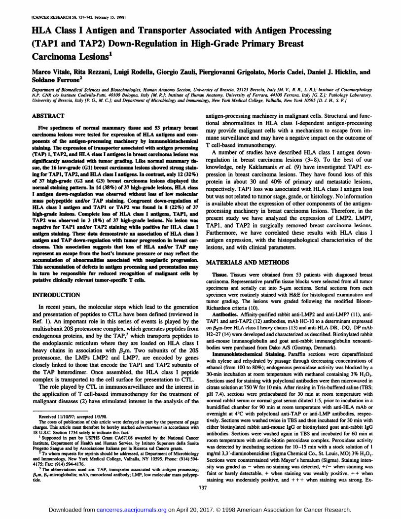
Fig. 1. HLA class I antigen. TAPI, and TAP2 expression innormal mammary tissue. Sections of a formalin-fixed, paraffin-embedded normal mammary tissue were stained in the immu-noperoxidase reaction with mouse anti-HLA Class I mAbHC-IO (A: X120). with affinity-purified anti-TAPI antibodies(B; X300) and with affinity-purified anti-TAP2 antibodies (C;XI20). Anti-HLA class I antigen. anti-TAPI. and anti-TAP2
immunoreactivity is located in the cytoplasm, and staining . ivaries from moderate to strong intensity. ^B
"^ "%N ^^ «
^W '•fc,<*Jvv^*r^
'é'' 5' •-•/.. •
738
on April 20, 2017. © 1998 American Association for Cancer Research. cancerres.aacrjournals.org Downloaded from

LOSS OF HLA CLASS I ANTIGEN IN BREAST CARCINOMA
pression of a marker was scored as down-regulated when staining was +/- or
-. Normal stroma and infiltrating lymphocytes were examined in each spec
imen as internal controls. Negative controls were performed by omittingprimary antibodies. Samples were analyzed and scored blindly.
Flow Cytometry. DNA content was measured by flow cytometry in 50-(j.M-thick formalin-fixed, paraffin-embedded tissue sections, using the meth
odology described by Hedley et al. (15). Briefly, tissue sections were sequentially deparaffmized with xylene. rehydrated by passage through decreasingethanol concentrations, and washed twice in distilled water. Tissue was thenenzymatically disrupted by resuspending in 1 ml of 0.5% pepsin, 0.9% NaCI,adjusted to pH 1.5 with 2 N HC1. and incubated at room temperature for 90 minwith intermittent mixing. Enzymatic digestion was stopped by adding anexcess of Tris buffer (pH 8.6). Disaggregated samples were washed in PBS.filtered to remove aggregates, and stained with 0.6 ¿tg/ml4',6-diamidino-2-
phenylindole (Calbiochem). DNA ploidy was measured by a Partee Call flowcytometer (Partee GMBH. Munster, Germany). Human lymphocytes wereused as an internal control.
RESULTS
Preliminary assays tested the staining of formalin-fixed, paraffin-
embedded normal mammary tissues with the panel of antibodies to beused in the present investigation. Anti-HLA class I mAb HC-10, aswell as affinity-purified anti-LMP2, anti-LMP7. anti-TAPl, and anti-
TAP2 antibodies, stained alveolar cells and epithelial cells of ducts inthe five normal mammary tissue samples tested (Fig. 1). The stainingintensity with anti-HLA class I mAb and with anti-LMP subunitantibodies was strong, whereas that with anti-TAP subunit antibodies
was moderate to strong. The five antibodies stained also the interlobular tissue but with weak intensity. Anti-HLA class II mAb H2-27
stained alveoli and ductal tissues with a focal pattern and strongintensity.
Fifty-three primary breast carcinoma lesions were tested for HLA
class I antigen, HLA class II antigen, and TAPI and TAP2 expression.Twenty-two lesions were also tested for LMP2 and LMP7 subunitexpression. Anti-HLA class I mAb and anti-TAPl and anti-TAP2
antibodies stained 28 (53%), 46 (87%), and 46 (87%) of the 53lesions, respectively. Expression of HLA class I antigens, TAPI, andTAP2 was coordinate in 28 lesions and was not detectable in 3 others.Among the 22 remaining lesions with HLA class I antigen down-
regulation, 14 expressed both TAPI and TAP2. 4 only TAPI, and 4only TAP2 (Table 1).
HLA class I antigen and TAP subunit down-regulation in breast
carcinoma lesions were significantly (P < 0.001 and P < 0.02,respectively) correlated with tumor grading. Down-regulation of HLA
class I antigen, TAPI, or TAP2 was not found in any of the 16low-grade (Gl) lesions tested (Fig. 2). In contrast. TAPI and/or TAP2down-regulation and HLA class I antigen down-regulation occurred in29 and 68% of high-grade (G2 and G3) lesions, respectively (Fig. 3).
The intensity of staining for HLA class I antigens and TAP subunitsdid not differ significantly between low- and high-grade lesions,
because the intensity was moderate to strong in greater than 50% oflow- and high-grade lesions. Lesions negative for HLA class I anti
gens and either one of the TAP subunits stained only with weak
Table 1 Phenotype of HLA class I, TAPI, and TAP2 in primary breast carciiutmalesions
TAPI TAP2 HLA IGrade"+
+ +Low++ +High++High+
High+High-
- - HighNo.
oflesions16/1612/3714/374/374/373/37%too323811118
' Low-grade lesion. Gl; high-grade lesion. G2 and G3.
intensity for the other TAP subunit. Neither HLA class I antigen norTAP down-regulation are significantly associated with histopatholog-
ical characteristics of the lesions, i.e., lesion dimensions, tumorploidy. cellular proliferation, and lymph node métastases.Data onsurvival were available for only 13 patients in this study. In this smallnumber of patients, no relationship was found between HLA class Iantigen and TAP subunit down-regulation and patients' survival.
LMP2 and LMP7 were expressed in the 9 low-grade and 13high-grade lesions tested. Intensity of staining for LMP2 and LMP7
ranged from weak to strongly positive with no relationship to tumorgrading. HLA class II antigen expression was detected in 37 (70%) ofthe lesions. Positive lesions were stained strongly for HLA class IIantigen expression with a focal pattern of staining similar to thatfound in normal alveoli and ductal breast tissue. HLA class II antigenexpression and staining intensity did not correlate with the degree ofdifferentiation of the lesions (data not shown), because HLA class IIantigens are expressed in 11 (69%) of 16 low-grade lesions and in 26(70%) of 37 high-grade lesions.
DISCUSSION
The present study has shown down-regulation of HLA class Iantigens in 47% and down-regulation of TAPI and/or TAP2 in 21%
of primary breast carcinoma lesions. HLA class I antigen and TAPdown-regulation are both associated with tumor grading. The resultsof our study differ in several respects from those published by Kak-lamanis et al. (9). The frequency of TAPI down-regulation found in
the present study is lower than that described by Kaklamanis et al. (9),who had not detected TAPI in 33% of primary breast carcinomalesions. Furthermore, Kaklamanis et al. (9) found coordinate expression of TAPI and HLA class I antigens, whereas we have found asignificantly more frequent loss of HLA class I antigens than of TAPIor TAP2. Lastly, Kaklamanis et al. (9) found no significant association between HLA class I antigen or TAPI down-regulation in pri
mary breast carcinoma lesions and tumor grading, whereas we did.Whether these differences reflect the characteristics of the anti-TAPl
xenoantibodies used in the two studies cannot be assessed, because noinformation is available about the anti-TAPl xenoantibodies used by
Kaklamanis et al. (9). Furthermore, we cannot exclude a role of thecriteria to classify the results and/or of the characteristics of thelesions used in the two investigations, because the earlier study (9)provides only limited information about these parameters. On theother hand, two lines of evidence argue against the use of differentsubstrates in the immunohistochemical reactions, i.e., frozen tissuesections by Kaklamanis et al. (9) and formalin-fixed tissue sections by
ourselves, as a major reason for the conflicting results. In the earlieranalysis of a large number of breast carcinoma lesions (9), identicalresults were obtained when the staining of sections from frozen andformalin-fixed portions of each lesion were compared with anti-HLA
class I mAb HCA2 and mAb W6/32, respectively. mAb HCA2 (13)and mAb W6/32 (16) recognize a linear and a conformational anti-
genie determinant of HLA class I antigens, respectively. Furthermore,we have not found significant differences in the immunohistochemicalstaining of frozen and autologous formalin-fixed sections of a number
of melanoma lesions (17 and unpublished results) with the mAbHC-10, which recognizes a linear determinant of HLA class I heavychains (13), and with anti-TAPl and anti-TAP2 xenoantibodies. The
discrepancy between the earlier results (9) and our own is also notlikely to be caused by the different specificity and characteristics ofthe anti-HLA class I mAb used in the two studies, because Kaklama
nis et al. (9) reported that the results of immunostaining of breastcarcinoma lesions with mAb W6/32 were identical to those obtained
739
on April 20, 2017. © 1998 American Association for Cancer Research. cancerres.aacrjournals.org Downloaded from

LOSS OF HLA CLASS I ANTIGEN IN BREAST CARCINOMA
, • ATT" ' ' ^. Ar F -«.' •*•.-
v>
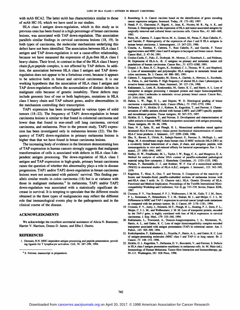
Fig. 2. HLA class I antigen. TAPI, and TAP2 expression ina low-grade (G1 ) breast carcinoma lesion. Sections of a formalin-fixed, paraffin-embedded low-grade (Gl) breast carcinoma
lesion were stained in the ¡mmunoperoxidase reaction withmouse anti-HLA class I mAb HC-10 (/l; X120), with affinity-purified anti-TAPl antibodies (S; X120), and with affinity-purified anti-TAP2 antibodies (C; X300). Staining intensitywith anti-HLA class 1 mAb is moderate to strong, whereas thatwith anti-TAPl and anti-TAP2 antibodies is weak to moderate.
740
on April 20, 2017. © 1998 American Association for Cancer Research. cancerres.aacrjournals.org Downloaded from

LOSS OF HLA CLASS I ANTIGEN IN BREAST CARCINOMA
Fig. 3. Reduced HLA class I antigen, TAPI, and TAP2expression in a high-grade (G2) breast carcinoma lesion. Sections of a formalin-fixed, paraffin-embedded high-grade (G2)
breast carcinoma lesion were stained in the immunoperoxidasereaction with mouse anti-HLA class I mAb HC-10 (A; X300),affinity-purified anti-TAPl antibodies (B; X120), and affinity-purified anti-TAP2 antibodies (C; XI20). Note weak, hetero
geneous staining for HLA class I antigens, TAPI, and TAP2.
.- : •••v u«-•.v Y* - ••*••-•' •* . i •••••t :' .. ':,v>v:..•¿;.-, "^'•»f*••
r^ •»•--' 1-»'' ; ., ,_ ¥ . . »,•»A ^- »- ' •y' - ' •'
741
on April 20, 2017. © 1998 American Association for Cancer Research. cancerres.aacrjournals.org Downloaded from
LOSS OF HLA CLASS I ANTIGEN IN BREAST CARCINOMA
with mAb HCA2. The latter mAb has characteristics similar to thoseof mAb HC-10, which we have used in our studies.
HLA class I antigen down-regulation, which in this study as in
previous ones has been found in a high percentage of breast carcinomalesions, was associated with TAP down-regulation. This association
parallels similar findings in cervical carcinoma lesions (18, 19). Inboth types of carcinoma, the molecular mechanisms underlying thisdefect have not been identified. The association between HLA class Iantigen and TAP down-regulation is not a cause-effect relationship,because we have measured the expression of /32m-free HLA class I
heavy chains. Their level, in contrast to that of the HLA class I heavychain:ß2m:peptide complex, is not affected by TAP defects. In addition, the association between HLA class I antigen and TAP down-
regulation does not appear to be a fortuitous event, because it appearsto be selective both in breast and cervical carcinomas. It is ourworking hypothesis that the association of HLA class I antigen andTAP down-regulation reflects the accumulation of distinct defects in
malignant cells because of genetic instability. These defects mayinclude genomic loss of one or both haplotypes, mutations in HLAclass I heavy chain and TAP subunit genes, and/or abnormalities inthe mechanism controlling their transcription.
TAPI expression has been investigated in various types of solidtumors (18-22). The frequency of TAPI down-regulation in breast
carcinoma lesions is similar to that found in colorectal carcinoma andlower than that found in non-small cell lung carcinoma, cervical
carcinoma, and melanoma. Prior to the present study, TAP2 expression has been investigated only in melanoma lesions (22). The frequency of TAP2 down-regulation in primary melanoma lesions is
higher than that we have found in breast carcinoma lesions.The increasing body of evidence in the literature demonstrating loss
of TAP expression in human cancers strongly suggests that malignanttransformation of cells is associated with defects in HLA class I-de-pendent antigen processing. The down-regulation of HLA class Iantigen and TAP expression in high-grade, primary breast carcinoma
raises the question of whether this abnormality plays a role in diseaseprogression. TAPI and/or TAP2 down-regulation in breast carcinomalesions were not associated with patients' survival. This finding par
allels similar results in colon carcinoma (18) but is at variance withthose in malignant melanoma.4 In melanoma. TAPI and/or TAP2
down-regulation was associated with a statistically significant de
crease in survival. It is tempting to speculate that the different resultsobtained in the three types of malignancies may reflect the differentrole that immunological events play in the pathogenesis and in theclinical course of the disease.
ACKNOWLEDGMENTS
We acknowledge the excellent secretarial assistance of Alicia N. Brammer,Harriet V. Harrison. Donna D. James, and Elba I. Osorio.
REFERENCES
1. Germain. R.N. MHC-dependent antigen processing and peptide presentation: providing ligands for T lymphocyte activation. Cell. 76: 287-299. 1994.
4 S. Ferrone, manuscript in preparation.
2. Rosenberg. S. A. Cancer vaccines based on the identification of genes encodingcancer regression antigens. Immunol. Today, 18: 175-182, 1997.
3. Natali, P. G.. Giacomini, P.. Bigotti, A., Imai, K.. Nicotra, M. R., Ng, A. K.. andFerrone, S. Heterogeneity in the expression of HLA and tumor-associated antigens bysurgically removed and cultured breast carcinoma cells. Cancer Res.. 43: 660-668,
1983.4. Perez, M., Cabrera, T., Lopez Nevot, M. A.. Gomez, M., Peran, F.. Ruiz Cabello. F.,
and Garrido. F. Heterogeneity of the expression of class I and II HLA antigens inhuman breast carcinoma. J. Immunogenet., 13: 247-253, 1986.
5. Concha, A., Esteban. F.. Cabrera. T., Ruiz Cabello, F., and Garrido, F. Tumoraggressiveness and MHC class I and II antigens in laryngeal and breast cancer. Semin.Cancer Biol.. 2: 47-54, 1991.
6. Cordon-Cardo, C., Fuks, Z., Drobnjak, M.. Moreno. C., Eisenbach. L., and Feldman,M. Expression of HLA-A, -B, -C antigens on primary and metastatic tumor cellpopulations of human carcinomas. Cancer Res., 5/: 6372-6380, 1991.
7. Goepel, J. R.. Rees, R. C.. Rogers, K., Stoddard, C. J., Thomas, W. E., and Shepherd,L. Loss of monomorphic and polymorphic HLA antigens in metastatic breast andcolon carcinoma. Br. J. Cancer, 64: 880-883, 1991.
8. Cabrera, T., Angustias Fernandez. M., Sierra. A.. Garrido. A.. Herruzo, A., Escobedo,A., Fabra, A., and Garrido. F. High frequency of altered HLA class I phenotypes ininvasive breast carcinomas. Hum. Immunol., 50: 127-134. 1996.
9. Kaklamanis, L.. Leek, R.. Koukourakis, M., Gatter, K. C.. and Harris, A. L. Loss oftransponer in antigen processing 1 transport protein and major histocompatibilitycomplex class 1 molecules in metastatic versus primary breast cancer. Cancer Res.,55: 5191-5194, 1995.
10. Dalton. L. W., Page. D. L., and Dupont, W. D. Histológica! grading of breastcarcinoma: a reproducibility study. Cancer (Phila.), 73: 2765-2770, 1994.
11. Dellaratta, D. V.. Hicklin, D. J.. Kishore, R.. Kageshita, T., and Ferrone, S. Characterization of rabbit antisera elicited with human LMP2- and LMP7-specific peptidesand recombinant proteins. Tissue Antigens, 50: 567-575. 1997.
12. Hicklin. D. J.. Kageshita. T., and Ferrone. S. Development and characterization ofrabbit antisera to human MHC-linked transporters associated with antigen processing.Tissue Antigens. 48: 38-46, 19%.
13. Slam, N. J., Spits, H., and Ploegh. H. L. Monoclonal antibodies raised againstdenatured HLA-B locus heavy chains permit biochemical characterization of certainHLA-C locus products. J. Immunol., 137: 2299-2306. 1986.
14. Zhu, X., Bavari. S., Ulrich. R.. Sadegh-Nasseri, S.. Ferrone, S., McHugh, L., andMage, M. A recombinant single chain human class II MHC molecule (HLA-DRI} as
a covalently linked heterotrimer of or chain, ßchain, and antigenic peptide, withimmunogenicity in vitro and reduced affinity for bacterial superantigens. Eur. J. Immunol., 27: 1993-1941, 1997.
15. Hedley. D. W.. Friedlander, M. L., Taylor, I. W.. Rugg, C. A., and Musgrove, E. A.Method for analysis of cellular DNA content of paraffin-embedded pathologicalmaterial using flow cytometry. J. Histochem. Cytochem., 31: 1333-1335, 1983.
16. Parham. P.. Bamstable. C. J.. and Bodmer, W. F. Use of a monoclonal antibody(W6/32) in structural studies of HLA-A.B.C, antigens. J. Immunol., 123: 342-349,
1979.17. Kageshita. T.. Hirai, S., Ono. T. and Ferrone, S. Comparison of the reactivity of
frozen and formalin-fixed, paraffin-embedded sections of melanoma lesions withanti-HLA class I mAb. In: D. Charron (ed.), HLA. Genetic Diversity of HLA.
Functional and Medical Implication. Proceedings of the Twelfth International Histocompatibility Workshop and Conference. Vol. II, pp. 737-739. Sevres. France: EDK,
1997.18. Cromme, F. V., Van Bommel, P. F. J.. Walboomers, J. M. M.. Galle, T. J. M., Stern,
P. L., Kenemans, P., Heimerhorst, T. J. M., Stukart, M. J., and Meijer, C. J. L. M.Differences in MHC and TAP-1 expression in cervical cancer lymph node métastasesas compared with the primary tumors. Br. J. Cancer. 69: 1176-1181. 1994.
19. Cromme. F. V., Airey. J.. Heemels, M-T., Ploegh, H. L.. Keating, P. J., Stern, P. L.,
Meijer, C. J. L. M., and Walboomers, J. M. M. Loss of transporter protein, encodedby the TAP-1 gene, is highly correlated with loss of HLA expression in cervicalcarcinomas. J. Exp. Med., 779: 335-340. 1994.
20. Kaklamanis. L., Townsend, A., Doussis-Anagnostopoulou, 1. A.. Mortensen, N.,Harris, A. L.. and Gatter, K. C. Loss of major histocompatibility complex-encoded
transporter associated with antigen presentation (TAP) in colorectal cancer. Am. J.Pathol., 145: 505-509, 1994.
21. Korkolopoulou. P.. Kaklamanis, L.. Pezzella, F., Harris, A. L.. and Gatter, K. C. Lossof antigen-presenting molecules (MHC class I and TAP-1) in lung cancer. Br. J.Cancer, 73: 148-153, 19%.
22. Hicklin, D. J., Kageshita. T., Dellaratta, D. V., Boccaletti, V., and Ferrone, S. Defectsin HLA class I antigen presentation machinery in melanoma cells. In: M. Maio (ed.).Immunology of Human Melanoma. Tumor-Host Interaction and Immunotherapy, pp.95-111. Washington. DC: IOS Press. 1996.
742
on April 20, 2017. © 1998 American Association for Cancer Research. cancerres.aacrjournals.org Downloaded from

1998;58:737-742. Cancer Res Marco Vitale, Rita Rezzani, Luigi Rodella, et al. Primary Breast Carcinoma LesionsProcessing (TAP1 and TAP2) Down-Regulation in High-Grade HLA Class I Antigen and Transporter Associated with Antigen
Updated version
http://cancerres.aacrjournals.org/content/58/4/737
Access the most recent version of this article at:
E-mail alerts related to this article or journal.Sign up to receive free email-alerts
Subscriptions
Reprints and
To order reprints of this article or to subscribe to the journal, contact the AACR Publications
Permissions
To request permission to re-use all or part of this article, contact the AACR Publications
on April 20, 2017. © 1998 American Association for Cancer Research. cancerres.aacrjournals.org Downloaded from