can exercise reduce the autonomic dysfunction of patients
TRANSCRIPT

Can Exercise Reduce the Autonomic Dysfunction of Patients With Cancer and Its Survivors? A Systematic Review and Meta-Analysis
LAVÍN-PÉREZ, Ana Myriam, COLLADO-MATEO, Daniel, MAYO, Xián, LIGUORI, Gary, HUMPHREYS, Liam and JIMENEZ GUTIERREZ, Alfonso
Available from Sheffield Hallam University Research Archive (SHURA) at:
http://shura.shu.ac.uk/29015/
This document is the author deposited version. You are advised to consult the publisher's version if you wish to cite from it.
Published version
LAVÍN-PÉREZ, Ana Myriam, COLLADO-MATEO, Daniel, MAYO, Xián, LIGUORI, Gary, HUMPHREYS, Liam and JIMENEZ GUTIERREZ, Alfonso (2021). Can Exercise Reduce the Autonomic Dysfunction of Patients With Cancer and Its Survivors? A Systematic Review and Meta-Analysis. Frontiers in Psychology, 12.
Copyright and re-use policy
See http://shura.shu.ac.uk/information.html
Sheffield Hallam University Research Archivehttp://shura.shu.ac.uk
SYSTEMATIC REVIEWpublished: 24 August 2021
doi: 10.3389/fpsyg.2021.712823
Frontiers in Psychology | www.frontiersin.org 1 August 2021 | Volume 12 | Article 712823
Edited by:
Rodrigo Ramirez-Campillo,
University of Los Lagos, Chile
Reviewed by:
David Cristóbal Andrade,
University of Antofagasta, Chile
Jairo Azócar Gallardo,
University of Los Lagos, Chile
*Correspondence:
Daniel Collado-Mateo
Specialty section:
This article was submitted to
Movement Science and Sport
Psychology,
a section of the journal
Frontiers in Psychology
Received: 09 June 2021
Accepted: 12 July 2021
Published: 24 August 2021
Citation:
Lavín-Pérez AM, Collado-Mateo D,
Mayo X, Liguori G, Humphreys L and
Jiménez A (2021) Can Exercise
Reduce the Autonomic Dysfunction of
Patients With Cancer and Its
Survivors? A Systematic Review and
Meta-Analysis.
Front. Psychol. 12:712823.
doi: 10.3389/fpsyg.2021.712823
Can Exercise Reduce the AutonomicDysfunction of Patients With Cancerand Its Survivors? A SystematicReview and Meta-AnalysisAna Myriam Lavín-Pérez 1,2,3, Daniel Collado-Mateo 2*, Xián Mayo 2, Gary Liguori 4,
Liam Humphreys 5 and Alfonso Jiménez 2,3,5
1 PhD International School, Program of Epidemiology and Public Health (Interuniversity), Rey Juan Carlos University,
Móstoles, Spain, 2Centre for Sport Studies, Rey Juan Carlos University, Fuenlabrada, Spain, 3GO fitLAB, Ingesport, Madrid,
Spain, 4College of Health Sciences, University of Rhode Island, Kingston, NY, United States, 5 Advanced Wellbeing Research
Centre, College of Health, Wellbeing and Life Sciences, Sheffield Hallam University, Sheffield, United Kingdom
Background: Cancer therapies have increased patient survival rates, but side
effects such as cardiotoxicity and neurotoxicity can lead to autonomic nervous and
cardiovascular system dysfunction. This would result in a decrease in parasympathetic
activity and the enhancement of sympathetic activity. Heart rate variability (HRV), which
reflects autonomic modulation, is a valuable physiological tool since it correlates with
cancer-related fatigue, stress, depression, and mortality in patients with cancer.
Objective: This study aimed to analyze the effects of exercise programs on the
autonomic modulation, measured by the HRV of patients with cancer and its survivors.
Methods: The Preferred Reporting Items for Systematic Reviews and Meta-Analyses
(PRISMA) guidelines were followed, and the quality of the articles was assessed with the
Physiotherapy Evidence Database (PEDro) scale. The meta-analysis statistic procedure
was performed by using RevMan software version 5.3.
Results: From the 252 articles found, six studies were included in the review
involving 272 participants aged 30–75 years. Exercise programs had a mean length
of 10.4 ± 4.6 weeks, a frequency of 3 ± 1.4 days/week, and a mean duration of
78 ± 23.9min. In time-domain HRV measures, exercise may increase in the SD of
normal-to-normal intervals [p < 0.00001, with a mean difference (MD) of 12.79ms
from 9.03 to 16.55] and a decreased root mean square of successive R–R interval
differences (p = 0.002, with an MD of 13.08ms from 4.90 to 21.27) in comparison
with control groups (CG). The frequency-domain data reveal that the exercise group
(EG) improve significantly more than the CGs in low frequency [absolute power: p
< 0.0001, with a standardized mean difference (SMD) of 0.97 from 0.61 to 1.34;
relative power: p = 0.04, with an MD = −7.70 from −15.4 to −0.36], high-frequency
[absolute power: p = 0.001, with a SMD of 1.49 from 0.32 to 2.66; relative power:
p = 0.04, with an MD of 8.00 normalized units (n.u.) from 0.20 to 15.80], and
low-to-high frequency ratio (p = 0.007 with an MD of −0.32 from −0.55 to −0.09).

Lavín-Pérez et al. Exercise, Oncology and HRV
Conclusion: Exercise programs could lead to positive effects on the autonomic
modulation of patients with cancer and its survivors. More beneficial changes may
occur with resistance and endurance workouts. However, due to the low number of
interventions performed, further research is needed to substantiate the findings and
to provide additional insights regarding the exercise intensity required to increase the
autonomic modulation of the patient.
Keywords: autonomic modulation, exercise programs, cardiovascular dysfunction, oncology, heart rate variability
INTRODUCTION
Modern advances in cancer therapy have resulted in increasedsurvival rates. However, patients are frequently affected byvarious negative side effects (Miller et al., 2019). In particular,both the autonomic control impairment and the increased riskof cardiovascular disease (Lakoski et al., 2015) are suffered by∼80% (Coumbe and Groarke, 2018) and 42% (Chen et al.,2012) of the patients, respectively. The autonomic nervoussystem (ANS) is the main homeostatic regulatory system of thebody, involved in the etiology and the clinical course of cancertherapies (Simó et al., 2018). Chemotherapy and radiotherapycause cardiotoxicity (Scott et al., 2014) and neurotoxicity (Parket al., 2013), thereby affecting ANS and cardiovascular function.The cardiovascular ANS needs to be controlled during the cancerphases to regulate levels of autonomic dysfunction of patients(Walsh and Nelson, 2002).
Heart rate variability (HRV) is a noninvasive tool to evaluatethe autonomic modulation of the sinus node in healthy,cardiac, and noncardiac disease populations (Lombardi andStein, 2011). In patients with cancer, HRV is a useful, effective,and more practical method to assess autonomic dysfunctionthan other validated methods, which are more complex to apply(Guo et al., 2013). HRV measures provide a multidimensionalregister of autonomic modulation, including the sympatheticand parasympathetic modulation of cardiac function (Arabet al., 2016), which is notably modified in patients withcancer compared with the healthy population. Studies includingwide sample sizes have found significantly lower values inthe root mean square of successive R–R interval measures(RMSSD), representing the parasympathetic activity, and theSD of the interbeat interval of normal sinus beats (SDNN)(i.e., overall HRV measure) of patients with cancer comparedwith the healthy population (De Couck and Gidron, 2013;Bijoor et al., 2016). Cancer-treating drug therapies inducecardiac abnormalities, such as heart failure, myocardial ischemia,myocarditis, hypertension, or arrhythmias, among others (Changet al., 2017), detected with HRV even with normal systolicleft ventricular function (Tjeerdsma et al., 1999) with anoveractivation of the sympathetic nervous system (SNS) and adecrease in the parasympathetic nervous system (PNS) activity(Coumbe and Groarke, 2018). Consequently, this imbalance,produced by cancer drugs, may stimulate the hypothalamic-pituitary-adrenal axis and the endocannabinoid and renin-angiotensin-aldosterone systems producing an increase inoxidative stress, chronic inflammation, and atherosclerosis with
a reduction in vasodilation, which affects the health of patients(Lakoski et al., 2015). Several types of research have focused onthe relationship between the global HRV modifications and thehealth of patients with cancer obtaining an inverse correlationwith cancer-related fatigue (Fagundes et al., 2011) and depression(Giese-Davis et al., 2006). Findings also show the role of HRV topredict survivorship being higher when the vagal nerve activationis stimulated (Zhou et al., 2016).
Palma et al. (2020) and Dias Reis et al. (2017) publisheda systematic review of supportive therapy modalities thathave been developed to reduce the HRV results. They foundinterventions based on music therapy, traditional Chinesemedicine-related treatments, exercise, relaxation, and myofascialrelease techniques and concluded that HRV seemed to bea safe and easily applicable method to assess cancer-relatedautonomic dysfunction (Palma et al., 2020). However, not onlythe consequences of cancer therapy need to be considered toimprove HRV but also the lifestyles of patients can influence itnegatively (Scott et al., 2014). Thus, a multifactorial interventionthat could modify the physiological changes mentioned earlierand other health and psychological parameters is crucial toglobally benefit the patients with cancer.
Exercise programs appear to promote physiological changesleading to reduce the decline of the autonomic modulationand improving its levels during (Mostarda et al., 2017) andafter cancer treatments (Shin et al., 2016). Scott et al. (2014)approximated the benefits of aerobic exercise training to theautonomic modulation of patients with cancer stating that itmay decrease sympathetic tone and increase vagal tone by theinfluence of exercise in attenuate cardiovascular abnormalitiesas heart failure or coronary artery disease. Aerobic exercisemodifies the renin-angiotensin-aldosterone system (Scott et al.,2014) and stimulates the hypothalamic-pituitary-adrenal axissuppressing angiotensin II expression, which promotes thesympathetic activity of the ANS (Routledge et al., 2010).Consequently, the stimulation of the hypothalamic-pituitary-adrenal axis and the endocannabinoid and renin-angiotensin-aldosterone systems is produced (Arab et al., 2016). Exercise alsoupregulates nitric oxide promoting vasodilation (Kingwell, 2000;Scott et al., 2014) reduces reactive oxygen species induced bychemotherapy toxicity (Scott et al., 2014). Moreover, regardingthe remaining physiological changes mentioned earlier, exercisecan also decrease chronic inflammation (Gleeson et al., 2011),improving the immune system and the stimulation of naturalkiller cells (Khosravi et al., 2019). Exercise can also reducebody mass index (BMI) (Thomas et al., 2017), which may be
Frontiers in Psychology | www.frontiersin.org 2 August 2021 | Volume 12 | Article 712823

Lavín-Pérez et al. Exercise, Oncology and HRV
correlated to an HRV increase (Arab et al., 2016). Althoughexercise may have the potential role to increase HRV, theresults presented by the different interventions developed arecontroversial considering all the variables inside HRV. Inthis way, this systematic review and meta-analysis aimed toevaluate the effects of exercise interventions on the autonomicfunction of patients with cancer and its survivors analyzing themeasures involved.
METHODS
The Preferred Reporting Items for Systematic Reviews andMeta-Analyses (PRISMA) guidelines have been followed to develop thesystematic review (Liberati et al., 2009). Before the data extractionwas performed, the study was registered in the InternationalProspective Register of Systematic Reviews (PROSPERO) by thefollowing identification number: CRD42020191041.
Search StrategyThe databases employed for searching the articles were Web ofScience (including studies indexed in the KCI-Korean JournalDatabase, MEDLINE, Russian Science Citation Index, SciELOCitation Index, and PubMed (MEDLINE). The terms used forthe search were as follows: cancer and neoplasms, separatedby OR; and HRV, autonomous nervous function, autonomousnervous system, separated by OR; and exercise, training, physicalactivity, also separated by OR. The only filter employed was therequirement of being articles written in English or Spanish. Thesearch was carried out from April 2020 to June 2020.
The articles found were included if they fulfilled the followingcriteria: (1) the target population was patients with cancer orsurvivors, (2) the program involves any physical exercise, (3)the investigation assesses and reports any HRV variable, (4) thestudy includes at least one control group (CG) whose results arecompared with an exercise group (EG). Moreover, the followingexclusion criteria were set: (1) the article was a review, letter tothe editor, conference abstract, case report, or study protocol, (2)the study did not evaluate the HRV variables directly after theintervention, and (3) the studies were completely written in alanguage different from English or Spanish.
Risk of Bias AssessmentThe analysis of the risk of bias was performed using thePhysiotherapy Evidence Database (PEDro) scale, known as avalid and reliable instrument to assess eligibility, allocation togroups, blinding of allocation, and comparison between groupsat baseline and its outcomes (Maher et al., 2003). The leadingreasons for its selection were due to it being the most used in thescientific area of the Sport Sciences for Health and it is a specifictool focused on physical therapies (Moseley et al., 2020).
Data ExtractionParticipants, Interventions, Comparisons, and Study
Designs InformationThe main information of the articles is reported in thetables and figures in the article and the supplementary data.Regarding participants, CG and EG baseline parameters
were extracted, such as sample size, mean age, BMI,physical activity level, cancer type, stage, type of treatment,and timing. The intervention characteristics included thelength of the program, duration of each session, weeklyfrequency, exercise description, intensity, progression, andadherence. The intervention of the comparison group wasalso extracted.
HRV Outcome MeasurementsInterventions included an HRV assessment before and after theintervention. For consistency in measures, participants were laidin a supine position (Niederer et al., 2013; Dias Reis et al., 2017;Zhou et al., 2018) or seated in chairs (Shin et al., 2016; Leeet al., 2018). HRV was recorded for 5min of adequate stationarysign (Niederer et al., 2013; Lee et al., 2018; Zhou et al., 2018)
with Polar S810© (Niederer et al., 2013), CANS 3000 (Laxtha,Daejeon, Korea) (Shin et al., 2016), or CheckMyHeart HandheldHRV (Lee et al., 2018) by using ECG (Zhou et al., 2018). Thespectral analysis from the artifact-free data was performed bythe fast Fourier transformation with the Kubios HRV Analysissoftware (Niederer et al., 2013; Dias Reis et al., 2017; Mostardaet al., 2017; Zhou et al., 2018) or AcqKnowledge (Zhou et al.,2018). The results of this review include two HRV variables inthe time domain, namely, SDNN and RMSSD. In the frequencydomain, the low-frequency (LF) band, the high-frequency (HF)band, the ratio of LF-to-HF (LF/HF), and the total power (TP),calculated as the sum of the energy in very-low-frequency (VLF),LF, and HF bands, were extracted (Shaffer and Ginsberg, 2017).The frequency-domain data were reported in absolute units byusing milliseconds squared (ms2) or normalized units (n.u.)obtained by diving the result between the TP and multipliedto 100 [i.e., LF or HF/(TP–VLF) × 100] (Task Force of theEuropean Society of Cardiology the North American Societyof Pacing Electrophysiology, 1996). SDNN and RMSSD weremeasured in 1/1,000 s (milliseconds) (Shin et al., 2016), and HFwas recorded from 0.15 to 0.4Hz and LF from 0.04 to 0.15Hz(Table 3) (Niederer et al., 2013; Shin et al., 2016; Dias Reis et al.,2017; Mostarda et al., 2017; Zhou et al., 2018).
Statistical AnalysisThe analysis was performed with the post-intervention meansand SDs of the EG and the CG collected from the articles.The meta-analysis statistical tool used was the Review ManagerSoftware (RevMan software version 5.3) (RevMan, 2014). Theselected method was the inverse variance with random effectsand a 95% CI (Schmidt et al., 2009). Additionally, the I2 modelwas used to calculate the heterogeneity of the results. The resultswere presented with a standardizedmean difference (SMD) whenthe same variable was measured in different units (i.e., ms2 andlog ms2) as occurred with the absolute power variables of HFand LF. In contrast, the mean difference (MD) was used whenall studies assessed the variable using the same units (n.u. orms). According to the Cochrane Handbook for Systematic Reviewsof Intervention, the SMD effects were interpreted as small withresults <0.4, moderate from 0.4 to 0.7, and large >0.7 (Higginsand Green, 2011).
Frontiers in Psychology | www.frontiersin.org 3 August 2021 | Volume 12 | Article 712823

Lavín-Pérez et al. Exercise, Oncology and HRV
FIGURE 1 | Study flow diagram following the preferred reporting items for systematic reviews and meta-analyses guidelines.
RESULTS
Study SelectionAbout 252 articles were identified in Web of Science (n = 166)and PubMed (n = 86) scientific databases. Figure 1 shows theflow diagram where 209 articles were analyzed after removingthe duplicates. Two hundred and three articles were excludedafter reading the title or the abstract (n = 164) or after the full-text evaluation (n = 39). Therefore, both the systematic reviewand the meta-analysis were developed with six studies publishedbetween 2012 and 2018.
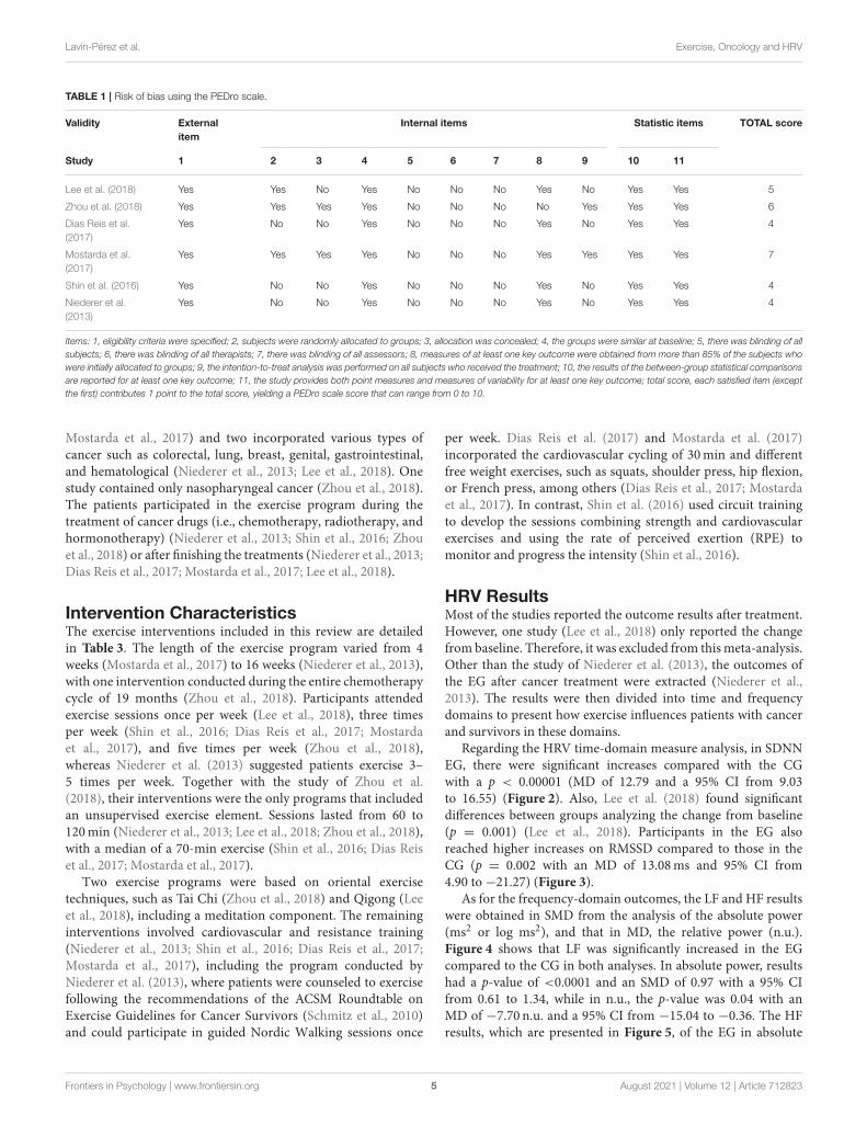
Risk of BiasIn the PEDro scale, as Table 1 shows the mean score obtainedwas 5, and the external validity was 5, ranging from 4–7 (10 beingthe highest possible mark). The five articles assessed positively
reached the external validity (item 1) and the statistic items(items 10 and 11). However, the internal validity punctuation wasmore heterogeneous. None of the evaluated studies met items5, 6, and 7 related to the blinding process, difficult to fulfill insport sciences.
Participants CharacteristicsThe baseline information of the participants is reported inTable 2. The total sample size of the systematic review was 272,out of which 126 were included in the CG and 146 in the EG,and the sample wasmainly composed of women. The ages rangedfrom 30 to 75 years, although most of the participants were olderthan 45 years of age. Different types of cancer were included inthis study as follows: three interventions contained only patientswith breast cancer (Shin et al., 2016; Dias Reis et al., 2017;
Frontiers in Psychology | www.frontiersin.org 4 August 2021 | Volume 12 | Article 712823
Lavín-Pérez et al. Exercise, Oncology and HRV
TABLE 1 | Risk of bias using the PEDro scale.
Validity External
item
Internal items Statistic items TOTAL score
Study 1 2 3 4 5 6 7 8 9 10 11
Lee et al. (2018) Yes Yes No Yes No No No Yes No Yes Yes 5
Zhou et al. (2018) Yes Yes Yes Yes No No No No Yes Yes Yes 6
Dias Reis et al.
(2017)
Yes No No Yes No No No Yes No Yes Yes 4
Mostarda et al.
(2017)
Yes Yes Yes Yes No No No Yes Yes Yes Yes 7
Shin et al. (2016) Yes No No Yes No No No Yes No Yes Yes 4
Niederer et al.
(2013)
Yes No No Yes No No No Yes No Yes Yes 4
Items: 1, eligibility criteria were specified; 2, subjects were randomly allocated to groups; 3, allocation was concealed; 4, the groups were similar at baseline; 5, there was blinding of all
subjects; 6, there was blinding of all therapists; 7, there was blinding of all assessors; 8, measures of at least one key outcome were obtained from more than 85% of the subjects who
were initially allocated to groups; 9, the intention-to-treat analysis was performed on all subjects who received the treatment; 10, the results of the between-group statistical comparisons
are reported for at least one key outcome; 11, the study provides both point measures and measures of variability for at least one key outcome; total score, each satisfied item (except
the first) contributes 1 point to the total score, yielding a PEDro scale score that can range from 0 to 10.
Mostarda et al., 2017) and two incorporated various types ofcancer such as colorectal, lung, breast, genital, gastrointestinal,and hematological (Niederer et al., 2013; Lee et al., 2018). Onestudy contained only nasopharyngeal cancer (Zhou et al., 2018).The patients participated in the exercise program during thetreatment of cancer drugs (i.e., chemotherapy, radiotherapy, andhormonotherapy) (Niederer et al., 2013; Shin et al., 2016; Zhouet al., 2018) or after finishing the treatments (Niederer et al., 2013;Dias Reis et al., 2017; Mostarda et al., 2017; Lee et al., 2018).
Intervention CharacteristicsThe exercise interventions included in this review are detailedin Table 3. The length of the exercise program varied from 4weeks (Mostarda et al., 2017) to 16 weeks (Niederer et al., 2013),with one intervention conducted during the entire chemotherapycycle of 19 months (Zhou et al., 2018). Participants attendedexercise sessions once per week (Lee et al., 2018), three timesper week (Shin et al., 2016; Dias Reis et al., 2017; Mostardaet al., 2017), and five times per week (Zhou et al., 2018),whereas Niederer et al. (2013) suggested patients exercise 3–5 times per week. Together with the study of Zhou et al.(2018), their interventions were the only programs that includedan unsupervised exercise element. Sessions lasted from 60 to120min (Niederer et al., 2013; Lee et al., 2018; Zhou et al., 2018),with a median of a 70-min exercise (Shin et al., 2016; Dias Reiset al., 2017; Mostarda et al., 2017).
Two exercise programs were based on oriental exercisetechniques, such as Tai Chi (Zhou et al., 2018) and Qigong (Leeet al., 2018), including a meditation component. The remaininginterventions involved cardiovascular and resistance training(Niederer et al., 2013; Shin et al., 2016; Dias Reis et al., 2017;Mostarda et al., 2017), including the program conducted byNiederer et al. (2013), where patients were counseled to exercisefollowing the recommendations of the ACSM Roundtable onExercise Guidelines for Cancer Survivors (Schmitz et al., 2010)and could participate in guided Nordic Walking sessions once
per week. Dias Reis et al. (2017) and Mostarda et al. (2017)incorporated the cardiovascular cycling of 30min and differentfree weight exercises, such as squats, shoulder press, hip flexion,or French press, among others (Dias Reis et al., 2017; Mostardaet al., 2017). In contrast, Shin et al. (2016) used circuit trainingto develop the sessions combining strength and cardiovascularexercises and using the rate of perceived exertion (RPE) tomonitor and progress the intensity (Shin et al., 2016).
HRV ResultsMost of the studies reported the outcome results after treatment.However, one study (Lee et al., 2018) only reported the changefrom baseline. Therefore, it was excluded from thismeta-analysis.Other than the study of Niederer et al. (2013), the outcomes ofthe EG after cancer treatment were extracted (Niederer et al.,2013). The results were then divided into time and frequencydomains to present how exercise influences patients with cancerand survivors in these domains.
Regarding the HRV time-domain measure analysis, in SDNNEG, there were significant increases compared with the CGwith a p < 0.00001 (MD of 12.79 and a 95% CI from 9.03to 16.55) (Figure 2). Also, Lee et al. (2018) found significantdifferences between groups analyzing the change from baseline(p = 0.001) (Lee et al., 2018). Participants in the EG alsoreached higher increases on RMSSD compared to those in theCG (p = 0.002 with an MD of 13.08ms and 95% CI from4.90 to−21.27) (Figure 3).
As for the frequency-domain outcomes, the LF and HF resultswere obtained in SMD from the analysis of the absolute power(ms2 or log ms2), and that in MD, the relative power (n.u.).Figure 4 shows that LF was significantly increased in the EGcompared to the CG in both analyses. In absolute power, resultshad a p-value of <0.0001 and an SMD of 0.97 with a 95% CIfrom 0.61 to 1.34, while in n.u., the p-value was 0.04 with anMD of −7.70 n.u. and a 95% CI from −15.04 to −0.36. The HFresults, which are presented in Figure 5, of the EG in absolute
Frontiers in Psychology | www.frontiersin.org 5 August 2021 | Volume 12 | Article 712823

Lavín-Pérez et al. Exercise, Oncology and HRV
TABLE 2 | Baseline characteristics of participants.
Study Design Group N (%
females)
Age Cancer type Cancer stage Cancer treatment (type or
timing)
Lee et al.
(2018)
RCT CG 26 (84.6%) 30–39 (3.8%), 40–49
(19.2%), 50–59 (57.7%),
60–69 (11.5%), and
70–75 (7.7%)
Colorectal (11.5%), lung
(3.8%), breast (69.2%),
gynecological (7.7%), and
other (7.7%)
I (42.3%), II
(26.9%), III
(26.9%), and
none (3.8%)
After treatment:
surgery, chemotherapy,
radiotherapy, and target therapy
EG 29 (86.2%) 30–39 (10.3%), 40–49
(24.1%), 50–59 (37.9%),
60–69 (17.2%), and
70–75 (10.3%)
Colorectal (6.9%), lung
(3.4%), malignant
lymphoma (3.4%), breast
(62.1%), gynecological
(6.9%), and other (17.2%)
I (24.1%), II
(51.7%), III
(20.7%), and
none (3.4%)
Zhou et al.
(2018)
RCT CG 57 (21.05%) <30 (12.3%), 30–50
(64.9%), and >50 (22.8%)
Nasopharyngeal III (40.3%), IV a/b
(59.6%)
During treatment:
chemotherapy and radiotherapy
EG 57 (33.33%) <30 (22.8%), 30–50
(59.6%), and >50 (17.5%)
III (35.1%), IV a/b
(64.9%)
Dias Reis
et al. (2017)
Controlled
trial
CG 9 (100%) 45 ± 7 Breast I, II, and III After treatment:
hormonal therapy,
chemotherapy, and radiotherapy
EG 9 (100%) 48 ± 7.2
Mostarda
et al. (2017)
RCT CG 9 (100%) Range 30–59 Breast I, II, and III After treatment: hormonal
therapy (33.3%), chemotherapy
(27.8%), and radiotherapy
(38.9%)
EG 9 (100%)
Shin et al.
(2016)
Controlled
trial
CG 10 (100%) 49.2 (range, 35–60) Breast 0 (10%), I (20%),
II (60%), and III
(10%)
During treatment: anticancer
treatment (80%) and
radiotherapy (30%)
EG 12 (100%) 46.3 (range, 35–66) Breast 0 (8.3%), I
(33.3%), II (50%),
and III (8.3%)
During treatment:
anticancer treatment (83.3%),
radiotherapy (91.7%)
Niederer et al.
(2013)
Controlled
trial
CG 15 (60%) 61.6 ± 10.6 Each group: Gastrointestinal
(20%), genital and breast
(26.67%), diagnosis
bronchial (13.33%),
hematological (6.67%), and
other (33.33%)
0, I, and II Chemotherapy 12,
Radiotherapy1, and Hormone
therapy2
EG
(during)
15 (60%) 59.6 ± 9.4 0, I, and II During treatment:
Chemotherapy 14 and
Radiotherapy1
EG (after) 15 (60%) 60.7 ± 6.7 0, I, and II After treatment
RCT, randomized controlled trial; CG, control group; EG, exercise group.
power measures, were significantly higher than the CG outcomes(p= 0.001, and an SMD of 1.49 with a 95% CI from 0.32 to 2.66)and their effects on the relative power units (p = 0.04, and an
MD of 8.00 n.u. with a 95% CI from 0.20 to 15.80). Moreover,Lee et al. (2018) stated that significant differences in the change
from baseline reveal but not between groups (Lee et al., 2018).The ratio LF/HF showed significant differences between EG and
CG with a p-value of 0.007 and an MD of −0.32 (95% CI from
−0.55 to −0.09), as represented in Figure 6. Finally, the TPdata was not analyzed by meta-analysis only two studies includethe measure (Niederer et al., 2013; Lee et al., 2018). Niedereret al. (2013) stated the significant differences in the interactionbetween groups (i.e., exercise during treatment group, exerciseafter treatment group, and CG) and time (before and afterintervention) with a p-value of 0.025. However, their post-hocanalysis showed high differences between after treatment group
and the CG (p = 0.012). As for the results of Lee et al. (2018),the Qigong EG significantly increases TP with a p-value of 0.002from baseline to after intervention.
DISCUSSION
This systematic review and meta-analysis aimed to evaluate theeffects of exercise interventions in the HRV of cancer patientsand survivors. The data obtained showed significant differencesbetween the EG and the CG in all the variables analyzed asfollows: SDNN, RMSSD, LF (ms2 and n.u.), HF (ms2 and n.u.),LF/HF ratio, and TP. Thus, exercise interventions may improvethe autonomic control in patients with cancer and reduce therisk of autonomic dysfunction of participants. However, somespecifications need to be considered due to the heterogeneity
Frontiers in Psychology | www.frontiersin.org 6 August 2021 | Volume 12 | Article 712823

Lavín-Pérez et al. Exercise, Oncology and HRV
TABLE 3 | Characteristics of exercise interventions and heart rate variability (HRV) measurements.
Study Group Length Sessions
duration
Weekly
frequency
Exercise description HRV measurement and analyses process
Lee et al. (2018) CG 12 weeks Tool: CheckMyHeart Handel HRV ECG
Duration: 5min
Position: seated or lying prone
HRV variables: SDNN, TP, and HF
Analysis information
EG 12 weeks 120min 1 time/week Qigong: standing position,
meditation, and leg massage.
Zhou et al.
(2018)
CG During the
chemotherapy
of 19 months
Tool: ECG
Duration:5min
Position: supine
HRV variables: LF, HF, and LF/HF
Analysis information: AcqKnowledge and
Kubios and relative power and normalized
units computation for frequency variables
EG 60min 5
times/week
Supervised or instructional video
Tai Chi exercise:
Warm up: 10min
Main part: 30min Tai Chi exercise and
10min of breathing and meditation
Relaxation: 10 min
Dias Reis et al.
(2017)
CG 12 weeks Tool: Tachogram
Duration: 5min
Position: supine
HRV variables: SDNN, RMSSD, LF, HF, and
LF/HF
Analysis information: beat-to-beat interval of
iR–R, manually and automatic Kubios
software filter, beat-by-beat sets were
converted to equidistant time series and then
applied the FFT
EG 12 weeks ≈70min 3
times/week
Cardiovascular training: 30min cycle
ergometer (60% VO2max )
Resistance training: free weight
exercise (hip flexion and extension,
shoulder press, free squad, French
triceps press, curved row
General stretching: maintained each
exercise 20–30 s
Mostarda et al.
(2017)
CG 4 weeks Tool: Tachogram
Duration: 5min
Position: NR
HRV variables: SDNN, RMSSD LF, HF, LF/HF
Analysis information: beat-to-beat interval of
iR-R, manually and automatic Kubios software
filter, FFT, and normalized units computation
EG 4 weeks 70min 3
times/week
Cardiovascular training: 30min cycle
ergometer (60% VO2max )
Resistance training: squat, shoulder
press, hip flexion, barbell bent over
row, and French press
Shin et al. (2016) CG 8 weeks Tool: CANS 3000 (wrists and ankles and
electrodes)
Duration: 5min
Position: sitting
HRV variables: SDNN, RMSSD, LF, HF, LF/HF
Analysis information: R–R interval (1/1,000 s)
EG 8 weeks 70min 3
times/week
Warm up: 10min gymnastics and
stretching (upper body)
Main exercises: 40min circuit
exercises (Shaking while running in
place, flank, running in place, squat,
walking in place, crunch, step, lunge,
running with open arms, back muscle
exercise)
Intensity control: RPE. Progression
from 9 to 14 RPE
Cool down: 15min gymnastics
and stretching
Niederer et al.
(2013)
CG 16 weeks Tool: Polar S810
Duration: 5min
Position: supine
HRV variables: TP, HF, LF
Analysis information: FFT, Kubios HRV
Analysis, Biosignal Analysis, and
logarithmic values.
EG (during) 16 weeks NR (only
recommen
dations)
NR (only
recommen
dations)
Home-based training counseling
(recommendation 3–5 times per
week, 1 h, at 70–90% of individual
anaerobic threshold, 13–14 RPE) and
the opportunity to participate in a
guided Nordic-Walking training
(1 time/week)
EG (after) 16 weeks
CG, control group; EG, exercise group; NR, not reported; VO2max , maximum oxygen consumption; RPE, the rating of perceived exertion; SDNN, SD of the interbeat interval of normal
sinus beats; RMSSD, root mean square of successive differences between normal heartbeats; LF, low-frequency band; HF, high-frequency band; LF/HF, Ratio of LF-to-HF; TP, Total
Power; FFT, fast Fourier transform.
Frontiers in Psychology | www.frontiersin.org 7 August 2021 | Volume 12 | Article 712823

Lavín-Pérez et al. Exercise, Oncology and HRV
FIGURE 2 | Effects of exercise in SDNN (standard deviation of the interbeat interval of normal sinus beats) heart rate variability measure.
FIGURE 3 | Effects of exercise in RMSSD (root mean square of successive differences between normal heartbeats) heart rate variability measure.
FIGURE 4 | Effects of exercise in HF (High Frequency) heart rate variability measure expressed in absolute (ms2) and relative (n.u) power.
of the programs and the physiological implications of thevariables analyzed.
Although we have included all the variables measured in theHRV, the physiological interpretation of some of these variablesof ANS recording is controversial. Only RMSSD and HF havebeen proven to reflect the PNS activity to date (Shaffer and
Ginsberg, 2017), whereas the reflection of SNS or PNS in SDNN,LF, and LF/HF is not clear. SDNN seems to measure the overallHRV with the contribution of sympathetic and parasympatheticmodulation, but in the short-term resting recordings, the mainsource of the variation could be provided from the SNP (Shafferand Ginsberg, 2017). The LF physiological interpretation is still
Frontiers in Psychology | www.frontiersin.org 8 August 2021 | Volume 12 | Article 712823

Lavín-Pérez et al. Exercise, Oncology and HRV
FIGURE 5 | Effects of exercise in LF (Low Frequency) heart rate variability measure expressed in absolute (ms2) and relative (n.u) power.
FIGURE 6 | Effects of exercise in Ratio HF/LF (ratio of Low Frequency to High Frequency) heart rate variability measure.
not universally agreed since some researchers assume it as anindex of cardiac sympathetic control (Reyes Del Paso et al., 2013),whereas more current literature state that it may principallyreflect the baroreflex activity (Goldstein et al., 2011) or even beingmainly determined by the PSN (Reyes Del Paso et al., 2013).However, it seems to depend on the band recording frequencyhaving a possible SNS implication if it reaches 0.1Hz (Shaffer andGinsberg, 2017). Consequently, the physiological interpretationof the LF/HF ratio is also uncertain due to LF not being a pureSNS index (Goldstein et al., 2011; Shaffer and Ginsberg, 2017).With this in mind, the following discussion will be focused onthe HF and RMSSD physiological values to argue the overallautonomic control effects.
In line with HRV parasympathetic activity variables ofthe CGs, several articles have revealed the effects of cancertreatments in RMSSD and HF and the overall measure ofSDNN. Surgery, for instance, significantly reduces RMSSDand SDNN even 14 days post-op (Hansen et al., 2013),
which is similar to what occurs after administration of ahigh dose of chemotherapy and its subsequent cardiotoxicity(Kloter et al., 2018). This ANS dysfunction reduces the releaseof catecholamine neurotransmitters, which could negativelyinfluence the regulation of the tumor microenvironment (Hannset al., 2019). Cancer decreases catecholamine production, witha concomitant rise in oxidative stress, inflammation, and cancerprogression (Cole et al., 2015). Under normal conditions, thePNS could regulate the inflammatory response, but the declineof the vagal nerve activity produced by cancer may inhibitinflammatory regulation (Williams et al., 2019). In this way, incontrast to healthy controls with similar characteristics, patientswith breast cancer have significantly lower RMSSD, HF, andSDNN values 1 year after treatment (Caro-Morán et al., 2016).Consequently, when comparing the HRV results with the normalvalues of healthy individuals, the differences are notable (Nunanet al., 2010). Thus, the role of PNS and its variance is so crucialin cancer prognosis that having a high HF power is positively
Frontiers in Psychology | www.frontiersin.org 9 August 2021 | Volume 12 | Article 712823

Lavín-Pérez et al. Exercise, Oncology and HRV
FIGURE 7 | Explication of the changes in the heart rate variability of cancer patients and exercise training effects in comparation to healthy population data. Healthy
population of HRV data from Nunan et al. (2010), cancer and treatments consequences and exercise effects data correspond to control and exercise group after
intervention results of the current systematic review. References from the physiological changes in cancer patients and its exercise effects are reported in the
systematic review discussion. SDNN, standard deviation of the interbeat interval of normal sinus beats; RMSSD, root mean square of successive RR interval
measures; HF, high frequency; LF, low frequency; LF/HF, low-to-high frequency ratio; PNS, parasympathetic nervous system; ROS, reactive oxygen species.
correlated with survival in patients with advanced breast cancer(Giese-Davis et al., 2015).
Exercise seems to have a positive influence on the ANS ofpatients with cancer and its related physiological consequences,as shown in Figure 7, illustrates, in this case, the recovery tothe normal values of HRV measures after the interventions.Exercise can induce the increase of catecholamines, which arecommonly reduced due to cancer and lead to positive changes intumor hypoxia, angiogenesis, metabolic stress, and cell immunity(Hojman et al., 2018) by the lactate production, according tothe Warburg effect (San-Millán and Brooks, 2016). This wouldincrease the parasympathetic responses and decrease the localoxidative stress and DNA damage, i.e., inflammatory reactions(De Couck et al., 2012). Consequently, the ability of cancercells to form tumors in distinct tissues (Hojman et al., 2018)and the risk of developing metabolic abnormalities (Licht et al.,2010) related to poor cancer prognosis (De Couck et al., 2012)could be reduced. Moreover, exercise may also benefit patients byincreasing the vagal nerve stimulation in the renin-angiotensin-aldosterone system (Miller and Arnold, 2019). When this occurs,there may be a reduction in the renin enzyme production (Cunhaet al., 2016), with subsequent angiotensin II reduction, therebyaffecting the cholinergic parasympathetic neurotransmissionto the heart (Miller and Arnold, 2019). These mechanismscontrol angiogenesis, tumorigenesis, metastasis, and cellularproliferation (Munro et al., 2017). Few articles relate the effectsof exercise in the renin-angiotensin-aldosterone of patients withcancer, but in other disease populations, it appears that exercise
could prevent the increase of angiotensin-converting enzyme andplasma angiotensin II levels (Nunes-Silva et al., 2017) (Figure 7).
The type of exercise and the intensity of exercise couldbe the important factors to consider for the PNS activation.First, to increase catecholamine production, moderate- or high-intensity exercise is needed (Zouhal et al., 2008). Additionally,in HF values, no significant differences between the controland the Qigong group were found (Lee et al., 2018), andthe Tai Chi intervention presented the lowest MDs results(Zhou et al., 2018), which could mean that the intensityperformed was too low to impact the PNS activity. Accordingly,low responses were also shown in the intervention in thestudy of Niederer et al. (2013), where participants engaged inunsupervised physical activity without a structured program(36). Still, studies carried out with other target populationsshow that low-intensity exercise modalities such as Tai Chiincreased the parasympathetic stimulation (Cole et al., 2016).However, a study with elderly women, which compared theeffects of the autonomic modulation of Tai Chi and walkingprograms, found no significant HRV differences between thegroups (Audette et al., 2006). Perhaps higher intensities maybe needed to increase the muscle recruitment associated withthe rise in circulating catecholamines (Spiering et al., 2008),which has been shown to decrease HRV (Zouhal et al., 2008).Moreover, the comparison between the effects of endurancetraining and resistance training on the autonomic modulation,measured by HRV, is still controversial in patients with chronicdiseases (Boudet et al., 2017). Although endurance training
Frontiers in Psychology | www.frontiersin.org 10 August 2021 | Volume 12 | Article 712823

Lavín-Pérez et al. Exercise, Oncology and HRV
seemed to be more effective in modifying the HRV activityin healthy populations, in patients with metabolic syndrome,the high-intensity resistance training together with enduranceseemed to have greater decreases in the heart rate and greaterincreases in the VLF domain compared with moderate resistancetraining with endurance workout (Boudet et al., 2017). Theseimprovements could be produced by the role of strength trainingin declining the inflammatory process, an aspect shared withcancer physiology (Gleeson et al., 2011). Besides, resistancetraining may be utilized to prevent or to regain the decline ofHRV considering that sarcopenia is a significant predictor oftoxicity and time to tumor progression (Prado et al., 2009). Moreinvestigation is needed to identify the type of optimal exerciseand to analyze the physiological process of resistance exercise inHRV physiology.
Most of the sport science investigations performed about HRVmeasure the effects of acute doses of exercise during the practiceand in the recovery phase. In cancer, the acute effects of exercisehave been analyzed with HF and RMSSD measures during andafter yoga practice obtaining significant HF alterations in allthe positions performed except in meditation and post-resting(Mackenzie et al., 2014). Hence, in line with the previousliterature, a higher muscle activation may be required duringexercise to stimulate the vagal nerve activity. An interventioncarried out with Tai Chi Qigong added that a minimum of4min of practice is required to achieve the effects in HF andLF (Fong et al., 2015). Moreover, HRV measures could providean opportunity to record how participants have responded totraining in the 12–24 h post-exercise session (Javaloyes et al.,2020). These HRV outcomes, usually measured by RMSSD,can guide to decide the intensity and volume of the followingsession of training (Kiviniemi et al., 2007; Javaloyes et al., 2020).Nevertheless, no investigations have been performed at presentwith patients with cancer.
The current meta-analysis and systematic review are the firstto explore the effects of exercise programs on the HRV of patientswith cancer and its survivors. Some limitations need to bementioned. The total sample size was moderate at best, althoughinterventions that involved all types of exercise were included.Consequently, the studies analyzed were heterogeneous, limitingthe generalization of the results, but still provide a wider reviewof the types of interventions investigated in the field. Finally, onlystudies written in English or Spanish, indexed in PubMed orWeb
of Science and articles with a before and after HRV measure orchanges from baseline outcomes were included.
CONCLUSION
Exercise programs may lead to positive effects on the overallautonomic control, measured byHRVof patients with cancer andits survivors. This systematic review and meta-analysis show thatexercise can increase SDNN (overall HRV), RMSSD, andHF (n.u.andms2), reflecting the stimulation of PNS activity. Furthermore,significant differences between EG and CGwere also found in theLF and the LF/HF ratio of HRV variables. Due to the low numberof interventions performed on HRV, exercise, and cancer, nofurther conclusions can be made. Thus, future research is neededto contrast the findings and to provide more specific informationabout the type and intensity of exercise required to improve theoverall autonomic control and to reduce the toxicity and futureautonomic dysfunction of the patient with cancer.
DATA AVAILABILITY STATEMENT
The raw data supporting the conclusions of this article will bemade available by the authors, without undue reservation.
AUTHOR CONTRIBUTIONS
AL-P, DC-M, and XM: conceptualization, resources, validation,and writing the original draft preparation. AL-P, DC-M, and AJ:methodology, writing the review, and editing. AL-P and DC-M:software, formal analysis, and data curation. GL, LH, and AJ:investigation. GL, LH, and XM: supervision. DC-M and AJ:project administration. All authors have read and approved thepublished version of the manuscript.
FUNDING
The authors declare that this study received funding from GOfit LAB-Ingesport and the Industrial Doctorate Spanish Nationalgrant program of the SpanishMinistry of Science, Innovation andUniversities. The funder was not involved in the study design,collection, analysis, interpretation of data, the writing of thisarticle or the decision to submit it for publication.
REFERENCES
Arab, C., Dias, D. P. M., De Almeida Barbosa, R. T., De Carvalho, T.
D., Valenti, V. E., Crocetta, T. B., et al. (2016). Heart rate variability
measure in breast cancer patients and survivors: a systematic review.
Psychoneuroendocrinology 68, 57–68. doi: 10.1016/j.psyneuen.2016.
02.018
Audette, J. F., Jin, Y. S., Newcomer, R., Stein, L., Duncan, G., and Frontera, W. R.
(2006). Tai Chi versus brisk walking in elderly women. Age Ageing 35, 388–393.
doi: 10.1093/ageing/afl006
Bijoor, S. N., Subbalakshmi, N., and Banerjee, S. (2016). Influence of cancer and its
severity on vagal nerve activity assessed by time domain measures of heart rate
variability. Res. J. Pharm. Biol. Chem. Sci. 7, 1215–1220.
Boudet, G., Walther, G., Courteix, D., Obert, P., Lesourd, B., Pereira, B.,
et al. (2017). Paradoxical dissociation between heart rate and heart rate
variability following different modalities of exercise in individuals with
metabolic syndrome: the RESOLVE study. Eur. J. Prev. Cardiol. 24, 281–296.
doi: 10.1177/2047487316679523
Caro-Morán, E., Fernández-Lao, C., Galiano-Castillo, N., Cantarero-Villanueva,
I., Arroyo-Morales, M., and Díaz-Rodríguez, L. (2016). Heart rate variability in
breast cancer survivors after the first year of treatments: a case-controlled study.
Biol. Res. Nurs. 18, 43–49. doi: 10.1177/1099800414568100
Chang, H.-M., Moudgil, R., Scarabelli, T., Okwuosa, T. M., and Yeh, E. T. (2017).
Cardiovascular complications of cancer therapy: best practices in diagnosis,
prevention, and management: part 1. J. Am. Coll. Cardiol. 70, 2536–2551.
doi: 10.1016/j.jacc.2017.09.1096
Frontiers in Psychology | www.frontiersin.org 11 August 2021 | Volume 12 | Article 712823

Lavín-Pérez et al. Exercise, Oncology and HRV
Chen, J., Long, J. B., Hurria, A., Owusu, C., Steingart, R. M., and Gross,
C. P. (2012). Incidence of heart failure or cardiomyopathy after adjuvant
trastuzumab therapy for breast cancer. J. Am. Coll. Cardiol. 60, 2504–2512.
doi: 10.1016/j.jacc.2012.07.068
Cole, A. R., Wijarnpreecha, K., Chattipakorn, S. C., and Chattipakorn, N. (2016).
Effects of Tai Chi exercise on heart rate variability. Complement. Ther. Clin.
Pract. 23, 59–63. doi: 10.1016/j.ctcp.2016.03.007
Cole, S. W., Nagaraja, A. S., Lutgendorf, S. K., Green, P. A., and Sood, A. K. (2015).
Sympathetic nervous system regulation of the tumour microenvironment. Nat.
Rev. Cancer 15, 563–572. doi: 10.1038/nrc3978
Coumbe, B. G. T., and Groarke, J. D. (2018). Cardiovascular autonomic
dysfunction in patients with cancer. Curr. Cardiol. Rep. 20:69.
doi: 10.1007/s11886-018-1010-y
Cunha, T. S., Silva, K. A. S., Sanches, A., Silva, S. D. Jr., Oliveira, V., et al.
(2016). “Exercise and renin angiotensin system,” in New Aspects of the Renin
Angiotensin System Cardiovascular and Renal Diseases (Sharjah: Bentham
Science Publishers), 275–321.
De Couck, M., and Gidron, Y. (2013). Norms of vagal nerve activity, indexed
by Heart Rate Variability, in cancer patients. Cancer Epidemiol. 37, 737–741.
doi: 10.1016/j.canep.2013.04.016
De Couck, M., Mravec, B., and Gidron, Y. (2012). You may need the vagus nerve
to understand pathophysiology and to treat diseases. Clin. Sci. 122, 323–328.
doi: 10.1042/CS20110299
Dias Reis, A., Silva Garcia, J. B., Rodrigues Diniz, R., Silva-Filho, A. C., Dias, C. J.,
Leite, R. D., et al. (2017). Effect of exercise training and detraining in autonomic
modulation and cardiorespiratory fitness in breast cancer survivors. J. Sports
Med. Phys. Fitness 57, 1062–1068. doi: 10.23736/S0022-4707.17.07012-8
Fagundes, C. P., Murray, D. M., Hwang, B. S., Gouin, J. P., Thayer, J.
F., Sollers, J. J. 3rd, Shapiro, C. L., et al. (2011). Sympathetic and
parasympathetic activity in cancer-related fatigue: more evidence for a
physiological substrate in cancer survivors. Psychoneuroendocrinology 36,
1137–1147. doi: 10.1016/j.psyneuen.2011.02.005
Fong, S. S. M., Wong, J. Y. H., Chung, L. M. Y., Yam, T. T. T., Chung, J. W.
Y., Lee, Y. M., et al. (2015). Changes in heart-rate variability of survivors of
nasopharyngeal cancer during Tai Chi Qigong practice. J. Phys. Ther. Sci. 27,
1577–1579. doi: 10.1589/jpts.27.1577
Giese-Davis, J.,Wilhelm, F. H., Conrad, A., Abercrombie, H. C., Sephton, S., Yutsis,
M., et al. (2006). Depression and stress reactivity in metastatic breast cancer.
Psychosom. Med. 68, 675–683. doi: 10.1097/01.psy.0000238216.88515.e5
Giese-Davis, J., Wilhelm, F. H., Tamagawa, R., Palesh, O., Neri, E., Taylor, C.
B., et al. (2015). Higher vagal activity as related to survival in patients with
advanced breast cancer: an analysis of autonomic dysregulation. Psychosom.
Med. 77:346. doi: 10.1097/PSY.0000000000000167
Gleeson, M., Bishop, N. C., Stensel, D. J., Lindley, M. R., Mastana, S. S., and
Nimmo, M. A. (2011). The anti-inflammatory effects of exercise: mechanisms
and implications for the prevention and treatment of disease. Nat. Rev.
Immunol. 11, 607–615. doi: 10.1038/nri3041
Goldstein, D. S., Bentho, O., Park, M.-Y., and Sharabi, Y. (2011). Low-frequency
power of heart rate variability is not a measure of cardiac sympathetic tone but
may be ameasure ofmodulation of cardiac autonomic outflows by baroreflexes.
Exp. Physiol. 96, 1255–1261. doi: 10.1113/expphysiol.2010.056259
Guo, Y., Palmer, J. L., Strasser, F., Yusuf, S. W., and Bruera, E. (2013). Heart
rate variability as a measure of autonomic dysfunction in men with advanced
cancer. Eur. J. Cancer Care 22, 612–616. doi: 10.1111/ecc.12066
Hanns, P., Paczulla, A. M., Medinger, M., Konantz, M., and Lengerke, C. (2019).
Stress and catecholamines modulate the bone marrow microenvironment to
promote tumorigenesis. Cell stress 3, 221–235. doi: 10.15698/cst2019.07.192
Hansen, M. V., Rosenberg, J., and Gögenur, I. (2013). Lack of circadian variation
and reduction of heart rate variability in women with breast cancer undergoing
lumpectomy: a descriptive study. Breast Cancer Res. Treat. 140, 317–322.
doi: 10.1007/s10549-013-2631-x
Higgins, J., and Green, S. (2011). “Chapter 17.8.2: Study summaries using more
than one patient-reported outcome,” in Cochrane Handbook for Systematic
Reviews of Interventions (London, UK: The Cochrane Collaboration and John
Wiley & Sons Ltd), 541–543.
Hojman, P., Gehl, J., Christensen, J. F., and Pedersen, B. K. (2018). Molecular
mechanisms linking exercise to cancer prevention and treatment. Cell Metab.
27, 10–21. doi: 10.1016/j.cmet.2017.09.015
Javaloyes, A., Sarabia, J. M., Lamberts, R. P., Plews, D., and Moya-Ramon,
M. (2020). Training prescription guided by heart rate variability vs. block
periodization in well-trained cyclists. J. Strength Cond. Res. 34, 1511–1518.
doi: 10.1519/JSC.0000000000003337
Khosravi, N., Stoner, L., Farajivafa, V., and Hanson, E. D. (2019). Exercise training,
circulating cytokine levels and immune function in cancer survivors: a meta-
analysis. Brain Behav. Immun. 81, 92–104. doi: 10.1016/j.bbi.2019.08.187
Kingwell, B. A. (2000). Nitric oxide as a metabolic regulator during exercise: effects
of training in health and disease. Clin. Exp. Pharmacol. Physiol. 27, 239–250.
doi: 10.1046/j.1440-1681.2000.03232.x
Kiviniemi, A. M., Hautala, A. J., Kinnunen, H., and Tulppo, M.
P. (2007). Endurance training guided individually by daily heart
rate variability measurements. Eur. J. Appl. Physiol. 101, 743–751.
doi: 10.1007/s00421-007-0552-2
Kloter, E., Barrueto, K., Klein, S. D., Scholkmann, F., and Wolf, U. (2018). Heart
rate variability as a prognostic factor for cancer survival - a systematic review.
Front. Physiol. 9:623. doi: 10.3389/fphys.2018.00623
Lakoski, S. G., Jones, L. W., Krone, R. J., Stein, P. K., and Scott, J. M.
(2015). Autonomic dysfunction in early breast cancer: Incidence, clinical
importance, and underlying mechanisms. Am. Heart J. 170, 231–241.
doi: 10.1016/j.ahj.2015.05.014
Lee, Y. H., Lai, G. M., Lee, D. C., Tsai Lai, L. J., and Chang, Y. P. (2018).
Promoting physical and psychological rehabilitation activities and evaluating
potential links among cancer-related fatigue, fear of recurrence, quality of
life, and physiological indicators in cancer survivors. Integr. Cancer Ther. 17,
1183–1194. doi: 10.1177/1534735418805149
Liberati, A., Altman, D. G., Tetzlaff, J., Mulrow, C., Gøtzsche, P. C.,
Ioannidis, J. P., et al. (2009). The PRISMA statement for reporting
systematic reviews and meta-analyses of studies that evaluate health care
interventions: explanation and elaboration. J. Clin. Epidemiol. 62, e1–e34.
doi: 10.1016/j.jclinepi.2009.06.006
Licht, C. M., Vreeburg, S. A., Van Reedt Dortland, A. K., Giltay, E. J.,
Hoogendijk, W. J., Derijk, R. H., et al. (2010). Increased sympathetic and
decreased parasympathetic activity rather than changes in hypothalamic-
pituitary-adrenal axis activity is associated with metabolic abnormalities. J.
Clin. Endocrinol. Metab. 95, 2458–2466. doi: 10.1210/jc.2009-2801
Lombardi, F., and Stein, P. K. (2011). Origin of heart rate variability and
turbulence: an appraisal of autonomic modulation of cardiovascular function.
Front. Physiol. 2:95. doi: 10.3389/fphys.2011.00095
Mackenzie, M. J., Carlson, L. E., Paskevich, D. M., Ekkekakis, P., Wurz, A.
J., Wytsma, K., et al. (2014). Associations between attention, affect and
cardiac activity in a single yoga session for female cancer survivors: an
enactive neurophenomenology-based approach. Conscious. Cogn. 27, 129–146.
doi: 10.1016/j.concog.2014.04.005
Maher, C. G., Sherrington, C., Herbert, R. D., Moseley, A. M., and Elkins,
M. (2003). Reliability of the PEDro scale for rating quality of randomized
controlled trials. Phys. Ther. 83, 713–721. doi: 10.1093/ptj/83.8.713
Miller, A. J., and Arnold, A. C. (2019). The renin-angiotensin system
in cardiovascular autonomic control: recent developments and clinical
implications. Clin. Auton. Res. 29, 231–243. doi: 10.1007/s10286-018-
0572-5
Miller, K. D., Nogueira, L., Mariotto, A. B., Rowland, J. H., Yabroff, K. R., Alfano, C.
M., et al. (2019). Cancer treatment and survivorship statistics, 2019. CA Cancer
J. Clin. 69, 363–385. doi: 10.3322/caac.21565
Moseley, A.M., Elkins,M. R., Van derWees, P. J., and Pinheiro,M. B. (2020). Using
research to guide practice: the physiotherapy evidence database (PEDro). Braz.
J. Phys. Ther. 24, 384–391. doi: 10.1016/j.bjpt.2019.11.002
Mostarda, C., Castro-Filha, J., Reis, A. D., Sevilio, M. Jr., Dias, C. J.,
Silva-Filho, A. C., et al. (2017). Short-term combined exercise training
improves cardiorespiratory fitness and autonomic modulation in cancer
patients receiving adjuvant therapy. J. Exerc. Rehabil. 13, 599–607.
doi: 10.12965/jer.1735048.524
Munro, M. J., Wickremesekera, A. C., Davis, P. F., Marsh, R., Tan, S. T., and
Itinteang, T. (2017). Renin-angiotensin system and cancer: a review. Integr.
Cancer Sci. Ther. 4, 1–6. doi: 10.15761/ICST.1000231
Niederer, D., Vogt, L., Thiel, C., Schmidt, K., Bernhöerster, M., Lungwitz, A., et al.
(2013). Exercise Effects on HRV in Cancer Patients. Int. J. Sports Med. 34,
68–73. doi: 10.1055/s-0032-1314816
Frontiers in Psychology | www.frontiersin.org 12 August 2021 | Volume 12 | Article 712823

Lavín-Pérez et al. Exercise, Oncology and HRV
Nunan, D., Sandercock, G. R., and Brodie, D. A. (2010). A quantitative
systematic review of normal values for short-term heart rate
variability in healthy adults. Pacing Clin. Electrophysiol. 33, 1407–1417.
doi: 10.1111/j.1540-8159.2010.02841.x
Nunes-Silva, A., Rocha, G. C., Magalhaes, D. M., Vaz, L. N., Salviano De Faria, M.
H., and Simoes E Silva, A. C. (2017). Physical exercise and ACE2-Angiotensin-
(1-7)-Mas receptor axis of the Renin Angiotensin System. Protein Pept. Lett. 24,
809–816. doi: 10.2174/0929866524666170728151401
Palma, S., Keilani, M., Hasenoehrl, T., and Crevenna, R. (2020).
Impact of supportive therapy modalities on heart rate variability in
cancer patients - a systematic review. Disabil. Rehabil. 42, 36–43.
doi: 10.1080/09638288.2018.1514664
Park, S. B., Goldstein, D., Krishnan, A. V., Lin, C. S. Y., Friedlander, M. L., Cassidy,
J., et al. (2013). Chemotherapy-induced peripheral neurotoxicity: a critical
analysis. CA Cancer J. Clin. 63, 419–437. doi: 10.3322/caac.21204
Prado, C. M. M., Baracos, V. E., Mccargar, L. J., Reiman, T., Mourtzakis, M.,
Tonkin, K., et al. (2009). Sarcopenia as a determinant of chemotherapy
toxicity and time to tumor progression in metastatic breast cancer
patients receiving capecitabine treatment. Clin. Cancer Res. 15, 2920–2926.
doi: 10.1158/1078-0432.CCR-08-2242
RevMan, R. (2014). The Nordic Cochrane Centre, the Cochrane Collaboration. Book
[computer program] version.
Reyes Del Paso, G. A., Langewitz, W., Mulder, L. J., Van Roon, A., and Duschek,
S. (2013). The utility of low frequency heart rate variability as an index of
sympathetic cardiac tone: a review with emphasis on a reanalysis of previous
studies. Psychophysiology 50, 477–487. doi: 10.1111/psyp.12027
Routledge, F. S., Campbell, T. S., Mcfetridge-Durdle, J. A., and Bacon, S. L. (2010).
Improvements in heart rate variability with exercise therapy. Can. J. Cardiol.
26, 303–312. doi: 10.1016/S0828-282X(10)70395-0
San-Millán, I., and Brooks, G. A. (2016). Reexamining cancer metabolism:
lactate production for carcinogenesis could be the purpose and explanation
of the Warburg Effect. Carcinogenesis 38, 119–133. doi: 10.1093/carcin/
bgw127
Schmidt, F. L., Oh, I. S., and Hayes, T. L. (2009). Fixed-versus random-
effects models in meta-analysis: Model properties and an empirical
comparison of differences in results. Br. J. Math. Stat. Psychol. 62, 97–128.
doi: 10.1348/000711007X255327
Schmitz, K. H., Courneya, K. S., Matthews, C., Demark-Wahnefried, W., Galvão,
D. A., Pinto, B. M., et al. (2010). American College of Sports Medicine
roundtable on exercise guidelines for cancer survivors. Med. Sci. Sports Exerc.
42, 1409–1426. doi: 10.1249/MSS.0b013e3181e0c112
Scott, J. M., Jones, L. W., Hornsby, W. E., Koelwyn, G. J., Khouri, M. G., Joy,
A. A., et al. (2014). Cancer therapy-induced autonomic dysfunction in early
breast cancer: implications for aerobic exercise training. Int. J. Cardiol. 171:e50.
doi: 10.1016/j.ijcard.2013.11.113
Shaffer, F., and Ginsberg, J. P. (2017). An overview of heart rate variability metrics
and norms. Front. Public Health 5:258. doi: 10.3389/fpubh.2017.00258
Shin, H. C., Yang, J. O., and Kim, S. R. (2016). Effects of circuit exercise on
autonomic nerve system of survivors after surgery of breast cancer. J. Phys.
Ther. Sci. 28, 2898–2903. doi: 10.1589/jpts.28.2898
Simó, M., Navarro, X., Yuste, V. J., and Bruna, J. (2018). Autonomic
nervous system and cancer. Clin. Auton. Res. 28, 301–314.
doi: 10.1007/s10286-018-0523-1
Spiering, B. A., Kraemer, W. J., Anderson, J. M., Armstrong, L. E., Nindl,
B. C., Volek, J. S., et al. (2008). Resistance exercise biology: manipulation
of resistance exercise programme variables determines the responses of
cellular and molecular signalling pathways. Sports Med. 38, 527–540.
doi: 10.2165/00007256-200838070-00001
Task Force of the European Society of Cardiology the North American Society
of Pacing Electrophysiology (1996). Heart rate variability: standards of
measurement, physiological interpretation, and clinical use. Circulation 93,
1043–1065. doi: 10.1161/01.CIR.93.5.1043
Thomas, G. A., Cartmel, B., Harrigan, M., Fiellin, M., Capozza, S., Zhou, Y., et al.
(2017). The effect of exercise on body composition and bone mineral density
in breast cancer survivors taking aromatase inhibitors. Obesity. 25, 346–351.
doi: 10.1002/oby.21729
Tjeerdsma, G., Meinardi, M. T., Van Der Graaf, W. T., Van Den Berg, M. P.,
Mulder, N. H., Crijns, H. J., et al. (1999). Early detection of anthracycline
induced cardiotoxicity in asymptomatic patients with normal left ventricular
systolic function: autonomic versus echocardiographic variables. Heart 81,
419–423. doi: 10.1136/hrt.81.4.419
Walsh, D., and Nelson, K. A. (2002). Autonomic nervous system
dysfunction in advanced cancer. Support. Care Cancer 10, 523–528.
doi: 10.1007/s00520-002-0376-x
Williams, D. P., Koenig, J., Carnevali, L., Sgoifo, A., Jarczok, M. N.,
Sternberg, E. M., et al. (2019). Heart rate variability and inflammation:
a meta-analysis of human studies. Brain Behav. Immun. 80, 219–226.
doi: 10.1016/j.bbi.2019.03.009
Zhou, W., Wan, Y. H., Chen, Q., Qiu, Y. R., and Luo, X. M. (2018). Effects
of Tai Chi exercise on cancer-related fatigue in patients with nasopharyngeal
carcinoma undergoing chemoradiotherapy: a randomized controlled trial. J.
Pain Symptom Manage. 55, 737–744. doi: 10.1016/j.jpainsymman.2017.10.021
Zhou, X., Ma, Z., Zhang, L., Zhou, S., Wang, J., Wang, B., et al. (2016).
Heart rate variability in the prediction of survival in patients with cancer:
a systematic review and meta-analysis. J. Psychosom. Res. 89, 20–25.
doi: 10.1016/j.jpsychores.2016.08.004
Zouhal, H., Jacob, C., Delamarche, P., and Gratas-Delamarche, A. (2008).
Catecholamines and the effects of exercise, training and gender. Sports Med.
38, 401–423. doi: 10.2165/00007256-200838050-00004
Conflict of Interest: AL-P and AJ were employed by GO fit LAB-Ingesport.
The remaining authors declare that the research was conducted in the absence of
any commercial or financial relationships that could be construed as a potential
conflict of interest.
Publisher’s Note: All claims expressed in this article are solely those of the authors
and do not necessarily represent those of their affiliated organizations, or those of
the publisher, the editors and the reviewers. Any product that may be evaluated in
this article, or claim that may be made by its manufacturer, is not guaranteed or
endorsed by the publisher.
Copyright © 2021 Lavín-Pérez, Collado-Mateo, Mayo, Liguori, Humphreys and
Jiménez. This is an open-access article distributed under the terms of the Creative
Commons Attribution License (CC BY). The use, distribution or reproduction in
other forums is permitted, provided the original author(s) and the copyright owner(s)
are credited and that the original publication in this journal is cited, in accordance
with accepted academic practice. No use, distribution or reproduction is permitted
which does not comply with these terms.
Frontiers in Psychology | www.frontiersin.org 13 August 2021 | Volume 12 | Article 712823