cambridge international examinations cambridge international general … · 2016-03-14 · † test...
TRANSCRIPT

This document consists of 11 printed pages and 1 blank page.
DC (RW/CGW) 77094/6© UCLES 2014 [Turn over
Cambridge International ExaminationsCambridge International General Certificate of Secondary Education
*7676012583*
BIOLOGY 0610/51
Paper 5 Practical Test May/June 2014
1 hour 15 minutes
Candidates answer on the Question Paper.
Additional Materials: As listed in the Confidential Instructions.
READ THESE INSTRUCTIONS FIRST
Write your Centre number, candidate number and name on all the work you hand in.Write in dark blue or black pen.You may use a pencil for any diagrams or graphs.Do not use staples, paper clips, glue or correction fluid.DO NOT WRITE IN ANY BARCODES.
Answer all questions.
Electronic calculators may be used.You may lose marks if you do not show your working or if you do not use appropriate units.
At the end of the examination, fasten all your work securely together.The number of marks is given in brackets [ ] at the end of each question or part question.
For Examiner’s Use
1
2
Total
The syllabus is approved for use in England, Wales and Northern Ireland as a Cambridge International Level 1/Level 2 Certificate.

2
0610/51/M/J/14© UCLES 2014
Read through all the questions on this paper carefully before starting work.
1 Starch is broken down into reducing sugars in the alimentary canal. The digested products are absorbed into the blood.
You will investigate the action of enzymes on the digestion of starch.
(a) Describe how you would carry out a test for starch.
...................................................................................................................................................
...............................................................................................................................................[2]
(b) To test for reducing sugars you will use the Benedict’s test. This test can indicate whether:
• reducing sugar is present in a high concentration;
• reducing sugar is present in a low concentration;
• reducing sugar is absent.
Describe how you could tell the difference between these possible results.
...................................................................................................................................................
...................................................................................................................................................
...................................................................................................................................................
...................................................................................................................................................
...................................................................................................................................................
...............................................................................................................................................[3]
(c) You are provided with four test-tubes supported in a test-tube rack.
• Label the three small test-tubes 0, 5 and 10.
You are also provided with a solution of starch labelled starch solution and an enzyme solution labelled enzyme solution.
• Use a syringe to put 5 cm3 of starch solution into the large test-tube. Rinse the syringe.
• Use the syringe to add 5 cm3 of enzyme solution to the starch solution in the large test-tube. Rinse the syringe.
• Shake gently to mix the contents.

3
0610/51/M/J/14© UCLES 2014 [Turn over
• Immediately use a pipette to remove a sample of the mixture from the large test-tube. Put one drop onto the white tile and place the rest of the solution in the test-tube labelled 0.
• Start the timer.
• Test the drop on the white tile for the presence of starch.
• Record your result in Table 1.1. This is the result for time = 0 minutes.
Now test the solution in the test-tube labelled 0 for the presence of reducing sugar.
• Use a syringe to add approximately 2 cm3 of Benedict’s solution to the test-tube labelled 0.
• Raise your hand to ask for hot water to pour into the beaker provided.
• Be careful when handling the apparatus. The water is very hot.
• Place the test-tube labelled 0 into the beaker of hot water and leave it for approximately two minutes.
• Record your observations and conclusions in Table 1.1.
• After 5 minutes, use a clean pipette to remove another sample of the mixture from the large test-tube. Put one drop onto the white tile and place the rest of the solution into the test-tube labelled 5.
• Test the drop on the white tile for the presence of starch.
• Record your result in Table 1.1. This is the result for time = 5 minutes.
Now test the solution in the test-tube labelled 5 for the presence of reducing sugar.
• Use a syringe to add approximately 2 cm3 of Benedict’s solution to the test-tube labelled 5.
• Place the test-tube labelled 5 into the beaker of hot water and leave it for approximately two minutes.
• Record your observations and conclusions in Table 1.1.
• After 10 minutes, use a clean pipette to remove another sample of the mixture from the large test-tube. Put one drop onto the white tile and place the rest of the solution into the test-tube labelled 10.
• Test the drop on the white tile for the presence of starch.
• Record your result in Table 1.1. This is the result for time = 10 minutes.
Now test the solution in the test-tube labelled 10 for the presence of reducing sugar.
• Use a syringe to add approximately 2 cm3 of Benedict’s solution to the test-tube labelled 10.
• Place the test-tube labelled 10 into the beaker of hot water and leave it for approximately two minutes.
• Record your observations and conclusions in Table 1.1.
4
0610/51/M/J/14© UCLES 2014
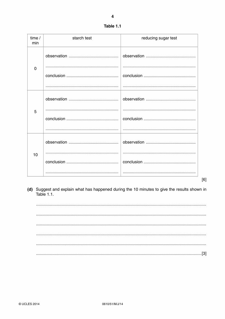
Table 1.1
time / min
starch test reducing sugar test
0
observation ...........................................
...............................................................
conclusion .............................................
...............................................................
observation ...........................................
...............................................................
conclusion .............................................
...............................................................
5
observation ...........................................
...............................................................
conclusion .............................................
...............................................................
observation ...........................................
...............................................................
conclusion .............................................
...............................................................
10
observation ...........................................
...............................................................
conclusion .............................................
...............................................................
observation ...........................................
...............................................................
conclusion .............................................
...............................................................
[6]
(d) Suggest and explain what has happened during the 10 minutes to give the results shown in Table 1.1.
...................................................................................................................................................
...................................................................................................................................................
...................................................................................................................................................
...................................................................................................................................................
...................................................................................................................................................
...............................................................................................................................................[3]

5
0610/51/M/J/14© UCLES 2014 [Turn over
(e) Explain why each of the following procedures was used:
(i) a clean pipette was used to remove each sample of the mixture;
...........................................................................................................................................
.......................................................................................................................................[1]
(ii) a white tile was used for the starch test.
...........................................................................................................................................
.......................................................................................................................................[1]
(f) Describe two further tests that would act as ‘controls’ for this experiment.
...................................................................................................................................................
...................................................................................................................................................
...................................................................................................................................................
...............................................................................................................................................[2]
(g) Suggest one improvement to the method that would make the results more reliable.
...................................................................................................................................................
...............................................................................................................................................[1]
(h) State the name of the enzyme that works in the alimentary canal to break down starch.
...............................................................................................................................................[1]
6
0610/51/M/J/14© UCLES 2014
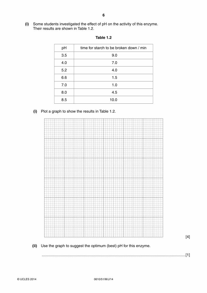
(i) Some students investigated the effect of pH on the activity of this enzyme. Their results are shown in Table 1.2.
Table 1.2
pH time for starch to be broken down / min
3.5 9.0
4.0 7.0
5.2 4.0
6.6 1.5
7.0 1.0
8.0 4.5
8.5 10.0
(i) Plot a graph to show the results in Table 1.2.
[4]
(ii) Use the graph to suggest the optimum (best) pH for this enzyme.
.......................................................................................................................................[1]

7
0610/51/M/J/14© UCLES 2014 [Turn over
(iii) Describe the effect of pH on the activity of this enzyme.
...........................................................................................................................................
...........................................................................................................................................
...........................................................................................................................................
...........................................................................................................................................
...........................................................................................................................................
.......................................................................................................................................[3]
[Total: 28]
8
0610/51/M/J/14© UCLES 2014
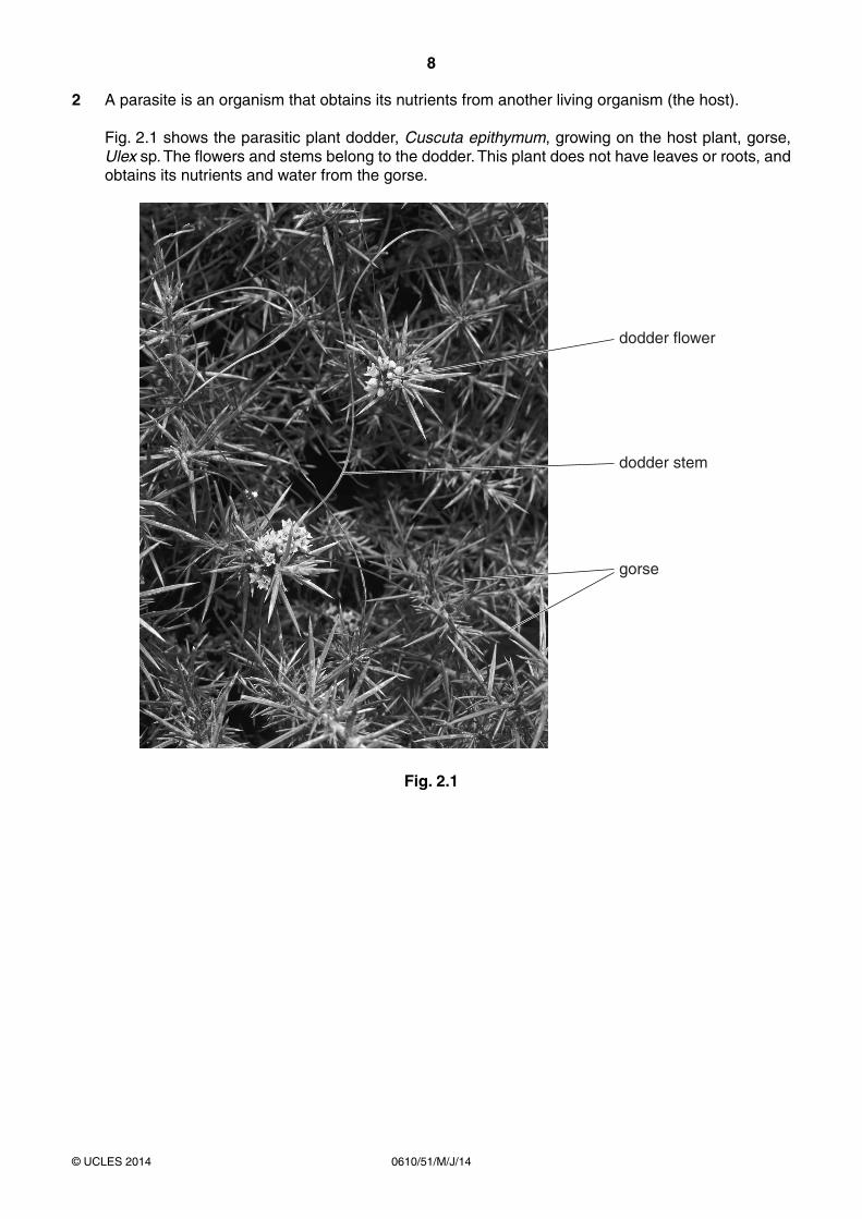
2 A parasite is an organism that obtains its nutrients from another living organism (the host).
Fig. 2.1 shows the parasitic plant dodder, Cuscuta epithymum, growing on the host plant, gorse, Ulex sp. The flowers and stems belong to the dodder. This plant does not have leaves or roots, and obtains its nutrients and water from the gorse.
dodder flower
dodder stem
gorse
Fig. 2.1

9
0610/51/M/J/14© UCLES 2014 [Turn over
Fig. 2.2 shows a section through the stem of gorse to show the attachment of the dodder as seen using a microscope.
stem ofdodder
× 50
stem ofgorse
M NM N
Fig. 2.2
(a) On Fig. 2.2, draw labelled lines to identify the position of:
(i) xylem of gorse;
(ii) phloem of gorse. [2]
(b) Suggest how dodder obtains minerals from the gorse.
...................................................................................................................................................
...............................................................................................................................................[1]
10
0610/51/M/J/14© UCLES 2014
(c) The structure that dodder uses to make contact with the gorse is called a haustorium. The width of the haustorium is marked by the line MN, on Fig. 2.2.
Measure the length of MN.
........................................................ mm
Calculate the actual width of the haustorium (MN).
Show your working.
actual width ........................................................ mm [3]
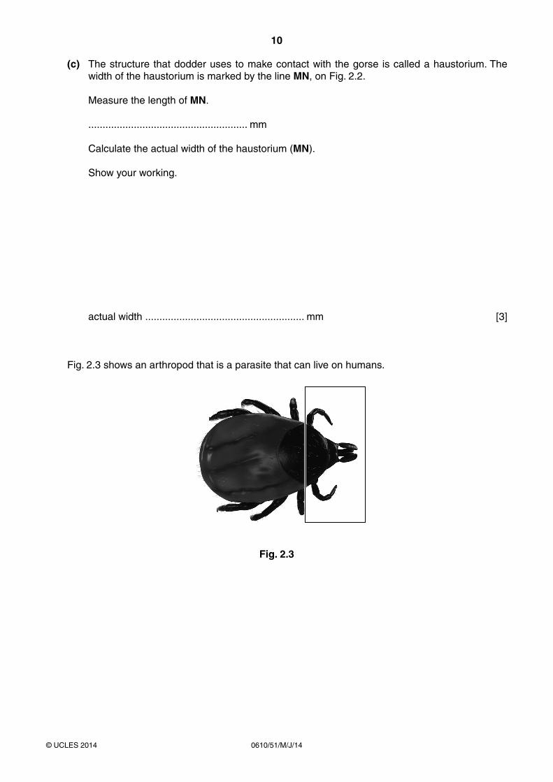
Fig. 2.3 shows an arthropod that is a parasite that can live on humans.
Fig. 2.3

11
0610/51/M/J/14© UCLES 2014
(d) (i) Make a large labelled drawing of the part of the parasite in the rectangle.
[4]
(ii) Name the group of arthropods to which this animal belongs.
Give a reason for your answer.
...........................................................................................................................................
.......................................................................................................................................[2]
[Total: 12]

12
0610/51/M/J/14© UCLES 2014
Copyright Acknowledgements:
Question 2 Figure 2.1Question 2 Figure 2.2Question 2 Figure 2.3
© Ref: B650/0051; Geoff Kidd/Science Photo Library; Dodder (Cuscuta epithymum); www.sciencephoto.com.© Ref: C004/8753; Garry Delong/Science Photo Library; Parasitic Dodder Plant; www.sciencephoto.com.© Ref: F004/2842; Sciepro/Science Photo Library; Tick; www.sciencephoto.com.
Permission to reproduce items where third-party owned material protected by copyright is included has been sought and cleared where possible. Every reasonable effort has been made by the publisher (UCLES) to trace copyright holders, but if any items requiring clearance have unwittingly been included, the publisher will be pleased to make amends at the earliest possible opportunity.
Cambridge International Examinations is part of the Cambridge Assessment Group. Cambridge Assessment is the brand name of University of Cambridge Local Examinations Syndicate (UCLES), which is itself a department of the University of Cambridge.
BLANK PAGE