calcium oscillations-coupled conversion of actin travelling waves to standing oscillations ·...
TRANSCRIPT

Calcium oscillations-coupled conversion of actintravelling waves to standing oscillationsMin Wu1,2, Xudong Wu, and Pietro De Camilli2
Howard Hughes Medical Institute, Department of Cell Biology and Program in Cellular Neuroscience, Neurodegeneration and Repair, Yale University Schoolof Medicine, New Haven, CT 06520
Contributed by Pietro De Camilli, December 11, 2012 (sent for review November 9, 2012)
Dynamic spatial patterns of signaling factors or macromolecularassemblies in the form of oscillations or traveling waves haveemerged as important themes in cell physiology. Feedback mech-anisms underlying these processes and theirmodulationby signalingevents and reciprocal cross-talks remain poorly understood. Here weshow that antigen stimulation of mast cells triggers cyclic changes inthe concentration of actin regulatory proteins and actin in the cellcortex that can be manifested in either spatial pattern. Recruitmentof FBP17 and active Cdc42 at the plasma membrane, leading to actinpolymerization, are involved in both events, whereas calcium oscil-lations, which correlatewith globalfluctuations of plasmamembranePI(4,5)P2, are tightly linked to standing oscillations and counteractwave propagation. These findings demonstrate the occurrence of acalcium-independent oscillator that controls the collective dynamicsof factors linking the actin cytoskeleton to the plasma membrane.Coupling between this oscillator and the one underlying globalplasma membrane PI(4,5)P2 and calcium oscillations spatially regu-lates actin dynamics, revealing an unexpected pattern-renderingmechanismunderlyingplastic changes occurring in the cortical regionof the cell.
curvature | pattern formation | plasticity | GTPase | frequency
Actin waves have emerged as important cell oscillators thatmay impact cell polarity (1, 2), motility (3–6), and division
(7). Another prominent example of rhythmic cell activity is theoscillations of cytosolic calcium, a second messenger that has alsobeen implicated in these actin-dependent processes, although itsprecise involvement remains debated (8–11). Calcium oscillationsare most strikingly observed in excitatory or secretory cells, butcan occur in all cells in response to hormones or growth factorsstimulation (12). Though such oscillations provide an attractivemechanism to encode information with frequency and amplitude(13, 14), calcium oscillation-specific functions remain elusive,partially because of the difficulty in dissecting the effects of cal-cium oscillations from those due to sustained calcium elevations.Considering that rhythmic changes of actin and calcium are likelyto have overlapping function in the regulation of fundamentalcellular events, here we have investigated their potential couplingin mast cells, a model system where stimulus-evoked changes incytosolic calcium and actin dynamics have been extensively docu-mented (15). We report the occurrence of both actin waves andactin oscillations in an activity-dependent manner, and the cou-pling of actin oscillations but not of waves with calcium oscillations.Our results suggest that calcium oscillations, rather than elevationof calcium levels per se, are key factors responsible for the con-version of actin waves into actin oscillations. Thus, calcium oscil-lations, coupled to PI(4,5)P2 oscillations, can be used specifically toregulate actin dynamics.
Results and DiscussionOscillations and Waves of FBP17, Actin, and Cdc42. We first charac-terized the dynamic actin rearrangements elicited by multivalentantigen cross-linking of Fc receptors on the mast cell surface.Cortical actin in mast cells is represented by a thick and densenetwork of actin bundles (16). To capture events proximal to
assembly sites at the plasma membrane, we imaged FBP17, apotent membrane-associated activator of N-WASP and Arp2/3-dependent actin polymerization (17–19), by total internal re-flection fluorescence microscopy (TIRFM). Antigen stimulationproduced a striking appearance of FBP17 hotspots in the eva-nescent field either as recurring waves of hotspots (Fig. 1 A and B;Movie S1) or as oscillations (Fig. 1 C and D; Movie S2). Thepropagation of the waves was due to the sequential formation ofnew spots at the front of the cluster and disappearance at theback (Fig. 1B), with the center of mass of the cluster moving at anaverage speed of 0.87 μm/s (n = 70) and a maximum speed of 1.8μm/s. During oscillations, hotspots also formed and disassembledsequentially, but the centers of mass of the hotspot ensemble wererelatively stationary (Fig. 1D). Strictly speaking, oscillations were infact oscillatory standing waves, although to avoid confusion be-tween the two events, we refer to them as standing oscillations. Theperiodicity of the two patterns were comparable, with cycles of29.4 ± 5 s (n = 12) or 31.4 ± 4.4 s (n = 16) for standing oscillationsand traveling waves, respectively.Oscillations and waves were also observed for actin, as reported
both by LifeAct-EGFP (Fig. 2 A and C; Movie S3) and EGFP-N-WASP (Fig. 2B). These cyclic changes did not require the over-expression of FBP17 but were dependent of the polymerization ofactin. Addition of latrunculin, which blocks actin polymerization,to cells with traveling waves completely abolished wave propa-gation (Fig. 2D; Movie S3). Antigen stimulation in the presenceof latrunculin also failed to trigger oscillations or waves (Fig.S1A). Thus, although the peak of actin was delayed by ∼3–6 scompared with that of FBP17 (Fig. 2C), an actin-dependentpositive feedback mechanism was required for pattern initiationand propagation.Biochemical studies have demonstrated the activation of the
small GTPase Cdc42 in mast cells in response to antigen stim-ulation (20). Cdc42 is a key regulator of actin dynamics, whosereported effectors in mediating such regulation include N-WASPand the FBP17 homologs Toca1 (21) and CIP4 (22). We havenow demonstrated that FBP17 as well is a Cdc42 effector, be-cause purified FBP17 binds to the active form of Cdc42, i.e., toGTPγS-bound Cdc42 or to a constitutively active Cdc42 mutant(Fig. 2E). Thus, we examined whether the activation of Cdc42 inresponse to stimulation occurred in pulses. TIRFM analysis ofa fluorescent activity sensor of Cdc42 (23) revealed that antigenstimulation of mast cells induced the rapid translocation of this
Author contributions: M.W. designed research; M.W. and X.W. performed research; M.W.contributed new reagents/analytic tools; M.W. analyzed data; and M.W. and P.D.C. wrotethe paper.
The authors declare no conflict of interest.
Freely available online through the PNAS open access option.1Present address: Department of Biological Sciences, Centre for BioImaging Sciences, andMechanobiology Institute, National University of Singapore, Republic of Singapore117546.
2To whom correspondence may be addressed. E-mail: [email protected] or [email protected].
This article contains supporting information online at www.pnas.org/lookup/suppl/doi:10.1073/pnas.1221538110/-/DCSupplemental.
www.pnas.org/cgi/doi/10.1073/pnas.1221538110 PNAS | January 22, 2013 | vol. 110 | no. 4 | 1339–1344
CELL
BIOLO
GY
Dow
nloa
ded
by g
uest
on
Feb
ruar
y 27
, 202
0

sensor to the plasma membrane, followed by either oscillationsor waves (Fig. 2F). Importantly, waves of active Cdc42 over-lapped with FBP17 waves both temporally and spatially (Fig. 2G;Movie S4), although the appearance of the fluorescence withinthe waves was diffuse for Cdc42 and punctate for FBP17. Thesefindings strongly support a role of transient local activation ofCdc42 in facilitating the recruitment and polymerization ofFBP17 at the plasma membrane.
Oscillations but Not Waves Are Coupled to Calcium Oscillations. Stand-ing oscillations of FBP17 were strictly stimulation-dependent.They started as early as minutes after the beginning of the stimulus(Fig. 3A) and were observed up to an hour. In contrast, FBP17 wavesoccasionally occurred spontaneously without stimuli, although thesespontaneous waves had slightly longer periods (40.6 ± 10.5 s, n = 9)than stimulus-evoked waves (31.4± 4.4 s, n= 16; P< 0.005, Student ttest. These differences led us to speculate that standing oscillationsmay require additional factors relative to traveling waves.Antigen stimulation of RBL-2H3 cells frequently induces cy-
tosolic calcium oscillations (24, 25), whose time of onset relativeto the stimulus, and duration, are heterogeneous (Fig. S2). In allcells where FBP17 oscillations occurred (n = 12), oscillatory cal-cium spikes were also detected, as shown by the calcium sensorGCaMP3 (26) (Fig. 3A; Movie S5). Fourier transforms of bothoscillations revealed a precisely matching frequency component(Fig. 3A), whereas the phases were roughly anticorrelated (Fig.3C). However, the elevation of cytosolic calcium alone was notsufficient to induce FBP17 recruitment, and regulated calciumdynamics was required for oscillations of FBP17. Presence of
thapsigargin (which depleted intracellular calcium stores) duringantigen stimulation completely abolished both calcium oscil-lations and FBP17 oscillations (Fig. S1B). Calcium oscillationsoften started after a broad and prolonged calcium peak due todepletion of intracellular stores (Fig. 3A), but this initial calciumelevation did not induce FBP17 recruitment; rather, in manycases, this corresponded to a decrease in the FBP17 intensity(Figs. 3A and 4). In addition, the rise and fall of the FBP17 signalduring each oscillation occurred when calcium level remained inthe baseline (Fig. 3C). These findings suggested that calciumitself was unlikely to be the rate-limiting step in the regulation ofthe FBP17 oscillations, and that factors linking both calcium andFBP17 may be responsible for their identical frequency.Calcium oscillations can be coupled with oscillations of IP3 via
a feedback loop that involves the calcium-dependent hydrolysisof PI(4,5)P2 by phospholipase C (12). PI(4,5)P2, which is con-centrated in the plasma membrane, plays a major role in therecruitment and activation at this membrane of actin regulatoryproteins and thus in actin nucleation (27); it is also thought tohave a direct role in the membrane recruitment of FBP17 (17,18). To determine whether PI(4,5)P2 links calcium spikes toFBP17 oscillations, levels of this phosphoinositide in the plasmamembrane were monitored by TIRFM using mRFP-PHPLCδ,a PI(4,5)P2 reporter. PI(4,5)P2 started to oscillate after an anti-gen stimulation-dependent initial PI(4,5)P2 drop. This initialdrop was shown to be due to the activation of phospholipase C(28), which is consistent with an absence of FBP17 recruitmentin this initial phase of stimulation. PI(4,5)P2 oscillations werecell-wide (Fig. 3B) and anticorrelated with calcium oscillation,
Fig. 1. Traveling waves or standing oscillations of FBP17 in antigen-stimulated mast cells visualized by TIRFM. (A and B) Waves. (A) Three individual frames froma movie of mCherry-FBP17 waves (Movie S1). An inverted grayscale lookup table was used, and fluorescence is shown in black. (Far Right) Color image showingsuperimposition of fluorescence signal from 10 different frames (3-s intervals) of the cell on the Far Left, where bright puncta in the same frame are shown inthe same color. (B) Montage of 35 frames (3 s or 0.2-s intervals) of a small region of the cell showing that wave propagation is through sequential formation anddisassembly of puncta. (C and D) Oscillations. (C) Three individual frames from a movie of a mCherry-FBP17 oscillation. (Far Right) Color image showing su-perimposition of fluorescence signal from 10 different frames (3-s intervals) of the cell shown on the Far Left, with color-coding based on time as in A. (D)Montage of 35 frames (3-s intervals) of a small region of the cell showing the cyclic assembly of mCherry-FBP17 puncta at the cell cortex. (Scale bar: 10 μm.)
1340 | www.pnas.org/cgi/doi/10.1073/pnas.1221538110 Wu et al.
Dow
nloa
ded
by g
uest
on
Feb
ruar
y 27
, 202
0

similar to FBP17 oscillations (Fig. 3C). Thus, PI(4,5)P2 mayrepresent a key upstream factor that coordinates calcium andFBP17/actin oscillations.Though calcium oscillations with matching frequencies were
observed in all cells with FBP17 oscillations (n = 12), demon-strating a tight coupling between the two processes, they onlyoccurred in 21% of the cells with FBP17 waves (5 of 24 cells). Ofthe other cells in this group (n = 19), 53% had no detectablecalcium fluctuations (n = 10; e.g., Fig. S3 A and B before stim-ulation) and 47% had intermittent and weak calcium pulseswhose frequencies did not match those of the FBP17 waves (n =9; e.g., Fig. S3B after stimulation). This lack of strong couplingbetween calcium oscillations and traveling waves of FBP17 addsfurther evidence for the lack of a direct cause–effect relationshipbetween levels of cytosolic calcium and FBP17 recruitment to thecell cortex.Consistent with the dissociation between traveling waves and
calcium oscillations, there were no global PI(4,5)P2 oscillationsor local PI(4,5)P2 waves (as detected by the PI(4,5)P2 reportermRFP-PHPLCδ) in cells with traveling waves. Dynamic turnover
of PI(4,5)P2, resulting in changes too shallow to be revealed bymRFP-PHPLCδ, may occur at waves, likely coordinated by cyclesof phosphorylation/dephosphorylation by PI4P 5-kinases andPI(4,5)P2 phosphatases, respectively. Importantly, however, thefeedback loop involving FBP17, Cdc42, and actin can apparentlyact independently from that controlling cell-wide calcium oscil-lations, which involves the activation of phospholipase C andresults in detectable PI(4,5)P2 oscillations (Fig. 5).
Interconversion Between Oscillations and Waves. To determinewhether these different actin dynamic spatial patterns (oscil-lations and waves) reflect heterogeneity of the cell populationor differential activity states of the cell, we examined if they caninterconvert within the same cell. Cells that displayed spontane-ous FBP17 waves with little or no calcium pulses were stimulatedwith antigen, to elicit calcium/PI(4,5)P2 oscillations. In cells thatgenerated robust calcium oscillations (Fig. 4A), FBP17 wavesbecame synchronized into a cell-wide standing oscillatory patternafter a brief pause (Fig. 4B; Movie S6). Conversely, in cells wheretraveling waves resumed after such pause, calcium oscillations
0 1 2 3 4
A B
C D
E GF
Lifeact N-WASPFBP17 FBP17
FBP17FBP17
merge merge
merge
FB
P17
Active Cdc42Active Cdc42
latrunculin
3 min 3 min
3 min
Bait: GST-Cdc42
G12
V, G
TPW
T, G
TPS
WT,
GD
P
inpu
t
FB
P17
Life
act
FB
P17
Time / min
Fig. 2. Stimulus-evoked waves of FBP17 are synchronized with both N-WASP and active Cdc42 and require actin polymerization. (A and B) Kymographs of actin(LifeAct-GFP), N-WASP-GFP, and mCherry-FBP17 waves. (C) Relative phase-shifts of FBP17 and actin. Peaks of FBP17 preceded actin by 3–6 s. (D) Montage of se-quential frames from a portion of a cell expressingmCherry-FBP17 showing that addition of latrunculin abolished waves (Movie S3). (E) FBP17 is a Cdc42 effector invitro. Binding assay showing that purified FBP17 interacts with the purified active form of Cdc42, i.e., with GTPγS-bound Cdc42 or with a constitutively active GTP-bound Cdc42 mutant, but not to GDP-bound Cdc42. (F) Snapshots of a cell with antigen-evoked traveling waves show that the diffuse but locally concentratedaccumulation of active Cdc42 at the plasmamembrane (Cdc42 CBD-EGFP) coincides with clusters of FBP17 puncta. (G) Kymograph from a movie of traveling wavesshowing coupled dynamics of FBP17 and active Cdc42 (Movie S4). An inverted lookup table was used for grayscale images and kymographs. (Scale bar: 10 μm.)
Wu et al. PNAS | January 22, 2013 | vol. 110 | no. 4 | 1341
CELL
BIOLO
GY
Dow
nloa
ded
by g
uest
on
Feb
ruar
y 27
, 202
0
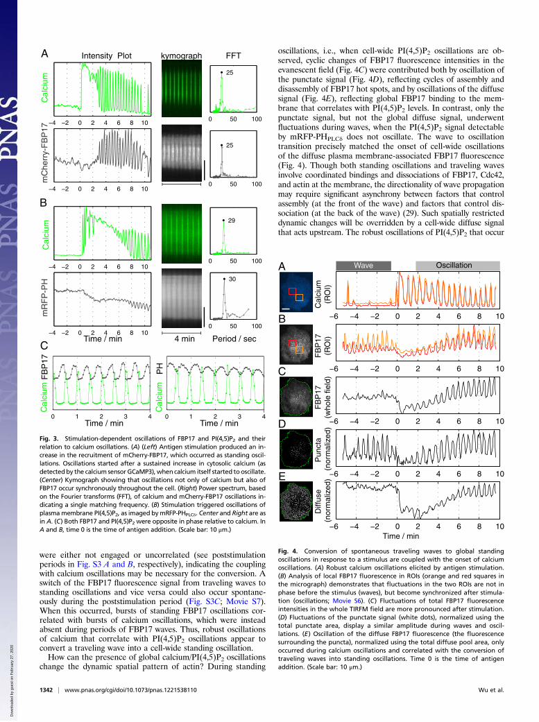
were either not engaged or uncorrelated (see poststimulationperiods in Fig. S3 A and B, respectively), indicating the couplingwith calcium oscillations may be necessary for the conversion. Aswitch of the FBP17 fluorescence signal from traveling waves tostanding oscillations and vice versa could also occur spontane-ously during the poststimulation period (Fig. S3C; Movie S7).When this occurred, bursts of standing FBP17 oscillations cor-related with bursts of calcium oscillations, which were insteadabsent during periods of FBP17 waves. Thus, robust oscillationsof calcium that correlate with PI(4,5)P2 oscillations appear toconvert a traveling wave into a cell-wide standing oscillation.How can the presence of global calcium/PI(4,5)P2 oscillations
change the dynamic spatial pattern of actin? During standing
oscillations, i.e., when cell-wide PI(4,5)P2 oscillations are ob-served, cyclic changes of FBP17 fluorescence intensities in theevanescent field (Fig. 4C) were contributed both by oscillation ofthe punctate signal (Fig. 4D), reflecting cycles of assembly anddisassembly of FBP17 hot spots, and by oscillations of the diffusesignal (Fig. 4E), reflecting global FBP17 binding to the mem-brane that correlates with PI(4,5)P2 levels. In contrast, only thepunctate signal, but not the global diffuse signal, underwentfluctuations during waves, when the PI(4,5)P2 signal detectableby mRFP-PHPLCδ does not oscillate. The wave to oscillationtransition precisely matched the onset of cell-wide oscillationsof the diffuse plasma membrane-associated FBP17 fluorescence(Fig. 4). Though both standing oscillations and traveling wavesinvolve coordinated bindings and dissociations of FBP17, Cdc42,and actin at the membrane, the directionality of wave propagationmay require significant asynchrony between factors that controlassembly (at the front of the wave) and factors that control dis-sociation (at the back of the wave) (29). Such spatially restricteddynamic changes will be overridden by a cell-wide diffuse signalthat acts upstream. The robust oscillations of PI(4,5)P2 that occur
−4 −2 0 2 4 6 8 10
−4 −2 0 2 4 6 8 10
−4 −2 0 2 4 6 8 10
−4 −2 0 2 4 6 8 10
0 1 2 3 4 0 1 2 3 4
Am
RF
P-P
H
B
mC
herr
y-F
BP
17
Time / min
FFTIntensity Plot
Period / sec
Cal
cium
FB
P17
Time / min Time / min
kymograph
4 minC
0 50 100
25
0 50 100
25
0 50 100
29
0 50 100
30
Cal
cium
PH
Cal
cium
Cal
cium
Fig. 3. Stimulation-dependent oscillations of FBP17 and PI(4,5)P2 and theirrelation to calcium oscillations. (A) (Left) Antigen stimulation produced an in-crease in the recruitment of mCherry-FBP17, which occurred as standing oscil-lations. Oscillations started after a sustained increase in cytosolic calcium (asdetectedby the calcium sensor GCaMP3),when calcium itself started tooscillate.(Center) Kymograph showing that oscillations not only of calcium but also ofFBP17 occur synchronously throughout the cell. (Right) Power spectrum, basedon the Fourier transforms (FFT), of calcium and mCherry-FBP17 oscillations in-dicating a single matching frequency. (B) Stimulation triggered oscillations ofplasmamembrane PI(4,5)P2, as imaged bymRFP-PHPLCδ. Center and Right are asin A. (C) Both FBP17 and PI(4,5)P2 were opposite in phase relative to calcium. InA and B, time 0 is the time of antigen addition. (Scale bar: 10 μm.)
−6 −4 −2 0 2 4 6 8 10
−6 −4 −2 0 2 4 6 8 10
−6 −4 −2 0 2 4 6 8 10
−6 −4 −2 0 2 4 6 8 10
−6 −4 −2 0 2 4 6 8 10
Diff
use
(nor
mal
ized
)P
unct
a (n
orm
aliz
ed)
FB
P17
(RO
I)F
BP
17(w
hole
fiel
d)C
alci
um(R
OI)
A
B
C
D
E
Time / min
Wave Oscillation
Fig. 4. Conversion of spontaneous traveling waves to global standingoscillations in response to a stimulus are coupled with the onset of calciumoscillations. (A) Robust calcium oscillations elicited by antigen stimulation.(B) Analysis of local FBP17 fluorescence in ROIs (orange and red squares inthe micrograph) demonstrates that fluctuations in the two ROIs are not inphase before the stimulus (waves), but become synchronized after stimula-tion (oscillations; Movie S6). (C) Fluctuations of total FBP17 fluorescenceintensities in the whole TIRFM field are more pronounced after stimulation.(D) Fluctuations of the punctate signal (white dots), normalized using thetotal punctate area, display a similar amplitude during waves and oscil-lations. (E) Oscillation of the diffuse FBP17 fluorescence (the fluorescencesurrounding the puncta), normalized using the total diffuse pool area, onlyoccurred during calcium oscillations and correlated with the conversion oftraveling waves into standing oscillations. Time 0 is the time of antigenaddition. (Scale bar: 10 μm.)
1342 | www.pnas.org/cgi/doi/10.1073/pnas.1221538110 Wu et al.
Dow
nloa
ded
by g
uest
on
Feb
ruar
y 27
, 202
0

during calcium oscillations likely represent the signal that modu-lates globally the dynamics of proteins associated with theplasma membrane.
Concluding Remarks. Feedback regulatory loops underlying oscil-lations or waves have been increasingly recognized as importantfactors in the organization of signaling events (30). The oscillatorynature of the recruitment and assembly of actin regulatory proteinsand actin, via the control of reversible and low-affinity interactions,may facilitate the occurrence of adaptive modifications in the actincortex and the plasma membrane. This plasticity may be particu-larly important following stimulation—an event that involves dra-matic cell shape changes, actin reorganization, regulated secretion,and compensatory endocytosis (15). In this context, it is importantto note that calcium oscillations induced by stimulation in mastcells and other secretory cells were shown to correlate with oscil-latory bursts of secretion (31–33). Interestingly, FBP17 is an F-BAR domain-containing protein (34) that can participate in en-docytosis (35, 36). Though feedback regulatory mechanisms me-diated by Cdc42 activation and actin polymerization are involvedin the collective events described here, irrespective of theirspatial pattern, they can be coupled to, or decoupled from, thoseunderlying calcium oscillations, resulting in either standingoscillations or traveling waves (Fig. 5). The integration of dis-tinct oscillatory systems may be of general importance in fine-tuning cell plasticity by spatially controlling membrane and cy-toskeleton dynamics.
MethodsMaterials. RBL-2H3 cells (tumor mast cells), mouse monoclonal anti–2,4-dini-trophenyl (DNP) IgE, and the multivalent antigen DNP-BSA were generousgifts from Barbara Baird and David Holowka (Cornell University, Ithaca, NY).The following reagents were obtained from commercial sources: GTP andGDP from Sigma; GTPgS from Roche; and thapsigargin and latrunculin B fromCalbiochem. Polyclonal anti-FBP17 antibodies were previously described (17).Constructs for the following proteins were kind gifts: mCherry-FBP17 fromPontus Aspenstrom (Karolinska Institutet, Stockholm); mRFP-PHPLCδ (pleck-strin homology domain of PLCδ) from Sergio Grinstein (University of Toronto,Toronto); LifeAct-EGFP from Roland Wedlich-Soldner (Max Planck Institute ofBiochemistry, Martinsried, Germany); GST-Cdc42 G12V and GST-Cdc42 WTfrom Tony Koleske (Yale University, New Haven, CT); CBD-GFP, a Cdc42 ac-tivity probe (generated by cloning bacterial expressing Cdc42 activity sensor
pET23-CBD-(-PP)-EGFP (23) (Addgene no. 12597) into pEGFP-N1 vector withrestriction sites XhoI and BamH1) from Hongying Shen (our laboratory).GCAMP3 (26) was from Addgene (no. 22692). FBP17-EGFP was prepared bycloning EGFP-FBP17 (17) into the pEGFP-N1 vector using the BamH1 andHindIII restriction sites. GST-FBP17 (17) was previously described.
Cell Culture and Transfection. RBL-2H3 cells were maintained in monolayercultures and harvested with trypsin-EDTA (Life Technologies) 3–5 d afterpassage. Transient transfections were carried out on cells in suspension usingprogram X-01 of the Amaxa electroporator (Lonza). After transfection, cellswere plated at subconfluent densities in 35-mm Petri dishes with coverglassbottoms (MatTek) and sensitized with anti-DNP IgE at 0.4 μg/mL overnight.Typically cells were cultured for 12–24 h and washed twice in buffered salinesolution [135 mM NaCl, 5.0 mM KCl, 1.8 mM CaCl2, 1.0 mM MgCl2, 5.6 mMglucose, 20 mM Hepes (pH 7.4)] containing 1 mg/mL BSA before imaging;they were subsequently transferred to a homemade heated microscopestage maintained at 37 °C through a bipolar temperature controller (Har-vard Apparatus). In all experiments, cells were stimulated with 100 ng/mLDNP-BSA. For experiments using inhibitors, latrunculin B and thapsigarginwere diluted from the stock solution and added to the adherent cells ata final concentration of 12.5 μM and 2 μM, respectively.
TIRFM. A Nikon Ti-E inverted microscope equipped with Perfect Focus system,motorized illumination unit, and iXon 897 Electron Multiplying ChargeCoupled Device (EMCCD) camera (Andor Technologies) was used. All imageswere acquired through an Olympus TIRFM objective (CFI Apochromat 100×,N.A. 1.49). Samples were excited with 488-nm/50-mW and 561-nm/50-mWdiode lasers sequentially, and two-color images were acquired througha dual-bandpass filter (exciter: 468/34, 553/24 nm; emitter: 512/23, 630/91 nm;Semrock). Andor IQ software was used to acquire data at binning 2.
Image Analysis. ImageJ (National Institutes of Health) was used for generatingmontages, kymographs, and quantification based onmanually selected regionsof interest (ROIs). MatLab (MathWorks) was used for frequency analysis andquantification requiring image segmentation. For the identification of thefrequency components of an intensityprofile, the built-in fast Fourier transformfunction inMatLabwas used. For quantification involving image segmentation,including global analysis of a single cell and analysis of puncta intensity vs.diffuse signals, custom-written algorithmswas used (37). Briefly, amedianfilterwas applied to each frame of the image series and the field of view wassegmented into two regions: “cell” and “background” by thresholding andmorphological operations. All pixels positive for cell were subsequently seg-mented into puncta (identified as pixels with intensity above their localbackground by a threshold value A) and diffuse (defined as pixels not greaterthan their local background by a threshold value B) by local backgroundsubtraction, peak identification, and thresholding. B is set at a smaller valuethan A to reduce the contamination of the signals from the puncta into diffusepool. The defined puncta and cell boundaries were overlaid with the originalmovie for visual inspection. The number of pixels defined as puncta or diffuse,and their average intensities, were plotted against time.
Statistical Analyses. Statistical analyses were performed with the unpairedtwo-tailed Student t test. Average data are expressed as mean ± SEM.
Protein Biochemistry. GST fusion proteins (Cdc42 wt, Cdc42 G12V, and FBP17)were bacterially expressed and purified using Glutathione Sepharose 4B beads(Amersham Biosciences) according to the manufacturer’s instructions. GST-Cdc42 WT and Cdc42 G12V were eluted from the beads and incubated withfresh beads to reach saturation of the bead capacity. For nucleotide loading,these GST-Cdc42–loaded beads were incubated in buffer A (PBS plus 2.5 mMMgCl2) with 2.5 mM GTP, GDP, or GTPγS at 30 °C for 20 min. Beads were thenwashed twice with buffer A, and the supernatant was removed. For in vitrobinding, the GST tag of FBP17 was removed using PreScission protease(Amersham Biosciences), and fresh beads were used to remove free GST anduncleaved GST-FBP17. Cleaved FBP17 (0.5–1 mg/mL, 200 μL) was incubatedwith GST-Cdc42 beads loaded with nucleotides in buffer A with 0.1% Triton Xat 4 °C for 2 h. Then, beads were washed once with buffer A + 0.25% Triton Xand twice with buffer A + 0.1% Triton X. Total protein was eluted with SDS/PAGE sample loading buffer. A total of 5% (vol/vol) of the eluted mixture and0.5% of input were loaded for Western blot analysis.
ACKNOWLEDGMENTS. We thank Hongying Shen, Francesca Giordano, andother members of P.D.C.’s laboratory for discussions, and F. Wilson, S. Wilson,and M. Graham for technical support. This work was supported in part byNational Institutes of Health Grants R37NS036251, DK45735, and DK082700.
Time
space
Time
space
TRAVELLING WAVES OSCILLATORY STANDING WAVES
PI(4,5)P2
Actin
Ca2+
IP3
PLC
Ca/PI(4,5)P2 OSCILLATIONS
Cdc42/FBP17/N-WASP
PI(4,5)P2
Actin
Cdc42/FBP17/N-WASP
A B
Fig. 5. Schematic representation of the mechanisms underlying the trav-eling waves and standing oscillations. (A) Traveling waves involve actin-de-pendent positive-feedback mechanisms that are calcium independent. (B)Actin oscillations require similar molecular components as actin waves, butare coupled with calcium oscillations, likely via PI(4,5)P2 oscillations.
Wu et al. PNAS | January 22, 2013 | vol. 110 | no. 4 | 1343
CELL
BIOLO
GY
Dow
nloa
ded
by g
uest
on
Feb
ruar
y 27
, 202
0

1. Ozbudak EM, Becskei A, van Oudenaarden A (2005) A system of counteracting feedbackloops regulates Cdc42p activity during spontaneous cell polarization. Dev Cell 9(4):565–571.
2. Millius A, Dandekar SN, Houk AR, Weiner OD (2009) Neutrophils establish rapid androbust WAVE complex polarity in an actin-dependent fashion. Curr Biol 19(3):253–259.
3. Giannone G, et al. (2004) Periodic lamellipodial contractions correlate with rearwardactin waves. Cell 116(3):431–443.
4. Schroth-Diez B, et al. (2009) Propagating waves separate two states of actin organi-zation in living cells. HFSP J 3(6):412–427.
5. Weiner OD, Marganski WA, Wu LF, Altschuler SJ, Kirschner MW (2007) An actin-basedwave generator organizes cell motility. PLoS Biol 5(9):e221.
6. Case LB, Waterman CM (2011) Adhesive F-actin waves: A novel integrin-mediatedadhesion complex coupled to ventral actin polymerization. PLoS ONE 6(11):e26631.
7. Mitsushima M, et al. (2010) Revolving movement of a dynamic cluster of actin fila-ments during mitosis. J Cell Biol 191(3):453–462.
8. Wei C, et al. (2009) Calcium flickers steer cell migration. Nature 457(7231):901–905.9. Wedlich-Soldner R, Li R (2003) Spontaneous cell polarization: Undermining de-
terminism. Nat Cell Biol 5(4):267–270.10. Traynor D, Milne JL, Insall RH, Kay RR (2000) Ca(2+) signalling is not required for
chemotaxis in Dictyostelium. EMBO J 19(17):4846–4854.11. Evans JH, Falke JJ (2007) Ca2+ influx is an essential component of the positive-
feedback loop that maintains leading-edge structure and activity in macrophages.Proc Natl Acad Sci USA 104(41):16176–16181.
12. Harootunian AT, Kao JP, Paranjape S, Tsien RY (1991) Generation of calcium oscillationsin fibroblasts by positive feedback between calcium and IP3. Science 251(4989):75–78.
13. Li W, Llopis J, Whitney M, Zlokarnik G, Tsien RY (1998) Cell-permeant caged InsP3ester shows that Ca2+ spike frequency can optimize gene expression. Nature 392(6679):936–941.
14. Dolmetsch RE, Xu K, Lewis RS (1998) Calcium oscillations increase the efficiency andspecificity of gene expression. Nature 392(6679):933–936.
15. Pfeiffer JR, Seagrave JC, Davis BH, Deanin GG, Oliver JM (1985) Membrane and cy-toskeletal changes associated with IgE-mediated serotonin release from rat basophilicleukemia cells. J Cell Biol 101(6):2145–2155.
16. Andrews NL, et al. (2008) Actin restricts FcepsilonRI diffusion and facilitates antigen-induced receptor immobilization. Nat Cell Biol 10(8):955–963.
17. Itoh T, et al. (2005) Dynamin and the actin cytoskeleton cooperatively regulateplasma membrane invagination by BAR and F-BAR proteins. Dev Cell 9(6):791–804.
18. Tsujita K, et al. (2006) Coordination between the actin cytoskeleton and membranedeformation by a novel membrane tubulation domain of PCH proteins is involved inendocytosis. J Cell Biol 172(2):269–279.
19. Takano K, Toyooka K, Suetsugu S, Suetsugu S (2008) EFC/F-BAR proteins and theN-WASP-WIP complex induce membrane curvature-dependent actin polymerization.EMBO J 27(21):2817–2828.
20. El-Sibai M, Backer JM (2007) Phospholipase C gamma negatively regulates Rac/Cdc42activation in antigen-stimulated mast cells. Eur J Immunol 37:261–270.
21. Ho H-YH, et al. (2004) Toca-1 mediates Cdc42-dependent actin nucleation by acti-
vating the N-WASP-WIP complex. Cell 118(2):203–216.22. Aspenström P (1997) A Cdc42 target protein with homology to the non-kinase do-
main of FER has a potential role in regulating the actin cytoskeleton. Curr Biol 7(7):
479–487.23. Nalbant P, Hodgson L, Kraynov V, Toutchkine A, Hahn KM (2004) Activation of en-
dogenous Cdc42 visualized in living cells. Science 305(5690):1615–1619.24. Millard PJ, Ryan TA, Webb WW, Fewtrell C (1989) Immunoglobulin E receptor cross-
linking induces oscillations in intracellular free ionized calcium in individual tumor
mast cells. J Biol Chem 264(33):19730–19739.25. Cohen R, Torres A, Ma H-T, Holowka D, Baird B (2009) Ca2+ waves initiate antigen-
stimulated Ca2+ responses in mast cells. J Immunol 183(10):6478–6488.26. Tian L, et al. (2009) Imaging neural activity in worms, flies and mice with improved
GCaMP calcium indicators. Nat Methods 6(12):875–881.27. Di Paolo G, De Camilli P (2006) Phosphoinositides in cell regulation and membrane
dynamics. Nature 443(7112):651–657.28. Stauffer TP, Ahn S, Meyer T (1998) Receptor-induced transient reduction in plasma
membrane PtdIns(4,5)P2 concentration monitored in living cells. Curr Biol 8(6):
343–346.29. Loose M, Fischer-Friedrich E, Herold C, Kruse K, Schwille P (2011) Min protein patterns
emerge from rapid rebinding and membrane interaction of MinE. Nat Struct Mol Biol
18(5):577–583.30. Brandman O, Meyer T (2008) Feedback loops shape cellular signals in space and time.
Science 322(5900):390–395.31. Tse A, Tse FW, Almers W, Hille B (1993) Rhythmic exocytosis stimulated by GnRH-
induced calcium oscillations in rat gonadotropes. Science 260(5104):82–84.32. Kim TD, Eddlestone GT, Mahmoud SF, Kuchtey J, Fewtrell C (1997) Correlating Ca2+
responses and secretion in individual RBL-2H3 mucosal mast cells. J Biol Chem 272(50):
31225–31229.33. Cohen R, Corwith K, Holowka D, Baird B (2012) Spatiotemporal resolution of mast cell
granule exocytosis reveals correlation with Ca2+ wave initiation. J Cell Sci 125(Pt 12):
2986–2994.34. Shimada A, et al. (2007) Curved EFC/F-BAR-domain dimers are joined end to end into
a filament for membrane invagination in endocytosis. Cell 129(4):761–772.35. Wu M, et al. (2010) Coupling between clathrin-dependent endocytic budding and
F-BAR-dependent tubulation in a cell-free system. Nat Cell Biol 12(9):902–908.36. Giuliani C, et al. (2009) Requirements for F-BAR proteins TOCA-1 and TOCA-2 in actin
dynamics and membrane trafficking during Caenorhabditis elegans oocyte growth
and embryonic epidermal morphogenesis. PLoS Genet 5(10):e1000675.37. Wu M, Holowka D, Craighead HG, Baird B (2004) Visualization of plasma membrane
compartmentalization with patterned lipid bilayers. Proc Natl Acad Sci USA 101(38):
13798–13803.
1344 | www.pnas.org/cgi/doi/10.1073/pnas.1221538110 Wu et al.
Dow
nloa
ded
by g
uest
on
Feb
ruar
y 27
, 202
0