calcium-dependent protein kinase 5 is required for release ... · that hyperactivation of the...
TRANSCRIPT

Calcium-Dependent Protein Kinase 5 Is Required for Releaseof Egress-Specific Organelles in Plasmodium falciparum
Sabrina Absalon,a,b Karin Blomqvist,a,b,c Rachel M. Rudlaff,a,b,d Travis J. DeLano,e Michael P. Pollastri,e Jeffrey D. Dvorina,b
aDivision of Infectious Diseases, Boston Children’s Hospital, Boston, Massachusetts, USAbDepartment of Pediatrics, Harvard Medical School, Boston, Massachusetts, USAcDepartment of Microbiology, Tumor and Cell Biology, Karolinska Institutet, Stockholm, SwedendBiological and Biomedical Sciences, Harvard Medical School, Boston, Massachusetts, USAeDepartment of Chemistry and Chemical Biology, Northeastern University, Boston, Massachusetts, USA
ABSTRACT The human malaria parasite Plasmodium falciparum requires efficientegress out of an infected red blood cell for pathogenesis. This egress event is highlycoordinated and is mediated by several signaling proteins, including the plant-like P.falciparum calcium-dependent protein kinase 5 (PfCDPK5). Knockdown of PfCDPK5results in an egress block where parasites are trapped inside their host cells. Themechanism of this PfCDPK5-dependent block, however, remains unknown. Here, weshow that PfCDPK5 colocalizes with a specialized set of parasite organelles known asmicronemes and is required for their discharge, implicating failure of this step as thecause of the egress defect in PfCDPK5-deficient parasites. Furthermore, we showthat PfCDPK5 cooperates with the P. falciparum cGMP-dependent kinase (PfPKG)to fully activate the protease cascade critical for parasite egress. The PfCDPK5-dependent arrest can be overcome by hyperactivation of PfPKG or by physical dis-ruption of the arrested parasite, and we show that both treatments facilitate the re-lease of the micronemes required for egress. Our results define the molecularmechanism of PfCDPK5 function and elucidate the complex signaling pathway ofparasite egress.
IMPORTANCE The signs and symptoms of clinical malaria result from the replica-tion of parasites in human blood. Efficient egress of the malaria parasite Plasmodiumfalciparum out of an infected red blood cell is critical for pathogenesis. The P. falci-parum calcium-dependent protein kinase 5 (PfCDPK5) is essential for parasite egress.Following PfCDPK5 knockdown, parasites remain trapped inside their host cell anddo not egress, but the mechanism for this block remains unknown. We show thatPfCDPK5 colocalizes with parasite organelles known as micronemes. We demonstratethat PfCDPK5 is critical for the discharge of these micronemes and that failure ofthis step is the molecular mechanism of the parasite egress arrest. We also showthat hyperactivation of the cGMP-dependent kinase PKG can overcome this arrest.Our data suggest that small molecules that inhibit the egress signaling pathwaycould be effective antimalarial therapeutics.
KEYWORDS Plasmodium falciparum, calcium-dependent protein kinase, egress,malaria, microneme
Malaria remains an important cause of global morbidity and mortality with anestimated 214 million cases and 438,000 deaths in 2015 (1). Most of these deaths
are due to infection by the Plasmodium falciparum parasite. The emergence and spreadof drug resistance, particularly of artemisinin-resistant parasites in southeastern Asia (2),threaten to limit the usefulness of antimalarial therapies. Therefore, there is an ongoing
Received 17 January 2018 Accepted 19January 2018 Published 27 February 2018
Citation Absalon S, Blomqvist K, Rudlaff RM,DeLano TJ, Pollastri MP, Dvorin JD. 2018.Calcium-dependent protein kinase 5 isrequired for release of egress-specificorganelles in Plasmodium falciparum. mBio9:e00130-18. https://doi.org/10.1128/mBio.00130-18.
Editor Louis H. Miller, NIAID, NIH
Copyright © 2018 Absalon et al. This is anopen-access article distributed under the termsof the Creative Commons Attribution 4.0International license.
Address correspondence to Jeffrey D. Dvorin,[email protected].
K.B. and R.M.R. contributed equally to thisarticle.
RESEARCH ARTICLE
crossm
January/February 2018 Volume 9 Issue 1 e00130-18 ® mbio.asm.org 1
on March 22, 2019 by guest
http://mbio.asm
.org/D
ownloaded from

need to identify additional critical biological pathways that might be targeted by a newgeneration of antimalarial medications.
The major clinical manifestations of malaria result from exponential expansion ofparasites during the blood stage of the life cycle (3–5). Asexual blood-stage develop-ment of Plasmodium parasites occurs via schizogony. In this process, multiple asyn-chronous nuclear divisions occur within a common cytoplasm. Mature and invasivemerozoites are not formed until a final segmentation (or budding) wherein the newlyformed daughter parasites are all formed simultaneously. Thus, the daughter merozo-ites are not fully mature until all daughter merozoites are mature (6). This requirementhighlights the importance of a coordinated egress signaling pathway to prevent releaseof immature parasites.
In apicomplexan organisms, including both Plasmodium spp. and Toxoplasma gon-dii, release of specialized apical organelles known as micronemes is a critical step inparasite egress. The triggered discharge of these organelles depends on the activationof calcium signaling, cGMP signaling, and phosphatidic acid signaling (7–10). Some ofthe protein mediators of microneme discharge have been identified, including calcium-dependent protein kinases (7, 11), C2 domain-containing proteins (12), and acylatedpleckstrin homology domain-containing proteins (10). However, the specific rolesplayed by these proteins in the activation of microneme discharge remain incompletelyunderstood.
Calcium-dependent protein kinases (CDPKs) (13–15) and the cyclic GMP-dependentprotein kinase (PKG) (8, 16, 17) are important for P. falciparum egress. Additionally, aprotease cascade mainly driven by the serine protease P. falciparum SUB1 (PfSUB1) isessential for egress (8, 18, 19). P. falciparum PKG (PfPKG) activity is required for PfSUB1release from specialized exoneme organelles (8), and once discharged into the parasi-tophorous vacuole, PfSUB1 proteolytically processes multiple parasite proteins, includ-ing the P. falciparum merozoite surface protein-1 (PfMSP1), which itself is essential foregress and allows PfMSP1 to interact with and destabilize the host erythrocyte cyto-skeleton (20). An important role for the perforin-like protein PfPLP1 in blood-stageparasite egress has been hypothesized (21). However, PfPLP1 can be knocked out in theblood stage without a detectable replication defect, suggesting that its role is nones-sential and/or redundant for asexual parasite egress (22). PfCDPK5 (PF3D7_1337800) isessential for P. falciparum egress (15). However, the mechanism behind the egress blockin PfCDPK5-deficient parasites has remained unknown.
In the current study, we perform a functional evaluation of the egress defect inPfCDPK5-deficient parasites. With superresolution microscopy, we demonstrate thedynamic localization of PfCDPK5 throughout schizogony and its colocalization with asubset of microneme organelles. We show that PfCDPK5-deficient parasites do notefficiently release a subset of micronemes and that this failure of microneme release isthe cause of the egress block. In response to baseline activation of PfPKG, PfCDPK5-deficient parasites initially show activation of the protease cascade that is similar toparasites without PfCDPK5 deficiency. However, this level of PfPKG activation is insuf-ficient to induce the release of the required subset of micronemes for egress, andprotease processing does not efficiently proceed to completion in PfCDPK5-deficientschizonts. Pharmacological enhancement of PfPKG activation facilitates micronemerelease and overcomes the PfCDPK5-mediated deficiency. Interestingly, mechanicalrelease of PfCDPK5-deficient merozoites also overcomes this block. We demonstratethat a major function of PfCDPK5 is to act cooperatively with the PfPKG signalingpathway. Our study provides a new layer in our understanding of the P. falciparumegress pathway.
RESULTSPfCDPK5 has dynamic localization during schizont development. We generated
a new transgenic P. falciparum strain, 3D7-PfCDPK5DDKnL, with three copies of thehemagglutinin (HA) epitope followed by the destabilizing domain (DD) at the endog-enous PF3D7_1337800 locus (Fig. 1A; also see Fig. S1 in the supplemental material). By
Absalon et al. ®
January/February 2018 Volume 9 Issue 1 e00130-18 mbio.asm.org 2
on March 22, 2019 by guest
http://mbio.asm
.org/D
ownloaded from
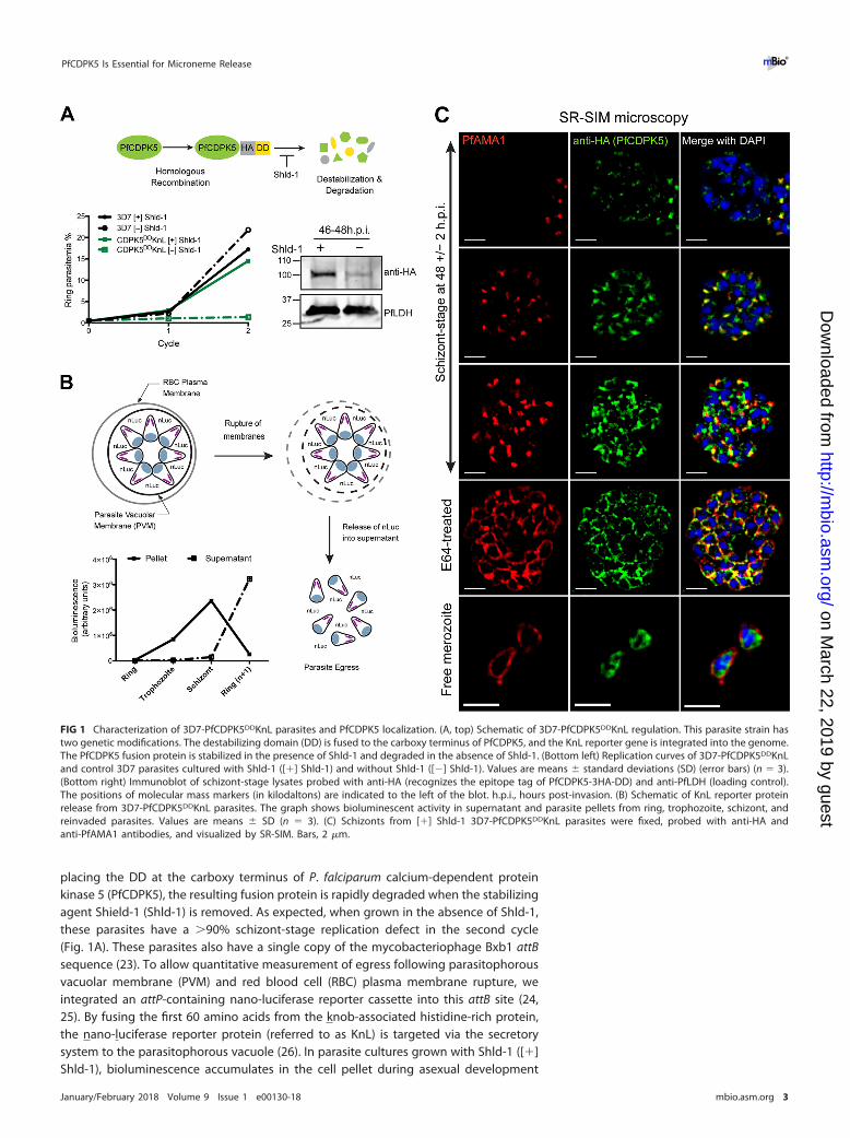
placing the DD at the carboxy terminus of P. falciparum calcium-dependent proteinkinase 5 (PfCDPK5), the resulting fusion protein is rapidly degraded when the stabilizingagent Shield-1 (Shld-1) is removed. As expected, when grown in the absence of Shld-1,these parasites have a �90% schizont-stage replication defect in the second cycle(Fig. 1A). These parasites also have a single copy of the mycobacteriophage Bxb1 attBsequence (23). To allow quantitative measurement of egress following parasitophorousvacuolar membrane (PVM) and red blood cell (RBC) plasma membrane rupture, weintegrated an attP-containing nano-luciferase reporter cassette into this attB site (24,25). By fusing the first 60 amino acids from the knob-associated histidine-rich protein,the nano-luciferase reporter protein (referred to as KnL) is targeted via the secretorysystem to the parasitophorous vacuole (26). In parasite cultures grown with Shld-1 ([�]Shld-1), bioluminescence accumulates in the cell pellet during asexual development
FIG 1 Characterization of 3D7-PfCDPK5DDKnL parasites and PfCDPK5 localization. (A, top) Schematic of 3D7-PfCDPK5DDKnL regulation. This parasite strain hastwo genetic modifications. The destabilizing domain (DD) is fused to the carboxy terminus of PfCDPK5, and the KnL reporter gene is integrated into the genome.The PfCDPK5 fusion protein is stabilized in the presence of Shld-1 and degraded in the absence of Shld-1. (Bottom left) Replication curves of 3D7-PfCDPK5DDKnLand control 3D7 parasites cultured with Shld-1 ([�] Shld-1) and without Shld-1 ([�] Shld-1). Values are means � standard deviations (SD) (error bars) (n � 3).(Bottom right) Immunoblot of schizont-stage lysates probed with anti-HA (recognizes the epitope tag of PfCDPK5-3HA-DD) and anti-PfLDH (loading control).The positions of molecular mass markers (in kilodaltons) are indicated to the left of the blot. h.p.i., hours post-invasion. (B) Schematic of KnL reporter proteinrelease from 3D7-PfCDPK5DDKnL parasites. The graph shows bioluminescent activity in supernatant and parasite pellets from ring, trophozoite, schizont, andreinvaded parasites. Values are means � SD (n � 3). (C) Schizonts from [�] Shld-1 3D7-PfCDPK5DDKnL parasites were fixed, probed with anti-HA andanti-PfAMA1 antibodies, and visualized by SR-SIM. Bars, 2 �m.
PfCDPK5 Is Essential for Microneme Release ®
January/February 2018 Volume 9 Issue 1 e00130-18 mbio.asm.org 3
on March 22, 2019 by guest
http://mbio.asm
.org/D
ownloaded from
and is released into the supernatant following egress (Fig. 1B). Thus, parasite biomasscan be quantified by bioluminescence in cultured cell pellet, and egress is monitoredby release of the KnL into the cell-free supernatant.
To evaluate the localization of PfCDPK5 during the asexual life cycle of P. falciparum,we utilized superresolution structured illumination microscopy (SR-SIM). Parasites weretightly synchronized, and at 44 h post-invasion (h.p.i.), schizonts were treated with E64,a cysteine protease inhibitor that prevents egress but still allows maturation of viablemerozoites (26–28). Samples were collected for immunofluorescence analysis (IFA) bySR-SIM at ~48 � 2 h.p.i. For an internal “timer” for parasite maturation, we monitoredthe expression and localization of the P. falciparum apical membrane antigen-1(PfAMA1) (29–31). Consistent with previous studies of PfAMA1, we initially detectedPfAMA1 expression in the micronemes and after a poorly characterized “egress trigger,”observed PfAMA1 translocation to the plasma membrane of the fully mature daughtermerozoites (8, 32, 33). In 3D7-PfCDPK5DDKnL parasites with undetected PfAMA1 ex-pression, we observed punctate cytoplasmic localization of PfCDPK5 (Fig. 1C, top row).In parasites with detected micronemal PfAMA1 localization, we observed two differentstaining patterns for PfCDPK5. The first was observed in parasites with weaker PfCDPK5staining, where there was strong colocalization with micronemal PfAMA1 (Fig. 1C,second row). Given that PfCDPK5 has no known signal sequence, we hypothesize thatit is located on the cytoplasmic side of these micronemes. In the second pattern,observed at higher levels of PfCDPK5 expression, PfCDPK5 retained colocalization withPfAMA1 but also exhibited additional diffuse apical staining (Fig. 1C, third row). Inparasites that were beyond the “egress trigger,” PfAMA1 was translocated to themerozoite surface, and PfCDPK5 relocalized to a peripheral cytoplasmic distributionwith increased signal near the plasma membrane (Fig. 1C, E64-treated row). Localiza-tion was similar in free merozoites isolated from parallel cultures grown without E64(Fig. 1C, Free merozoite row). Complete z-stacks for a representative parasite from eachlocalization type are provided (see Movies S1 to S5 in the supplemental material). Toverify that PfCDPK5 was localized inside the cytoplasm of “post-egress trigger” mero-zoites, we costained the parasites with antibodies directed against the PfGAP45 proteinassociated with the inner membrane complex (34) and confirmed that PfCDPK5 stain-ing is located within PfGAP45 staining (Fig. S2A). This result demonstrates that PfCDPK5is located within the parasite cytoplasm.
Previous reports have identified specialized apical organelles, including the PfSUB1-containing exoneme (18) and the PfROM1-containing mononeme (35), that do notoverlap with classic micronemes, rhoptries, and dense granules. In order to furtheridentify the localization of PfCDPK5, we costained parasites with markers directedagainst proteins in the rhoptries and micronemes. Results from SR-SIM showed thatPfCDPK5 did not consistently colocalize with the rhoptry neck marker PfRON4 (36) orthe exoneme marker PfSUB1 (Fig. S2B). However, we note that there are limited areasof overlap between PfCDPK5 and all of the tested apical organelle markers, mostlyobserved when PfCDPK5 had strong expression levels. Given the strong colocalizationwith PfAMA1-containing micronemes, we expected to see strong colocalization withPfEBA175, an invasion ligand residing in the micronemes (37–39). Interestingly, wefound that PfCDPK5 did not colocalize as strongly with PfEBA175 as it did with PfAMA1(Fig. 2A). The limited regions of colocalization seen with high levels of PfCDPK5expression largely correspond to the diffuse apical staining of PfCDPK5 and not themore intense punctate regions of PfCDPK5 staining. To confirm this result, we costainedparasites with antibodies against PfAMA1 and PfEBA175. As has been hypothesizedbefore by epifluorescence and immunoelectron microscopy (40), we found that atsuperresolution, these two proteins show limited regions of colocalization and largelydefine different subsets of micronemes (Fig. 2B). Thus, when visualized at superreso-lution, we found that PfCDPK5 and PfAMA1 colocalize but that PfAMA1 and PfEBA175appeared in different subsets of micronemes. These results provide further evidence forthe existence of subsets of micronemes in P. falciparum, potentially with differentfunctions.
Absalon et al. ®
January/February 2018 Volume 9 Issue 1 e00130-18 mbio.asm.org 4
on March 22, 2019 by guest
http://mbio.asm
.org/D
ownloaded from

PfCDPK5-deficient parasites do not release micronemes. Studies from bothPlasmodium spp. and T. gondii have demonstrated a role for calcium-dependent proteinkinases in apical organelle discharge. PfCDPK1 has been hypothesized to be importantfor microneme discharge (7). However, these experiments relied on chemical andpeptide inhibition of the kinase in extracellular merozoites. In addition, inducibleexpression of the PfCDPK1 autoinhibitory junction domain caused a replication defectat the early schizont stage of P. falciparum (13). A recent characterization of a P. falci-parum strain with an inducible knockdown of PfCDPK1 demonstrated no detecteddefects in parasite egress or PfAMA1 release but did demonstrate decreased invasionand release of PfEBA175 (41). The Plasmodium berghei ortholog of PfCDPK1 is notessential for blood-stage replication and therefore unlikely to be essential for mi-croneme discharge (42). Recently, PfCDPK1 was successfully knocked out in P. falci-parum, demonstrating that the protein is important but not essential for asexualgrowth (43). In T. gondii, CDPK1 (TgCDPK1) (11) and TgCDPK3 (44–46) are essential andimportant, respectively, for microneme secretion. Given these precedents, we evaluatedthe requirement of PfCDPK5 for microneme secretion.
As noted above, PfAMA1 translocates from micronemes to the plasma membrane ofdaughter merozoites during normal parasite egress, and these “post-egress” merozoitescan be obtained by inhibiting red blood cell plasma membrane rupture with E64treatment (28, 47). Synchronized 3D7-PfCDPK5DDKnL ring-stage parasites were main-tained in the presence and absence of Shld-1 until 46 h.p.i., E64 was added, and cultureswere incubated for an additional 6 h. Harvested schizonts were evaluated for thelocalization of PfAMA1 (Fig. 3A). In schizonts grown in the presence of Shld-1 ([�]
FIG 2 Localization of PfEBA175, PfAMA1, and PfCDPK5. (A and B) Schizonts from [�] Shld-1 3D7-PfCDPK5DDKnL parasites were fixed, probed with anti-HA and anti-PfEBA175 (A) or anti-PfAMA1 andanti-PfEBA175 (B) antibodies, and visualized by SR-SIM. The xz plane and yz plane are shown at the topand right, respectively, for panel B. The crosshairs indicate one of the limited sites of colocalization. Bars,2 �m.
PfCDPK5 Is Essential for Microneme Release ®
January/February 2018 Volume 9 Issue 1 e00130-18 mbio.asm.org 5
on March 22, 2019 by guest
http://mbio.asm
.org/D
ownloaded from
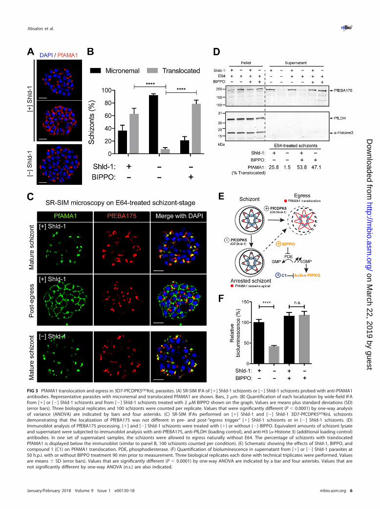
FIG 3 PfAMA1 translocation and egress in 3D7-PfCDPK5DDKnL parasites. (A) SR-SIM IFA of [�] Shld-1 schizonts or [�] Shld-1 schizonts probed with anti-PfAMA1antibodies. Representative parasites with micronemal and translocated PfAMA1 are shown. Bars, 2 �m. (B) Quantification of each localization by wide-field IFAfrom [�] or [�] Shld-1 schizonts and from [�] Shld-1 schizonts treated with 2 �M BIPPO shown on the graph. Values are means plus standard deviations (SD)(error bars). Three biological replicates and 100 schizonts were counted per replicate. Values that were significantly different (P � 0.0001) by one-way analysisof variance (ANOVA) are indicated by bars and four asterisks. (C) SR-SIM IFAs performed on [�] Shld-1 and [�] Shld-1 3D7-PfCDPK5DDKnL schizontsdemonstrating that the localization of PfEBA175 was not different in pre- and post-“egress trigger” [�] Shld-1 schizonts or in [�] Shld-1 schizonts. (D)Immunoblot analysis of PfEBA175 processing. [�] and [�] Shld-1 schizonts were treated with (�) or without (�) BIPPO. Equivalent amounts of schizont lysateand supernatant were subjected to immunoblot analysis with anti-PfEBA175, anti-PfLDH (loading control), and anti-H3 (�-Histone 3) (additional loading control)antibodies. In one set of supernatant samples, the schizonts were allowed to egress naturally without E64. The percentage of schizonts with translocatedPfAMA1 is displayed below the immunoblot (similar to panel B, 100 schizonts counted per condition). (E) Schematic showing the effects of Shld-1, BIPPO, andcompound 1 (C1) on PfAMA1 translocation. PDE, phosphodiesterase. (F) Quantification of bioluminescence in supernatant from [�] or [�] Shld-1 parasites at50 h.p.i. with or without BIPPO treatment 90 min prior to measurement. Three biological replicates each done with technical triplicates were performed. Valuesare means � SD (error bars). Values that are significantly different (P � 0.0001) by one-way ANOVA are indicated by a bar and four asterisks. Values that arenot significantly different by one-way ANOVA (n.s.) are also indicated.
Absalon et al. ®
January/February 2018 Volume 9 Issue 1 e00130-18 mbio.asm.org 6
on March 22, 2019 by guest
http://mbio.asm
.org/D
ownloaded from

Shld-1 schizonts), 37% � 8% had micronemal PfAMA1, and 63% � 8% showed plasmamembrane localization. In sharp contrast, 92% � 2% of the schizonts grown in theabsence of Shld-1 ([�] Shld-1 schizonts) had micronemal PfAMA1, and only 8% � 2%demonstrated plasma membrane localization (Fig. 3B). It is important to note that the3D7-PfCDPK5DDKnL parasites allow knockdown but do not completely knock outPfCDPK5. Thus, we find rare [�] Shld-1 schizonts with plasma membrane-localizedPfAMA1. These [�] Shld-1 schizonts with translocated PfAMA1 may be from residuallow levels of PfCDPK5 or from a less utilized PfCDPK5-independent pathway. Nonethe-less, these results show a bona fide defect in microneme release in PfCDPK5-deficientparasites.
To simultaneously evaluate the status of the PfAMA1- and PfEBA175-containingsubsets of micronemes, we costained [�] and [�] Shld-1 schizonts for the two markers(Fig. 3C). In both [�] and [�] Shld-1 schizonts, when PfAMA1 was apical, we found thatPfEBA175 was also apical. Interestingly, in the “post-egress trigger” [�] Shld-1 schizonts,we found that while PfAMA1 had been translocated, on the merozoite surface,PfEBA175 apparently remained apical. When released, PfEBA175-containing mi-cronemes expose the ectodomain of PfEBA175 onto the surface of the apical end of themerozoite. The exposed PfEBA175 is processed by the rhomboid-like protease PfROM4on the surface of the merozoite, shedding the ectodomain into the culture supernatant(48, 49). Shedding of processed forms of invasion ligands is detected in the supernatantfrom E64-treated schizonts (8). Therefore, to directly evaluate whether PfEBA175-containing micronemes had been released, we measured the discharge of the pro-cessed protein into the supernatant. With E64-treated schizonts, we found that pro-cessed PfEBA175 was readily detected in the supernatant of [�] Shld-1 schizonts butwas largely absent from the supernatant of [�] Shld-1 schizonts (Fig. 3D). Thus,although the sensitivity of superresolution microscopy was insufficient to detect dis-charge of PfEBA175-containing micronemes, their discharge was detected by measure-ment of the release of the processed form of PfEBA175. Together, these resultsdemonstrate that the signaling pathways controlling the release of PfAMA1-containingand PfEBA175-containing micronemes are regulated by PfCDPK5.
Pharmacologically enhanced PKG activity allows for microneme discharge inPfCDPK5-deficient schizonts. PKG signaling is known to be essential for both exon-eme and microneme release (8). PKG enzymatic activity is amenable to small-moleculeinhibition and activation in live parasites (schematic in Fig. 3E). The kinase itself isdirectly and reversibly blocked by treatment with 4-[2-(4-fluorophenyl)-5-(1-methylpiperidine-4-yl)-1H-pyrrol-3-yl]pyridine (compound 1 [C1]) (50). In contrast, PKGactivity is enhanced by increasing cyclic GMP levels with the specific phosphodiesteraseinhibitor 5-benzyl-3-isopropyl-1H-pyrazolo[4,3-d]pyrimidin-7(6H)-one (BIPPO) (51). Weexamined whether BIPPO-enhanced PKG activity could overcome the PfCDPK5-mediated defect in PfAMA1 translocation. Following treatment with 2 �M BIPPO,PfAMA1 translocation was observed in 78% � 6% of [�] Shld-1 schizonts (Fig. 3B).Similar to our findings with PfAMA1, processed PfEBA175 was robustly detected in thesupernatants of both [�] and [�] Shld-1 schizonts following BIPPO treatment (Fig. 3D).
Utilizing our bioluminescent transgenic reporter parasites (Fig. 1B), we found that[�] Shld-1 parasites, even with a �90% block as schizonts, still release some KnL intothe supernatant. We note that the amount of bioluminescence in the supernatant of[�] Shld-1 parasites was not further decreased when these parasites were treated witha mixture of protease inhibitors (including serine, cysteine, and metalloprotease inhib-itors) at 44 h.p.i. (Fig. S3A). Thus, it is likely that the PfCDPK5-deficient arrested schizontshave partial permeabilization of their surrounding PVM and RBC plasma membranethat is not dependent on ongoing protease activity. This finding is consistent withprevious reports that the PVM and RBC plasma membrane of segmented schizonts arepartially permeable to small proteins even in the presence of E64 (8, 28). Nonetheless,the significant difference in the quantity of bioluminescence in the supernatant be-tween [�] and [�] Shld-1 schizonts allowed a quantitative measure of membranepermeability as a surrogate for relative egress efficiency. To test whether BIPPO
PfCDPK5 Is Essential for Microneme Release ®
January/February 2018 Volume 9 Issue 1 e00130-18 mbio.asm.org 7
on March 22, 2019 by guest
http://mbio.asm
.org/D
ownloaded from

activation of PKG induced PfCDPK5-deficient parasites to egress, we measured thebioluminescence in the supernatant of treated parasites. As expected, the amount ofbioluminescent reporter released into the supernatant from [�] and [�] Shld-1 para-sites was equivalent following BIPPO treatment (Fig. 3F). Pretreatment of [�] Shld-1parasites with C1 prior to BIPPO prevented this release (Fig. S3A). For additionalconfirmation of this result, we treated PfCDPK5-deficient parasites with zaprinast, astructurally distinct phosphodiesterase inhibitor that increases cGMP levels and acti-vates PfPKG, and obtained similar release of bioluminescent activity into the superna-tant (Fig. S3B). This result demonstrates that enhanced PKG activity induces micronemesecretion and secondarily mediates full permeabilization of the PVM and RBC plasmamembrane.
PKG activity has been linked to downstream calcium release in both Plasmodiumspecies and T. gondii (52–54). It is formally possible that PKG-induced increases incytoplasmic calcium may be sufficient to activate a residual amount of PfCDPK5 in [�]Shld-1 parasites. To test whether the BIPPO-induced microneme discharge in PfCDPK5-deficient parasites was entirely dependent on calcium release, we evaluated PfAMA1localization (Fig. 4A) and release of the KnL reporter in the supernatant (Fig. 4B) in three
FIG 4 Calcium and cGMP signaling pathways in 3D7-PfCDPK5DDKnL parasites. (A) PfAMA1 translocation was visualized by wide-field IFA from [�] Shld-1 or [�]Shld-1 parasites treated with 1 �M A23187 and/or 2 �M BIPPO as indicated. Three biological replicates were performed, and 100 schizonts counted perreplicate. Values are means plus SD (error bars). The percentage translocated from the small-molecule-treated [�] Shld1 samples was compared to the valuefor the untreated [�] Shld1 condition by one-way ANOVA. Values that were significantly different (P � 0.001) by one-way ANOVA are indicated by an asterisk.Values that were not significantly different (#) by one-way ANOVA are also indicated. (B) Bioluminescence activity released in culture supernatants from thesame conditions as in panel A (three biological replicates each done with technical triplicates; mean � SD). Each of the [�] Shld1 with small-moleculetreatments was compared to the untreated [�] Shld1 condition by one-way ANOVA (*, P � 0.001; #, not significantly different). (C) 44 h.p.i. schizonts from [�]or [�] Shld-1 parasites were isolated by magnetic purification and lysed immediately or incubated for an 6 additional hours with the indicated compounds priorto lysis. Protein lysates were subjected to immunoblot analysis with anti-PfMSP142, anti-PfLDH (loading control), and anti-H3 (additional loading control)antibodies. Full-length, partially processed, and fully processed PfMSP142 are labeled. The quantitative ratio of fully processed PfMSP142 relative to theunprocessed form was calculated by volumetric measurement of fluorescence intensity with the Li-Cor Odyssey CLx system.
Absalon et al. ®
January/February 2018 Volume 9 Issue 1 e00130-18 mbio.asm.org 8
on March 22, 2019 by guest
http://mbio.asm
.org/D
ownloaded from

biological replicates of [�] and [�] Shld-1 3D7-PfCDPK5DDKnL schizonts treated witheither BIPPO, a calcium ionophore (A23187), or both. In PfCDPK5-deficient schizonts,A23187 treatment did not induce PfAMA1 translocation, suggesting that calciumrelease by itself was not sufficient for microneme discharge. In [�] Shld-1 controlparasites, A23187 treatment did not inhibit PfAMA1 translocation, showing that theelevated level of calcium was not toxic or inhibitory for microneme discharge. Thisconclusion is further supported by the observation that the combination of A23187 andBIPPO in the parasites had the same result as that for BIPPO alone in both [�] and [�]Shld-1 schizonts. Therefore, the BIPPO-induced microneme release and resulting egressin PfCDPK5-deficient schizonts are not solely due to a secondary PfPKG-inducedcalcium release.
Egress protease cascade is slowed in PfCDPK5-deficient parasites. Previously,we observed that the egress protease cascade was similarly initiated in both [�] and[�] Shld-1 schizonts (15). However, that observation was done using a single timepoint. Thus, we evaluated the extent to which the protease cascade in PfCDPK5-deficient parasites progressed with time—as has been demonstrated for wild-typeparasites (8). For this set of experiments, we chose to use the previously published3D7-PfCDPK5-DD parasites without an epitope tag (15). This strain was selected,because it has the strongest replication block of our existing strains. To evaluate theprotease cascade, we focused on PfSUB1 processing of the P. falciparum merozoitesurface protein-1 (PfMSP1), as this protein is known to be important for parasite egress(20). Following exoneme release, PfSUB1 proteolytically processes the ~200-kDaPfMSP1 precursor and releases a 42-kDa product, PfMSP142. We isolated 44 h.p.i.schizonts from parallel cultures that had been maintained with and without Shld-1.These isolated schizonts were sampled immediately and then allowed to incubate foran additional 6 h in the presence of E64. As seen previously, PfMSP142 was alreadydetected at the starting time point in both [�] and [�] Shld-1 samples (Fig. 4C). In the[�] Shld-1 schizonts, the amount of fully processed PfMSP142 relative to the unpro-cessed precursor increased from �20% at 44 h.p.i. to �80% 6 h later. In contrast, in the[�] Shld-1 parasites, the relative amount of fully processed PfMSP142 remained �20%at the later time point.
To evaluate whether exoneme release was completed at the initial time point, [�]Shld-1 parasites were treated with C1 to prevent any further PKG activity, and sampleswere collected at the later time point. Compared to the untreated [�] Shld-1 schizontsand the starting samples, the relative amount of fully processed PfMSP142 was lower,indicating that protease processing was incomplete at the starting time point and thatprocessing required PfPKG activity. As expected, treatment of [�] Shld-1 schizonts withC1 did not change the relative amount of the fully processed PfMSP142. However, whenthe [�] Shld-1 schizonts were treated with BIPPO, the relative amount of fully processedPfMSP142 increased to �40% (Fig. 4C). These results demonstrate a previously unrec-ognized finding—that protease processing is incomplete in [�] Shld-1 schizonts andthat full processing is increased when PfPKG activity is chemically enhanced by BIPPO.We evaluated PfSERA5, another substrate of PfSUB1 processing (18), in parallel(Fig. S3C). While the processing differences are smaller, we observed an increase in therelative amount of the mature (p50/p56) forms of PfSERA5 in the [�] Shld-1 schizontsafter 6 h and did not observe the same increase in the [�] Shld-1 schizonts (Fig. S3D).As was seen with PfMSP1, the amount of mature PfSERA5 increased in both [�] and [�]Shld-1 schizonts following BIPPO treatment.
Physical release of merozoites overcomes microneme release defect. A recentlydeveloped procedure to obtain significant quantities of viable merozoites has allowedfor direct studies of mechanically released parasites (27, 55, 56). We have previouslyshown that PfCDPK5-deficient merozoites were capable of reinvasion following phys-ical disruption of the blocked schizonts (15). However, the invasion obtained by thismethod was inefficient compared to [�] Shld-1 schizonts. We therefore modified thepublished “viable merozoite isolation” (27) procedure to directly evaluate the status of
PfCDPK5 Is Essential for Microneme Release ®
January/February 2018 Volume 9 Issue 1 e00130-18 mbio.asm.org 9
on March 22, 2019 by guest
http://mbio.asm
.org/D
ownloaded from

PfCDPK5-deficient merozoites (Fig. 5A). Applying this technique, we found that invasionefficiency of physically released merozoites from [�] and [�] Shld-1 schizonts wassimilar and more robust than previously reported (Fig. 5B). We evaluated PfAMA1localization in the schizont prior to shearing and in the merozoites following physicalrelease. Once again, the [�] Shld-1 schizonts had populations of both micronemal andtranslocated PfAMA1, while [�] Shld-1 schizonts had almost exclusively micronemallocalization. Following shearing, translocated PfAMA1 was readily detected on physi-cally released merozoites from both [�] and [�] Shld-1 schizonts (Fig. 5C). We repeatedthe shearing in the presence of C1 to evaluate whether PfAMA1 translocation inducedby physical release required PfPKG activity. By IFA, PfAMA1 remained micronemal inreleased merozoites from C1-treated [�] or [�] Shld-1 schizonts (Fig. 5C; shown withnuclear counterstain in Fig. S4). Thus, the process of physical disruption and/or expo-sure of PfCDPK5-deficient merozoites to the extracellular milieu was sufficient tofacilitate microneme release, as measured by PfAMA1 translocation, and this releaserequired PfPKG activity. By preventing PfAMA1 translocation, inhibition of PfPKG pre-vented reinvasion and new ring formation. The mechanism responsible for the en-hanced PfPKG activity in the sheared merozoites remains unknown. However, thesefindings convincingly demonstrate that a primary role of PfCDPK5 is to trigger mi-croneme release, allowing parasite egress.
DISCUSSION
PfCDPK5 is essential for P. falciparum egress out of an infected RBC. It localizes to theapical ends of newly forming daughter merozoites, and in mature, segmented schi-zonts, PfCDPK5 colocalizes with PfAMA1; later, PfCDPK5 has a diffuse apical staining
FIG 5 Physically released merozoites are invasive. (A) Schematic of the method to generate viable free merozoitesefficiently from [�] Shld-1 and [�] Shld-1 schizonts. 28G, 28-gauge; HCT, hematocrit. (B) Ring parasitemia fromphysically released [�] or [�] Shld-1 schizonts. The ring parasitemia was not different between the [�] and [�]Shld-1 conditions (n � 3; mean � SD; no significant difference by Student’s t test). (C) Wide-field IFA of PfAMA1localization before and after shearing from [�] and [�] Shld-1 parasites. Treatment of schizonts with 2.5 �M C1prior to shearing prevents PfAMA1 translocation.
Absalon et al. ®
January/February 2018 Volume 9 Issue 1 e00130-18 mbio.asm.org 10
on March 22, 2019 by guest
http://mbio.asm
.org/D
ownloaded from

that overlaps with several apical markers. PfCDPK5 is present in the membrane-associated fraction following sodium carbonate extraction, and this localization may bedue to potential palmitoylation sites within the protein (15). However, the traffickingdeterminants for PfCDPK5 remain unknown. By superresolution microscopy, we dem-onstrate multiple nonoverlapping subsets of micronemes. This result has been previ-ously noted for PfSUB1 (the “exoneme”) and PfROM4 (the “mononeme”) (18, 35).However, we suggest that the current list of apical organelles should include additionalsubsets of egress-specific micronemes. We hypothesize that a subset of micronemes istriggered for release by cooperative activation of PfPKG by PfCDPK5. This defect is mostconvincingly demonstrated for the PfAMA1-containing micronemes. The completeidentities of which parasite proteins reside in these micronemes remain unknown butlikely include proteases, perforin-like proteins, and potentially other unknown proteins.We hypothesize that PfCDPK5 interacts with and likely phosphorylates proteins that areassociated with apical organelles to transmit a “release” signal.
We note that parasites physically released following PfPKG-blockade by compound1 (C1) do not translocate PfAMA1 to the merozoite surface (Fig. 5C; also see Fig. S4 inthe supplemental material) and are, therefore, not invasive. In contrast, PfCDPK5-deficient parasites can reinvade new RBCs following physical release. Thus, blockingparasites prior to exoneme release prevents both egress and invasion. Our currentfindings demonstrate an essential role of PfCDPK5 for a downstream release of egress-mediating micronemes, and we show that chemically enhanced PfPKG activation canovercome the egress block in PfCDPK5-deficient parasites.
We, therefore, propose a model of egress wherein PfCDPK5 cooperates with PfPKGactivation (Fig. 6). In this model, a basal level of PfPKG activation allows some releaseof exonemes and partial activation of the protease cascade. This initial step is inde-pendent of PfCDPK5. In a later step, PfCDPK5 cooperates with PfPKG to facilitatePfAMA1 translocation and discharge of micronemes required for egress. This secondstep, requiring PfPKG and PfCDPK5, induces full activation of the protease cascade andallows parasite egress. Finally, supraphysiological activation of PfPKG by BIPPO treat-ment can bypass the requirement for PfCDPK5. One alternative to the model is that
FIG 6 Model for cooperativity between PfCDPK5 and PfPKG. The progress along the parasite egresspathway is promoted by activation of PfPKG. At low levels of PfPKG activation, the protease cascade andcalcium signaling pathway are initiated. PfCDPK5 activation, together with PfPKG, leads to furtherprogression of the protease cascade and triggered release of micronemes required for parasite egress(detected by PfAMA1 translocation, shown in red). Under physiological conditions, this step requires bothPfCDPK5 and PfPKG. The discharge of micronemes completes the egress process with the release ofinvasive merozoites. The requirement for PfCDPK5 can be bypassed by supraphysiological activation ofPfPKG by either BIPPO or physical disruption. PVM, parasitophorous vacuolar membrane.
PfCDPK5 Is Essential for Microneme Release ®
January/February 2018 Volume 9 Issue 1 e00130-18 mbio.asm.org 11
on March 22, 2019 by guest
http://mbio.asm
.org/D
ownloaded from

PfCDPK5 activity does not affect exoneme secretion directly but rather functions toincrease the efficiency and/or completeness of protease processing after exonemerelease.
Our methods to obtain free PfCDPK5-deficient merozoites require either BIPPO (orzaprinast) treatment or physical disruption. These methods facilitate PfAMA1 translo-cation and likely the release of other egress-specific factors. We cannot formally rule outa role for PfCDPK5 in the invasion process that was not also “overcome” by thesetreatments. Nonetheless, PfCDPK5 is the only calcium-dependent protein kinase inPlasmodium spp. that has a direct role in the release of micronemes required forparasite egress.
Synergy between calcium-dependent kinases and PKG has been suggested byprevious studies with both T. gondii and Plasmodium parasites (57–59). Our currentresults provide strong genetic evidence for this hypothesis. Brochet and colleaguesevaluated the relationship between P. berghei PKG (PbPKG) and calcium signaling,providing a molecular pathway for connection of the two pathways (52). Alam andcolleagues demonstrated that PfCDPK1 was a likely substrate of PfPKG and thatphosphorylated PfCDPK1 localized to the apical area of daughter merozoites (60).Bansal and colleagues demonstrated that increased PfPKG activity was able to com-pensate for a less active PfCDPK1 (57). Similarly, Moon and colleagues demonstratedthat increased PbPKG signaling was able to overcome a defect in gliding motilityobserved in PbCDPK3-deficient ookinetes (59). Our current study adds to the complex-ity of the egress signaling pathway by demonstrating a cooperative relationshipbetween PfCDPK5 and PfPKG activity.
In summary, we have demonstrated a functional explanation for the egress blockobserved in PfCDPK5-deficient parasites. In the absence of normal PfCDPK5 activity,physiologic activation of the PfPKG pathway is insufficient to release a subset ofmicronemes that are essential for parasite egress. Thus, the molecular mechanism forthe egress block observed in PfCDPK5-deficient schizonts is secondary to the failure todischarge micronemes that are required for egress.
MATERIALS AND METHODSSmall molecules and antibodies. Synthesis of 5-benzyl-3-isopropyl-1H-pyrazolo[4,3-d]pyrimidin-
7(6H)-one (BIPPO) was adapted from published methods (51, 61) and dissolved at 10 mM in dimethylsulfoxide (DMSO). See Text S1 in the supplemental material for details. Compound 1 (C1) {4-[2-(4-fluorophenyl)-5-(1-methylpiperidine-4-yl)-1H-pyrrol-3-yl]pyridine} (50 mM), E64 (10 mM), A23187(50 mM), and zaprinast (10 mg/ml) were dissolved in DMSO. Commercially available antibodies wereobtained from Roche Applied Science (rat antihemagglutinin [anti-HA] [3F10]), Life Technologies (mouseanti-HA [clone 2-2.2.14]), and Abcam (rabbit anti-H3). Other antibodies were generously provided byMichael Makler at Flow Inc. (mouse anti-P. falciparum lactate dehydrogenase [anti-PfLDH]), Julian Raynerat the Wellcome Trust Sanger Institute (rabbit anti-PfGAP45), Robin Anders at The Walter & Eliza HallInstitute of Medical Research (mouse anti-PfAMA1 [clone 1FG]), Jean-Francois Dubremetz at UniversitèMontpellier (mouse anti-PfSERA5 [clone 24C6.1F1]), Alan Cowman, Jenny Thompson, and Kaye Wycher-ley at The Walter & Eliza Hall Institute of Medical Research (rabbit anti-PfEBA175 and mouse anti-PfRON4),Michael Blackman at the London School of Hygiene and Tropical Medicine and the Francis Crick Institute(rabbit anti-PfSUB1), Carole Long at NIAID, NIH (rabbit anti-PfMSP142), and Kim Lee Sim via BEI Resources,NIAID, NIH (mouse anti-PfEBA175 [clone R218]).
Parasite culture and transfection. The 3D7 strain (Walter & Eliza Hall Institute) and transgenicderivatives were cultured in human red blood cells (RBCs) in RPMI 1640 supplemented with 0.5%Albumax II, 50 mg/liter hypoxanthine, 0.21% sodium bicarbonate, and 25 mM HEPES as previouslydescribed (62). To generate the 3D7-PfCDPK5DD strain, sorbitol-synchronized ring-stage parasites wereelectroporated with 100 �g of plasmid DNA of the single-crossover plasmid (see Text S1 for details).Following transfection, parasites were maintained with 250 nM Shield-1 (Shld-1), and stable single-crossover parasites were selected by cycling on and off WR99210 (Jacobus Pharmaceutical Company).Individual transgenic clones were obtained by limiting dilutions. To generate the 3D7-PfCDPK5DDKnLparasites with the KnL reporter, a clone was transfected with pJDD250 that expresses the Bxb1 integrase(23) and the KnL reporter and selected on blasticidin (while maintaining WR99210 and Shld-1).
Parasite phenotypic assays. For replication curves, ring-stage parasites were washed to removeShld-1, replated in the presence or absence of 0.25 mM Shld-1 at 0.4% parasitemia and 1% hematocrit,and parasitemia was monitored by flow cytometry using SYBR green I staining. For measurements ofbioluminescence throughout the asexual development cycle, 100 �l of resuspended culture wasremoved from ring, trophozoite, schizont, and reinvaded rings and centrifuged at 14,000 rpm in amicrocentrifuge. The supernatant was transferred to a fresh tube, and 20 �l was mixed 1:1 with nano-Globuffer/substrate (Promega). The pelleted cells were resuspended in 100 �l of phosphate-buffered saline
Absalon et al. ®
January/February 2018 Volume 9 Issue 1 e00130-18 mbio.asm.org 12
on March 22, 2019 by guest
http://mbio.asm
.org/D
ownloaded from

(PBS), and then 20 �l of the PBS solution containing cells was mixed 1:1 with nano-Glo buffer/substrate.Light output was measured on a SpectraMax L instrument.
For all indirect immunofluorescence assays (IFAs), parasites were synchronized by Percoll purificationof late-stage schizonts, followed by sorbitol synchronization of newly invaded rings 2 h later. In thefollowing cycle, sorbitol synchronization was repeated, and the cultures were washed and replated withor without Shld-1. IFAs were performed as previously described (47) with minor modifications. Thinsmears were made on glass slides, air dried, and fixed with 1% paraformaldehyde, permeabilized with0.1% Triton 100 for 10 min, and blocked with 3% bovine serum albumin (BSA) overnight at 4°C. Primaryantibodies were incubated overnight in a cold room at the dilutions indicated: anti-PfAMA1 (1:200),anti-PfEBA175 (1:500), anti-PfGAP45 (1:5000), anti-HA (1:50), anti-PfSUB1 (1:500), and anti-PfRON4 (1:200).Subsequently, cells were washed three times with PBS and incubated for 45 min with the Alexa Fluor 488or 555 secondary antibodies (1:1,000) (Molecular Probes). After removal of unbound antibodies withthree PBS washes, slides were mounted with Vectashield containing 4=,6=-diamidino-2-phenylindole(DAPI) (Vector Laboratories Inc.) with coverslips and kept at 4°C until evaluation. Wide-field images wereobtained with a Nikon E800 epifluorescence microscope using a 100� (oil) objective, and images werecaptured using SPOT Imaging software and then processed using Adobe Photoshop. Superresolutionstructured illumination microscopy (SR-SIM) Z-stacks were captured using an ELYRA PS.1 microscope(Carl Zeiss Microscopy). The ELYRA microscope was used with a 100� (oil) objective and excitationwavelengths of 405, 488, and 561 nm. SIM images were collected at 100- to 200-nm z-axis steps, with fiverotations of the structured illumination grid per channel. The resulting stacks were processed usingdefault reconstruction parameters in ZEN 2012 Black software.
For the P. falciparum AMA1 (PfAMA1) translocation IFAs shown in Fig. 3, 10 �M E64 was added to theschizonts 46 h postinvasion (h.p.i.) and incubated for 4 h, then 2 �M BIPPO was added for an additional2 h, and schizonts were harvested for analysis. For the bioluminescence and PfAMA1 translocation IFAsshown in Fig. 4, 10 �M E64 was added to 46 h.p.i. schizonts and incubated for 150 min, 1 �M A23187(or nothing) was added for 90 min, then 2 �M BIPPO was added for 2 h, and schizonts/supernatants wereharvested for analysis. A total of 100 schizonts were scored for micronemal or plasma membranetranslocation, and bioluminescence was measured as described above. For PfEBA175 processing exper-iments, synchronized ring-stage parasites were washed three times to remove Shld-1, and grown at 4%hematocrit in the presence and absence of 250 nM Shld-1. At 44 h.p.i., schizonts were purified by passagethrough a magnetically activated cell sorting (MACS) column, spun down to a pellet, and resuspendedin 100 �l of medium containing 10 �M E64 in the presence or absence of Shld-1, and plated in a 96-wellplate. Heparin (50 �g/ml) was added to all wells to prevent reinvasion. BIPPO (2 �M) was added 4 h afterplating, and parasites were collected 2 h after the addition of BIPPO. Samples were spun down toseparate supernatant and pellet. Parasite pellets were washed once with RMPI 1640, and supernatantswere spun again to clear any remaining parasites. Washed pellets and cleared supernatants were boiledin sample buffer, analyzed by immunoblotting with anti-PfEBA175 antibodies or anti-PfLDH and anti-H3antibodies to control for loading, probed with Li-Cor secondary antibodies, and visualized on a Li-CorOdyssey CLx system. Concurrently, samples of parasites were prepared for immunofluorescence assaysby preserving 300 �l each of Shld-1 cultures grown with Shld-1 ([�] Shld-1 cultures) and Shld-1 culturesgrown without Shld-1 ([�] Shld-1 cultures) prior to MACS purification. Cultures were plated along withMACS-purified schizonts, and samples were treated as described above. At the time of sample collection,cultures were spun down, and smears were made from the RBC pellet. IFA preparation was performedas described above, and PfAMA1 translocation was determined.
For PfMSP1 and PfSERA5 processing assays, 44 h.p.i. schizonts from [�] and [�] Shld-1 cultures werepurified by magnetic separation, placed in medium without Albumax II with or without Shld-1. A sampleof the purified schizonts were lysed immediately in sample buffer, and the remaining schizonts wereincubated for an additional 6 h before harvesting. E64 (10 �M) was added to all samples at 44 h.p.i. (forC1-treated samples, 2.5 �M C1 was also added). At 48 h.p.i., 2 �M BIPPO was added to indicated samples.All treated samples were lysed at 50 h.p.i. Lysates were separated on a TGX 4 to 20% gradient cell(Bio-Rad), transferred to polyvinylidene difluoride (PVDF) by wet transfer, immunoblotted with rabbitanti-PfMSP142 or mouse anti-PfSERA5 (and anti-PfLDH and anti-H3 as loading controls), probed withLi-Cor secondary antibodies, and visualized on a Li-Cor Odyssey CLx system.
For physical disruption assays, viable merozoites were performed as previously described (27, 55)with minor modifications. Late-stage schizonts were magnet purified, incubated in medium with 10 �ME64 and without Albumax II for 6 h, and sheared with 20 strokes through a 28-gauge needle. The shearedmerozoites were incubated with or without fresh RBCs at 37C for 15 min. Reinvasion was determined byflow cytometry, and PfAMA1 translocation was determined by IFA.
SUPPLEMENTAL MATERIALSupplemental material for this article may be found at https://doi.org/10.1128/mBio
.00130-18.TEXT S1, DOCX file, 1.5 MB.FIG S1, TIF file, 22 MB.FIG S2, TIF file, 14.8 MB.FIG S3, TIF file, 23 MB.FIG S4, TIF file, 17.7 MB.MOVIE S1, MOV file, 0.6 MB.
PfCDPK5 Is Essential for Microneme Release ®
January/February 2018 Volume 9 Issue 1 e00130-18 mbio.asm.org 13
on March 22, 2019 by guest
http://mbio.asm
.org/D
ownloaded from
MOVIE S2, MOV file, 1 MB.MOVIE S3, MOV file, 0.8 MB.MOVIE S4, MOV file, 0.8 MB.MOVIE S5, MOV file, 0.2 MB.
ACKNOWLEDGMENTSThis work was supported by funds from NIH grants R01 AI 102907 and DP2 AI
112219 (to J.D.D.) and R01 AI 114685 (M.P.P.) and the Swedish Research CouncilDNR2013-367 (to K.B.).
We thank Anthony Hill at the Boston Children’s Hospital Cellular Imaging Core andDouglas Richardson at the Harvard Center for Biological Imaging for core facilitymanagement and technical advice. We thank the providers of antibodies detailed inMaterials and Methods. We thank James McGee and Tanya Labunska for technicalassistance and Jonathan Robbins for helpful discussions.
REFERENCES1. World Health Organization. 2016. World malaria report 2015. World
Health Organization, Geneva, Switzerland.2. Ashley EA, Dhorda M, Fairhurst RM, Amaratunga C, Lim P, Suon S, Sreng
S, Anderson JM, Mao S, Sam B, Sopha C, Chuor CM, Nguon C, Sovann-aroth S, Pukrittayakamee S, Jittamala P, Chotivanich K, Chutasmit K,Suchatsoonthorn C, Runcharoen R, Hien TT, Thuy-Nhien NT, Thanh NV,Phu NH, Htut Y, Han K-T, Aye KH, Mokuolu OA, Olaosebikan RR, Folar-anmi OO, Mayxay M, Khanthavong M, Hongvanthong B, Newton PN,Onyamboko MA, Fanello CI, Tshefu AK, Mishra N, Valecha N, Phyo AP,Nosten F, Yi P, Tripura R, Borrmann S, Bashraheil M, Peshu J, Faiz MA,Ghose A, Hossain MA, Samad R, Rahman MR, et al. 2014. Spread ofartemisinin resistance in Plasmodium falciparum malaria. N Engl J Med371:411– 423. https://doi.org/10.1056/NEJMoa1314981.
3. White NJ, Pukrittayakamee S, Hien TT, Faiz MA, Mokuolu OA, DondorpAM. 2014. Malaria. Lancet 383:723–735. https://doi.org/10.1016/S0140-6736(13)60024-0.
4. Miller LH, Ackerman HC, Su X-Z, Wellems TE. 2013. Malaria biology anddisease pathogenesis: insights for new treatments. Nat Med 19:156 –167.https://doi.org/10.1038/nm.3073.
5. Cowman AF, Healer J, Marapana D, Marsh K. 2016. Malaria: biology anddisease. Cell 167:610 – 624. https://doi.org/10.1016/j.cell.2016.07.055.
6. Francia ME, Striepen B. 2014. Cell division in apicomplexan parasites. NatRev Microbiol 12:125–136. https://doi.org/10.1038/nrmicro3184.
7. Bansal A, Singh S, More KR, Hans D, Nangalia K, Yogavel M, Sharma A,Chitnis CE. 2013. Characterization of Plasmodium falciparum calcium-dependent protein kinase 1 (PfCDPK1) and its role in microneme secre-tion during erythrocyte invasion. J Biol Chem 288:1590 –1602. https://doi.org/10.1074/jbc.M112.411934.
8. Collins CR, Hackett F, Strath M, Penzo M, Withers-Martinez C, Baker DA,Blackman MJ. 2013. Malaria parasite cGMP-dependent protein kinaseregulates blood stage merozoite secretory organelle discharge andegress. PLoS Pathog 9:e1003344. https://doi.org/10.1371/journal.ppat.1003344.
9. Carruthers VB, Sibley LD. 1999. Mobilization of intracellular calciumstimulates microneme discharge in Toxoplasma gondii. Mol Microbiol31:421– 428. https://doi.org/10.1046/j.1365-2958.1999.01174.x.
10. Bullen HE, Jia Y, Yamaryo-Botté Y, Bisio H, Zhang O, Jemelin NK, Marq JB,Carruthers V, Botté CY, Soldati-Favre D. 2016. Phosphatidic acid-mediated signaling regulates microneme secretion in Toxoplasma. CellHost Microbe 19:349 –360. https://doi.org/10.1016/j.chom.2016.02.006.
11. Lourido S, Shuman J, Zhang C, Shokat KM, Hui R, Sibley LD. 2010.Calcium-dependent protein kinase 1 is an essential regulator of exo-cytosis in Toxoplasma. Nature 465:359 –362. https://doi.org/10.1038/nature09022.
12. Farrell A, Thirugnanam S, Lorestani A, Dvorin JD, Eidell KP, Ferguson DJP,Anderson-White BR, Duraisingh MT, Marth GT, Gubbels M-J. 2012. ADOC2 protein identified by mutational profiling is essential for apicom-plexan parasite exocytosis. Science 335:218 –221. https://doi.org/10.1126/science.1210829.
13. Azevedo MF, Sanders PR, Krejany E, Nie CQ, Fu P, Bach LA, WunderlichG, Crabb BS, Gilson PR. 2013. Inhibition of Plasmodium falciparumCDPK1 by conditional expression of its J-domain demonstrates a key
role in schizont development. Biochem J 452:433– 441. https://doi.org/10.1042/BJ20130124.
14. Kato N, Sakata T, Breton G, Le Roch KG, Nagle A, Andersen C, BursulayaB, Henson K, Johnson J, Kumar KA, Marr F, Mason D, McNamara C,Plouffe D, Ramachandran V, Spooner M, Tuntland T, Zhou Y, Peters EC,Chatterjee A, Schultz PG, Ward GE, Gray N, Harper J, Winzeler EA. 2008.Gene expression signatures and small-molecule compounds link a pro-tein kinase to Plasmodium falciparum motility. Nat Chem Biol 4:347–356.https://doi.org/10.1038/nchembio.87.
15. Dvorin JD, Martyn DC, Patel SD, Grimley JS, Collins CR, Hopp CS, BrightAT, Westenberger S, Winzeler E, Blackman MJ, Baker DA, Wandless TJ,Duraisingh MT. 2010. A plant-like kinase in Plasmodium falciparumregulates parasite egress from erythrocytes. Science 328:910 –912.https://doi.org/10.1126/science.1188191.
16. Billker O, Lourido S, Sibley LD. 2009. Calcium-dependent signaling andkinases in apicomplexan parasites. Cell Host Microbe 5:612– 622. https://doi.org/10.1016/j.chom.2009.05.017.
17. Taylor HM, McRobert L, Grainger M, Sicard A, Dluzewski AR, Hopp CS,Holder AA, Baker DA. 2010. The malaria parasite cyclic GMP-dependentprotein kinase plays a central role in blood-stage schizogony. EukaryotCell 9:37– 45. https://doi.org/10.1128/EC.00186-09.
18. Yeoh S, O’Donnell RA, Koussis K, Dluzewski AR, Ansell KH, Osborne SA,Hackett F, Withers-Martinez C, Mitchell GH, Bannister LH, Bryans JS,Kettleborough CA, Blackman MJ. 2007. Subcellular discharge of a serineprotease mediates release of invasive malaria parasites from host eryth-rocytes. Cell 131:1072–1083. https://doi.org/10.1016/j.cell.2007.10.049.
19. Arastu-Kapur S, Ponder EL, Fonovic UP, Yeoh S, Yuan F, Fonovic M,Grainger M, Phillips CI, Powers JC, Bogyo M. 2008. Identification ofproteases that regulate erythrocyte rupture by the malaria parasitePlasmodium falciparum. Nat Chem Biol 4:203–213. https://doi.org/10.1038/nchembio.70.
20. Das S, Hertrich N, Perrin AJ, Withers-Martinez C, Collins CR, Jones ML,Watermeyer JM, Fobes ET, Martin SR, Saibil HR, Wright GJ, Treeck M, EppC, Blackman MJ. 2015. Processing of Plasmodium falciparum merozoitesurface protein MSP1 activates a spectrin-binding function enablingparasite egress from RBCs. Cell Host Microbe 18:433– 444. https://doi.org/10.1016/j.chom.2015.09.007.
21. Garg S, Agarwal S, Kumar S, Yazdani SS, Chitnis CE, Singh S. 2013.Calcium-dependent permeabilization of erythrocytes by a perforin-likeprotein during egress of malaria parasites. Nat Commun 4:1736. https://doi.org/10.1038/ncomms2725.
22. Yang ASP, O’Neill MT, Jennison C, Lopaticki S, Allison CC, Armistead JS,Erickson SM, Rogers KL, Ellisdon AM, Whisstock JC, Tweedell RE, Dingla-san RR, Douglas DN, Kneteman NM, Boddey JA. 2017. Cell traversalactivity is important for Plasmodium falciparum liver infection in hu-manized mice. Cell Rep 18:3105–3116. https://doi.org/10.1016/j.celrep.2017.03.017.
23. Nkrumah LJ, Muhle RA, Moura PA, Ghosh P, Hatfull GF, Jacobs WR,Fidock DA. 2006. Efficient site-specific integration in Plasmodium falci-parum chromosomes mediated by mycobacteriophage Bxb1 integrase.Nat Methods 3:615– 621. https://doi.org/10.1038/nmeth904.
24. Hall MP, Unch J, Binkowski BF, Valley MP, Butler BL, Wood MG, Otto P,
Absalon et al. ®
January/February 2018 Volume 9 Issue 1 e00130-18 mbio.asm.org 14
on March 22, 2019 by guest
http://mbio.asm
.org/D
ownloaded from
Zimmerman K, Vidugiris G, Machleidt T, Robers MB, Benink HA, EggersCT, Slater MR, Meisenheimer PL, Klaubert DH, Fan F, Encell LP, Wood KV.2012. Engineered luciferase reporter from a deep sea shrimp utilizing anovel imidazopyrazinone substrate. ACS Chem Biol 7:1848 –1857.https://doi.org/10.1021/cb3002478.
25. Azevedo MF, Nie CQ, Elsworth B, Charnaud SC, Sanders PR, Crabb BS,Gilson PR. 2014. Plasmodium falciparum transfected with ultra brightNanoLuc luciferase offers high sensitivity detection for the screening ofgrowth and cellular trafficking inhibitors. PLoS One 9:e112571. https://doi.org/10.1371/journal.pone.0112571.
26. Wickham ME, Culvenor JG, Cowman AF. 2003. Selective inhibition of atwo-step egress of malaria parasites from the host erythrocyte. J BiolChem 278:37658 –37663. https://doi.org/10.1074/jbc.M305252200.
27. Boyle MJ, Wilson DW, Richards JS, Riglar DT, Tetteh KKA, Conway DJ,Ralph SA, Baum J, Beeson JG. 2010. Isolation of viable Plasmodiumfalciparum merozoites to define erythrocyte invasion events and ad-vance vaccine and drug development. Proc Natl Acad Sci U S A 107:14378 –14383. https://doi.org/10.1073/pnas.1009198107.
28. Hale VL, Watermeyer JM, Hackett F, Vizcay-Barrena G, van Ooij C, ThomasJA, Spink MC, Harkiolaki M, Duke E, Fleck RA, Blackman MJ, Saibil HR.2017. Parasitophorous vacuole poration precedes its rupture and rapidhost erythrocyte cytoskeleton collapse in Plasmodium falciparum egress.Proc Natl Acad Sci U S A 114:3439 –3444. https://doi.org/10.1073/pnas.1619441114.
29. Le Roch KG, Zhou Y, Blair PL, Grainger M, Moch JK, Haynes JD, De LaVega P, Holder AA, Batalov S, Carucci DJ, Winzeler EA. 2003. Discovery ofgene function by expression profiling of the malaria parasite life cycle.Science 301:1503–1508. https://doi.org/10.1126/science.1087025.
30. Bozdech Z, Llinás M, Pulliam BL, Wong ED, Zhu J, DeRisi JL. 2003. Thetranscriptome of the intraerythrocytic developmental cycle of Plasmo-dium falciparum. PLoS Biol 1:E5. https://doi.org/10.1371/journal.pbio.0000005.
31. Aurrecoechea C, Brestelli J, Brunk BP, Dommer J, Fischer S, Gajria B, GaoX, Gingle A, Grant G, Harb OS, Heiges M, Innamorato F, Iodice J, KissingerJC, Kraemer E, Li W, Miller JA, Nayak V, Pennington C, Pinney DF, RoosDS, Ross C, Stoeckert CJ, Treatman C, Wang H. 2009. PlasmoDB: afunctional genomic database for malaria parasites. Nucleic Acids Res37:D539 –D543. https://doi.org/10.1093/nar/gkn814.
32. Narum DL, Thomas AW. 1994. Differential localization of full-length andprocessed forms of PF83/AMA-1 an apical membrane antigen of Plas-modium falciparum merozoites. Mol Biochem Parasitol 67:59 – 68.https://doi.org/10.1016/0166-6851(94)90096-5.
33. Triglia T, Healer J, Caruana SR, Hodder AN, Anders RF, Crabb BS, CowmanAF. 2000. Apical membrane antigen 1 plays a central role in erythrocyteinvasion by Plasmodium species. Mol Microbiol 38:706 –718. https://doi.org/10.1046/j.1365-2958.2000.02175.x.
34. Jones ML, Kitson EL, Rayner JC. 2006. Plasmodium falciparum erythro-cyte invasion: a conserved myosin associated complex. Mol BiochemParasitol 147:74 – 84. https://doi.org/10.1016/j.molbiopara.2006.01.009.
35. Singh S, Plassmeyer M, Gaur D, Miller LH. 2007. Mononeme: a newsecretory organelle in Plasmodium falciparum merozoites identified bylocalization of rhomboid-1 protease. Proc Natl Acad Sci U S A 104:20043–20048. https://doi.org/10.1073/pnas.0709999104.
36. Richard D, Macraild CA, Riglar DT, Chan J-A, Foley M, Baum J, Ralph SA,Norton RS, Cowman AF. 2010. Interaction between Plasmodium falci-parum apical membrane antigen 1 and the rhoptry neck protein com-plex defines a key step in the erythrocyte invasion process of malariaparasites. J Biol Chem 285:14815–14822. https://doi.org/10.1074/jbc.M109.080770.
37. Adams JH, Sim BK, Dolan SA, Fang X, Kaslow DC, Miller LH. 1992. A familyof erythrocyte binding proteins of malaria parasites. Proc Natl Acad SciU S A 89:7085–7089.
38. Sim BK, Chitnis CE, Wasniowska K, Hadley TJ, Miller LH. 1994. Receptor andligand domains for invasion of erythrocytes by Plasmodium falciparum.Science 264:1941–1944. https://doi.org/10.1126/science.8009226.
39. Thompson JK, Triglia T, Reed MB, Cowman AF. 2001. A novel ligand fromPlasmodium falciparum that binds to a sialic acid-containing receptor onthe surface of human erythrocytes. Mol Microbiol 41:47–58. https://doi.org/10.1046/j.1365-2958.2001.02484.x.
40. Healer J, Crawford S, Ralph S, McFadden G, Cowman AF. 2002. Indepen-dent translocation of two micronemal proteins in developing Plasmo-dium falciparum merozoites. Infect Immun 70:5751–5758. https://doi.org/10.1128/IAI.70.10.5751-5758.2002.
41. Kumar S, Kumar M, Ekka R, Dvorin JD, Paul AS, Madugundu AK, Gilberger
T, Gowda H, Duraisingh MT, Keshava Prasad TS, Sharma P. 2017. PfCDPK1mediated signaling in erythrocytic stages of Plasmodium falciparum. NatCommun 8:63. https://doi.org/10.1038/s41467-017-00053-1.
42. Jebiwott S, Govindaswamy K, Mbugua A, Bhanot P. 2013. Plasmodiumberghei calcium dependent protein kinase 1 is not required for hostcell invasion. PLoS One 8:e79171. https://doi.org/10.1371/journal.pone.0079171.
43. Bansal A, Molina-Cruz A, Brzostowski J, Liu P, Luo Y, Gunalan K, Li Y,Ribeiro JMC, Miller LH. 2018. PfCDPK1 is critical for malaria parasitegametogenesis and mosquito infection. Proc Natl Acad Sci U S A 115:774 –779. https://doi.org/10.1073/pnas.1715443115.
44. Garrison E, Treeck M, Ehret E, Butz H, Garbuz T, Oswald BP, Settles M,Boothroyd J, Arrizabalaga G. 2012. A forward genetic screen reveals thatcalcium-dependent protein kinase 3 regulates egress in Toxoplasma.PLoS Pathog 8:e1003049. https://doi.org/10.1371/journal.ppat.1003049.
45. McCoy JM, Whitehead L, van Dooren GG, Tonkin CJ. 2012. TgCDPK3regulates calcium-dependent egress of Toxoplasma gondii from hostcells. PLoS Pathog 8:e1003066. https://doi.org/10.1371/journal.ppat.1003066.
46. Lourido S, Tang K, Sibley LD. 2012. Distinct signalling pathways controlToxoplasma egress and host-cell invasion. EMBO J 31:4524 – 4534.https://doi.org/10.1038/emboj.2012.299.
47. Absalon S, Robbins JA, Dvorin JD. 2016. An essential malaria proteindefines the architecture of blood-stage and transmission-stage parasites.Nat Commun 7:11449. https://doi.org/10.1038/ncomms11449.
48. O’Donnell RA, Hackett F, Howell SA, Treeck M, Struck N, Krnajski Z,Withers-Martinez C, Gilberger TW, Blackman MJ. 2006. Intramembraneproteolysis mediates shedding of a key adhesin during erythrocyteinvasion by the malaria parasite. J Cell Biol 174:1023–1033. https://doi.org/10.1083/jcb.200604136.
49. Baker RP, Wijetilaka R, Urban S. 2006. Two Plasmodium rhomboid pro-teases preferentially cleave different adhesins implicated in all invasivestages of malaria. PLoS Pathog 2:e113. https://doi.org/10.1371/journal.ppat.0020113.
50. Gurnett AM, Liberator PA, Dulski PM, Salowe SP, Donald RGK, AndersonJW, Wiltsie J, Diaz CA, Harris G, Chang B, Darkin-Rattray SJ, Nare B,Crumley T, Blum PS, Misura AS, Tamas T, Sardana MK, Yuan J, Biftu T,Schmatz DM. 2002. Purification and molecular characterization of cGMP-dependent protein kinase from Apicomplexan parasites. A novel che-motherapeutic target. J Biol Chem 277:15913–15922. https://doi.org/10.1074/jbc.M108393200.
51. Howard BL, Harvey KL, Stewart RJ, Azevedo MF, Crabb BS, Jennings IG,Sanders PR, Manallack DT, Thompson PE, Tonkin CJ, Gilson PR. 2015.Identification of potent phosphodiesterase inhibitors that demonstratecyclic nucleotide-dependent functions in apicomplexan parasites. ACSChem Biol 10:1145–1154. https://doi.org/10.1021/cb501004q.
52. Brochet M, Collins MO, Smith TK, Thompson E, Sebastian S, Volkmann K,Schwach F, Chappell L, Gomes AR, Berriman M, Rayner JC, Baker DA,Choudhary J, Billker O. 2014. Phosphoinositide metabolism links cGMP-dependent protein kinase G to essential Ca2� signals at key decisionpoints in the life cycle of malaria parasites. PLoS Biol 12:e1001806.https://doi.org/10.1371/journal.pbio.1001806.
53. Sidik SM, Hortua Triana MA, Paul AS, El Bakkouri M, Hackett CG, Tran F,Westwood NJ, Hui R, Zuercher WJ, Duraisingh MT, Moreno SNJ, LouridoS. 2016. Using a genetically encoded sensor to identify inhibitors ofToxoplasma gondii Ca2� signaling. J Biol Chem 291:9566 –9580. https://doi.org/10.1074/jbc.M115.703546.
54. Stewart RJ, Whitehead L, Nijagal B, Sleebs BE, Lessene G, McConvilleMJ, Rogers KL, Tonkin CJ. 2017. Analysis of Ca2� mediated signalingregulating Toxoplasma infectivity reveals complex relationships be-tween key molecules. Cell Microbiol 19:e12685. https://doi.org/10.1111/cmi.12685.
55. Riglar DT, Richard D, Wilson DW, Boyle MJ, Dekiwadia C, Turnbull L,Angrisano F, Marapana DS, Rogers KL, Whitchurch CB, Beeson JG, Cow-man AF, Ralph SA, Baum J. 2011. Super-resolution dissection of coordi-nated events during malaria parasite invasion of the human erythrocyte.Cell Host Microbe 9:9 –20. https://doi.org/10.1016/j.chom.2010.12.003.
56. Weiss GE, Gilson PR, Taechalertpaisarn T, Tham W-H, de Jong NWM,Harvey KL, Fowkes FJI, Barlow PN, Rayner JC, Wright GJ, Cowman AF,Crabb BS. 2015. Revealing the sequence and resulting cellular morphol-ogy of receptor-ligand interactions during Plasmodium falciparum inva-sion of erythrocytes. PLoS Pathog 11:e1004670. https://doi.org/10.1371/journal.ppat.1004670.
57. Bansal A, Ojo KK, Mu J, Maly DJ, Van Voorhis WC, Miller LH. 2016.
PfCDPK5 Is Essential for Microneme Release ®
January/February 2018 Volume 9 Issue 1 e00130-18 mbio.asm.org 15
on March 22, 2019 by guest
http://mbio.asm
.org/D
ownloaded from

Reduced activity of mutant calcium-dependent protein kinase 1 is com-pensated in Plasmodium falciparum through the action of protein ki-nase G. mBio 7:e02011-16. https://doi.org/10.1128/mBio.02011-16.
58. McCoy JM, Stewart RJ, Uboldi AD, Li D, Schröder J, Scott NE, PapenfussAT, Lehane AM, Foster LJ, Tonkin CJ. 2017. A forward genetic screenidentifies a negative regulator of rapid Ca2�-dependent cell egress(MS1) in the intracellular parasite Toxoplasma gondii. J Biol Chem 292:7662–7674. https://doi.org/10.1074/jbc.M117.775114.
59. Moon RW, Taylor CJ, Bex C, Schepers R, Goulding D, Janse CJ, Waters AP,Baker DA, Billker O. 2009. A cyclic GMP signalling module that regulatesgliding motility in a malaria parasite. PLoS Pathog 5:e1000599. https://doi.org/10.1371/journal.ppat.1000599.
60. Alam MM, Solyakov L, Bottrill AR, Flueck C, Siddiqui FA, Singh S, Mistry S,Viskaduraki M, Lee K, Hopp CS, Chitnis CE, Doerig C, Moon RW, Green JL,Holder AA, Baker DA, Tobin AB. 2015. Phosphoproteomics reveals malariaparasite protein kinase G as a signalling hub regulating egress and invasion.Nat Commun 6:7285. https://doi.org/10.1038/ncomms8285.
61. Wang C, Ashton TD, Gustafson A, Bland ND, Ochiana SO, Campbell RK,Pollastri MP. 2012. Synthesis and evaluation of human phosphodies-terases (PDE) 5 inhibitor analogs as trypanosomal PDE inhibitors. Part 1.Sildenafil analogs. Bioorg Med Chem Lett 22:2579 –2581. https://doi.org/10.1016/j.bmcl.2012.01.119.
62. Trager W, Jensen JB. 1976. Human malaria parasites in continuousculture. Science 193:673– 675. https://doi.org/10.1126/science.781840.
Absalon et al. ®
January/February 2018 Volume 9 Issue 1 e00130-18 mbio.asm.org 16
on March 22, 2019 by guest
http://mbio.asm
.org/D
ownloaded from