calcaneal fracture management - orthoclips.com · calcaneus fracture extensile lateral approach...
TRANSCRIPT

Calcaneal FractureManagement
Extensile Lateral Approach Versus Small IncisionTechnique
Nathan J. Kiewiet, MDa,*, Bruce J. Sangeorzan, MDb
KEYWORDS
� Calcaneus fracture � Extensile lateral approach � Percutaneous fixation calcaneus
KEY POINTS
� Calcaneal fracture management continues to be an area of sustained interest.
� Extensile lateral approach to the calcaneus carries significant risks of wound complica-tions and infection.
� Small incision techniques may reduce risks and improve recovery.
� Less invasive techniques have been shown to reduce risk of wound-healingcomplications.
INTRODUCTION
Intra-articular calcaneus fractures have long been a vexing problem for the treating or-thopedic surgeon. First described by Malgaigne in 1843, calcaneus fractures were notconsistently diagnosed until the development of radiography in the late 1890s. Themost common tarsal bone fracture, calcaneal fractures currently account for approx-imately 2% of all fractures; displaced intra-articular fractures represent 60% to 75% ofall calcaneal fractures.Historically these fractures were treated nonoperatively; but over the past few de-
cades, surgical fixation has become more prevalent. Cotton1 identified the pooroutcome associatedwith treatmentwithout reduction and favored closedmanipulationusing a hammer to reduce the lateral wall and reimpact the fracture and suggested thatopen reduction is contraindicated. By the 1920s, Cotton2 reported on his treatment of
Disclosures: The authors have nothing to disclose.a Drisko, Fee, and Parkins Orthopedic Surgery, 19550 East 39th Street, Suite 410, Independence,MO 64057, USA; b Department of Orthopedics and Sports Medicine, Harborview Medical Cen-ter, University of Washington, 325 9th Avenue, Seattle, WA 98104, USA* Corresponding author.E-mail address: [email protected]
Foot Ankle Clin N Am 22 (2017) 77–91http://dx.doi.org/10.1016/j.fcl.2016.09.013 foot.theclinics.com1083-7515/17/ª 2016 Elsevier Inc. All rights reserved.

Kiewiet & Sangeorzan78
healedmalunions. He continued to endorse initial reduction in acute cases of calcanealfractures to reduce the morbidity seen with malunions. In 1952, Essex-Lopresti3
showed good results with open reduction through a lateral approach and stated thatjoint-depression fractures require formal open reduction with internal fixation. Opera-tive management again fell into disfavor in the 1950s after Lindsay and Dewar4 pre-sented results that suggested primary subtalar fusions were being performedunnecessarily and that operative intervention of acute calcaneus fractures had manycomplications. Kitaoka and colleagues5 evaluated gait analysis outcomes of 16 of 27patients treated conservatively with casting. Most patients exhibited altered gait pat-terns, particularly on uneven ground, confirming nonoperative management led to atleast some persistent functional impairment. Crosby and Fitzgibbons6 reviewed theirresults of conservative management with casting. They showed good results of closedtreatment of nondisplaced fractures and poor results of displaced fractures of the pos-terior facet based on computed tomography (CT) scans. They suggested operativetreatment was indicated for displaced fractures of the posterior facet.
NONOPERATIVE VERSUS OPERATIVE MANAGEMENT
Several studies have been published comparing nonoperative and operative manage-ment, many with contradicting results. Jarvholm and colleagues7 and Parmar and col-leagues8 compared operative versus nonoperative treatment and found no differencein clinical outcome and that problems associated with internal fixation did not justifyoperative management. There were several limitations to their studies making mean-ingful conclusions difficult to reach. Studies by Agren and colleagues9 and Ibrahim andcolleagues10 reported no significant advantage to surgical management. Agren andcolleagues9 found that surgical intervention was associated with a higher risk of com-plications and no improvement in outcome measures with surgical management at1 year. However, at an 8- to 12-year follow-up there was a trend toward better out-comes with regard to patient-reported visual analog scale (VAS) pain and functionscores and better physical component of the 36-Item Short Form Health Survey(SF-36) scores in the operative group. These results did not reach significance. Therewas also an increased prevalence of radiographically documented posttraumatic sub-talar arthritis in the nonoperative group; however, the need for secondary subtalararthrodesis was not increased. Ibrahim and colleagues10 showed no difference at a15-year follow-up between surgical and nonsurgical management. On the otherhand, studies by O’Farrell and colleagues,11 Leung and colleagues,12 and Crosbyand Fitzgibbons13 showed better results with surgical intervention. A randomized, pro-spective study by Thordarson and Krieger14 compared operative versus nonoperativemanagement for displaced fractures. This study showed statistically significantimprovement in functional results and overall outcome in the surgically treated group,confirming that operative intervention could lead to improved outcomes. Buckley andcolleagues15 reported on a prospective, randomized controlled trial comparing oper-ative versus nonoperative treatment of displaced intra-articular calcaneal fractures.Their results showed no significant difference in outcome measures, including SF-36 and VAS scores, between operative and nonoperative management. However,nonoperative treatment did result in a subtalar fusion rate for failed outcomes 6 timeshigher than the operative group.
SALVAGE OF CALCANEAL MALUNION
Nonoperative management of intra-articular calcaneus fractures increases the risk formalunion and posttraumatic subtalar arthrosis. Fractures left untreated result in

Calcaneal Fracture Management 79
significant displacement leading to altered morphology and function. The resultantmorphology can adversely affect the function of the surrounding joints and soft tis-sues. Sequelae following nonoperative treatment of calcaneus fractures include lossof height, heel widening, subfibular and calcaneocuboid joint impingement, varusheel alignment, and posttraumatic subtalar arthrosis (Fig. 1).4,5 Symptomatic com-plaints following calcaneal malunionmay include lateral hindfoot pain due to subfibularimpingement, anterior ankle pain due to loss of height resulting in a more horizontaltalus and anterior ankle impingement, and pain due to posttraumatic arthrosis of thesubtalar or calcaneocuboid joints. Functionally, the ankle, subtalar, and transversetarsal joints can all be affected by calcaneal malunion.The reconstructive procedures to correct calcaneal malunions are technically
demanding and carry significant risks of complications as well. Distraction bone-block subtalar arthrodesis was originally described by Gallie16 and modified by Carrand Benirschke17 to help correct hindfoot alignment (Fig. 2). Stephens and Sanders18
reported a CT-based classification system for calcaneal malunions based on the cor-onal CT images and a treatment protocol based on this classification system. Surgicaltreatment included lateral wall exostectomy with peroneal tenolysis for type I mal-unions, lateral wall exostectomy with peroneal tenolysis and distraction subtalararthrodesis for type II malunions, and lateral wall exostectomy with peroneal tenolysisand distraction subtalar arthrodesis with calcaneal osteotomy for type III malunions.Clare and colleagues19 then reported long-term results using this classification andtreatment protocol. In their series, they treated 40 type II and III malunions with aninitial fusion rate of 93%. Overall their protocol was shown to be effective at relievingpain, improving patient function, and reestablishing a plantigrade foot. They noted sig-nificant difficulty with restoring talocalcaneal height in type III nonunions; given thetechnical difficulties encountered, they concluded that patients with displaced intra-articular calcaneal fractures benefit from initial operative intervention. Radney and col-leagues20 reported their results for a series of patients who underwent subtalararthrodesis for painful posttraumatic subtalar arthrosis. One group had been treatedsurgically initially and underwent an in situ subtalar fusion while the second grouphad been treated nonoperatively and developed a painful malunion, which was treatedwith distraction subtalar arthrodesis. Their results showed improved outcomes andfewer wound complications in patients undergoing subtalar fusion after originally be-ing treated surgically with open reduction internal fixation compared with patients
Fig. 1. Heel widening and varus alignment is noted at the left heel following nonoperativemanagement of a calcaneus fracture.

Fig. 2. (A) Lateral view of a calcaneal malunion with flattening of Bohler angle. (B) Lateralview following distraction bone block subtalar arthrodesis for salvage of calcanealmalunion.
Kiewiet & Sangeorzan80
originally being treated nonoperatively. Given these results, the authors recommendopen reduction internal fixation of displaced calcaneal fractures when appropriate.
SURGICAL MANAGEMENTExtensile Lateral Approach
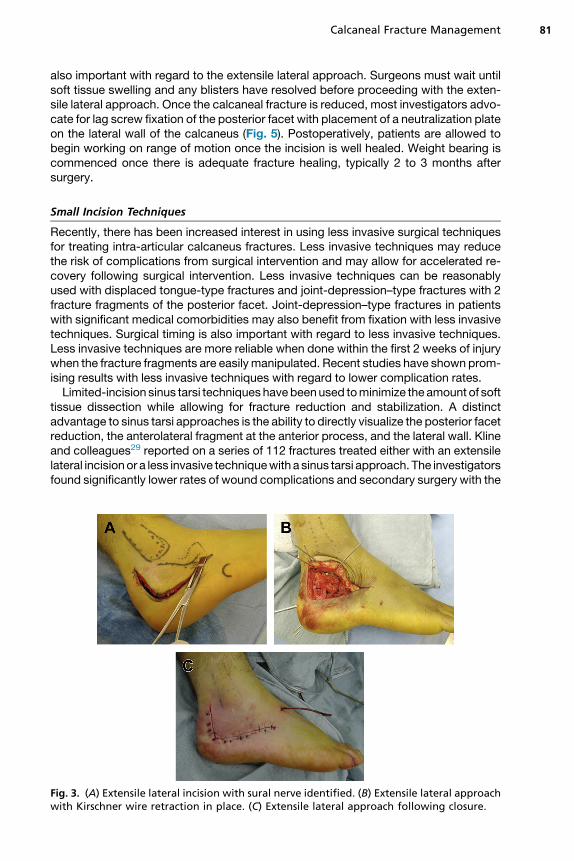
Surgical management of intra-articular calcaneus fractures can be technicallydemanding with regard to reduction and fixation and also carries a high risk of com-plications.9,10,21–25 The traditional extensile lateral approach for open reduction inter-nal fixation of calcaneus fractures creates an L-shaped soft tissue flap that dependson the lateral calcaneal branch of the peroneal artery, which is vulnerable to injury dur-ing the extensile approach.24–27 When the extensile lateral approach is inappropriatelyplaced, the sural nerve is also in danger (Fig. 3). Although the extensile approach of-fers good visualization of the posterior facet for fracture reduction and direct access tothe lateral wall, expertise is required to minimize high rates of complications with re-gard to wound healing and infection (Fig. 4).21–23,28 Timing of surgical intervention is
Calcaneal Fracture Management 81
also important with regard to the extensile lateral approach. Surgeons must wait untilsoft tissue swelling and any blisters have resolved before proceeding with the exten-sile lateral approach. Once the calcaneal fracture is reduced, most investigators advo-cate for lag screw fixation of the posterior facet with placement of a neutralization plateon the lateral wall of the calcaneus (Fig. 5). Postoperatively, patients are allowed tobegin working on range of motion once the incision is well healed. Weight bearing iscommenced once there is adequate fracture healing, typically 2 to 3 months aftersurgery.
Small Incision Techniques
Recently, there has been increased interest in using less invasive surgical techniquesfor treating intra-articular calcaneus fractures. Less invasive techniques may reducethe risk of complications from surgical intervention and may allow for accelerated re-covery following surgical intervention. Less invasive techniques can be reasonablyused with displaced tongue-type fractures and joint-depression–type fractures with 2fracture fragments of the posterior facet. Joint-depression–type fractures in patientswith significant medical comorbidities may also benefit from fixation with less invasivetechniques. Surgical timing is also important with regard to less invasive techniques.Less invasive techniques are more reliable when done within the first 2 weeks of injurywhen the fracture fragments are easily manipulated. Recent studies have shown prom-ising results with less invasive techniques with regard to lower complication rates.Limited-incision sinus tarsi techniques havebeenused tominimize the amount of soft
tissue dissection while allowing for fracture reduction and stabilization. A distinctadvantage to sinus tarsi approaches is the ability to directly visualize the posterior facetreduction, the anterolateral fragment at the anterior process, and the lateral wall. Klineand colleagues29 reported on a series of 112 fractures treated either with an extensilelateral incisionor a less invasive techniquewith a sinus tarsi approach. The investigatorsfound significantly lower rates of wound complications and secondary surgery with the
Fig. 3. (A) Extensile lateral incision with sural nerve identified. (B) Extensile lateral approachwith Kirschner wire retraction in place. (C) Extensile lateral approach following closure.

Fig. 4. (A) Wound necrosis at the apex of the extensile lateral approach with exposed hard-ware. (B) Extensive skin necrosis following the extensile lateral approach.
Kiewiet & Sangeorzan82

Fig. 5. (A) Preoperative lateral view of joint-depression calcaneus fracture. (B) Preoperativeaxial view of joint-depression calcaneus fracture. (C) Postoperative lateral view of joint-depression calcaneus fracture. (D) Postoperative axial view of joint-depression calcaneusfracture.
Calcaneal Fracture Management 83
less invasive technique. Outcomes reported were similar between the two groups, andboth techniques had a union rate of 100%. Xia and colleagues30 reported on a random-ized controlled trial of 117 calcaneus fractures comparing an extensile lateral approachwith a limited sinus tarsi approach. Their study showed decreased surgical times andlower wound complications with the less invasive technique. Significantly higher Mary-land Foot Scores were also found in the less invasive group.
SMALL INCISION FIXATION SURGICAL TECHNIQUE
The authors use a small incision technique for some displaced tongue-type fracturesand joint-depression–type fractures without significant comminution of the posteriorfacet. During surgical intervention, patients are placed in the lateral decubitus posi-tion using a beanbag or alternative positioner with all bony prominences well padded(Fig. 6). The authors do place a thigh tourniquet, but this is not routinely used during

Fig. 6. Patient is placed in the lateral decubitus position on a beanbag with all bony prom-inences well padded.
Fig. 7. Markings are made for the lateral extensile incision, and small incisions are made inline with the extensile incision.
Kiewiet & Sangeorzan84

Calcaneal Fracture Management 85
the case unless there is substantial bleeding. The distal fibula and base of the fifthmetatarsal are outlined, and the standard extensile lateral approach is outlined incase it is necessary to transition to this approach following an attempt at reductionthrough small incisions (Fig. 7). Fluoroscopy is brought in from the end of the bed sothat lateral and axial images can be obtained (Fig. 8). Using fluoroscopy, a lateral im-age is obtained to mark the level of the displaced posterior facet fragment. A smallsubcentimeter incision is made in the skin at this level along the horizontal limb of theextensile lateral approach, and blunt dissection is carried out down to the lateral wallof the calcaneus. A second small incision is made at the posterior heel, and a 4.0- or5.0-mm Schanz pin is placed posteriorly into the calcaneal tuberosity. With a tongue-type fracture, the Schanz pin is placed into the tongue fragment to assist in
Fig. 8. (A) Fluoroscopy is brought in from the end of the bed to allow for obtaining a lateralview. (B) An axial view can also be obtained by bringing fluoroscopy in from the end of thebed.

Kiewiet & Sangeorzan86
reduction; with a joint-depression–type fracture, the Schanz pin is placed into the tu-berosity to assist in reduction of the tuberosity fragment. A freer or arbeitsgemein-schaft fur osteosynthesefragen (AO) elevator is then inserted through the initialsmall incision on the horizontal limb to assist in reduction of the posterior facet
Fig. 9. (A) Site of the displaced posterior facet fragment is identified with fluoroscopy, and asubcentimeter incision is made in line with the plantar limb of the extensile lateral incision.(B) An elevator is inserted to reduce the displaced posterior facet fragment. (C) Axial view ofa joint-depression calcaneus fracture before placement of a Schanz pin and reduction. (D)With joint-depression–type fractures, a Schanz pin is placed into the tuberosity fragmentto assist in reduction of the tuberosity. (E) Once the posterior facet is reduced, 2 guidewiresare placed just below the posterior facet and critical angle of Gissane. The knife blade ismarking the posterior facet for placement of the lag screw across the posterior facet. (F)For tongue-type fractures, a Schanz pin is placed into the tongue fragment to assist inreduction.

Calcaneal Fracture Management 87
fragment. The posterior facet fragment is reduced; joint-depression fractures, the tu-berosity is reduced with a combination of distraction for length, rotation out of varus,and medialization. Once reduced, 2 guide pins or Kirschner wires are placed from thesuperior aspect of the calcaneal tuberosity beneath the posterior facet and criticalangle of Gissane and into the anterior process of the calcaneus (Fig. 9). Adequatereduction and placement of guide pins are confirmed with fluoroscopic views,
Fig. 10. (A) Lateral view with screws in place following small incision technique. (B) Axialview showing screw placement with small incision technique. (C) AP foot view showingscrew placement with small incision technique.

Kiewiet & Sangeorzan88
including a lateral and axial view of the calcaneus, an AP view of the foot, and Brodenviews of the calcaneus to evaluate the posterior facet reduction. Once reduction isconfirmed, the authors then proceed with placement of screws in the same pathas the guidewires. The authors prefer to use cannulated 4.5- or 5.5-mm fullythreaded screws. The lateral hindfoot is marked just plantar to the posterior facet us-ing fluoroscopy, and a small incision is made in the skin with blunt dissection throughthe subcutaneous tissue down to the lateral calcaneus. A 3.5-mm cortical screw isthen placed using lag technique across the posterior facet fracture fragment securingthe lateral posterior facet fragment to the medial sustentaculum fragment and givingcompression across the posterior facet fragment. A small incision is then made at theplantar aspect of the heel for placement of a 3.5-mm screw as a kickstand from theplantar aspect of the calcaneal tuberosity to just inferior to the posterior facet. Injoint-depression–type fractures, the placement of 2 more 3.5-mm fully threadedscrews from the plantar aspect of the tuberosity into the anterior process of thecalcaneus may be warranted to assist in holding reduction of the tuberosity frag-ment. Fluoroscopy is used to obtain final lateral and axial views of the calcaneus,an anteroposterior (AP) view of the foot, and Broden views of the calcaneus to assureadequate reduction and good placement of hardware (Fig. 10). The incisions areclosed with a 4-0 nylon suture (Fig. 11). The incisions are dressed, and patientsare placed in a posterior splint or a tall controlled ankle motion (CAM) boot. Postop-eratively, range-of-motion exercises are started either immediately or 1 to 2 weeksafter surgery. Weight bearing is commenced at 6 to 8 weeks after surgery or whenfracture healing seems adequate to begin weight bearing on follow-up radiographs.
EXTENSILE LATERAL VERSUS SMALL INCISION TECHNIQUE
The senior author and his colleagues have undertaken a prospective study evaluatingthe extensile lateral approach versus minimally invasive reduction and small fragmentfixation for tongue-type calcaneus fractures. These data remain unpublished to date;however, their study showed a significant difference in length of hospital stay and timeto weight bearing between the two groups. They also showed improved musculoskel-etal function assessment (MFA) scores at 1 year in the small incision group (Table 1).
Fig. 11. Closed incisions used for small incision calcaneus fixation technique.

Table 1Results of unpublished data from senior author comparing an extensile lateral approach witha small incision technique for tongue-type calcaneus fractures
Small Incision Extensile Lateral Overall P Value
Hospital stay (d) 1.6 3.7 2.8 <.001
Time to weight bearing (wk) 12.3 20.5 16.8 .03
Pain (VAS) (1–10) 2.6 3.1 2.9 NS
MFA scores (6 wk) 41.1 46.1 — —
MFA scores (3 mo) 36.0 40.9 — —
MFA scores (6 mo) 23.5 29.9 — —
MFA scores (12 mo) 17.0 28.9 — —
Abbreviation: NS, no significance.
Calcaneal Fracture Management 89
SUMMARY
Treatment of displaced intra-articular calcaneus fractures has long been a controversialtopic amongorthopedicsurgeons.Historically, nonoperativemanagementwas the treat-mentofchoice; but over thepast fewdecades, operativemanagementhasbecomemoreprevalent, with studies showing improved outcomes following surgical management.Surgical intervention with an extensile lateral approach continues to be most prev-
alent; however, there is recent interest in less invasive techniques for fixation of dis-placed calcaneal fractures. The extensile lateral approach allows for directvisualization of the posterior facet reduction; however, it carries risks of wound-healing complications and infection. Less invasive techniques have been shown tohave less wound-healing complications. Small incision techniques can be used forcertain tongue-type fractures and for joint-depression fractures with minimal commi-nution. Joint-depression fractures with extensive posterior facet comminution are lessamenable to small incision techniques. The authors allow for an accelerated recoveryfollowing fixation with a small incision technique in a hope to reduce the significance ofloss of range of motion.Studies have shown advantages to less invasive techniques recently, and unpub-
lished data by the senior author and his colleagues show advantages with regard tohospital length of stay and time to weight bearing. Although the authors recognizethese techniques may be a compromise between reducing the risks associated withthe extensile lateral approach and accepting a possible imperfect reduction, they thinkthat the benefits of the small incision techniques outweigh the drawbacks. Given theresults of recent studies and the authors’ experience, they do think that small incisiontechniques are a viable option for surgical management of specific calcaneus fracturetypes, including tongue-type calcaneal fractures and joint-depression fractureswithout significant comminution of the posterior facet.
REFERENCES
1. Cotton FJ. Fractures of the os calcis. Boston Med Surg J 1908;18:559–65.2. Cotton FJ. Old os calcis fractures. Ann Surg 1921;74:294–303.3. Essex Lopresti P. The mechanism, reduction technique, and results in fractures of
the os calcis. Br J Surg 1952;39:395–419.4. Lindsay WRN, Dewar FP. Fractures of the os calcis. Am J Surg 1958;95:555–76.5. Kitaoka HB, Schaap EJ, Chao EY, et al. Displaced intra-articular fractures of
the calcaneus treated non-operatively: clinical results and analysis of motion

Kiewiet & Sangeorzan90
and ground-reaction and temporal forces. J Bone Joint Surg Am 1994;76:1531–40.
6. Crosby LA, Fitzgibbons T. Computerized tomography scanning of acute intra-articular fractures of the calcaneus. J Bone Joint Surg Am 1990;72:852–9.
7. Jarvholm U, Korner L, Thoren O, et al. Fractures of the calcaneus. Acta OrthopScand 1984;55:652–6.
8. Parmar HV, Triffitt PD, Gregg PJ. Intra-articular fractures of the calcaneum treatedoperatively or conservatively: a prospective study. J Bone Joint Surg Br 1993;75:932–7.
9. Agren PH, Wretenberg P, Sayed-Noor AS. Operative versus nonoperative treat-ment of displaced intra-articular calcaneal fractures: a prospective, randomized,controlled multicenter trial. J Bone Joint Surg Am 2013;95:1351–7.
10. Ibrahim T, Roswell M, Rennie W, et al. Displaced intra-articular calcaneal frac-tures: 15 year follow-up of a randomised controlled trial of conservative versusoperative treatment. Injury 2007;38:848–55.
11. O’Farrell DA, O’Byrne JM, McCabe JP, et al. Fractures of the os calcis: improvedresults with internal fixation. Injury 1993;24:263–5.
12. Leung KS, Yuen KM, Chan WS. Operative treatment of displaced intra-articularfractures of the calcaneum: medium-term results. J Bone Joint Surg Br 1993;75:196–201.
13. Crosby LA, Fitzgibbons TC. Open reduction and internal fixation of Type II intra-articular calcaneus fractures. Foot Ankle Int 1996;17:253–8.
14. Thordarson DB, Krieger LE. Operative vs. nonoperative treatment of intra-articular fractures of the calcaneus: a prospective randomized trial. Foot AnkleInt 1996;17:2–9.
15. Buckley R, Tough S, McCormack R, et al. Operative compared with non-operativetreatment of displaced intra-articular calcaneal fractures: a prospective, random-ized, controlled multicenter trial. J Bone Joint Surg Am 2002;84:1733–44.
16. Gallie WE. Subastragalar arthrodesis in fractures of the os calcis. J Bone JointSurg Am 1943;25:731–6.
17. Carr JB, Benirschke SK. Subtalar distraction bone block fusion for late complica-tions of os calcis fractures. Foot Ankle 1988;9:81–6.
18. Stephens HM, Sanders R. Calcaneal malunions: results of a prognosticcomputed tomography classification system. Foot Ankle Int 1996;17:395–401.
19. Clare MP, Lee WE 3rd, Sanders RW. Intermediate to long-term results of a treat-ment protocol for calcaneal fracture malunions. J Bone Joint Surg Am 2005;87:963–73.
20. Radney CS, Clare MP, Sanders RW. Subtalar fusion after displaced intra-articularcalcaneal fractures: does initial operative treatment matter? J Bone Joint Surg Am2009;91:541–6.
21. Gardner MJ, Nork SE, Barei DP, et al. Secondary soft tissue compromise intongue-type calcaneus fractures. J Orthop Trauma 2008;22(7):439–45.
22. Gougoulias N, Khanna A, McBride DJ, et al. Management of calcaneal fractures:systematic review of randomized trials. Br Med Bull 2009;92:153–67.
23. Swanson SA, Clare MP, Sanders RW. Management of intra-articular fractures ofthe calcaneus. Foot Ankle Clin 2008;13(4):659–78.
24. Cavadas PC, Landin L. Management of soft tissue complications of the lateralapproach for calcaneal fractures. Plast Reconstr Surg 2007;120(2):459–66.
25. Bibbo C, Ehrlich DA, Nguyen HM, et al. Low wound complication rates for thelateral extensile approach for calcaneal ORIF when the lateral calcaneal arteryis patent. Foot Ankle Int 2014;35(7):650–6.

Calcaneal Fracture Management 91
26. Gould N. Lateral approach to the os calcis. Foot Ankle 1984;4(4):218–20.27. Benirschke SK, Sangeorzan BJ. Extensive intraarticular fractures of the foot. Sur-
gical management of calcaneal fractures. Clin Orthop Relat Res 1993;292:128–34.
28. Abidi NA, Dhawan S, Gruen GS, et al. Wound-healing risk factors after openreduction and internal fixation of calcaneal fractures. Foot Ankle Int 1998;19(12):856–61.
29. Kline AJ, Anderson RB, Davis WH, et al. Minimally invasive technique versus anextensile lateral approach for intra-articular calcaneal fractures. Foot Ankle Int2013;34(6):773–80.
30. Xia S, Lu Y, Wang H, et al. Open reduction and internal fixation with conventionalplate via L-shaped lateral approach versus internal fixation with a percutaneousplate via a sinus tarsi approach for calcaneal fractures: a randomized controlledtrail. Int J Surg 2014;12(5):475–80.