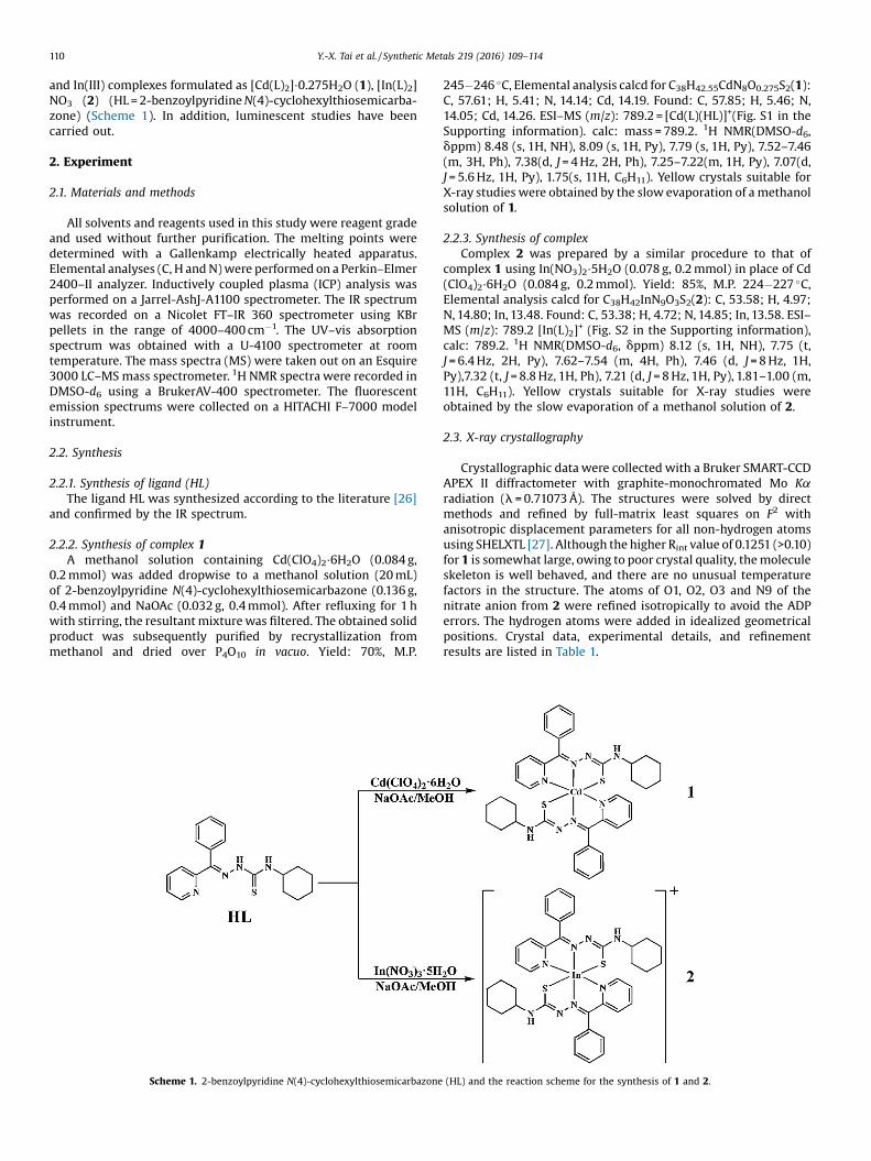
cadmium(ii) and indium(iii) complexes derived from 2...
TRANSCRIPT

Synthetic Metals 219 (2016) 109–114
Cadmium(II) and indium(III) complexes derived from2-benzoylpyridine N(4)-cyclohexylthiosemicarbazone: Synthesis,crystal structures, spectroscopic characterization and cytotoxicity
Yan-Xue Tai, Yu-Mei Ji, Yan-Li Lu, Ming-Xue Li*, Yuan-Yuan Wu, Qiu-Xia Han*Key Laboratory of Polyoxometalates of Henan Province, Institute of Molecular and Crystal Engineering, College of Chemistry and Chemical Engineering, HenanUniversity, Kaifeng 475004, PR China
A R T I C L E I N F O
Article history:Received 21 March 2016Received in revised form 6 May 2016Accepted 14 May 2016Available online xxx
Keywords:ThiosemicarbazoneCrystal structureCytotoxicityFluorescent probe
A B S T R A C T
Two metal complexes [Cd(L)2]�0.275H2O(1) and [In(L)2]NO3 (2) (HL = 2-benzoylpyridine N(4)-cyclo-hexylthiosemicarbazone) have been synthesized and structurally characterized by elemental analysis, anumber of spectroscopic methods (IR, UV-vis, NMR), mass spectrometry, and X-ray crystallography. TheSchiff’s base ligand forms hexacoordinated complexes having octahedral geometry for Cd(II) and In(III)complexes, respectively. The synthesized compounds were tested for antiproliferative activity andshowed the ability to kill HepG2 cells (human hepato cellular carcinoma) significantly, especially 2 withIC50 = 2.02 � 0.14 mM. Of particular note is the fact that complex 1 has ca 12-fold lower toxicity in thenormal hepatocyte QSG7701 cells than in the hepatocellular carcinoma HepG2 cells. In addition, complex2 also exhibited excellent luminescent property. Upon the addition of 1 equiv of In3+ ion, 200-foldfluorescence enhancement of HL at lem = 504 nm has been observed. Moreover, the fluorescent colorchange (from transparent to light-green) could be observed by naked eyes under the light of 365 nm.These findings can expand the applications of thiosemicarbazone derivatives in the fields of colorimetricand fluorescent probes.
ã 2016 Elsevier B.V. All rights reserved.
Contents lists available at ScienceDirect
Synthetic Metals
journal homepage: www.elsevier .com/ locate /sy nmet
1. Introduction
From the early 1950s to the present, the chemistry ofthiosemicarbazones has shown considerable interest in analyticalchemistry, pharmacological properties and spectrophotometry[1–5]. The best known member of this family, 3-aminopyridinecarboxaldehyde thiosemicarbazone (3-AP), is a potent ribonucle-otide reductase inhibitor that is currently in phase II clinical trialsfor the treatment of a number of forms of cancer, including non-small-cell lung cancer and renal carcinoma [6]. This compoundshows therapeutic activity over a certain range of dosages inpreclinical tumor models without imposing intolerable hosttoxicity [7]. In more cases, the involvement of mixed nature ofthe N and S donor atoms in coordination with metal ions isresponsible for increasing biological activities of thiosemicarba-zones [8,9]. Their mechanism of action is still controversial in manyrespects, including ribonucleotide reductase inhibition, metal
* Corresponding authors.E-mail addresses: [email protected] (M.-X. Li), [email protected]
(Q.-X. Han).
http://dx.doi.org/10.1016/j.synthmet.2016.05.0150379-6779/ã 2016 Elsevier B.V. All rights reserved.
dependent radical damage, DNA binding, and inhibition of proteinsynthesis [10,11].
Cadmium is an extremely toxic element whose deleteriousactions influence the majority of human tissues and is oftenpresent in the environment [12]. Its toxicity derives from the factthat it is rapidly localized intracellularly, mainly in the liver, andthen is bound to metallothionein forming a complex that is slowlytransferred to the bloodstream to be deposited in the kidneys. Evenso, cadmium complexes have attracted more and more attentionsdue to their widely reported bioactivities, such as DNA bindingability [13], antibacterial activities [14] and antitumor activities[15,16]. Moreover, the compounds which are able to form stablecomplexes with cadmium could be employed as detoxificatingagents [14].
Indium is an auger electron emitter, potentially enabling itscomplexes to be dual imaging-therapeutic agents [17]. However,indium complexes remain relatively unexplored to date [18–21].Therefore, detailed investigations of indium complexes will bevaluable.
As part of our systematic studies on heterocyclic thiosemi-carbazones and their metal complexes [8,22–25], here we reportthe synthesis, characterization and in vitro cytotoxicity of Cd(II)
110 Y.-X. Tai et al. / Synthetic Metals 219 (2016) 109–114
and In(III) complexes formulated as [Cd(L)2]�0.275H2O (1), [In(L)2]NO3 (2) (HL = 2-benzoylpyridine N(4)-cyclohexylthiosemicarba-zone) (Scheme 1). In addition, luminescent studies have beencarried out.
2. Experiment
2.1. Materials and methods
All solvents and reagents used in this study were reagent gradeand used without further purification. The melting points weredetermined with a Gallenkamp electrically heated apparatus.Elemental analyses (C, H and N) were performed on a Perkin–Elmer2400–II analyzer. Inductively coupled plasma (ICP) analysis wasperformed on a Jarrel-AshJ-A1100 spectrometer. The IR spectrumwas recorded on a Nicolet FT–IR 360 spectrometer using KBrpellets in the range of 4000–400 cm�1. The UV–vis absorptionspectrum was obtained with a U-4100 spectrometer at roomtemperature. The mass spectra (MS) were taken out on an Esquire3000 LC–MS mass spectrometer. 1H NMR spectra were recorded inDMSO-d6 using a BrukerAV-400 spectrometer. The fluorescentemission spectrums were collected on a HITACHI F–7000 modelinstrument.
2.2. Synthesis
2.2.1. Synthesis of ligand (HL)The ligand HL was synthesized according to the literature [26]
and confirmed by the IR spectrum.
2.2.2. Synthesis of complex 1A methanol solution containing Cd(ClO4)2�6H2O (0.084 g,
0.2 mmol) was added dropwise to a methanol solution (20 mL)of 2-benzoylpyridine N(4)-cyclohexylthiosemicarbazone (0.136 g,0.4 mmol) and NaOAc (0.032 g, 0.4 mmol). After refluxing for 1 hwith stirring, the resultant mixture was filtered. The obtained solidproduct was subsequently purified by recrystallization frommethanol and dried over P4O10 in vacuo. Yield: 70%, M.P.
Scheme 1. 2-benzoylpyridine N(4)-cyclohexylthiosemicarbazone
245�246 �C, Elemental analysis calcd for C38H42.55CdN8O0.275S2(1):C, 57.61; H, 5.41; N, 14.14; Cd, 14.19. Found: C, 57.85; H, 5.46; N,14.05; Cd, 14.26. ESI–MS (m/z): 789.2 = [Cd(L)(HL)]+(Fig. S1 in theSupporting information). calc: mass = 789.2. 1H NMR(DMSO-d6,dppm) 8.48 (s, 1H, NH), 8.09 (s, 1H, Py), 7.79 (s, 1H, Py), 7.52–7.46(m, 3H, Ph), 7.38(d, J = 4 Hz, 2H, Ph), 7.25–7.22(m, 1H, Py), 7.07(d,J = 5.6 Hz, 1H, Py), 1.75(s, 11H, C6H11). Yellow crystals suitable forX-ray studies were obtained by the slow evaporation of a methanolsolution of 1.
2.2.3. Synthesis of complexComplex 2 was prepared by a similar procedure to that of
complex 1 using In(NO3)2�5H2O (0.078 g, 0.2 mmol) in place of Cd(ClO4)2�6H2O (0.084 g, 0.2 mmol). Yield: 85%, M.P. 224�227 �C,Elemental analysis calcd for C38H42InN9O3S2(2): C, 53.58; H, 4.97;N, 14.80; In, 13.48. Found: C, 53.38; H, 4.72; N, 14.85; In, 13.58. ESI–MS (m/z): 789.2 [In(L)2]+ (Fig. S2 in the Supporting information),calc: 789.2. 1H NMR(DMSO-d6, dppm) 8.12 (s, 1H, NH), 7.75 (t,J = 6.4 Hz, 2H, Py), 7.62–7.54 (m, 4H, Ph), 7.46 (d, J = 8 Hz, 1H,Py),7.32 (t, J = 8.8 Hz, 1H, Ph), 7.21 (d, J = 8 Hz, 1H, Py), 1.81–1.00 (m,11H, C6H11). Yellow crystals suitable for X-ray studies wereobtained by the slow evaporation of a methanol solution of 2.
2.3. X-ray crystallography
Crystallographic data were collected with a Bruker SMART-CCDAPEX II diffractometer with graphite-monochromated Mo Karadiation (l = 0.71073 Å). The structures were solved by directmethods and refined by full-matrix least squares on F2 withanisotropic displacement parameters for all non-hydrogen atomsusing SHELXTL [27]. Although the higher Rint value of 0.1251 (>0.10)for 1 is somewhat large, owing to poor crystal quality, the moleculeskeleton is well behaved, and there are no unusual temperaturefactors in the structure. The atoms of O1, O2, O3 and N9 of thenitrate anion from 2 were refined isotropically to avoid the ADPerrors. The hydrogen atoms were added in idealized geometricalpositions. Crystal data, experimental details, and refinementresults are listed in Table 1.
(HL) and the reaction scheme for the synthesis of 1 and 2.

Table 1Crystallographic data and structural refinements for 1 and 2.
1 2
Empirical formula C38H42.55CdN8O0.275S2 C38H42InN9O3S2Formula weight (g�mol�1) 792.29 851.74T (K) 293(2) 296(2)Crystal system Monoclinic Monoclinicspace group C2/c Cca (Å) 30.057(18) 17.9751(14)b (Å) 14.596(9) 13.6158(11)c (Å) 18.951(11) 16.8775(13)a (deg) 90.00 90.00b (deg) 109.860(11) 107.673(2)g (deg) 90.00 90.00V (Å3) 7820(8) 3935.7(5)Z 8 4Dc (g�cm�3) 1.338 1.437m (mm�1) 0.702 0.755Limiting indices �35 � h � 35
�17 � k � 17�22 � l � 20
�21 � h � 8�16 � k � 16�20 � l � 20
Rint 0.1251 0.0283Restrains/parameters 37/443 333/455u range (�) 2.081–25.099 1.911–25.095Goodness-of-fit on F2 0.872 1.029R1, wR2 [I > 2s(I)] 0.0663, 0.1599 0.0724, 0.1948R1, wR2 [all data] 0.1371, 0.1823 0.0985, 0.2227
Table 2Selected bond lengths (Å) and bond angles (�) of complexes 1 and 2.
1 2
Cd(1)–S(1) 2.564(3) In(1)–S(1) 2.506(4)Cd(1)–S(2) 2.565(2) In(1)–S(2) 2.493(5)Cd(1)–N(3) 2.410(6) In(1)–N(3) 2.262(13)Cd(1)–N(4) 2.406(7) In(1)–N(4) 2.323(14)Cd(1)–N(7) 2.389(6) In(1)–N(7) 2.268(13)Cd(1)–N(8) 2.390(6) In(1)–N(8) 2.326(13)S(1)–C(7) 1.735(8) S(1)–C(7) 1.743(16)S(2)–C(26) 1.730(7) S(2)–C(26) 1.756(18)N(3)–C(8) 1.328(9) N(3)–C(8) 1.29(2)N(7)–C(27) 1.287(8) N(7)–C(27) 1.33(2)N(8)–Cd(1)–N(4) 92.3(2) N(7)–In(1)–N(8) 71.5(4)N(4)–Cd(1)–N(3) 67.6(2) N(3)–In(1)–N(4) 70.5(5)N(3)–Cd(1)–S(1) 73.40(18) N(7)–In(1)–N(4) 90.4(5)N(4)–Cd(1)–S(1) 137.31(17) N(4)–In(1)–N(8) 91.8(5)N(7)–Cd(1)–S(2) 73.59(15) N(4)–In(1)–S(2) 93.5(4)N(8)–Cd(1)–S(2) 137.50(15) N(3)–In(1)–S(1) 77.9(3)S(1)–Cd(1)–S(2) 97.10(8) N(7)–In(1)–S(1) 121.4(4)C(7)–S(1)–Cd(1) 99.0(3) N(8)–In(1)–S(1) 93.4(4)C(26)–S(2)–Cd(1) 98.5(3) N(4)–In(1)–S(1) 147.7(4)C(27)–N(7)–Cd(1) 121.1(5) C(7)–S(1)–In(1) 94.9(6)N(6)–N(7)–Cd(1) 121.4(5) C(26)–S(2)–In(1) 96.2(6)
C(8)–N(3)–In(1) 120.0(11)N(2)–N(3)–In(1) 122.4(10)
Y.-X. Tai et al. / Synthetic Metals 219 (2016) 109–114 111
2.4. Fluorescence measurements
For fluorescence behavior, stock methanol solutions (10�3mol�L�1) of HL, In(NO3)�5H2O, 1 and 2 were prepared. The solution ofHL, 1 and 2 were then diluted to 10�4mol�L�1 with methanol. Inconcentration gradients experiments, HL (1 mL, 10�3mol�L�1) andappropriate amounts of In3+(0, 125, 250, 500, 750, 1000 mL) wereadded to a volumetric flask(10 mL), and then added methanol to10 mL, respectively. For fluorescence measurements, excitationwas provided at 361 nm, and emission was acquired from 371 nmto 730 nm.
2.5. Cell culture
The hepatocellular carcinoma HepG2 cells and the normalhepatocyte QSG7701 cells were cultured in Dulbecco’s minimalessential medium supplemented with 10% fetal bovine serum, 100IU�mL�1 penicillin, and 100 mg�mL�1 streptomycin at 37 �C in ahumidified atmosphere with 5% CO2. The cells were harvested with0.02% EDTA and 0.025% trypsin and then rinsed three times inphosphate-buffered saline (PBS). The resulting cell suspension wasused in following experiment.
2.6. Cytotoxicity assay
3-(4,5-Dimethylthiazol-2-yl)-2,5-diphenyltetrazolium bro-mide (MTT) assay was carried out to evaluate cytotoxicity. Cellswere plated into 96-well plates at a cell density of 1 �104 cells perwell and allowed to grow in a CO2 incubator. After 24 h, themedium was removed and replaced by fresh medium containingthe tested compounds which were dissolved in DMSO at 0.01 Mand diluted to various concentrations with PBS before theexperiment, and the final concentration of DMSO is lower than1%. After 24 h incubation, cultures were incubated in 100 mL ofmedium with 10 mL of 5 mg/mL MTT solution for 4 h at 37 �C. Themedium with MTT was removed, and 100 mL of dimethyl sulfoxide(DMSO) was added to each well to dissolve the formazan. Theabsorbance at 570 nm was measured with microplate reader (Bio-Tek ELX800, USA). The inhibitory percentage of each compound atvarious concentrations was calculated, and the IC50 value was
determined. All data are expressed as the mean � SD from threeseparate determinations.
3. Results and discussion
3.1. X-ray crystallography
Selected bond distances and angles of 1 and 2 are given inTable 2. The molecular structures along with the atom numberingscheme and the unit cell packing are depicted in Figs. 1 and 2,respectively.
3.1.1. Crystal structure of 1As shown in Fig. 1a, the two N,N,S-coordinated ligands give the
Cd(II) ion a highly distorted octahedral coordination polyhedron.One sulfur atom, one imine nitrogen atom and one pyridinenitrogen atom from one ligand and one imine nitrogen atom fromanother ligand occupy the basal positions, the two remainingpositions in the octahedral geometry are the axial ones which areoccupied by one sulfur atom and one pyridine nitrogen atom fromthe second ligand. The pseudo-macrocyclic coordination mode ofeach ligand affords two five-membered chelate rings, the dihedralangle between the chelate rings are 12.6 and 12.8�, respectively.Both C–S bond lengths suggest evolution towards the thiol form.The C–N and N–N bond lengths in L� are intermediate betweenformal single and double bonds, pointing to an extensive electrondelocalization over the entire molecular skeleton [28]. Similarly,intermolecular hydrogen bonds of complex 1 also link the differentcomponents to stabilize the crystal structure (Fig. 1b and c). Thehydrogen bond involves the terminal nitrogen atom N(5) and thecoordinated sulfur atom S(2) with N(5)� � �S(2) 3.422(7) Å and theangle N(5)–H(5A)� � �S(2) being 161.9� (symmetry code: �x + 1/2,�y + 1/2, �z), respectively.
3.1.2. Crystal structure of 2As shown in Fig. 2a, the molecular structure of 2 contains one
[In(L)2]+ cation and one nitrate ion acting as the counterion. Theindium(III) ion has the same coordination model as cadmium(II) incomplex 1. The pseudo-macrocyclic coordination mode of eachligand affords two five-membered chelate rings, which are nearplanar, the dihedral angle between the chelate rings are 3.4 and

Fig. 1. (a) Structure of complex 1 with atomic numbering scheme. (b) Hydrogen bond indicated by dashed line. (c) The molecular packing projected along the b-axis.
112 Y.-X. Tai et al. / Synthetic Metals 219 (2016) 109–114
5.1�, respectively. Fig. 2b is the molecular packing projected alongthe b-axis.
3.2. Infrared spectra
The characteristic vibrational bands of HL, 1 and 2 correspond-ing to the important functions in the system are presented inTable 3. The free ligand shows two bands at 3336 and 3162 cm�1
Fig. 2. (a) Structure of complex 2 with atomic numbering sche
observed at 3285 and 3236 cm�1 in complexes 1 and 2 instead,respectively. These bands may be assigned for N–H stretchingvibrations. The n(C=N) bands of thiosemicarbazone in twocomplexes undergoes a red shift of wavenumber compared tothat of the ligand (1523 cm�1), a clear sign of coordination via theimine nitrogen atom. Additionally, the increase in the frequency ofn(N–N) band of the thiosemicarbazone in the spectra of complexesis due to the increase in the bond strength, again confirms the
me. (b) The molecular packing projected along the b-axis.

Table 3Infrared spectral assignments for the ligand HL, 1 and 2.
Compound n(N–H) n(C=N) n(N–N) n(C=S) n(py)
HL 3336, 3162 1523 1119 800 6081 3285 1494 1147 787 6272 3236 1496 1152 781 643
Fig. 4. Fluorescence emission spectra of HL, 1 and 2 in methanol solution at roomtemperature.
Y.-X. Tai et al. / Synthetic Metals 219 (2016) 109–114 113
coordination via the imine nitrogen [4]. The thioamide band, whichcontains considerable n(C = S) character, is less intense in thecomplexes and is found at a lower frequency, suggestingcoordination of the metal through sulfur [29]. The pyridyl nitrogenresulting breathing motion of the pyridine ring is shifted to ahigher frequency upon complexation and is consistent withpyridine ring nitrogen coordination. These observations haveindicated the participation of imine nitrogen, thiolate sulfur andpyridine nitrogen of the ligand in the coordination.
3.3. UV–vis spectra
The UV–vis spectra of these compounds were studied inmethanol solution. As shown in Fig. 3 the ligand HL gives two bandat 322 and 410 nm, while complex 1 exhibits two peaks of 302 and398 nm in ultraviolet region and complex 2 exhibits one band of302 nm in ultraviolet region and one peak of 408 nm in visibleregion. These compounds have a band in the range 302–322 nmdue to p!p* transitions, while the strong absorption at 398 (1)and 408 (2) nm may be attributed to the LMCT transitions. Such afeature provides a good evidence for the chelation of the ligand tothe metal centre.
3.4. Fluorescence spectral studies
The fluorescence behaviors of these compounds (HL, 1 and 2)were studied in 1 �10�4M methanol solution. Fluorescenceexperiments were carried out with 361, 354 and 333 nm asexcitation wavelength of HL, 1 and 2, respectively. The spectralchanges of HL, 1 and 2 are shown in Fig. 4. It can be seen that thethree compounds show different emission peaks at 406, 400 and537, 371 and 508 nm, respectively and the complex 2 exhibits astrong fluorescence emission at 508 nm. The results indicated thatdifferent metal complexes had a significant effect not only on theemission peak but also on the emission intensity [30]. The titrationexperiments were carried out by adding different concentrationsof indium(III) ion. Upon the addition of 1 equiv of In3+ ion, 200-foldfluorescence enhancement of HL has been observed. Furthermore,
Fig. 3. UV–vis absorption spectra of the HL, 1 and 2 in methanol solution at roomtemperature.
the visual fluorescence response can be seen under UV light(Fig. 5b). This phenomenon indicates that this ligand can be used asa sensitive probe for In3+.
3.5. Cytotoxicity assay
In terms of the cytotoxic activity of thiosemicarbazones [31],firstly, we have evaluated the ability of HL and complexes 1 and 2 toinhibit tumor cell growth against HepG2 cells. To explore thetoxicity of these compounds, their effect on normal QSG7701 cellsis also described. In our experiments, IC50 values (compoundconcentration that produces 50% of cell death) in micromolar unitsare calculated (Fig. 6). Mitoxantrone (Mito) is a syntheticantineoplastic drug and widely used as a potent chemotherapeuticagent in the treatment of various types of cancer because of itsapparent lower risk of cardio-toxic effects. Therefore, mitoxan-trone was employed as the reference compound for comparison.
Fig. 5. (a) Fluorescence emission spectra of HL in the absence and presence ofIn3+(methanol solvent) at 504 nm, lex = 361 nm, voltage 700 V, Concentration of HL10�4M. INSET Changes in the emission intensity at 504 nm with incrementaladdition of In3+ (b) Visual changes observed for HL in the presence of differentequivalents In3+ under UV lamp (365 nm).
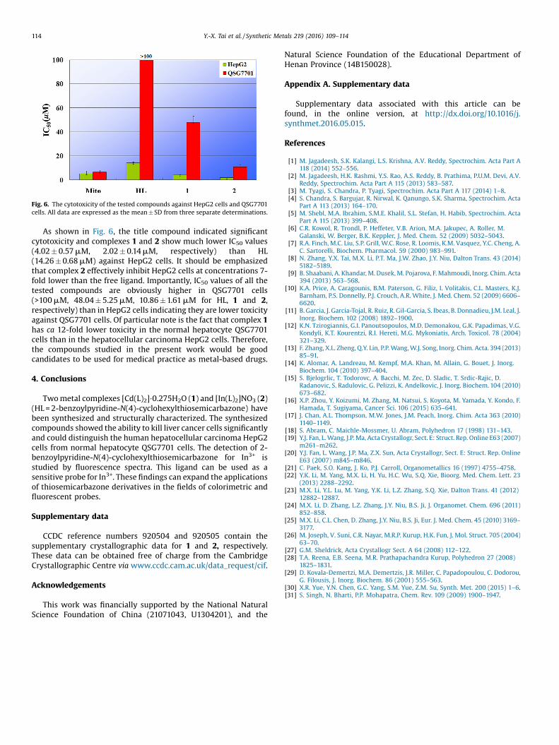
Fig. 6. The cytotoxicity of the tested compounds against HepG2 cells and QSG7701cells. All data are expressed as the mean � SD from three separate determinations.
114 Y.-X. Tai et al. / Synthetic Metals 219 (2016) 109–114
As shown in Fig. 6, the title compound indicated significantcytotoxicity and complexes 1 and 2 show much lower IC50 values(4.02 � 0.57 mM, 2.02 � 0.14 mM, respectively) than HL(14.26 � 0.68 mM) against HepG2 cells. It should be emphasizedthat complex 2 effectively inhibit HepG2 cells at concentrations 7-fold lower than the free ligand. Importantly, IC50 values of all thetested compounds are obviously higher in QSG7701 cells(>100 mM, 48.04 � 5.25 mM, 10.86 � 1.61 mM for HL, 1 and 2,respectively) than in HepG2 cells indicating they are lower toxicityagainst QSG7701 cells. Of particular note is the fact that complex 1has ca 12-fold lower toxicity in the normal hepatocyte QSG7701cells than in the hepatocellular carcinoma HepG2 cells. Therefore,the compounds studied in the present work would be goodcandidates to be used for medical practice as metal-based drugs.
4. Conclusions
Two metal complexes [Cd(L)2]�0.275H2O (1) and [In(L)2]NO3 (2)(HL = 2-benzoylpyridine-N(4)-cyclohexylthiosemicarbazone) havebeen synthesized and structurally characterized. The synthesizedcompounds showed the ability to kill liver cancer cells significantlyand could distinguish the human hepatocellular carcinoma HepG2cells from normal hepatocyte QSG7701 cells. The detection of 2-benzoylpyridine-N(4)-cyclohexylthiosemicarbazone for In3+ isstudied by fluorescence spectra. This ligand can be used as asensitive probe for In3+. These findings can expand the applicationsof thiosemicarbazone derivatives in the fields of colorimetric andfluorescent probes.
Supplementary data
CCDC reference numbers 920504 and 920505 contain thesupplementary crystallographic data for 1 and 2, respectively.These data can be obtained free of charge from the CambridgeCrystallographic Centre via www.ccdc.cam.ac.uk/data_request/cif.
Acknowledgements
This work was financially supported by the National NaturalScience Foundation of China (21071043, U1304201), and the
Natural Science Foundation of the Educational Department ofHenan Province (14B150028).
Appendix A. Supplementary data
Supplementary data associated with this article can befound, in the online version, at http://dx.doi.org/10.1016/j.synthmet.2016.05.015.
References
[1] M. Jagadeesh, S.K. Kalangi, L.S. Krishna, A.V. Reddy, Spectrochim. Acta Part A118 (2014) 552–556.
[2] M. Jagadeesh, H.K. Rashmi, Y.S. Rao, A.S. Reddy, B. Prathima, P.U.M. Devi, A.V.Reddy, Spectrochim. Acta Part A 115 (2013) 583–587.
[3] M. Tyagi, S. Chandra, P. Tyagi, Spectrochim. Acta Part A 117 (2014) 1–8.[4] S. Chandra, S. Bargujar, R. Nirwal, K. Qanungo, S.K. Sharma, Spectrochim. Acta
Part A 113 (2013) 164–170.[5] M. Shebl, M.A. Ibrahim, S.M.E. Khalil, S.L. Stefan, H. Habib, Spectrochim. Acta
Part A 115 (2013) 399–408.[6] C.R. Kowol, R. Trondl, P. Heffeter, V.B. Arion, M.A. Jakupec, A. Roller, M.
Galanski, W. Berger, B.K. Keppler, J. Med. Chem. 52 (2009) 5032–5043.[7] R.A. Finch, M.C. Liu, S.P. Grill, W.C. Rose, R. Loomis, K.M. Vasquez, Y.C. Cheng, A.
C. Sartorelli, Biochem. Pharmacol. 59 (2000) 983–991.[8] N. Zhang, Y.X. Tai, M.X. Li, P.T. Ma, J.W. Zhao, J.Y. Niu, Dalton Trans. 43 (2014)
5182–5189.[9] B. Shaabani, A. Khandar, M. Dusek, M. Pojarova, F. Mahmoudi, Inorg. Chim. Acta
394 (2013) 563–568.[10] K.A. Price, A. Caragounis, B.M. Paterson, G. Filiz, I. Volitakis, C.L. Masters, K.J.
Barnham, P.S. Donnelly, P.J. Crouch, A.R. White, J. Med. Chem. 52 (2009) 6606–6620.
[11] B. Garcia, J. Garcia-Tojal, R. Ruiz, R. Gil-Garcia, S. Ibeas, B. Donnadieu, J.M. Leal, J.Inorg. Biochem. 102 (2008) 1892–1900.
[12] K.N. Tzirogiannis, G.I. Panoutsopoulos, M.D. Demonakou, G.K. Papadimas, V.G.Kondyli, K.T. Kourentzi, R.I. Hereti, M.G. Mykoniatis, Arch. Toxicol. 78 (2004)321–329.
[13] F. Zhang, X.L. Zheng, Q.Y. Lin, P.P. Wang, W.J. Song, Inorg. Chim. Acta. 394 (2013)85–91.
[14] K. Alomar, A. Landreau, M. Kempf, M.A. Khan, M. Allain, G. Bouet, J. Inorg.Biochem. 104 (2010) 397–404.
[15] S. Bjelogrlic, T. Todorovc, A. Bacchi, M. Zec, D. Sladic, T. Srdic-Rajic, D.Radanovic, S. Radulovic, G. Pelizzi, K. Andelkovic, J. Inorg. Biochem. 104 (2010)673–682.
[16] X.P. Zhou, Y. Koizumi, M. Zhang, M. Natsui, S. Koyota, M. Yamada, Y. Kondo, F.Hamada, T. Sugiyama, Cancer Sci. 106 (2015) 635–641.
[17] J. Chan, A.L. Thompson, M.W. Jones, J.M. Peach, Inorg. Chim. Acta 363 (2010)1140–1149.
[18] S. Abram, C. Maichle-Mossmer, U. Abram, Polyhedron 17 (1998) 131–143.[19] Y.J. Fan, L. Wang, J.P. Ma, Acta Crystallogr, Sect. E: Struct. Rep. Online E63 (2007)
m261–m262.[20] Y.J. Fan, L. Wang, J.P. Ma, Z.X. Sun, Acta Crystallogr, Sect. E: Struct. Rep. Online
E63 (2007) m845–m846.[21] C. Paek, S.O. Kang, J. Ko, P.J. Carroll, Organometallics 16 (1997) 4755–4758.[22] Y.K. Li, M. Yang, M.X. Li, H. Yu, H.C. Wu, S.Q. Xie, Bioorg. Med. Chem. Lett. 23
(2013) 2288–2292.[23] M.X. Li, Y.L. Lu, M. Yang, Y.K. Li, L.Z. Zhang, S.Q. Xie, Dalton Trans. 41 (2012)
12882–12887.[24] M.X. Li, D. Zhang, L.Z. Zhang, J.Y. Niu, B.S. Ji, J. Organomet. Chem. 696 (2011)
852–858.[25] M.X. Li, C.L. Chen, D. Zhang, J.Y. Niu, B.S. Ji, Eur. J. Med. Chem. 45 (2010) 3169–
3177.[26] M. Joseph, V. Suni, C.R. Nayar, M.R.P. Kurup, H.K. Fun, J. Mol. Struct. 705 (2004)
63–70.[27] G.M. Sheldrick, Acta Crystallogr Sect. A 64 (2008) 112–122.[28] T.A. Reena, E.B. Seena, M.R. Prathapachandra Kurup, Polyhedron 27 (2008)
1825–1831.[29] D. Kovala-Demertzi, M.A. Demertzis, J.R. Miller, C. Papadopoulou, C. Dodorou,
G. Filousis, J. Inorg. Biochem. 86 (2001) 555–563.[30] X.R. Yue, Y.N. Chen, G.C. Yang, S.M. Yue, Z.M. Su, Synth. Met. 200 (2015) 1–6.[31] S. Singh, N. Bharti, P.P. Mohapatra, Chem. Rev. 109 (2009) 1900–1947.