c sepstodeont studies - septodont-fr.be studies collection... · dr. a. segovia ramírez ......
TRANSCRIPT

Case StudiesSeptodont
No. 14 - October 2016 Collection
BIODENTINE™
Retrofilling in apicectomyDr. A. Segovia Ramírez
BIODENTINE™
Deep caries treatment Dr. M. Federlin
BIODENTINE™
Perforation repair treatmentDr. J. Abarca Reveco
BIOROOT™ RCS
Upper molarwith periradicularperforation Dr. E. Ambu

EditorialSince its foundation Septodont has developed, manufacturedand distributed a wide range of high quality products fordental professionals.
Septodont recently innovated in the field of gingival prepa-ration, composites and dentine care with the introductionof Racegel, the N’Durance® line and Biodentine™, whichare appreciated by clinicians around the globe.
Septodont created the “Septodont Case Studies Collection”- a series of case reports - in 2012 to share with you theirexperience and the benefits of using these innovations indaily practice.Over the past 4 years, authors from more than 15 countrieshave generously contributed to the success of our magazinethat is now distributed on the 5 continents.Each new issue of the Case Studies Collection is theopportunity to discover new clinical challenges and theirtreatment solutions.
This 14th issue features three Biodentine™ cases and a newcase report on the most recent Septodont Innovation :BioRoot™ RCS.
BioRoot™ RCS is the new paradigm for endodontic obtu-rations. Its outstanding sealing properties combined withantimicrobial and bioactive properties allow to get ahigh seal of the endodontium without having to usecomplex warm gutta techniques.
Biodentine™, the first biocompatible and bioactive dentinreplacement material. Biodentine™ uniqueness not onlylies in its innovative bioactive and “pulp-protective”chemistry, but also in its universal application, both inthe crown and in the root.

Endodontic retreatment of an upper molarwith periradicular perforation and lesionDr. Emanuele Ambu
04
09
16
20
Retrofilling with Biodentine™ in apicectomyDr. Alberto Segovia Ramírez
Clinical case report of the use of Biodentine™
for deep caries treatment Dr. Marianne Federlin, Birger Thonemann, Kerstin Galler, Wolfgang Buchalla, Regensburg
Perforation repair with Biodentine™
Dr. Jaime Abarca Reveco
3
Content

4
Endodontic retreatment of anupper molar with periradicularperforation and lesion Dr. Emanuele Ambu, M.D., D.D.S., Clinical Assistant Professor, Tuscan School of Dental Medicine University of Siena,Italy; Dept. of Endodontics (Chairman: Prof. Simone Grandini);Visiting Professor at: University of Valencia for the Master's Degree in Endodontics(Chairman: Prof. Leopoldo Forner).
Endodontic retreatment diseases are almostalways accompanied by periradicular bonelesions, often with pain and apostematous mani-festations. Retreatment is the best therapy tosolve this problem, achieving positive results inmore than 85% of cases. The aim of endodonticretreatment is to reduce intracanal bacterialcontamination below the critical threshold follo-wing these procedures:• Isolation with rubber dam;• Proper opening of access cavity and canalorifices;
• Complete shaping of all canal systems;• Cleaning with appropriate irrigants;• Complete filling of the root canal system;• Filling the access cavity with permanentmaterial.
Retreatment prognosis is less favourable thanthat of conventional endodontic treatmentbecause several bacteria, like Enterococcusfaecalis and Treponema denticola appear to bemore resistant to the chemical agents used toclean, and can penetrate the dentinal tubules.They are also resistant to long periods of“starvation”. Under these conditions, the risk of recontami-nation can be reduced using materials ortechniques ensuring more effective sealing ofthe root canal system, increasing the percentageof success of the endodontic therapy. Warm gutta percha is considered the "goldstandard" to fill the root canal system; but itcontracts while cooling down and this forms agap between the root canal walls and gutta
Introduction

5
percha so that the bacteria of dentinal tubulescan contaminate canals again.To solve this problem, a cold-filling techniquebased on bioceramics has been recently intro-duced. This material was initially suggested toreplace mineral trioxide aggregate in variouscases (radicular perforations, apexifications,direct capping of the pulp, etc.), because MTAhad some problems: it took long to harden andit was unsuitable to be in touch with the oralenvironment.
More recently, a new preparation has permittedthe use of bioceramics as an endodontic fillingmaterial.
The aim of this paper is to describe a complexexample of molar retreatment treated by meansof BioRoot™ RCS, a canal filling material soldby Septodont (Saint-Maur-des-fossés, France).
A male Caucasian patient VL aged 48 wasreferred by a colleague who did not manage toprobe the root canals, after many attempts,had not succeeded in probing the canals of hisright upper molar, which had been unadequatelyfilled during previous treatments. There wasalso a perforation altering the pathway of themesio-vestibular canal. The clinical picture wasfurther complicated by two vestibular fistulaesecreting pus. Endoral RX and CBCT examination(Fig. 1) -- performed to better understand thefunctioning of root canals – showed widespreadperiradicular lesions and an inflammation of themale breast, with likely odontogenous origin.
While the palatal and mesio-vestibular canalswere easily renegotiable, it was not possible torestore the correct course of the mesio-vestibularcanal. Using a surgical microscope, it waspossible to locate the orifice of the mesio-palatal canal (Fig. 2, arrow), not detected in thetwo preceding treatments. This canal was foundto be confluent with the mesio-vestibular, thuspermitting recovery of the entire course, whichappeared anomalous compared to the usualendodontic anatomy of the first upper molar.Following a thorough cleaning of the root canalsystem, the practitioner filled the entire systemusing cold tapered gutta percha points (06) and
Clinical Case
Fig. 1 Fig. 2

6
Bioceramics are today quite well-known materialsin the literature, especially because they canreplace mineral trioxide aggregate. Biodentine™, defined as an active biosilicate, isbioactive and continues to produce hydroxya-patites long after blending. It can therefore fitthe dentinal walls and improve its sealing capacity. BioRoot™ RCS -- a product obtained after testingthese materials -- can ensure complete sealingof the root canal system after full cleaning andshaping. It is also an effective antibacterial agent.Its biocompatibility has been recently evaluatedin several studies, but there is no research publi-shed about the clinical effects of this material. Itis a very interesting material because it perfectlyadheres to both dentine and gutta percha, sothat it can be used in cold sealing techniques.
This cannot be considered a monocone tech-nology: in this technique, a single gutta perchacone is inserted into the canal -- which has[already] been filled with endodontic cement --in an attempt to occupy the major part of thevolume of the root canal. Since the endodonticcement is absorbable with time, this technologywas [traditionally] considered highly ineffective,and not recommended by many practotioners.On the other hand, in cold sealing technologyusing bioceramics, the cement plays an importantrole because it is truly active in sealing thecanal. The gutta percha cone -- which must beinserted quite deeply, near the apex -- is theonly material which permits retreatments in theevent of technical errors.
Discussion
Fig. 4Fig. 3
The case described above shows how it ispossible to have excellent canal filling even inanatomically complex or compromised situations.Thanks to the excellent sealing capacity ofbioceramics the periradical tissues can recover
rapidly. Prospective clinical studies must in anycase be conducted to evaluate the effectivenessof this technique, both in the medium and thelong term.
Conclusion
BioRoot™ RCS cement (Fig. 3). The control afterseven months showed that the periradicularlesion had almost disappeared and and the
renewed pneumatisation of the maxillary sinus(Fig. 4). Subsequent examination is anticipated12 months after completion of treatment.

7
Author:Dr. Emanuele Ambu1989, Graduated from Medicine and Surgery at the University of Bologna;1998, Attend advanced post-graduate courses at the University of Florence. 2002-2003, Teaching of dental subjects at the University of Modena-Reggio Emilia2005-2006, Coordinator of the Post-graduate Course in endodontics2006-2008, obtain Master Degree in Endodontics at the University of Bologna2011-2013, visiting professor at the Tuscan School of Medicine, University of Siena.
2013, visiting professor at the University of Cagliari.
Dr. Emanuele Ambu is currently (2016) a visiting professor in the teaching of “Retrograde Endodontics”at the Tuscan School of Medicine, University of Siena. He is also a visiting professor for the Master'sDegree in Endodontics and the Master's Degree in Endodontics at the Universitat de Valencia.
Dr. Emanuele Ambu has published various articles on endodontic subjects for both Italian andinternational journals.He is a reviewer for the journal International Journal of Pediatric Dentistry, [aswell as] for the journal Advances in Radiology and for the site Online Endo Academy.Speaker in courses and congresses in Italy and foreign countries since 1995.He works in his independent praxis in bologna as an endodontist and oral surgeon.
Download the complete series
www.septodont.com
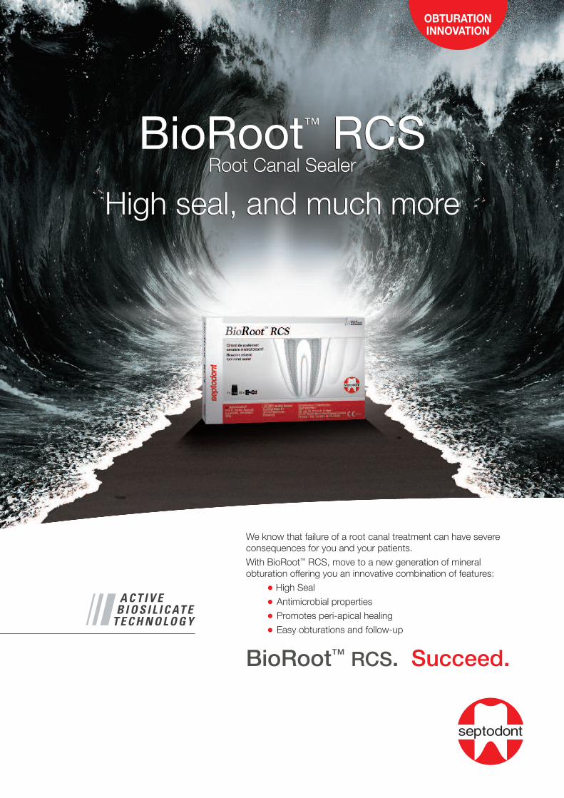
We know that failure of a root canal treatment can have severe consequences for you and your patients.
With BioRoot™ RCS, move to a new generation of mineral obturation offering you an innovative combination of features:
• High Seal
• Antimicrobial properties
• Promotes peri-apical healing
• Easy obturations and follow-up
BioRoot™ RCS. Succeed.
BioRoot™ RCSRoot Canal Sealer
High seal, and much more
BioRoot™ RCSRoot Canal Sealer
High seal, and much more

9
Retrofilling with Biodentine™in apicectomyC.D.E.E Alberto Segovia RamírezDental Surgeon, Universidad Autónoma de Nuevo León, Master in Endodontics,Universidad Autónoma de San Luis Potosí
Introduction: apicoectomy is a complex surgicalprocedure undertaken in order to eliminate theetiology and enucleate the lesion in an affectedtooth; a proper diagnosis is important in thedevelopment of a proper treatment plan and afavorable prognosis. Frank proposes that apicoec-tomy indications should be divided into threegroups. Methods: This case report describesthe surgical treatment of a tooth with a historyof root canal treatments associated with chronicperiapical lesion; the surgical approach taken
was an apicoectomy with retrofilling, and a xeno-graft was implanted. Discussion: The resultswere favorable because the symptoms andapical lesion presented by the patient disap-peared, and the tooth was preserved. The lesionwas sent in for histopathology, which showedapical granuloma ICD/WHO CODE 10: K04.5.Conclusion: pure tricalcium silicate (Biodentine™)is an ideal material for use in Apicoectomy withretrofilling due to its characteristics of biocom-patibility and apical sealing capacity.
Abstract
Periapical surgery is a surgical procedure usedto treat teeth with periapical lesions that cannotbe resolved using conventional endodontic treat-ments (root canal treatment or retreatment). It
must allow the curettage of the pathologicalperiapical tissue, apicoectomy of the affectedroot, and good retrograde sealing of the dentalcanal.(1)
Introduction

10
Indications: We follow the criteria proposed byFrank, grouping indications into 3 categories:1.- surgery due to technical errors2.- surgery due to dental anomalies3.- surgery due to dental pathology (2)
Diagnosis: In most cases, analysis of the dataobtained from anamnesis, physical examination,and radiology allow for an accurate diagnosis.Some cases may resist an accurate diagnosis;diagnosis may thus be presumptive(2)
Pulp vitality tests make it possible to distinguishradicular cysts from other non-endodontic peria-pical pathologies (cementomas, globulomaxillarycysts) where the vitality of the pulp itself ispreserved, while also differentiating the teeththat are affected by the cystic pathology andthose that are not. Radiologically, it is notpossible to establish an absolute and objectivedistinction between a radicular cyst and anapical granuloma.(3)
A lesion that is clinically and radiographicallysuspected to constitute a cyst must undergosurgical treatment. The choice of a specificsurgical method depends heavily on radiological
examination, which defines the extent and rela-tions of the cystic lesion with the neighboringorgans.(3)
During the procedure, between 2 to 3 mmshould be resectioned, and a retrograde cavityperformed with ultrasound and subsequentlyobturated, since statistically this shows greatersuccess than simple apicoectomy(4). It is alsonecessary to choose the right materials for usein the retrofilling procedure(5,6).We have studied the benefits of using puretricalcium silicate (Biodentine™) as comparedwith Mineral Trioxide Aggregate (MTA), as itpresents greater biostimulation(7). Garrido M.presents a study of biocompatibility for puretricalcium silicate, comparing it with MineralTrioxide Aggregate and zinc oxide-eugenol insubcutaneous tissues. They demonstrated thebiocompatibility of pure tricalcium silicate, after14 days with mild to negligible inflammation(8).Furthermore the use of guided tissue regenera-tion, membranes, and grafting in this type ofsurgery facilitates regenerative processes relativeto the alterations caused by the periapicaldisease and its sequelae(9).
Female patient, 25 years of age, presented forendodontic consultation on March 7, 2016. Thepatient reported pain upon chewing in the leftcentral incisor (OD 21), with a history of rootcanal treatment at 8 years old.First appointment: Physical examination: swellingwas observed in the left genian area; uponexploration of the soft tissue inflammation andflushing were observed in the apical area oftooth 21. Upon palpation of the gingiva buccalto the maxilla at tooth 21 a sharp pain was felt,and sharp pain presented as well upon horizontaland vertical percussion; pulp vitality tests wereconducted on the teeth adjacent to tooth 21 toassess the absence of devitalization in theseteeth. Radiographic exploration: Tooth 21 wasobserved, with root canal treatment, withoutapicogenesis. Radiolucent unilocular lesion,
apical third well-defined, diameter of5 mm; tooth presentsa crown-root ratio of1:1 (Fig. 1).Presumptive diag-nosis: Chronic Non-suppurative ApicalPeriodontitis.Second appointment:Occlusion was asses-sed in tooth 21, whichpresented attrition due to bruxism and thepresence of loose high class (class 4) resin inthe mesio-incisal. The treatment plan was deter-mined with a reserved prognosis for tooth 21.Anesthesia was applied using 3% Mepivacaine,and isolation conducted with a rubber dam at
Clinical case
Fig. 1: Initial radiography.

11
tooth 21; the resin was eliminated from tooth 21;the gutta-percha was removed from the cervicalthird of the root; the canal was cleaned withhypochlorite 5%; the canal was then dried. Atype 2 resin-modified ionomer was then placedin the cervical third of the canal, and we proceededto reconstruct the tooth using resin incrementally;the resin was then polished and a periapicaltomography conducted (Fig. 2). Third appoint-ment: Disinfection of teeth and mucousmembranes was performed with an oral antiseptic,and anesthesia performed with mepivacaine3%. A triangular flap was created from tooth 22
to tooth 11 using a #15 scalpel blade (Fig. 3and 4). At the time of flap elevation, the epitheliumwas found to be integrated with the mucosaand submucosa; maintaining tension on the flap,the knife blade was placed parallel to the bonesurface, and the flap was lifted without perforatingthe mucosa; once the trajectory was dissectedwe proceeded with the flap elevation (Fig. 5and 6). Curettage cleaning was performed witha Lucas curette, and while irrigating with salinesolution, the foreign body excision process wasconducted apical to tooth 21 (Fig. 7, 8, 9 and 10).Subsequently, the inflamed tissue was sent in
Fig. 2: Radiography withplacement of Resin-ModifiedGlass Ionomer.
Fig. 5: Granulomatous tissue observed. Fig. 6: Separation of granulomatoustissue from flap.
Fig. 7: Excision of inflamed tissue.
Fig. 8: Excision of inflamed tissue. Fig. 9: Cleaning and excision of inflamedtissue.
Fig. 10: Cleaning and excision of inflamedtissue.
Fig. 3: Triangular flap incision. Fig. 4: Flap elevation.

12
for histopathological examination, and weproceeded to conduct the resection of the apicalthird, removing 3 mm with a 0° tapered diamondbur. (Fig. 11). A retrograde cavity was preparedusing ultrasound, removing 3 mm of gutta-percha apically (Fig. 12 and 13). The area wasthen washed with saline, and the cavity driedwith paper points; and retrograde cavity accesswas inspected to ensure the complete removalof gutta-percha from the canal walls apically to
a depth of 3 mm for obturation with Biodentine™
(Fig. 14 and 15).One capsule of (Biodentine™) was prepared for30 seconds, and was placed in the retrogradecavity using a thin carrier (Fig. 16, 17, 18 and 19).Lyophilized bone was placed in saline in a sterileglass dish for 20 minutes; the osseous defectwas subsequently filled with bone, free of anypathological tissue, apical to tooth 21 (Fig. 20).The collagen membrane was then placed over
Fig. 11: Apical resection with diamond bur. Fig. 12: 3 mm retrograde cavity withultrasound.
Fig. 13: 3 mm retrograde cavity withultrasound.
Fig. 14: 3 mm retrograde cavity observed. Fig. 15: 3 mm retrograde cavity observed. Fig. 16: Preparation of pure tricalciumsilicate (Biodentine™) 30 sec.
Fig. 17: Tricalcium silicate(Biodentine™).
Fig. 18: Carrier for puretricalcium silicate.
Fig. 19: Retrograde filling with(biodentine™).
Fig.20 : Lyophilized xenograftbone in saline solution for20 min.
13
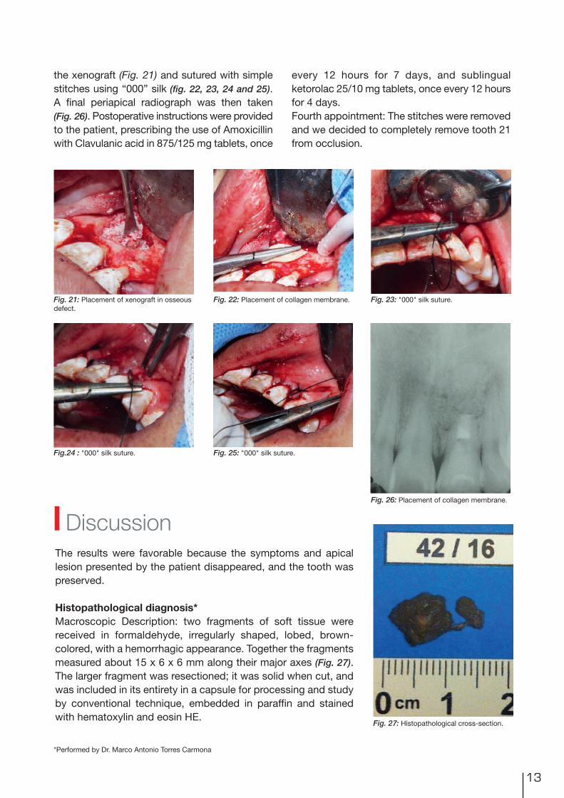
the xenograft (Fig. 21) and sutured with simplestitches using “000” silk (fig. 22, 23, 24 and 25). A final periapical radiograph was then taken(Fig. 26). Postoperative instructions were providedto the patient, prescribing the use of Amoxicillinwith Clavulanic acid in 875/125 mg tablets, once
every 12 hours for 7 days, and sublingualketorolac 25/10 mg tablets, once every 12 hoursfor 4 days.Fourth appointment: The stitches were removedand we decided to completely remove tooth 21from occlusion.
Fig. 21: Placement of xenograft in osseousdefect.
Fig. 22: Placement of collagen membrane. Fig. 23: "000" silk suture.
Fig.24 : "000" silk suture. Fig. 25: "000" silk suture.
Fig. 26: Placement of collagen membrane.
The results were favorable because the symptoms and apicallesion presented by the patient disappeared, and the tooth waspreserved.
Histopathological diagnosis*Macroscopic Description: two fragments of soft tissue werereceived in formaldehyde, irregularly shaped, lobed, brown-colored, with a hemorrhagic appearance. Together the fragmentsmeasured about 15 x 6 x 6 mm along their major axes (Fig. 27).The larger fragment was resectioned; it was solid when cut, andwas included in its entirety in a capsule for processing and studyby conventional technique, embedded in paraffin and stainedwith hematoxylin and eosin HE.
Discussion
*Performed by Dr. Marco Antonio Torres Carmona
Fig. 27: Histopathological cross-section.

Microscopic description: nine cuts were analyzed. Thespecimens studied were formed entirely of connective tissuepresenting edema and diffuse infiltration by mononuclearleukocytes and epithelioid histiocytes. Towards the interior,countless cholesteric fissures were observed. A deposit wasobserved containing a granular foreign body of diffuse orintracytoplasmic distribution in histiocytes (Fig. 28), originalmagnification X100. / (Fig. 29) original magnification X400).
No malignancy was observed in the cross-sectionsstudied.Diagnosis: apical granuloma. icd/who code 10: k04.5.
14
Pure tricalcium silicate (biodentine™) meets thecore characteristics for a retrofilling material,such as possessing a high degree of biocom-patibility, not causing cytotoxicity or inflammation,and antibacterial activity as provided by a highlyalkaline ph of 12.Its other properties include biostimulation ofthe mesenchymal cells in the supporting perio-dontal tissues, which promote the regenerationof the periapical environment, not containingother materials like mta.As regards mechanical characteristics, puretricalcium silicate (biodentine™) is far superiorto the other materials used for these treatments,
as it has a high degree of mechanical resistance(300 mpa at 30 days) thus preventing materialrupture and bacterial infiltration.Another important feature is the ease of handlingof the powder and liquid, providing the clinicianwith enough time to place and adapt the material,which leads to a reduction in surgical operatingtime favorable to both the patient and surgeon.We can thus say that pure tricalcium silicate(biodentine™) is an ideal material for surgicalendodontic operations.
Conclusion
Control at 6 monthsRX control at 6 months
Fig. 28: Original magnification X 100.
Fig. 29: Original magnification X 400.
RX control at 6 months

15
Author:C.D.E.E Alberto Segovia Ramírez1996-1998 Master’s in Endodontics from Faculty of Dentistry of the U.A.S.L.P.San Luis Potosí, S.L.P.Mexico.1988-1993 Dental surgeon from Faculty of Dentistry of the U.A.N.L. Monterrey,N.L. México.Certified as a specialist endodontist by the Mexican Council of Endodontics(no. 369) in May 2004, May 2009 and May 2014 in Mexico City D.F.
Professional and teaching experienceNational Endodontics Research Prize from the Mexican Endodontics Association, 1999.February-December 1997: Professor of Endodontics and Clinical Adviser in the undergraduateteaching staff at the Faculty of Dentistry of the U.A.S.L.P. September 1998 - May 2003: Postgraduate Professor of Endodontics in the Dentistry StudiesCenter of Querétaro.January 2001 to date: Professor of Endodontics at the Universidad del Valle de México(Querétaro campus).Guest professor for postgraduate Endodontics at the Universidad San Carlos de Guatemala(Faculty of Dentistry) from 2003 to 2009. President of the Scientific Committee of the Dentistry Association of Querétaro (2003-2005 and2005-2007).Private practice in Endodontics, from 1995 to date, in Monterrey, San Luis Potosí and NuevoLaredo, and since 1998 to date in Querétaro.
References1. Peñarocha M, Peñarocha M. Atlas de cirugía periapical. . 1a ed. Barcelona España: editorial océano; 2014.
2. Canalda C, Brau E. Endodoncia Técnicas clínicas y bases científicas. 3ª ed. Barcelona España: ElsevierMasson; 2014.
3. García A, Bujaldón A, Rodríguez A. Lesiones periapicales. Diagnóstico y tratamiento [periapical lesions.diagnosis and treatment] [internet] Universidad Complutense de Madrid Medicina Bucal. Departamento deEstomatología. Universidad de Granada AVANCES EN ODONTOESTOMATOLOGÍA, Vol. 31, 31-42. 2015.http://scielo.isciii.es/scielo.php?script=sci_arttext&pid=S0213-12852015000100005
4. Gómez V, Giner J, Maniegas L, Gaite J, Castro A, Ruiz J, “et all”. Apicectomía quirúrgica: propuesta de unprotocolo basado en la evidencia [Surgical apicoectomy: proposed evidence-based protocol.] [internet]Revista Española de Cirugía Oral y Maxilofacial, vol.33 no.2 Madrid, 2 pág. (Abr.-jun. 2011).http://scielo.isciii.es/scielo.php?pid=S1130-05582011000200002&script=sci_arttext&tlng=e
5. Borisova T, Panov V, Peev S, Papanchev G. ROOT END FILLING MATERIALS REVIEW. Scientifica MedicinaeDentalis, Medical University of Varna. (2013). vol. 1, �1, 13-17.
6. Priyanka S. A Literature Review of Root-End Filling Materials IOSR Journal of Dental and Medical Sciences(IOSR-JDMS) e-ISSN: 2279-0853, p-ISSN: 2279-0861. Volume 9, Issue 4 (Sep.- Oct. 2013), PP 20-25www.iosrjournals.org
7. Correa M, Castrillón N. Comparación de microfiltración apicocoronal entre MTA y Biodentine™ en dientesunirradiculares. [Comparison of microfiltration apicocoronal between MTA and Biodentine™ in singlerootedteeth]. [Internet] Universidad San Francisco Quito; Publicado: 2015/03/01. http://www.usfq.edu.ec/publicaciones/odontoinvestigacion/Documents/odontoinvestigacion_n001/oi_001_001.pdf
8. Garrido G, Moraes L, Louzada D, Menegucci L. and Rodrígues S. BIOCOMPATIBILITY EVALUATON OFBIODENTINE IN SUBCUTANEOUS TISSUE OF RATS. JOE. VOLUMEN 40, NUMBER 9, SEPTEMBRE 2014.
9. Calzada-Bandomo A, Calzada-Bandomo A, Mora-Pérez C. Terapia periodontal regenerativa: antecedentesy perspectivas. Medisur [revista en Internet]. 2013 [citado 2016 Jun 23]; 11(5):[aprox. 8 p.]. Disponible en:http://medisur.sld.cu/index.php/medisur/article/view/2360.

16
Clinical case report of the useof Biodentine™ for deep cariestreatment Marianne Federlin, Birger Thonemann, Kerstin Galler, Wolfgang Buchalla, Regensburg
Biodentine™ is a bioactive calcium silicate cementand is suitable as a dentin replacement materialfor a wide range of indications in dental therapy,both for restorative measures in the region ofthe tooth crown and for endodontic treatments.The indications for restorative measures include,in addition to treatment of exposed dentin(underfilling; deep caries treatment for extensivecaries lesions), purely provisional treatment inthe enamel and dentin regions as well as directand indirect capping.1 In the following casereport, Biodentine™ was used for deep cariestreatment and as an underfilling material as partof direct treatment of a class II cavity with acomposite filling.
A 72-year-old patient presented to the OutpatientClinic for Tooth Preservation and Periodontologyof the University of Regensburg in October
2013 for a routine checkup. When the findingswere recorded, radiographs were taken in theregion of the second quadrant becauseapproximal caries was suspected. In the regionof tooth 26 distally, “deep caries” was diagnosed(Fig. 1), and an appointment for filling therapywas therefore scheduled.
Biodentine™ for deep caries treatment/underfilling beneath a directcomposite restoration for deep caries of tooth 26
Fig. 1: Radiograph: Approximal caries tooth 26 distally.

17
Sensitivity testing of tooth 26 to cold with dryice was positive. Treatment was performedwithout local anesthesia at the patient’s request.Before dissection, a rubber dam was placedonto teeth 24 to 27. From the occlusal aspect,access to the defect was first created using arotary diamond bur with water cooling. Thedistal cavity region was dissected using oscillatinginstruments (SonicFlex, KaVo, Biberach) (Fig. 2).The cavity was created as delicately as possible.Caries excavation was then performed usinground burs with visual control using a surgicalmicroscope. The cavity was checked for residualcaries multiple times during the course of theexcavation by means of FACE (fluorescence-aided caries excavation, SIROinspect, Sirona,Bensheim).2
During complete caries removal, the pulp wasnot exposed (Fig. 3). A metal matrix (HaweTof-flemire matrix, Kerr Dental, Rastatt) was theninserted and wedged. The cavity was cleanedand dried.Biodentine™ was mixed according to the manu-facturer’s instructions and applied in the regionof the distal pulp wall using a small ball plugger(Dycal-Instrument) and adapted using a Heide-mann spatula. Biodentine™ was thussimultaneously used for deep caries treatmentand for underfilling. After a 12-minute bindingtime for Biodentine™, the defect was restoredusing a composite filling (Fig. 4).For filling therapy, Etch & Rinse adhesive (ExciTE,Ivoclar Vivadent, Schaan. Liechtenstein) wasused in combination with a fine-particle hybridcomposite (Tetric EvoCeram, Ivoclar Vivadent).After etching of the enamel with phosphoric
acid (Total Etch, Ivoclar Vivadent) (Fig. 5) andapplication of the adhesive (ExciTE DC, IvoclarVivadent) (Fig. 6), the first layer, consisting of anopaque, flowable composite, was inserted intothe cavity (Venus Flow Baseliner, Heraeus Kulzer,Hanau). The cavity lining with a flowable compo-site facilitates good primary adaptation and
Fig. 2: Visualization of the defect.
Fig. 3: Status post caries extraction, control by means of FACE.
Fig. 4: Biodentine™ inserted as underfillingand for deep caries therapy.
Fig. 5: Enamel etching with phosphoric acid. Fig. 6: Application of the adhesive system.

18
Fig. 7: Insertion of an opaque flowable composite (lining technique).
Fig. 9: Radiograph 7 months after deep caries therapy: White arrow:dimensions of Biodentine™.
Fig. 8: Composite filling.
Fig. 10: Composite filling 7 months after deep caries therapy.
stabilizes the bonding strength to dentin duringthe onset of the polymerization shrinkage of thecomposite that was placed next.3 In addition,the opaque liner facilitates demarcation of thetransition to the natural hard tooth structures(Fig. 7). The restoration was then constructedusing an incremental technique. After removalof the rubber dam, occlusion and articulationwere checked, and finishing and polishing (Fig. 8)were done using a fine-grit diamond bur.
In April 2014 (7 months after the deep cariestreatment), the patient presented again for adental examination. Tooth 26 still exhibited apositive reaction to sensitivity testing with dryice. The patient did not report any postoperativehypersensitivity after treatment. Radiographsshowed close adaptation of Biodentine™ todentin; because of the similar radio-opacity ofdentin and Biodentine™, it is not easy to distinguishbetween the two layers (Fig. 9 and 10). On thebasis of the radiographs, there was no evidenceof reactive tertiary dentin formation, as has also
been reported in the literature for Biodentine™.1
In this case, Biodentine™ was used as underfillingin terms of dentin replacement and for deepcaries treatment. When the residual dentinstrength is greatly reduced after caries excavation,microperforations to the pulp can be present.This can lead to direct contact of the componentsof the methacryl-based adhesive system withthe pulp tissue, resulting – in the worst-casescenario – in irreversible damage to the pulp.4
Therefore, in this case, Biodentine™ was usedas an underfilling material because of its biologicalactivity, on one hand, and for its dentin-likemechanical properties, on the other hand. Clini-cally, it was shown that the calcium-silicatecement, compared with other materials for deepcaries treatment, can be applied without anypressure because of its consistency after mixingand can be adapted closely to the dentin andforms a mechanically stable barrier after binding.This was also evident in the radiographs takenafter 7 months. A two-stage procedure was notindicated in this case.

19
Author:Priv.-Doz. Dr. med. dent. Marianne Federlin1980-1985 Studies in dentistry in Gießen, 1986-1988 Resident in independentpractice, since 1988 scientific staff member at the Outpatient Clinic for ToothPreservation and Periodontology, Hospital of the University of Regensburg(Director: Prof. Dr. W. Buchalla), 1992 Graduation (Edge quality of Cerecrestorations in vitro), since 1993 Senior Physician, 2009 Postdoctoral thesis(Micromorphology of the binding zone and adhesive bond to root canal dentin);Scientific focus: ceramics – adhesive systems – adhesive bond to root canaldentin.
Address for correspondence:University Hospital of RegensburgCenter for Dental and Oral MedicineOutpatient Clinic for Tooth Preservation and PeriodontologyPD Dr. Marianne FederlinFranz-Josef-Strauß-Allee 1193053 RegensburgTelephone 09 41/9 44-60 20E-mail [email protected]
References• Septodont R&D Department: Biodentine™ - Active Biosilicate Technology: ScientificFile.http://www.septodont.fr/fichiers_upload/biodentinescientificfile.pdf.
• Buchalla W, Lennon AM. Die fluoreszenzunterstütze Kariesexkavation - Funktionsprinzip und Empfehlungenfür den Gebrauch. Oralprophylaxe und Kinderzahnheilkunde, 2013; 35 (4): 162–167.
• Haller B. Direkte Seitenzahnrestauration mit Komposit. Zahnmedizin up2date 6, 2009: 579–598.
• Schmalz G. Kompositkunststoffe: Lokale Toxizität und Gewebeverträglichkeit - Pulpareaktionen. In:Biokompatibilität zahnärztlicher Werkstoffe Hrsg.: G. Schmalz, D. Arenholt-Bindslev, Elsevier GmbH, Urban& Fischer Verlag, 2005 München, S. 111–115.

20
Perforation repair withBiodentine™Dr. Jaime Abarca RevecoMSc DDS and Endodontics Specialist
A perforation is an artificial communicationbetween the root canal system and the toothsupport tissue. This communication may occurdue to dental hard tissue reabsorption, cariesor by iatrogenic endodontic therapy.1 Pulpchamber floor perforation is a iatrogenic compli-cation that can be produced during accesspreparation or in the instrumentation of thecervical third of the root canal, causing endo-dontic failure.2 It can also cause an inflammatoryresponse of the periodontal tissue that maylead to an irreversible loss of periodontal insertionin the affected area.3 The reparation procedurewill be determined by the perforation positionand the area involved. Treatment can be with orwithout surgery and the prognosis is usuallyexcellent if the problem is correctly diagnosedand repaired, using a material that gives propersealing and is biocompatible.2
Various materials have been used for root repair,
including silver amalgam, zinc oxide–eugenol,calcium hydroxide, composites, and glass iono-mers. However, none of them are ideal for thespecial conditions and requirements of this typeof treatment.4 An ideal material should bebiocompatible, dimensionally stable, insoluble,radiopaque, allow easy manipulation and place-ment and provide a proper seal.5
Biodentine™ (Septodont, Saint Maur des Fosses,France) is a high-purity calcium silicate–basedmaterial composed of tricalcium silicate, calciumcarbonate, zirconium oxide, and a water-basedliquid containing calcium chloride as the settingaccelerator and water-reducing agent.Biodentine™ is recommended as a dentin subs-titute under resin composite restorations andas an endodontic repair material because of itsgood sealing ability, high resistance to compres-sive strength, short setting time, biocompatibility,bioactivity, and biomineralization properties.6

21
Case ReportA male patient of 48 years of age was referredbecause of failure to locate the buccal canalsof the 2.6 tooth. (Fig. 1).
Clinical Examination: A tooth with endodonticaccess and temporary filling is observed. It isunresponsive to thermal testing, and presentspain to both vertical and horizontal percussionand also to palpation in the palatal area next tothe gingival margin.
Diagnosis: Apical Symptomatic Periodontitiswith Previously Initiated Therapy.
Treatment: On the first appointment, the tempo-rary filling is removed and a distopalatalperforation in the cameral floor becomes visible.(Fig. 2 and 3) The periodontal hemorrhagingunder the perforation was stabilized usingCalcium Hydroxide mixed with Propylene Glycol. Immediately after, the Palatal Canal is located(Fig. 4) and previous to the localization of thebuccal canals, the perforation is blocked with agingival barrier. (Fig. 5) Once the buccal canalsare located, all canals are prepared with recipro-cating rotary files. The perforation is sealed usingBiodentine™ (Fig. 6) and an intracanal dressing ofCalcium Hydroxide is left in place for 15 days.
Fig. 1: Preoperative Radiograph. Fig. 2: DP perforation. Fig. 3: DP perforation.
Fig. 4: Periodontal tissue stabilization and Palatal canal localization.
Fig. 7: Biodentine™ seal appearance on the second appointment
Fig. 5: Perforation blockage using gingivalbarrier.
Fig. 6: Biodentine™ placement.

22
References01 Lodiene G, Kleivmyr M, Bruzell E, Orstavik D. Sealing ability of mineral trioxide aggregate, glass ionomercement and composite resin when repairing large furcal perforations. British dental journal. 2011;210:E7.Epub 2011/03/12.
02 da Silva EJ, Andrade CV, Tay LY, Herrera DR. Furcal-perforation repair with mineral trioxide aggregate: Twoyears follow-up. Indian journal of dental research : official publication of Indian Society for Dental Research.2012;23:542-5. Epub 2012/12/22.
03 Samiee M, Eghbal MJ, Parirokh M, Abbas FM, Asgary S. Repair of furcal perforation using a newendodontic cement. Clinical oral investigations. 2010;14:653-8. Epub 2009/11/06.
04 Zhou HM, Shen Y, Wang ZJ, Li L, Zheng YF, Hakkinen L, Haapasalo M. In vitro cytotoxicity evaluation of anovel root repair material. Journal of endodontics. 2013;39:478-83. Epub 2013/03/26.
05 Aggarwal V, Singla M, Miglani S, Kohli S. Comparative evaluation of push-out bond strength of ProRootMTA, Biodentine, and MTA Plus in furcation perforation repair. Journal of conservative dentistry : JCD.2013;16:462-5. Epub 2013/10/02.
06 Guneser MB, Akbulut MB, Eldeniz AU. Effect of various endodontic irrigants on the push-out bond strengthof biodentine and conventional root perforation repair materials. Journal of endodontics. 2013;39:380-4.Epub 2013/02/14.
Author:Dr. Jaime Abarca RevecoMSc DDS and Endodontics SpecialistCoordinating Teacher of the Endodontics Graduate Program in San SebastianUniversityTeacher in the Rehabilitation Department of the Undergraduate Program at SanSebastian University Active Member of the Chilean Endodontics SocietyMember of the AAE
Biodentine™ proved to be a clinically reliablematerial in the reparation of pulp chamber floorperforations. It is biocompatible, like the wellstudied MTA, but with improved mechanical resis-
tance, making it an ideal material to endureocclusal forces in cases with large losses of hardtissue that occur during the search of a root canal.
Conclusion
To the second appointment the patient arriveswith a distopalatal cusp fracture. After toothisolation and temporary filling removal, theBiodentine™ was observed to be hard andstable. (Fig. 7) A final irrigation protocol wasperformed with 5% Sodium Hypochlorite and17% EDTA using sonic passive activation andthe canals were obturated using a heat carrier.(Fig. 8) A double-seal temporary filling of CalciumSulfate and Glass Ionomer is placed and thepatient is referred back to his practitioner fordefinitive restoration.
Fig. 8: Postoperative Radiograph.

The � rst and only dentin in a capsule
As the � rst all-in-one biocompatible and bioactive dentin substitute, Biodentine™ fully replaces dentin wherever it’s damaged.
Biodentine™ helps the remineralization of dentin, preserves the pulp vitality and promotes pulp healing. It replaces dentin with similar biological and mechanical properties.
Improving on Biodentine™ clinical implementation, you can now bond the composite onto Biodentine™ in the same visit and perform the full restoration in a single session.
To enjoy the clinical bene� ts of the � rst and only dentin in a capsule, ask your dental distributor for Biodentine™.
Learn more withthe Biodentine™ brochure
