c-reactive protein, cea and il-6 in occult common
TRANSCRIPT
Page 1/13
Diagnostic Value of Endoscopic RetrogradeCholangiopancreatography Combined with SerumC-Reactive Protein, CEA and IL-6 in Occult CommonBile Duct StonesShiJun Lu
funan county hospitalXueLi Liu ( [email protected] )
funan county hospital https://orcid.org/0000-0002-7573-2447WeiGuo Wu
funan county hospital
Research Article
Keywords: Endoscopic retrograde cholangiopancreatography, Serum, Occult Common bile duct stones,Diagnostic value
Posted Date: November 8th, 2021
DOI: https://doi.org/10.21203/rs.3.rs-1019584/v1
License: This work is licensed under a Creative Commons Attribution 4.0 International License. Read Full License
Page 2/13
AbstractObjective: To investigate the diagnostic value of ERCP combined with CRP, CEA and IL-6 in serum inOccult Common bile duct stones (CBDS).
Methods: 80 cases of patients with occult CBDS treated in our hospital were selected as the researchsubjects, and the diagnosis rates of ERCP alone or ERCP combined with CRP, CEA and IL-6 in serum werecompared.
Results: Versus ERCP alone, the diagnosis rate of combined diagnosis was clearly higher (P < 0.05).Versus patients without CBDS, the levels of CRP, CEA and IL-6 in serum in CBDS patients were apparentlyelevated (P < 0.05). The levels of CRP, CEA and IL-6 in serum were of high value in the diagnosis of CBDSin patients with occult CBDS (P < 0.05).
Conclusion: ERCP combined with CRP, CEA and IL-6 in serum is of high value in re-diagnosis of OccultCBDS.
BackgroundCommon bile duct stones (CBDs) are calculi located in the common duct, which can be divided intoprimary and secondary types in line with their sources [1]. There are severe upper abdominal pain, chills,fever, jaundice and other symptoms during the attack of CBDS in patients,while no symptoms exist inabout one-third of patients with CBDS, which is clinically called Occult CBDS [2, 3]. Similar with CBDS,occult CBDS can also lead to severe infectious diseases in patients, such as cholangitis, pancreatitis orchronic liver disease, and even result in patient death [4]. Therefore, it is crucial of timely treatment ofpatients with occult CBDS, but there are not any symptoms in patients, which is di�cult to attractattention from them, and also strengthens the di�culty of clinical diagnosis and treatment, leading tolimited prognosis of patients [5]. At present, the main methods for clinical diagnosis of patients withCBDS include laboratory tests such as white blood cell, neutrophil count and liver function, ultrasoundexamination, magnetic resonance cholangiopancreatography (MRCP), endoscopic ultrasonography(EUS), Endoscopic retrograde cholangiopancreatography (ERCP) and enhanced computed tomography(CT) or Magnetic Resonance Imaging (MRI), among which duodenoscopy is applied by ERCP to injectcontrast agent into the common bile duct for imaging, which exerts a high value in the diagnosis ofoccult CBDS, but there are still misdiagnosis and missed diagnosis [6, 7]. In�ammation is one of the vitalfactors causing CBDS or occult CBDS lesions, and a crucial manifestation of its lesions. C-reactiveprotein (CRP), carcinoembryonic antigen (CEA), and interleukin-6 (IL-6) are all major in�ammatorymarkers in the body, and their expression can effectively re�ect the level of in�ammatory activity in localor systemic parts [8–10]. Therefore, ERCP test with CRP, CEA and IL-6 in serum was combined in this studyto explore its diagnostic value in occult CBDS, aiming to �nd more reliable obstruction of occult CBDStest. The report was as follows.
Page 3/13
Materials And Methods1.1 General information
From January 2019 to February 2021, a total of 80 patients with occult CBDS treated in our hospital wereselected as the research subjects. This study has been approved in advance by the children and theirfamilies, and informed consent has been signed.
1.2 Inclusion and exclusion criteria
Inclusion criteria: (1) Patients had preoperative high-risk factors for occulting CBDS such as a history ofobstructive jaundices, or biliary pancreatitis in 14 cases, preoperative B-ultrasound CBD diameter ≥ 10mm in 10 cases, preoperative biochemical indexes alkaline phosphatase (AKP) or gamma-glutamyltransferase (r-GT) increased by more than 50% with multiple small calculi in the gallbladder [11,12]; (2)Patients with complete clinical data; (3) Patients with high cooperation.
Exclusion criteria :(1) patients with a de�nite preoperative diagnosis of CBDS; (2) Patients suffering frommental illness or poor mental condition; (3) Patients with severe abnormal heart, liver and kidneyfunctions; (4) Pregnant women.
1.3 methods
(1) ERCP checking method: Duodenoscopy (ED-450XT5, FUJINON) and digital gastrointestinalangiography machine (500 mA, Shimazu) were applied. The patients were intravenously injected with 5-10 mg diazepam (H12020957, KingYork, Tianjin, China) + 40 mg phloroglozol injection (Hengshengcompany, Nanjing, China, H20046766) 10-15 min before operation. Fifty mL ultravist 300 (Bayer AG,Berlin, Germany, H20171332) was set as contrast agent, and routine X-ray was performed. Routinefasting was urged for patients 12 ~ 24 h after ERCP, and color Doppler ultrasound (Voluson 730) wasapplied for ultrasound examination in patients. MRCP examination was performed by (3. 0T) nuclearmagnetic resonance (NMR) instrument, and if necessary, CT examination was constructed by 16-slicespiral CT (Siemens Ltd, Erlangen, Germany) [13,14]. (2) Serum indexes detection method: Serum indexes:Fastening venous blood 5 mL from the patients was extracted in the morning and stood for 30 min. Afterthe whole blood was naturally coagulated and the serum was precipitated, the supernatant was obtainedby centrifugation at about 1000-2000 g at 4℃ for 10 min. The expression of CEA, CRP and IL-6 in theserum was detected by Enzyme-linked immunosorbent assay (ELISA) method. The blank well, standardwell and control wells were set. The standard solution was diluted to protein content of 5 μg/ mL bycoating with carbonate buffer at 0.05 M pH9. The reaction well of each polystyrene plate was added with0.1 mL overnight at 4℃. The next day, the solution in the well was discarded and washed with washingbuffer for 3 times, 3 minutes each time (washing for short, the same below). The above coated reactionwell was joined with certainly diluted 0.1 mL samples to be tested, incubated at 37℃ for 1 h and washed(additionally blank wells and contrast wells were made). Each reaction well was added with 0.1 mLfreshly diluted enzyme-labelled antibody, incubated at 37℃ for 1 h, washed, then joined with 0.1 mL
Page 4/13
Tetramethylbenzidine (TMB) substrate solution temporarily prepared at 37℃ for 30 min, and 0.05 mL 2M sulfuric acid. On the ELISA detector, the optical density (OD) value of each well was measured afterzero setting with blank control well at 450 nm, and the concentration was calculated. The reagents andkits used were purchased from MSK (Wuhan, China) and were operated in strict accordance with the kitinstructions. All ELISA detection reagents were purchased from MSK [15,16].
1.4 Observation Indexes
(1) Diagnosis rate: the diagnosis of occult CBDS patients was compared between ERCP alone and ERCPcombined with CRP, CEA and IL-6 in serum; (2) The levels of CRP, CEA and IL-6 in serum: comparison ofCRP, CEA and IL-6 levels in serum in patients with CBDS or without; (3) Diagnostic value: the sensitivityand speci�city of CRP, CEA and IL-6 levels in serum in the diagnosis of CBDS was analyzed via ROC curvein occult CBDS patients.
1.5 Statistical analysis
In this study, SPSS22.0 software package was applied for statistical analysis of the collected data, andGraphPad 7 software package for drawing the required pictures. The measurement data were expressedas mean ± standard deviation (x̄ ± s), and t test was performed. The enumeration data were expressed ascase number (n) or percentage (%) by χ2 test. P < 0.05 indicated a statistical difference
Results2.1 Diagnostic results
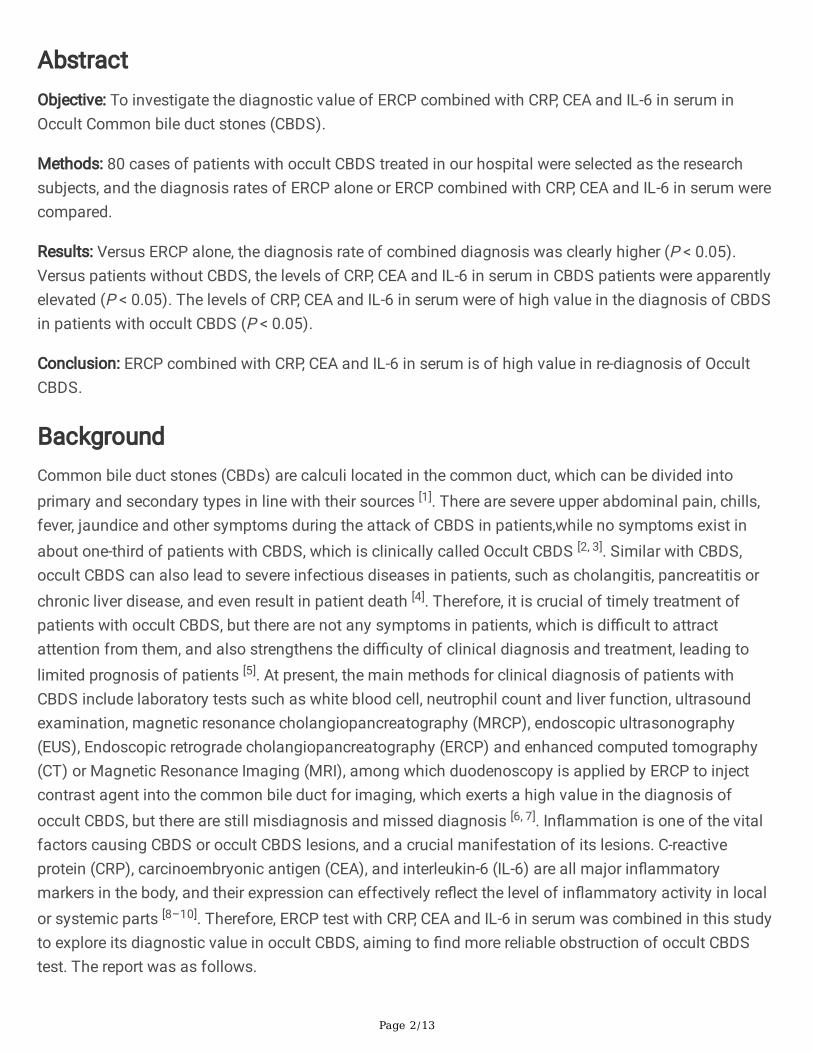
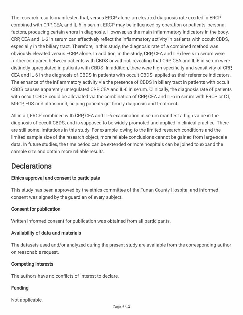
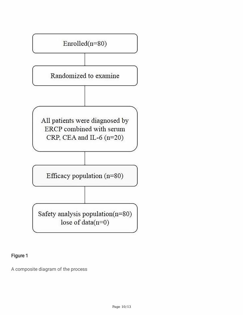
A total of 67 patients with CBDS were included in 80 patients with occulted CBDS, and 13 patientswithout CBDS after operation. Fifty-eight patients with CBDS and 22 patients without CBDS werediagnosed via ERCP alone, with a diagnosis rate of 88.75%. Fifty-seven patients with CBDS and 14without CBDS were diagnosed via ERCP combined with CRP, CEA and IL-6 in serum, with a diagnosis rateof 98.75%. The diagnosis rate of a combined way was clearly elevated versus ERCP alone (P < 0.05), asshown in Figure 1, 2.
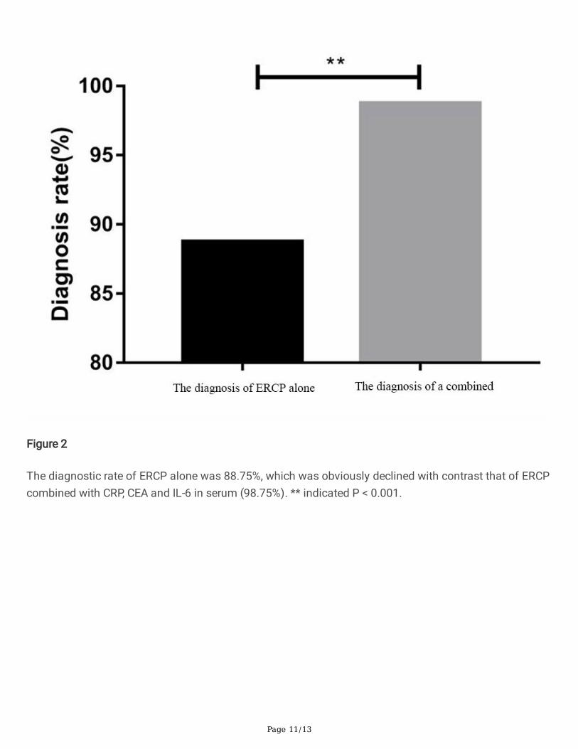
2.2 Elevated CRP, CEA and IL-6 exist in serum of CBDS patients
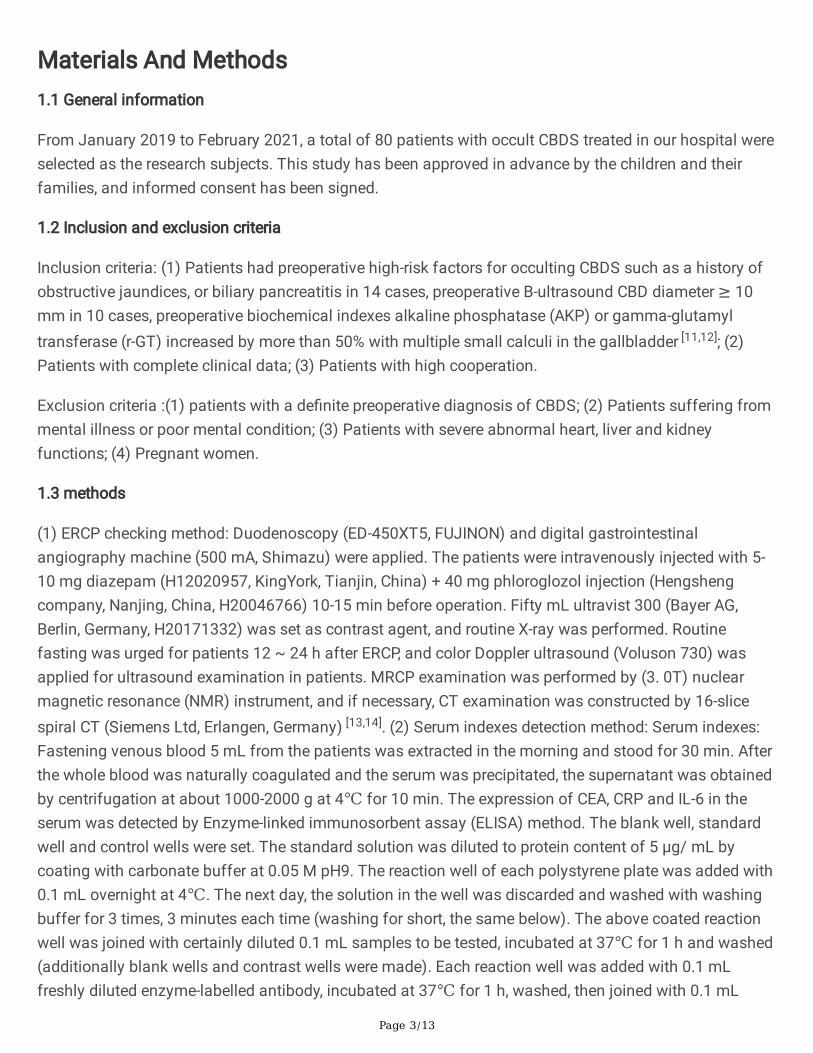
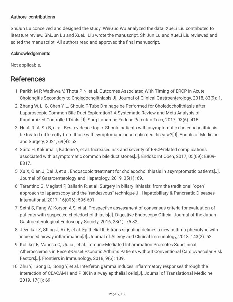
Versus patients without CBDS, CRP, CEA and IL-6 in serum in CBDS patients were apparently up-regulated(P < 0.05) (Table 1, Figure 3). CRP, CEA and IL-6 in serum might be involved in the pathological process ofCBDS.
2.3 ROC curve analysis
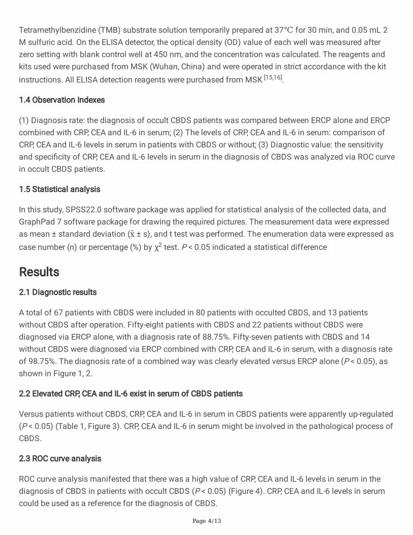
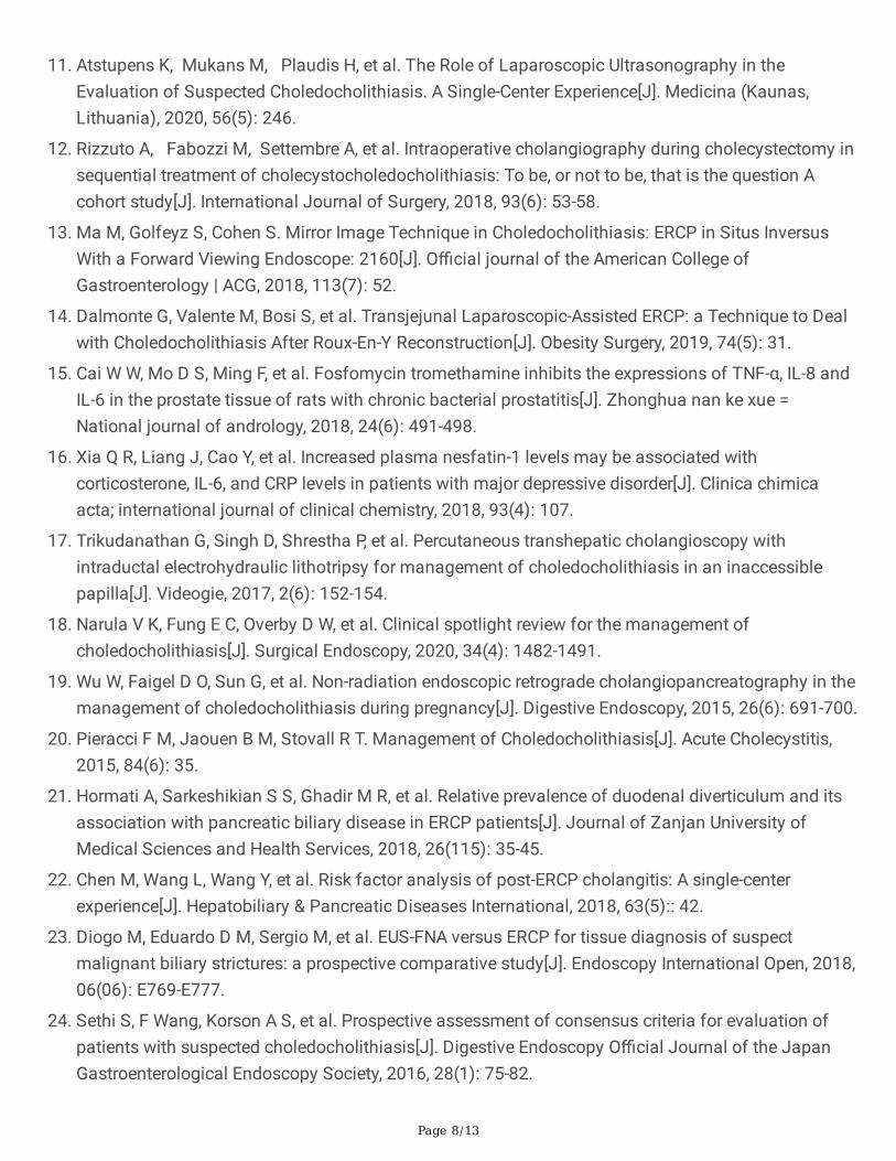
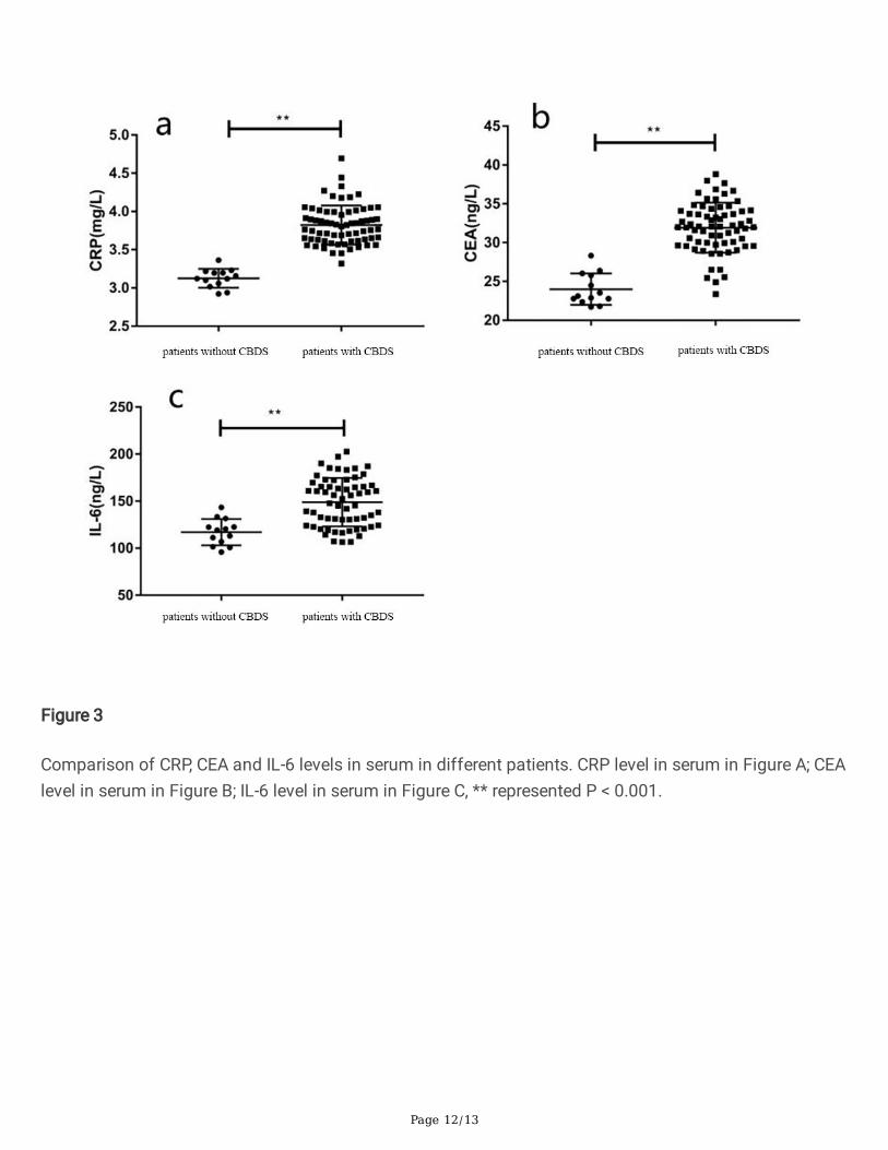
ROC curve analysis manifested that there was a high value of CRP, CEA and IL-6 levels in serum in thediagnosis of CBDS in patients with occult CBDS (P < 0.05) (Figure 4). CRP, CEA and IL-6 levels in serumcould be used as a reference for the diagnosis of CBDS.
Page 5/13
3. Results
CBDS is one of the most general clinical diseases of the digestive system and is also one of the mostprincipal gallstone diseases. It is statistical that CBDS accounts for more than 40% of gallstonediseases [17,18]. There are the triad of abdominal pain, chills, high fever and jaundice in most CBDSpatients, but no symptoms are taken place in 30% ~ 35% CBDS patients, clinically calling occult CBDS,thus increasing the di�culty of diagnosis and treatment of patients [19]. It is manifested in a clinical studythat regardless of symptoms, patients with CBDS should take timely diagnosis and treatment forprevention of further deterioration of the disease and occurrence of complications such as biliary tractinfection, which may endanger the health and even lives of patients [20]. Nowadays, clinically preoperativehematologic examination with ultrasound, MRCP, EUS, CT and other non-invasive examinations aremainly employed for occult CBDS, and the results of surgical examination are taken as the gold standard.Low sensitivity and speci�city are manifested in preoperative examination, which reveals certaindifferences with the results of postoperative gold standard examination.
The contrast agent is injected into the common bile duct through dodecoscopy in ERCP technology, andX-ray is applied for photography to �nally reveal the pancreatic bile duct, and the technology has beenwidely applied in the diagnosis of a variety of pancreatic bile duct diseases and minimally invasivesurgical treatment due to its characteristics such as less trauma, time period and fewer complications,manifesting a high clinical application value [21-23]. Sethi Set al. [24] found that ERCP diagnostictechnology is applied to the diagnosis of suspected choledocholithiasis, achieving a good diagnosticeffect, helping to de�ne the criteria of low, medium and high-risk choledocholithiasis. Whereas the studyof Borgosz J et al. [25] suggested that ERCP technology in the diagnosis of CBDS may be affected by bileduct deposits, resulting in misdiagnosis. However, few clinical studies have discussed the diagnosticvalue of ERCP technology in occult CBDS, therefore, ERCP checking with CRP, CEA and IL-6 test in serumwas combined in this study to observe its diagnostic value in patients with occult CBDS, thus �nding amore reliable diagnostic method for patients with occult CBDS.
In�ammatory activity is one of the crucial reactions involved in occult CBDS lesions, and clinicaldiagnosis and evaluation of occult CBDS and its prognostic effect can be conducted by detectingin�ammatory factors [26] . CRP in serum is an acute protein synthesized by the body liver, and its level isobviously increased when the body is infected or injured, effectively re�ecting the in�ammatory activitystate in the body. Although CEA in serum is often applied in the early diagnosis and assessment of colonand rectal cancer and other cancers, its pathological up-regulation can also be seen in the in�ammatorydiseases of hepatobiliary pancreas such as colitis, pancreatitis, cirrhosis and hepatitis, effectivelysuggesting the level of in�ammatory activity in patients with such diseases. Usually secreted by�broblasts and T cells, IL-6 is a multifunctional in�ammatory factor affecting the growth of all kinds ofcells in the body, and can produce a large number of in�ammatory cells in the active state to acceleratethe process of in�ammatory response [27-30]. In this study, CRP, CEA and IL-6 detection in serum werecombined with ERCP to observe their combined value in the diagnosis of occult CBDS.
Page 6/13
The research results manifested that, versus ERCP alone, an elevated diagnosis rate exerted in ERCPcombined with CRP, CEA, and IL-6 in serum. ERCP may be in�uenced by operation or patients' personalfactors, producing certain errors in diagnosis. However, as the main in�ammatory indicators in the body,CRP, CEA and IL-6 in serum can effectively re�ect the in�ammatory activity in patients with occult CBDS,especially in the biliary tract. Therefore, in this study, the diagnosis rate of a combined method wasobviously elevated versus ECRP alone. In addition, in the study, CRP, CEA and IL-6 levels in serum werefurther compared between patients with CBDS or without, revealing that CRP, CEA and IL-6 in serum weredistinctly upregulated in patients with CBDS. In addition, there were high speci�city and sensitivity of CRP,CEA and IL-6 in the diagnosis of CBDS in patients with occult CBDS, applied as their reference indicators.The enhance of the in�ammatory activity via the presence of CBDS in biliary tract in patients with occultCBDS causes apparently unregulated CRP, CEA and IL-6 in serum. Clinically, the diagnosis rate of patientswith occult CBDS could be alleviated via the combination of CRP, CEA and IL-6 in serum with ERCP or CT,MRCP, EUS and ultrasound, helping patients get timely diagnosis and treatment.
All in all, ERCP combined with CRP, CEA and IL-6 examination in serum manifest a high value in thediagnosis of occult CBDS, and is supposed to be widely promoted and applied in clinical practice. Thereare still some limitations in this study. For example, owing to the limited research conditions and thelimited sample size of the research object, more reliable conclusions cannot be gained from large-scaledata. In future studies, the time period can be extended or more hospitals can be joined to expand thesample size and obtain more reliable results.
DeclarationsEthics approval and consent to participate
This study has been approved by the ethics committee of the Funan County Hospital and informedconsent was signed by the guardian of every subject.
Consent for publication
Written informed consent for publication was obtained from all participants.
Availability of data and materials
The datasets used and/or analyzed during the present study are available from the corresponding authoron reasonable request.
Competing interests
The authors have no con�icts of interest to declare.
Funding
Not applicable.
Page 7/13
Authors' contributions
ShiJun Lu conceived and designed the study. WeiGuo Wu analyzed the data. XueLi Liu contributed toliterature review. ShiJun Lu and XueLi Liu wrote the manuscript. ShiJun Lu and XueLi Liu reviewed andedited the manuscript. All authors read and approved the �nal manuscript.
Acknowledgements
Not applicable.
References1. Parikh M P, Wadhwa V, Thota P N, et al. Outcomes Associated With Timing of ERCP in Acute
Cholangitis Secondary to Choledocholithiasis[J]. Journal of Clinical Gastroenterology, 2018, 83(9): 1.
2. Zhang W, Li G, Chen Y L. Should T-Tube Drainage be Performed for Choledocholithiasis afterLaparoscopic Common Bile Duct Exploration? A Systematic Review and Meta-Analysis ofRandomized Controlled Trials.[J]. Surg Laparosc Endosc Percutan Tech, 2017, 93(6): 415.
3. Hn A, Ri A, Sa B, et al. Best evidence topic: Should patients with asymptomatic choledocholithiasisbe treated differently from those with symptomatic or complicated disease?[J]. Annals of Medicineand Surgery, 2021, 69(4): 52.
4. Saito H, Kakuma T, Kadono Y, et al. Increased risk and severity of ERCP-related complicationsassociated with asymptomatic common bile duct stones[J]. Endosc Int Open, 2017, 05(09): E809-E817.
5. Xu X, Qian J, Dai J, et al. Endoscopic treatment for choledocholithiasis in asymptomatic patients[J].Journal of Gastroenterology and Hepatology, 2019, 35(1): 69.
�. Tarantino G, Magistri P, Ballarin R, et al. Surgery in biliary lithiasis: from the traditional "open"approach to laparoscopy and the "rendezvous" technique[J]. Hepatobiliary & Pancreatic DiseasesInternational, 2017, 16(006): 595-601.
7. Sethi S, Fang W, Korson A S, et al. Prospective assessment of consensus criteria for evaluation ofpatients with suspected choledocholithiasis[J]. Digestive Endoscopy O�cial Journal of the JapanGastroenterological Endoscopy Society, 2016, 28(1): 75-82.
�. Jevnikar Z, Stling J, Ax E, et al. Epithelial IL-6 trans-signaling de�nes a new asthma phenotype withincreased airway in�ammation[J]. Journal of Allergy and Clinical Immunology, 2018, 143(2): 52.
9. Kolliker F, Vanesa C, Julia , et al. Immune-Mediated In�ammation Promotes SubclinicalAtherosclerosis in Recent-Onset Psoriatic Arthritis Patients without Conventional Cardiovascular RiskFactors[J]. Frontiers in Immunology, 2018, 9(6): 139.
10. Zhu Y, Song D, Song Y, et al. Interferon gamma induces in�ammatory responses through theinteraction of CEACAM1 and PI3K in airway epithelial cells[J]. Journal of Translational Medicine,2019, 17(1): 69.
Page 8/13
11. Atstupens K, Mukans M, Plaudis H, et al. The Role of Laparoscopic Ultrasonography in theEvaluation of Suspected Choledocholithiasis. A Single-Center Experience[J]. Medicina (Kaunas,Lithuania), 2020, 56(5): 246.
12. Rizzuto A, Fabozzi M, Settembre A, et al. Intraoperative cholangiography during cholecystectomy insequential treatment of cholecystocholedocholithiasis: To be, or not to be, that is the question Acohort study[J]. International Journal of Surgery, 2018, 93(6): 53-58.
13. Ma M, Golfeyz S, Cohen S. Mirror Image Technique in Choledocholithiasis: ERCP in Situs InversusWith a Forward Viewing Endoscope: 2160[J]. O�cial journal of the American College ofGastroenterology | ACG, 2018, 113(7): 52.
14. Dalmonte G, Valente M, Bosi S, et al. Transjejunal Laparoscopic-Assisted ERCP: a Technique to Dealwith Choledocholithiasis After Roux-En-Y Reconstruction[J]. Obesity Surgery, 2019, 74(5): 31.
15. Cai W W, Mo D S, Ming F, et al. Fosfomycin tromethamine inhibits the expressions of TNF-α, IL-8 andIL-6 in the prostate tissue of rats with chronic bacterial prostatitis[J]. Zhonghua nan ke xue =National journal of andrology, 2018, 24(6): 491-498.
1�. Xia Q R, Liang J, Cao Y, et al. Increased plasma nesfatin-1 levels may be associated withcorticosterone, IL-6, and CRP levels in patients with major depressive disorder[J]. Clinica chimicaacta; international journal of clinical chemistry, 2018, 93(4): 107.
17. Trikudanathan G, Singh D, Shrestha P, et al. Percutaneous transhepatic cholangioscopy withintraductal electrohydraulic lithotripsy for management of choledocholithiasis in an inaccessiblepapilla[J]. Videogie, 2017, 2(6): 152-154.
1�. Narula V K, Fung E C, Overby D W, et al. Clinical spotlight review for the management ofcholedocholithiasis[J]. Surgical Endoscopy, 2020, 34(4): 1482-1491.
19. Wu W, Faigel D O, Sun G, et al. Non-radiation endoscopic retrograde cholangiopancreatography in themanagement of choledocholithiasis during pregnancy[J]. Digestive Endoscopy, 2015, 26(6): 691-700.
20. Pieracci F M, Jaouen B M, Stovall R T. Management of Choledocholithiasis[J]. Acute Cholecystitis,2015, 84(6): 35.
21. Hormati A, Sarkeshikian S S, Ghadir M R, et al. Relative prevalence of duodenal diverticulum and itsassociation with pancreatic biliary disease in ERCP patients[J]. Journal of Zanjan University ofMedical Sciences and Health Services, 2018, 26(115): 35-45.
22. Chen M, Wang L, Wang Y, et al. Risk factor analysis of post-ERCP cholangitis: A single-centerexperience[J]. Hepatobiliary & Pancreatic Diseases International, 2018, 63(5):: 42.
23. Diogo M, Eduardo D M, Sergio M, et al. EUS-FNA versus ERCP for tissue diagnosis of suspectmalignant biliary strictures: a prospective comparative study[J]. Endoscopy International Open, 2018,06(06): E769-E777.
24. Sethi S, F Wang, Korson A S, et al. Prospective assessment of consensus criteria for evaluation ofpatients with suspected choledocholithiasis[J]. Digestive Endoscopy O�cial Journal of the JapanGastroenterological Endoscopy Society, 2016, 28(1): 75-82.
Page 9/13
25. Borgosz J, Kupczak-Wiśniowska B, Podsiadło B, et al. Role of Endoscopic RetrogradeCholangiopancreatography in the Diagnosis of Choledocholithiasis[J]. Pielegniarstwo XXI wieku /Nursing in the 21st Century, 2016, 15(4): 48-52.
2�. Shen Y Z, Peng X H, Bai Y, et al. Clinical Observation of the E�cacy of Endoscopic RetrogradeCholangiopancreatography on Elder Choledocholithiasis and Its Effects on the Levels of TNF-α, IL-1,and IL-6[J]. Revista da Associao Médica Brasilra, 2018, 64(11): 1012-1016.
27. Kyle B, Line R, Terrie M, et al. Stressful Life Events and In�ammation in Midlife: ComparingAssociations With suPAR, CRP, and IL-6[J]. Innovation in Aging, 2020, 37(5): 43.
2�. Denis M, Fouzi M, Sha Ed Ah D, et al. Wide-range CRP versus high-sensitivity CRP on Rocheanalyzers: focus on low-grade in�ammation ranges and high-sensitivity cardiac troponin T levels[J].Scandinavian Journal of Clinical and Laboratory Investigation, 2018, 93(4): 1-6.
29. Kerr D, Laber D, Visweshwar N, et al. Case Report: CEA Elevation Can Be a Marker of IncreasedIn�ammation During Treatment with Oxaliplatin[J]. Anticancer Research, 2018, 38(3): 1711.
30. Wang Z, Zhang Y, Zhou Q, et al. Noninvasive imaging of hepatocyte IL-6/STAT3 signaling pathwayfor evaluating in�ammation responses induced by end-stage stored whole blood transfusion[J].Biotechnology Letters, 2019, 41(6-7): 733-742.
TablesTable 1 Comparison of CRP, CEA and IL-6 levels in serum in different patients
Groups Without CBDS group n = 13 CBDS group n = 67 T value P
CRP mg/L 3.86±0.29 3.14±0.21 5.382 0.009
CEA ng/L 31.64±3.25 24.17±2.84 6.351 0.001
IL-6 ng/L 149.25±31.47 136.52±28.49.72 6.174 0.001
Figures
Page 10/13
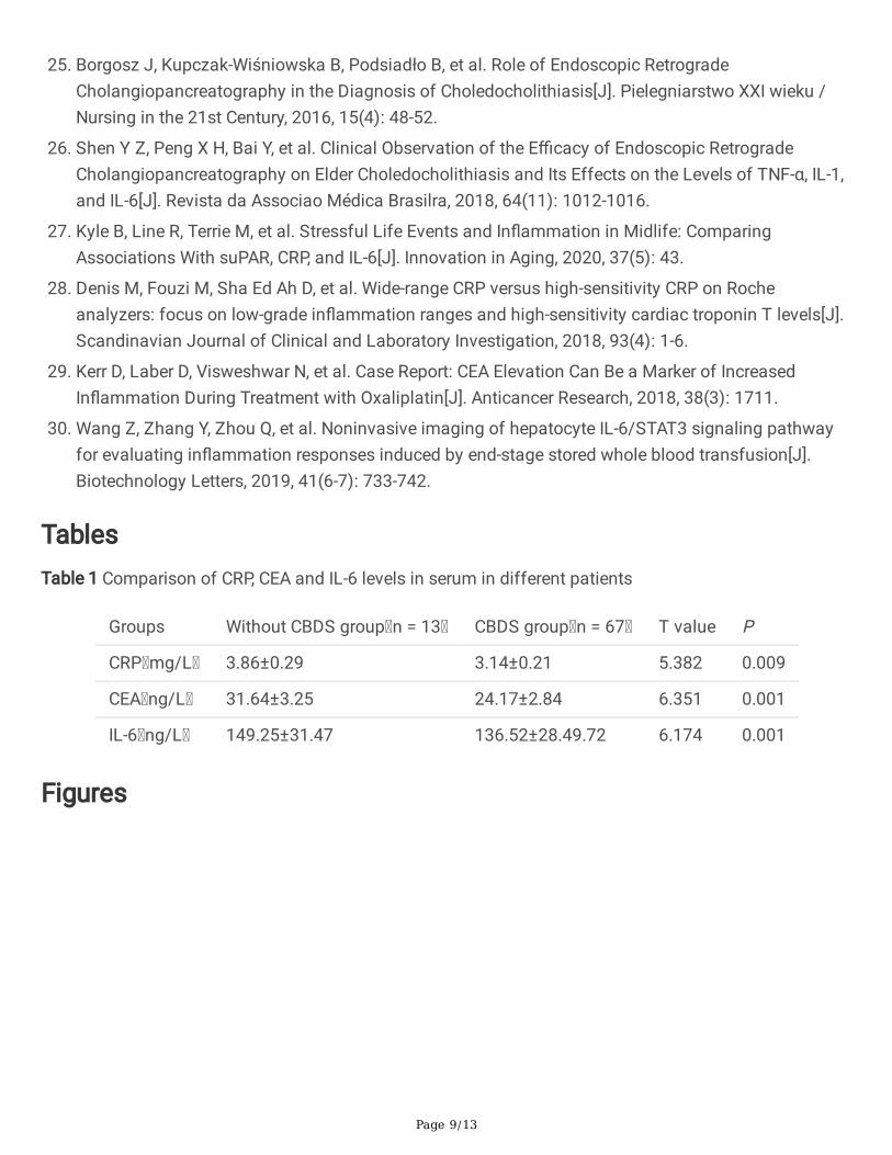
Figure 1
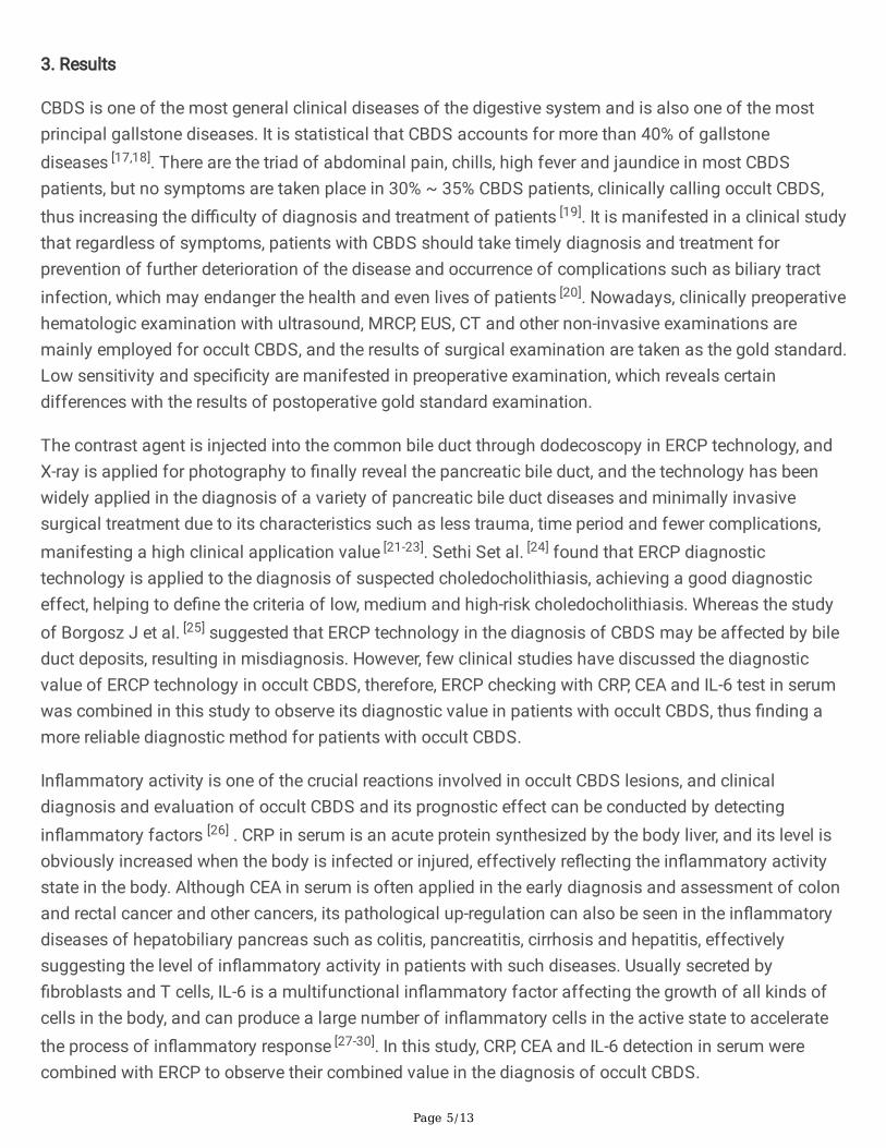
A composite diagram of the process
Page 11/13
Figure 2
The diagnostic rate of ERCP alone was 88.75%, which was obviously declined with contrast that of ERCPcombined with CRP, CEA and IL-6 in serum (98.75%). ** indicated P < 0.001.
Page 12/13
Figure 3
Comparison of CRP, CEA and IL-6 levels in serum in different patients. CRP level in serum in Figure A; CEAlevel in serum in Figure B; IL-6 level in serum in Figure C, ** represented P < 0.001.
Page 13/13
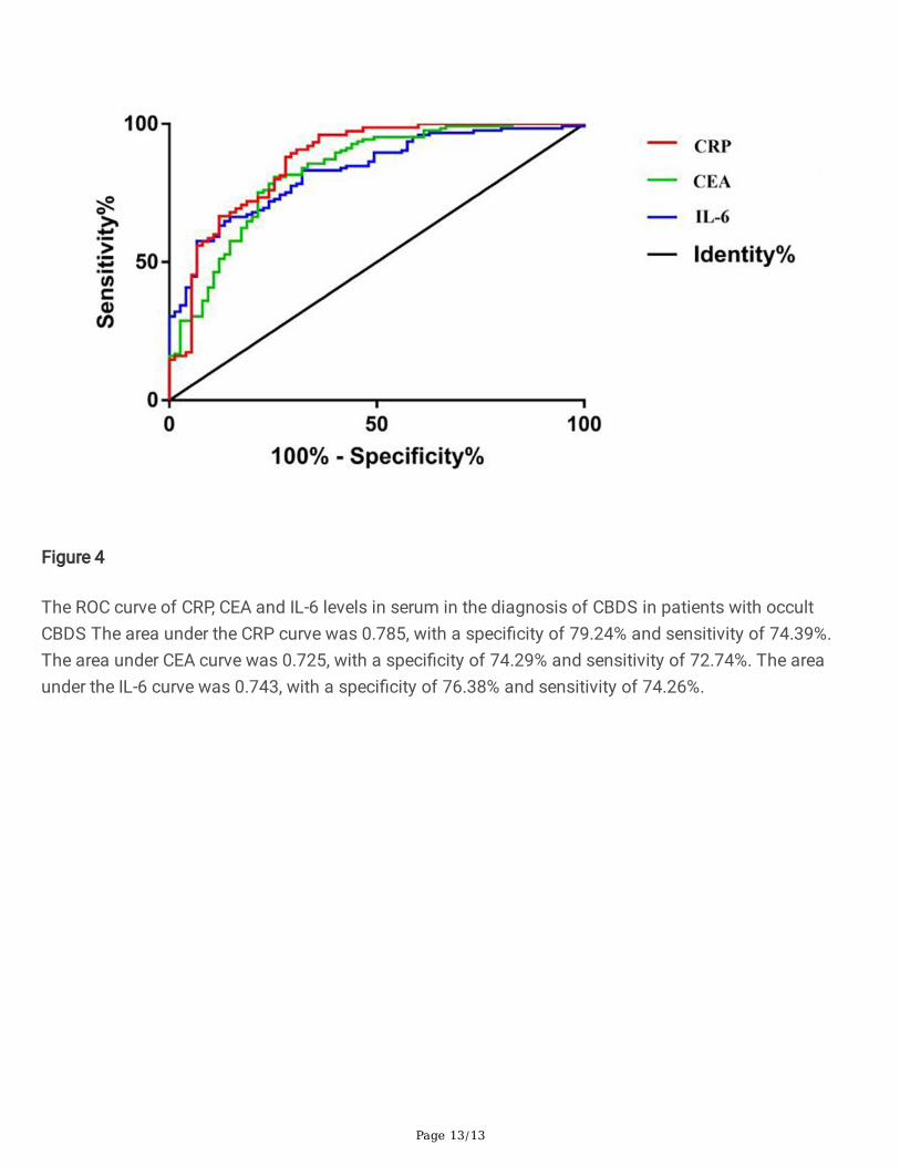
Figure 4
The ROC curve of CRP, CEA and IL-6 levels in serum in the diagnosis of CBDS in patients with occultCBDS The area under the CRP curve was 0.785, with a speci�city of 79.24% and sensitivity of 74.39%.The area under CEA curve was 0.725, with a speci�city of 74.29% and sensitivity of 72.74%. The areaunder the IL-6 curve was 0.743, with a speci�city of 76.38% and sensitivity of 74.26%.