by nwaji njemuwa - rhodes university · prof. chandrashekhara venkata raman discovered the...
TRANSCRIPT

BY
NWAJI NJEMUWA
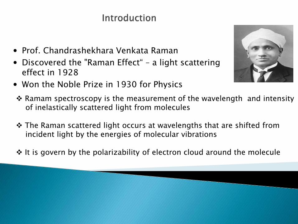
Prof. Chandrashekhara Venkata Raman Discovered the "Raman Effect“ – a light scattering
effect in 1928 Won the Noble Prize in 1930 for Physics Ramam spectroscopy is the measurement of the wavelength and intensity of inelastically scattered light from molecules
The Raman scattered light occurs at wavelengths that are shifted from incident light by the energies of molecular vibrations
It is govern by the polarizability of electron cloud around the molecule

Ener
gy
Rayleigh Scattering (elastic)
Stokes Scattering
Anti-Stokes Scattering
hν0 hν0 hν0
hν0 hν0−hνm hν0+hνm
E0
E0+hνm
Raman (inelastic)
Virtual State
IR Absorption


Examples of Laser wavelengths used for Raman spectroscopy: The most obvious difference is the excitation efficiency. The choice of laser wavelength has an important impact on experimental capabilities: Sensitivity.
◦ R is proportional to λ-4 (where R is the raman scattering efficiency/intensity).
◦ For example, Raman scattering at 532nm is a factor of 4.7 more efficient than at 785nm and 16 times better than at 1064nm, effectively meaning that scan time is much longer at higher wavelengths as compared to 532nm, assuming that all other conditions remain the same.


[http://www.foxnews.com/images/268784/1_61_zawahri_ayman.jpg]

Carbon Materials Purity of carbon nanotubes (CNTs) sp2 and sp3 structure in carbon materials
Pharmaceuticals and Cosmetics Compound distribution in tablets Polymorphic forms Contaminant identification
Life Sciences Bio-compatibility DNA/RNA analysis Drug/cell interactions Photodynamic therapy (PDT)
Geology and Mineralogy Gemstone and mineral identification Fluid inclusions Mineral and phase distribution in rock sections Mineral behaviour under extreme conditions

1360 1440 1520 1600-0.00005
0.00000
0.00005
0.00010
D(1446)
G(1530) GaBT-GQD
1400 1500 1600
0.000
0.001
0.002
0.003
0.004
D(1457)
G(1591)
GQD
Inte
nsity
(a.u
)
Raman shift (cm-2)
Raman spectra of (top to bottom) olivine, apatite, garnet and gypsum illustrating how Raman can be used for fast mineral ID.
Raman spectral image of a whole pharmaceutical tablet, showing the distribution of aspirin (red), caffeine (green) and paracetamol (blue)

Conclusion It is due to the scattering of light by vibrating molecules Little or no sample preparation is required for analysis It has a wide range of applications Raman spectroscopy is a complimentary method to IR

Thank You
‘’Success can only come to you by courageous devotion to the task lying ahead of you’’