by dr. zahoor 1. two major forms of ibd are recognized 1. crohn’s disease (cd) – it can affect...
TRANSCRIPT

1
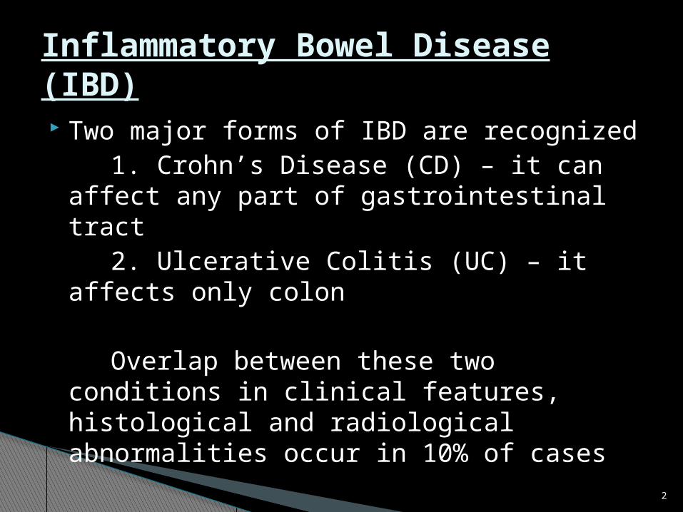
Inflammatory Bowel Disease (IBD)
By Dr. Zahoor
2
Two major forms of IBD are recognized 1. Crohn’s Disease (CD) – it can affect any
part of gastrointestinal tract 2. Ulcerative Colitis (UC) – it affects only
colon
Overlap between these two conditions in clinical features, histological and radiological abnormalities occur in 10% of cases
Inflammatory Bowel Disease (IBD)

3
Aetiology Not known, but interaction between several
cofactors e.g. genetic, environmental, intestinal microbiota and host immune response occurs
Inflammatory Bowel Disease (IBD)

4

5
AetiologyGenetic factors NOD2 gene (nucleotide oligomerization domain)
NOD2 protein on chromosome 16 has increased risk of development of ilial Crohn’s disease
Environmental & Other factors Smoking – smoking exacerbate CD, most patients
having Crohn’s disease are smokers By contrast, smoking (nicotine) has been effective
treatment in ulcerative colitis in small clinical trial
Inflammatory Bowel Disease (IBD)

6
Environmental & Other factors (cont) NSAID – associated with both onset of IBD
and flares of disease Nutritional factor – breast feeding may
provide protection against IBD Psychological factors – Chronic stress and
depression increase relapses in IBD patients Intestinal immune system – IBD occurs when
mucosal immune system gives inappropriate response to luminal antigen e.g. bacteria
Inflammatory Bowel Disease (IBD)

7
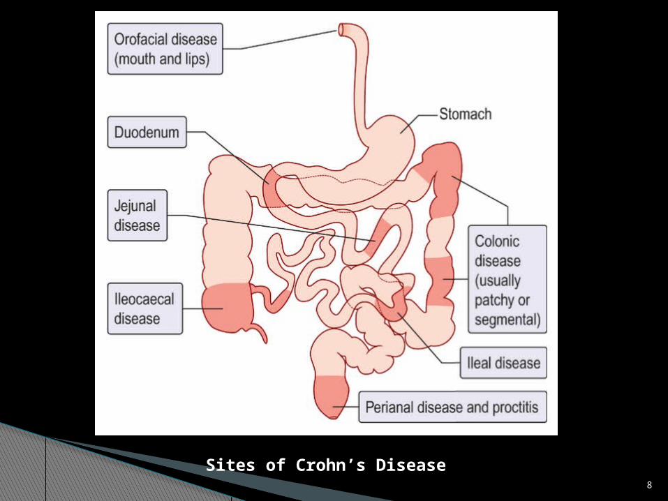
Pathology Crohn’s disease (CD) is a chronic
inflammatory condition that may affect any part of GIT from mouth to the anus but has tendency to affect terminal ileum and ascending colon
CD can involve small area or multiple areas with relatively normal bowel in between (skip lesion)
Inflammatory Bowel Disease (IBD)
8
Sites of Crohn’s Disease

9
Pathology – Ulcerative Colitis UC can affect the rectum alone (proctitis),
but can extend proximally to involve sigmoid and descending colon (left sided colitis) or may involve whole colon
Inflammatory Bowel Disease (IBD)

10
A. Extensive Colitis
B. Left sided Colitis + rectum
C. Proctitis (rectum)
Sites of UC

11
PathologyMacroscopic changes – Crohn’s Disease The involved bowel is usually thickened and
narrowed Deep ulcers and fissures in the mucosa
produce a cobble stone appearance Fistulae and abscesses may be seen Large and deep ulcers appear in a patchy
distribution producing cobble stone appearance
Inflammatory Bowel Disease (IBD)

12
PathologyMacroscopic changes – Ulcerative Colitis Mucosa looks reddened, inflamed and bleeds
easily (friability) In severe disease, there is extensive
ulceration with adjacent mucosa showing inflammation
Note – In fulminant colonic disease, in both CD and UC, most of the mucosa is lost and toxic dilatation occurs
Inflammatory Bowel Disease (IBD)

13
Pathology Microscopic changes - Crohn’s Disease Inflammation extends through all layers of
bowel (transmural)
Microscopic changes – Ulcerative Colitis Superficial inflammation is seen in the
bowel mucosa
Inflammatory Bowel Disease (IBD)

14

15
Serology Test UC – ANCA (Anti Neutrophil Cytoplasmic
Antibodies) occur
CD – Anti – Saccharomyces Cerevisiae Antibodies (ASCA) occur
Inflammatory Bowel Disease (IBD)
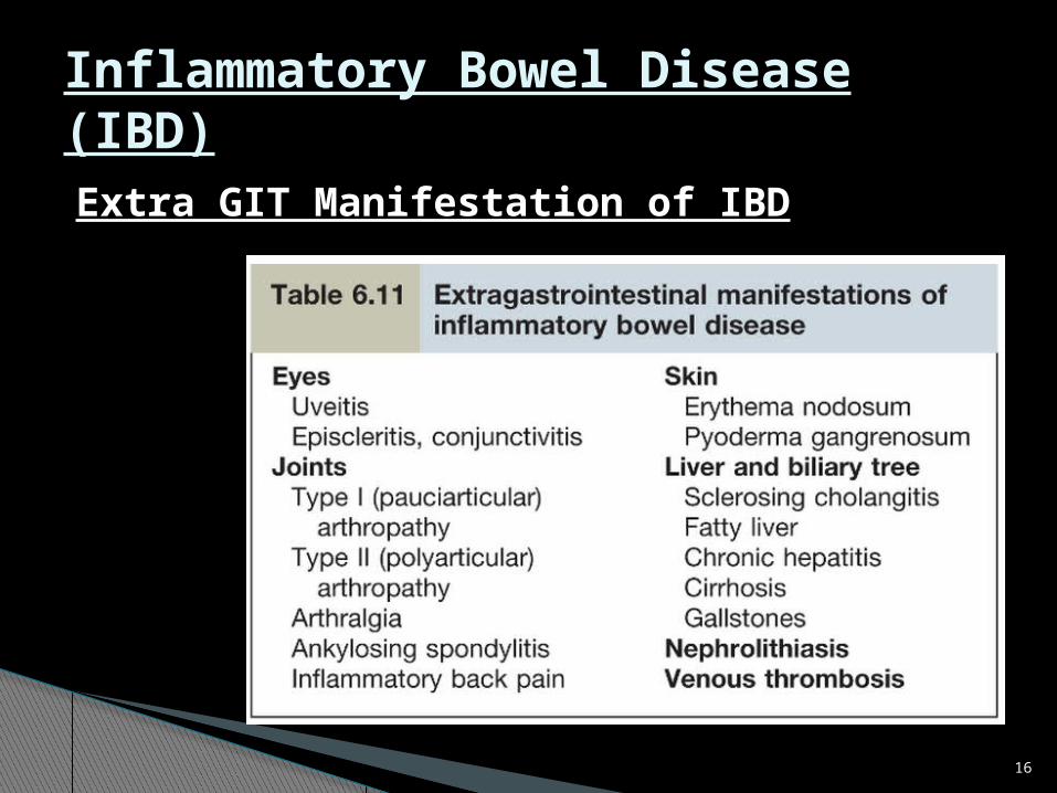
16
Extra GIT Manifestation of IBD
Inflammatory Bowel Disease (IBD)
17
Inflammatory Bowel Disease (IBD)
Crohn’s Disease

18
Clinical FeaturesMajor symptoms Diarrhea – present in 80% of cases and
contains blood if due to colonic disease Abdominal pain Weight loss
Constitutional symptoms Malaise, Lethargy, Anorexia, Nausea,
Vomiting, Low grade fever
Crohn’s Disease

19
Crohn’s disease may present with anal and perianal disease in 25% cases
Crohn’s Disease

20
Examination Loss of weight Signs of mal nutrition Aphthous ulceration of mouth – often seen Abdominal examination – normal But right iliac fossa mass is found
occasionally (due to inflamed loops of bowel or abcess)
Crohn’s Disease

21
Investigation Anaemia – normocytic Normochromic
anaemia of chronic disease Iron, Folate deficiency also occur (despite
terminal ileum involvement vitamin B12 deficiency is unusual )
Increased ESR and C-reactive protein (CRP) Increased WBC and platelet count
Crohn’s Disease

22
Investigation Hypoalbumiaemia Liver biochemistry may be abnormal Serology test - PANCA is negative,
- ASCA is positive
Crohn’s Disease

23
Endoscopy and Radiological imagingSigmoidoscopy Finding vary from mild Patchy superficial ulceration
to wide spread large and deeper ulcers producing cobble stone appearance
Colonoscopy It is done if colonic involvement is suspected
Upper GI endoscopy To exclude oesophageal and gastro duodenal
disease
Crohn’s Disease

24
Small Bowel Imaging Barium meal follow through CT – scan with oral contrast Small bowel ultrasound MRI Finding of imaging may be deep ulceration,
narrowing or stricture, skip lesion with normal bowel between the affected sites. Terminal ileum is commonly affected
Crohn’s Disease

25
Medial Management General consideration Aim is to induce and than maintain
remession. Stop smoking For Diarrhoea – Loperamide, codeine-
phosphate For Anaemia- If due to Vit B12, Folic acid,
Iron should be treated accordingly
Crohn’s Disease

26
Induction of Remission 1. Glucocoticosteriods 2.Aminosalicylates- less useful 3. Antibiotics- Ciprofloxan and Metranidozole 4. Entral Nutrition- For moderate to severe
cases
Crohn’s Disease

27
Refractory Cases If disease is limited to Terminal ileum,
surgical resection is done If patient has extensive Disease, remission
is achieved by Anti- TNF antibodies Infliximab anti-NF&IgG1 monoclonal antibody
Crohn’s Disease

28
Maintenance of Remission -Azathioprine - Mercaptopurine - Methotrexate
CHECK FOR LEUCOPENIA, BONE MARROW SUPPRESSION
CROHN’S DISEASE

29
ULCERATIVE COLITIS (UC)

30
Clinical features:◦General features of UC: Malaise, Lethargy, Anorexia with weight
loss Aphthous ulcers in mouth may be seen Disease can be mild, moderate or
severe Disease runs course of remission and
exacerbations
ULCERATIVE COLITIS (UC)

31
Disease extend is defined 1. Limited to rectum (Proctitis)2. Affecting left side of the colon 3. Extensive
• Proctitis is characterized by frequent passage of blood and mucus, urgency and tenesmus
• Stool passed may be solid
ULCERATIVE COLITIS

32
In left sided or extensive Ulcerative colitis Patient may have bloody diarrhoea passing 10-20 liquid stools per day
ULCERATIVE COLITIS

33
Toxic Megacolon It is serious complication of severe colitis Plain abdominal X-ray shows dilated colon
with diameter of more than 6 cm It is gas filled and there is danger of
perforation and high mortality (15-25 %)
ULCERATIVE COLITIS

34
X-Ray Abdomen Toxic Megacolon: Transverse and Descending colon affected
ULCERATIVE COLITIS

35
Examination Abdominal examination- Abdomen may be
distended or tender on palpation Tachycardia and pyrexia are signs of severe
colitis Rectal Examination with rigid
sigmoidoscope shows inflamed, bleeding, friable mucosa
ULCERATIVE COLITIS

36
Investigations:Blood tests
◦WBC, Platelet counts are raised◦Iron deficiency anemia is commonly
present◦ESR and CRP are often raised ◦Liver Biochemistry may be abnormal with
hypoalbumiaemia in severe disease◦PANCA may be positive
ULCERATIVE COLITIS

37
Investigations (cont) Stool cultures should always be done to
exclude infective cause of colitis Stool microscopy to exclude Amoebiasis Colonoscopy Endoscopy with mucosal biopsy is gold
standard test for diagnosis of UC
ULCERATIVE COLITIS

38
Investigations (cont) Imaging Plain X-ray abdomen is essential to exclude
colonic dilatation Ultrasound Abdomen – Inflammation of
colonic wall can be detected Technitium-labelled white cell scan – helps
to assess the extent of disease
ULCERATIVE COLITIS
39
Medical Management: For mild to moderate cases of UC
1. Aminosalicylate – the active substance of these drugs is 5-aminosalicylic acid (5-ASA) which is absorbed in small intestine
2. Drugs used are 1. Sulfasalazine2. Asacol
These drugs induce remission
ULCERATIVE COLITIS

40
Medical Management ( cont) For Proctitis and left sided colitis Rectal 5-ASA enema are first line of
treatment Oral 5-ASA will increase rate of remission Patient who don’t respond may require oral
prednisolone
ULCERATIVE COLITIS
41
Medical Management ( cont) Sever colitis – patient should be admitted to
the hospital and treated with Hydrocortisone 100 mg IV 6 hourly s/c Low molecular weight Heparin(LMWH) to
prevent thromboembolism IV fluids Nutritional support via enteral route
ULCERATIVE COLITIS

42
Medical Management: Monitor clinical status daily – fever,
tachycardia & stool frequency Do FBC, CRP, Urea & Electrolyte daily
ULCERATIVE COLITIS

43
Indication for surgery: Failure of medical treatment Toxic dilatation Hemorrhage Danger of perforation
ULCERATIVE COLITIS

44
Inflammatory Bowel disease (IBD) and cancer:◦ Patients with UC and CD have increased incidence
of developing colon cancer
Pregnancy and IBD◦ Women with inactive IBD have normal fertility◦ If there is active IBD, fertility may be reduced and
they are likely to suffer spontaneous abortion◦ ASA, steroid and Azathioprine are safe during
pregnancy but Methotrexate is Teratogenic and is contraindicated
ULCERATIVE COLITIS

45
Important note:◦In male Sulfapyridine moiety present in sulfasalazine impairs spermatogenesis therefore alternate aminosalicylate should be used in patients who want to have children
ULCERATIVE COLITIS

46
Thank You