bvdv in alpacas clayton l. kelling clayton l. kelling professor professor department of veterinary...
TRANSCRIPT

BVDV in Alpacas
Clayton L. Kelling
ProfessorDepartment of Veterinary
and Biomedical Sciences
University of Nebraska-Lincoln

BVDV in Alpacas
- Prevalence
- Infections - Fetal- Postnatal

BVDV in North American Alpaca Herds
- Multiple confirmed cases of PI alpacas and reproductive failure in N. America
- Prevalence unknown
- ARF RFP: determine the prevalence of BVDV

Collaborators
University of Nebraska: Christina L. Topliff David R.
Smith Sharon L. Clowser David J. SteffenJamie N. Henningson Kent M. EskridgeGary P. Rupp Bruce W.
Brodersen Clayton L. Kelling
Daniela Bedenice Tufts University
Robert J. Callan Colorado State University
Carlos Reggiardo University of Arizona
Kathy L. Kurth University of Wisconsin

Objective
to determine the current prevalence of BVDV-infected US alpaca herds

Approach
Tested 5 crias from a randomly selected sample of herds for:
BVDV antibodies BVDV RNA BVDV
US alpaca herds with 12 or more registered females
250/562 eligible herds (AOBA Directory)

Distribution of 562 eligible alpaca herds
12
US States-www.50states.com

Distribution of 250 selected alpaca herds
US States-www.50states.com
1
US States-www.50states.com
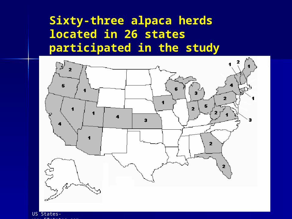
Sixty-three alpaca herds located in 26 states participated in the study

Results: Seropositive herds
Herds with seropositive crias 16/63 (25.4%)
95% Confidence interval of 15.3% - 37.9%

Case Studies of the 16 Herds with Seropositive Crias
- Individually contacted
- Detailed information
- Scheduled follow-up testing of the dams
- Whole herd RT-PCR testing:confirmed BVDV-free status
- Seropositive dams in BVDV-free herd: indicated prior BVDV exposure

Basis for herd BVDV seropositive status
BVDV Seropositive Herds
Colostrum
- DamPI exposure within herd
Off farm exposure - Supplemental

SUMMARY Case Studies of 16 Seropositive HerdsNo.Of Herds BVDV Antibody Source
6 Dam prior BVDV infection
6 Colostrum supplements(4 bovine, 1 goat, 1 both)
4 BVDV-infected herd

Basis for herd BVDV seropositive status
BVDV Seropositive Herds (n=6)
Colostrum
- DamPrior off farm exposure

Herd No. 10 BVDV Seropositive Seropositive Dam Prior Infection
Cria Bovine Antibody TiterNo. Age Colostrum BVDV 1 BVDV21 129d NO <2 <22 164d NO 8 <23 30d NO <2 <24 39d NO <2 <25 171d NO NA <26 23d NO <2 <2
DamNo.2 256 32
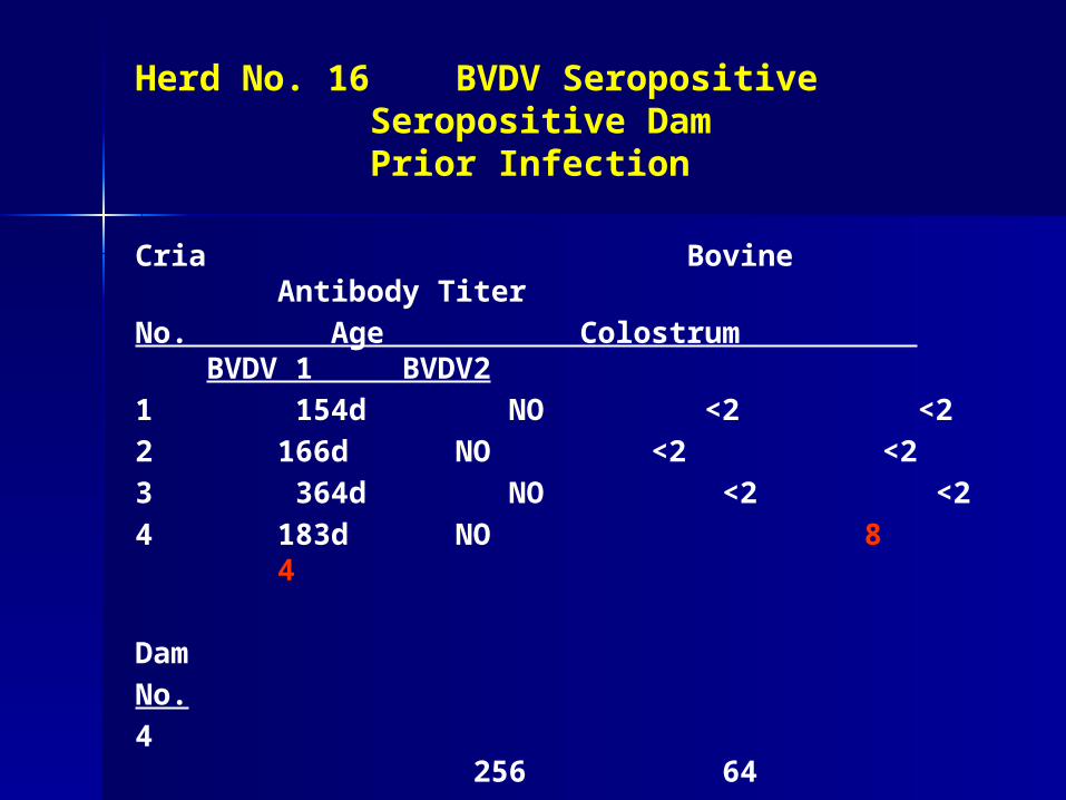
Herd No. 16 BVDV Seropositive Seropositive Dam Prior Infection
Cria Bovine Antibody Titer
No. Age Colostrum BVDV 1 BVDV2
1 154d NO <2 <22 166d NO <2 <23 364d NO <2 <24 183d NO 8 4
DamNo.4 256
64
Basis for herd BVDV seropositive status
BVDV Seropositive Herds (n=6)
Colostrum - Supplemental

Herd No. 7 BVDV Seropositive: Bovine Colostrum Fed to Crias
Cria Bovine Antibody TiterNo. Age Colostrum BVDV 1 BVDV21 207d NO <2 <22 11d YES 8 23 61d NO <2 <24 1d YES 4 4
Tested Dams 2 and 4: <2 <2

Herd No. 14 BVDV Seropositive: Bovine Colostrum Fed to
Cria
Cria Bovine Antibody Titer
No. Age Colostrum BVDV 1 BVDV2
1 60d No <2 <22 203d No <2 <23 37d Yes 32 164 61d No <2 <25 57d No <2 <2
DamNo.3 <2 <2

Basis for herd BVDV seropositive status
BVDV Seropositive Herds (n=4)
Colostrum
- DamPI exposure within
herd
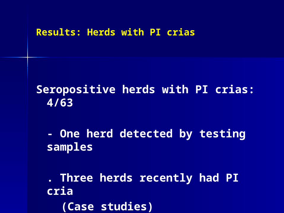
Results: Herds with PI crias
Seropositive herds with PI crias: 4/63
- One herd detected by testing samples
. Three herds recently had PI cria(Case studies)

Herd No. 4 BVDV Seropositive: PI Cria
Cria Bovine Antibody TiterNo. Age Colostrum BVDV 1 BVDV21 1d NO ≥256 322 1d NO <2 <23 25d NO <2 <24 3d NO ≥256 16

Herd No. 13 BVDV Seropositive: PI Cria
Cria Bovine Antibody TiterNo. Age Colostrum BVDV 1
BVDV21 167d NO ≥ 256 ≥ 2562 131d NO ≥ 256 ≥ 1283 141d NO ≥ 256 ≥ 2564 132d NO 16 25 84d NO 64 8

Source of infection in 3 of the 4 infected herds was linked
- Based on genetic homologies of viruses
- PCR amplified BVDV 5’ untranslated region (UTR) PCR products from PI animals
- Sequencing - 283 base pair (bp) fragment of
the 5’ UTR (nucleotides 107 and 389)

Source of infection in 3 herds with PI crias were linked:
- High genetic homologies (99.2 and 99.6%) confirmed the common source of infection
- Gestating females in 2 herds contacted a PI cria in the same herd

Source of BVDV infection in the fourth herd (No. 15) was unique
- Nucleotide identity:- 91.1% with Herd Nos, 4, 11 and
13
- Exposed to a breeding female with a PI
cria at side from another farm.
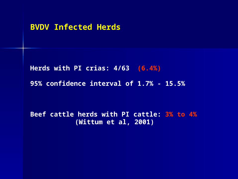
BVDV Infected Herds
Herds with PI crias: 4/63 (6.4%)
95% confidence interval of 1.7% - 15.5%
Beef cattle herds with PI cattle: 3% to 4% (Wittum et al, 2001)
Discussion
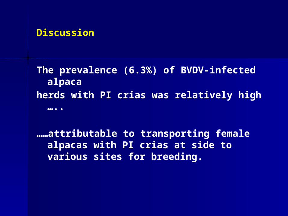
The prevalence (6.3%) of BVDV-infected alpaca
herds with PI crias was relatively high …..
……attributable to transporting female alpacas with PI crias at side to various sites for breeding.

The farms that had PI crias suffered severe economic losses due to:
- abortions, birth of weak crias that died
- expenses associated with diagnostic/treatmentregimens
- loss of sales

PI Crias from the BVDV infected herds

Objectives
Determine the extent of lesions and antigen distribution in PI alpacas and compare to PI calves

Materials and Methods
– 10 PI alpacas– 5 PI calves– Gross, microscopic, and IHC– Scored antigen deposition
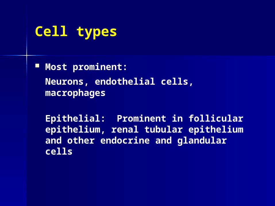
Cell types
Most prominent:
Neurons, endothelial cells, macrophages
Epithelial: Prominent in follicular epithelium, renal tubular epithelium and other endocrine and glandular cells

Kidney Salivary gland
Pancreas Thyroid

IHC. PI Ear Notch
Alpaca

PI alpaca vs. PI calf

Distribution of BVDV antigen in lymphoid organs of PI alpacas and calves
BVDV Antigen
__ Distribution Intensity Associated Lesions
PI calves Wide Marked None
PI alpacas Wide Moderate None
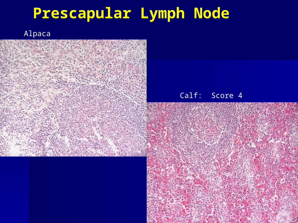
Prescapular Lymph NodeAlpaca
Calf: Score 4


Conclusions
PI alpacas:
– Widespread distribution of BVDV antigen affecting multiple organ systems and cell types
– Widespread epithelial staining suggests nasal, oral, and urinary fluids may be infectious

Conclusions
Fetal BVDV infections:
AbortionsPI crias

Acute BVDV infections in alpacas
Consequences of postnatal infections of alpacas?

Acute BVDV infections in alpacas
Experimental acute BVDV infections
Alpacas vs. cattle
BVDV NY-1
BVDV CO-06

Acute BVDV Infection in Alpacas and Cattle
96
98
100
102
104
106
108
-1 0 1 2 3 4 5 6 7 8 9
Days Post-infection
Rec
tal
Tem
per
atu
re (o
F)
Alpaca
Cattle

Lymphocytes
0
1
2
3
4
5
6
7
0 1 2 3 4 5 6 7 8 9
Days Post-infection
Lym
ph
ocy
tes
(x 1
03 /ul)
CO-06 Alpaca
NY-1 Cattle
NY-1 Alpaca

Alpaca GALT. IHC

Distribution of BVDV antigen in lymphoid tissues of acutely-infected alpacas and calves
BVDV Antigen
Distribution Associated lesions
Calves PP,MLN,Thy Marked
Alpacas (C3, PP, Colon) Very Mild Variable
***Preliminary findings

Acute BVDV infections in alpacas
Summary: Consequences of postnatal infections of alpacas:
Systemic infection - Marked leukopenia &
lymphopenia
- Very mild lymphodepletion - Antigen deposition

Acute BVDV infections in alpacas
Consequences of postnatal BVDV infections of alpacas:
Alpacas are susceptible
but are differences compared to calves

Comparative permissiveness of
alpaca and bovine cells to BVDV infections

BVDV NY-1 and CO-06 in alpaca and bovine cells
4
1 2 3
5 6
BT
AT
(-) NY-1 CO-06
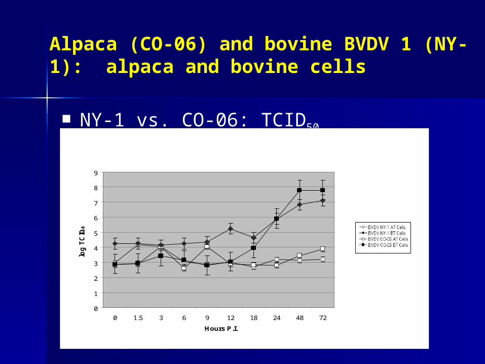
Alpaca (CO-06) and bovine BVDV 1 (NY-1): alpaca and bovine cells
NY-1 vs. CO-06: TCID50
0
1
2
3
4
5
6
7
8
9
0 1.5 3 6 9 12 18 24 48 72
Hours P.I.
log
TC
ID5
0

BVDV2: Alpaca vs. bovine cells
4
1 2 3
5 6
BT
AT
(-) 890 7937

BVDV2: Alpaca vs. bovine cells
890 vs. 7937: TCID50
0
1
2
3
4
5
6
7
8
9
0 1.5 3 6 9 12 18 24 48 72
Hours P.I.
log
TC
ID5
0

Alpaca cells: BVDV1 vs. BVDV2
0
1
2
3
4
5
6
0 1.5 3 6 9 12 18 24 48 72
Hours P.I.
log
TC
ID50
BVDV NY-1 A
BVDV CO-06 A
BVDV 890 A
BVDV 7937 A

Bovine cells: Alpaca BVDV vs. Bovine BVDV
0
1
2
3
4
5
6
7
8
9
0 1.5 3 6 9 12 18 24 48 72
Hours P.I.
log
TC
ID50
BVDV 890 B
BVDV 7937 B
BVDV NY-1 B
BVDV CO-06 B

Comparative permissiveness of alpaca and bovine cells to BVDV infections
Alpaca cells less permissive than bovine cells to BVDV infections
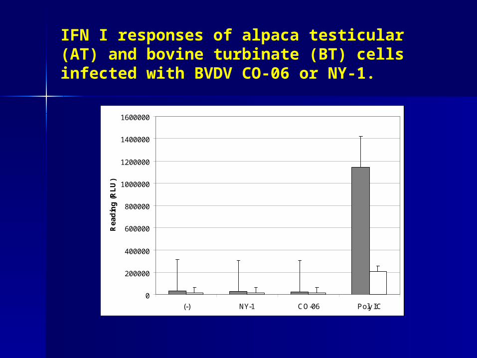
IFN I responses of alpaca testicular (AT) and bovine turbinate (BT) cells infected with BVDV CO-06 or NY-1.
0
200000
400000
600000
800000
1000000
1200000
1400000
1600000
(-) NY-1 CO-06 Poly I:C
Re
ad
ing
(R
LU
)

Inhibition of IFN synthesis by NY-1 and
Co-06 BVDV infection of AT and BT cells
0
200000
400000
600000
800000
1000000
1200000
(-) NY-1 (- PolyI:C)
NY-1 (+ PolyI:C)
CO-06 (-PolyI:C)
CO-06 (+ PolyI:C)
Poly I:C (- PolyI:C)
Poly I:C (+Poly I:C)
Re
ad
ing
(R
LU
)

Conclusions
Elevated IFN responses in BVDV infected alpaca cellscorrelates with:
–Reduced permissiveness of AT cells.

Conclusions
Elevated IFN responses in BVDV infected alpaca cellscorrelates with:
– Reduced permissiveness of AT cells.
– Mild pathologic effects of BVDV in lymphatic organs of alpacas infected postnatally.

Clinical Relevance
BVDV is important to the US alpaca industry

Clinical Relevance
BVDV is important to the US alpaca industry
Herd prevalence of 6.4%.
95% confidence interval of 1.7% - 15.5%

Clinical Relevance
BVDV is important to the US alpaca industry
Herd prevalence of 6.4%.
BVDV is pathogenic in alpacas

Clinical Relevance
BVDV is important to the US alpaca industry
Herd prevalence of 6.4%.
BVDV is pathogenic in alpacas
- fetal infections: PI, abortions
-

Clinical Relevance
BVDV is important to the US alpaca industry
Herd prevalence of 6.4%.
BVDV is pathogenic in alpacas
- fetal infections: PI, abortions
- postnatal infections

Clinical Relevance
BVDV is important to the US alpaca industry
Herd prevalence of 6.4%.
BVDV is pathogenic in alpacas- fetal infections: PI,
abortions- postnatal infections
Risk factors for BVDV exposure: Movement and commingling with unknown BVDV status

Clinical Relevance
BVDV is important to the US alpaca industry
Herd prevalence of 6.4%.
BVDV is pathogenic in alpacas- fetal infections: PI,
abortions- postnatal infections
Risk factors for BVDV exposure: Movement and commingling
Determine BVDV PI status (PCR tests) of alpacas before movement

Clinical Relevance
BVDV is important to the US alpaca industry
Herd prevalence of 6.4%.
BVDV is pathogenic in alpacas- fetal infections: PI,
abortions- postnatal infections
Risk factors for BVDV exposure: Movement and commingling
Determine BVDV PI status (PCR tests) of alpacas before movement

Acknowledgments
– Funding:
Mid-Atlantic Alpaca Association through the
Alpaca Research Foundation
University of Nebraska-LincolnAgricultural Research
DivisionInstitute of Agriculture and
Natural Resources


IHC and Histopathology Scales
IHC – 0: None– 1: Positive single or small
group– 2: Scattered staining, clearly
positive– 3: Widespread, multifocal,
many– 4: Intense widespread,
diffuse, most follicles
Lymphocytolysis– 1: Scattered cells (normal)– 2: Many cells, one or more
follicles affected– 3: Moderate (25-50%)– 4: Widespread, most
follicles (>50%)
Lymphoid depletion
– 0: None– 1: 1-10%– 2: 10-25%– 3: 25-50%– 4>50%
Inflammation and Necrosis– 0: None – 1: Mild– 2: Moderate– 3: Severe

Photomicrograph of sections of Peyer’s patches of calf acutely infected with BVDV 890, 9 days PI.

Interferon response in bovine and alpaca cells
NCL1 Luc ISRE cells
Gil et al., 2004

Interferon response in bovine and alpaca cells
Luciferase

Interferon response in bovine and alpaca cells
Interferon production assay– BT or AT cells infected at MOI of 10, mock infection
control with uninfected growth media– Incubated 24 hours – BVDV inactivated with changes in pH
Stimulation of NCL1 ISRE-Luc cells– Cells grown on 12 well plate and overlaid with test
sample Luciferase assay
– Cells lysed and fluorescence measured– NCL1 Luc ISRE cells express luciferase with an
interferon responsive element

Interferon response in bovine and alpaca cells
Interferon inhibition assay– AT and BT cells infected at an m.o.i. of
1, incubated 48 hours, then stimulated with poly I:C, a dsRNA analogue
– Assayed for interferon response using reporter cell line