busin, valentina and wells, beth and kersaudy-kerhoas
TRANSCRIPT

Busin, Valentina and Wells, Beth and Kersaudy-Kerhoas, Maïwenn and
Shu, Wenmaio and Burgess, Stewart T.G. (2016) Opportunities and
challenges for the application of microfluidic technologies in point-of-
care veterinary diagnostics. Molecular and Cellular Probes. ISSN 0890-
8508 , http://dx.doi.org/10.1016/j.mcp.2016.07.004
This version is available at https://strathprints.strath.ac.uk/57451/
Strathprints is designed to allow users to access the research output of the University of
Strathclyde. Unless otherwise explicitly stated on the manuscript, Copyright © and Moral Rights
for the papers on this site are retained by the individual authors and/or other copyright owners.
Please check the manuscript for details of any other licences that may have been applied. You
may not engage in further distribution of the material for any profitmaking activities or any
commercial gain. You may freely distribute both the url (https://strathprints.strath.ac.uk/) and the
content of this paper for research or private study, educational, or not-for-profit purposes without
prior permission or charge.
Any correspondence concerning this service should be sent to the Strathprints administrator:
The Strathprints institutional repository (https://strathprints.strath.ac.uk) is a digital archive of University of Strathclyde research
outputs. It has been developed to disseminate open access research outputs, expose data about those outputs, and enable the
management and persistent access to Strathclyde's intellectual output.
MA
NU
SC
RIP
T
AC
CE
PTE
D
ACCEPTED MANUSCRIPT
ACCEPTED MANUSCRIPT
1
Opportunities and challenges for the application of microfluidic 1
technologies in point-of-care veterinary diagnostics 2
3
Valentina Busina,b*, Beth Wellsa, Maïwenn Kersaudy-Kerhoasb, Wenmaio Shub,c and Stewart T. G. Burgessa 4
5
aMoredun Research Institute, Pentlands Science Park, Bush Loan, Edinburgh. EH26 0PZ. United Kingdom. 6
bSchool of Engineering and Physical Sciences, Heriot-Watt University, Edinburgh. EH14 4AS. United 7
Kingdom. 8
C Department of Biomedical Engineering, University of Strathclyde, Glasgow. G4 0NW. United Kingdom 9
10
*Corresponding author. Moredun Research Institute, Pentlands Science Park, Bush Loan, Edinburgh. EH26 11
0PZ. United Kingdom. Email: [email protected] (V. Busin). 12
13
Email addresses: 14
VB: [email protected] 15
BW: [email protected] 16
MKK: [email protected] 17
WS: [email protected] 18
STGB: [email protected] 19
20
21
22
23
24
25
26

MA
NU
SC
RIP
T
AC
CE
PTE
D
ACCEPTED MANUSCRIPT
ACCEPTED MANUSCRIPT
2
Abstract 27
There is a growing need for low-cost, rapid and reliable diagnostic results in veterinary medicine. Point-of-28
care (POC) tests have tremendous advantages over existing laboratory-based tests, due to their intrinsic 29
low-cost and rapidity. A considerable number of POC tests are presently available, mostly in dipstick or 30
lateral flow formats, allowing cost-effective and decentralised diagnosis of a wide range of infectious 31
diseases and public health related threats. Although, extremely useful, these tests come with some 32
limitations. Recent advances in the field of microfluidics have brought about new and exciting opportunities 33
for human health diagnostics, and there is now great potential for these new technologies to be applied in 34
the field of veterinary diagnostics. This review appraises currently available POC tests in veterinary 35
medicine, taking into consideration their usefulness and limitations, whilst exploring possible applications 36
for new and emerging technologies, in order to widen and improve the range of POC tests available. 37
Keywords 38
Point-of-care, veterinary diagnostics, microfluidics, lateral flow immunoassays, paper-based microfluidics, 39
dipstick test. 40
41
42
43
44
45
46
47
48
49
1. Introduction 50

MA
NU
SC
RIP
T
AC
CE
PTE
D
ACCEPTED MANUSCRIPT
ACCEPTED MANUSCRIPT
3
51
Point-of-care (POC) diagnostics is an area that has attracted considerable attention in the last decade. 52
Testing at POC means that analytical procedures are carried out at the side of or near to the patient [1], for 53
this reason, it is also sometimes referred to as “bed-side” testing [2]. The reasons for the considerable 54
interest in the field of POC testing are numerous: the potential to decrease costs of diagnosis [3], increasing 55
the accessibility of these types of test to disadvantaged populations [4], and reducing the time between 56
sampling and a treatment decision [5]. 57
Following the global trend towards more affordable and accessible diagnostic testing, the Sexually 58
Transmitted Diseases Diagnostics Initiative (SDI) within the World Health Organization (WHO) recently 59
established a set of benchmark criteria for the ideal rapid test, under the acronym “ASSURED” [6]: 60
Affordable, Sensitive, Specific, User-friendly (simple to perform in a few steps, with minimal technical 61
training), Robust and rapid (results available in less than 30 min), Equipment-free, Deliverable to those who 62
need them. Ideally, POC tests should respect all or as many as possible of these characteristics [7]. 63
64
2. Point of care testing and its scope in veterinary medicine 65
66
In the veterinary area, there is a similar need for low-cost, reliable and rapid diagnostic tests to be 67
carried out at the POC [8]. So called on-site or animal-side tests will have considerable advantages over 68
laboratory-based testing, which usually involves laborious and expensive laboratory techniques and 69
dedicated technical personnel. All of the analytical processes involved in testing, from collection of the 70
sample to communication of the results, could potentially be performed in a single step, considerably 71
reducing the time between testing and treatment [9]. This can translate into more affordable veterinary 72
care, reduced handling of animals, targeted treatments and rapid testing in more remote geographic areas. 73
The need for more affordable, rapid and accessible tests is a recurrent theme in the literature, in 74
particular as an invaluable tool in dealing with diseases that either represent a threat to public health [10], 75
have substantial impact on animal welfare [11] and/or are of economic importance [12], with particular 76

MA
NU
SC
RIP
T
AC
CE
PTE
D
ACCEPTED MANUSCRIPT
ACCEPTED MANUSCRIPT
4
relevance to situations where laboratory facilities and funds are limited [13]. Furthermore, the general 77
globalisation of trade of animals and animal products has greatly increased the risk of rapid and wide-78
ranging spread of emerging and exotic diseases, requiring timely and efficient ways of dealing with diseases 79
that could have catastrophic repercussions for the individual farmer, as well as economic implications for 80
the entire country and international trade [14]. In situations concerning disease outbreaks, where rapid 81
propagation of infectious agents and/or high mortality are salient features, as in the case of the highly 82
pathogenic H5N1 strain of avian influenza virus, a rapid “animal-side” test would represent a critical tool 83
for both collecting surveillance data and for assisting in the control of outbreaks [11, 15]. Currently 84
available veterinary POC tests offer a good opportunity for a truly “animal-side” diagnosis, but the 85
analytical performances of “on-site” testing are still considered limited compared with laboratory-based 86
testing [16], whilst the possibility offered by the support of a central laboratory in the interpretation of the 87
results is still perceived as critical [17]. Recent advances in microfluidic technologies for POC testing in the 88
human field could overcome these hurdles and might be applied in veterinary medicine. This review aims 89
to appraise the current status of POC testing in veterinary medicine, describing their advantages and 90
limitations, whilst also assessing the potential of microfluidic technologies to improve existing POC tests 91
and solve some of their intrinsic limitations. 92
93
3. Point-of-care devices currently available in veterinary diagnostics 94
95
At present, the most widely used technologies for POC testing in veterinary medicine are: dipstick tests 96
and lateral flow immunoassays. 97
98
3.1 Dipstick and strip test 99
100
These assays are based on the principle of immunoblotting and are made of paper strips with pads to 101
analyse specific fluids. After the sample is introduced, the results are compared with a colour-coded chart 102
to provide a semi-quantitative determination of the analyte(s). The most commonly used are test strips 103

MA
NU
SC
RIP
T
AC
CE
PTE
D
ACCEPTED MANUSCRIPT
ACCEPTED MANUSCRIPT
5
developed for human urine analysis, allowing the simultaneous detection or monitoring of leukocytes, 104
nitrite, urobilinogen, protein, pH, haemoglobin, specific gravity, ketones, bilirubin and/or glucose (Fig. 1) 105
[18]. While it has been developed for human patients, there is a high correlation between the dipstick 106
results and other routinely used methods for urine analysis, which has resulted in this test being widely 107
used in small animal private practice for first-line diagnosis of chronic kidney disease, mainly through an 108
assessment of proteinuria [19, 20]. However, care should be taken when interpreting positive test results 109
with low levels of proteins (traces) due to the high rate of false positive results [21]. 110
A smaller version of the urine dipstick, restricted to detection of glucose and ketone bodies, is also 111
largely applied for at-home management of pets with diabetes. This test is also widely used in farmed 112
ruminants for the diagnosis of ketosis in cattle [22] and pregnancy toxaemia in sheep. Due to some 113
variation in results, a further advance in the diagnosis of these diseases is the use of appropriate strips 114
combined with electronic hand-held meters to accurately measure both glucose and one of the main 115
ketone bodies, ß- bhydroxybutyrate (BHB) in blood, making diagnosis more reliable as well as sampling 116
potentially more successful and less stressful [23]. This POC test has shown great potential for the 117
quantitative detection of BHB, with improved sensitivity and specificity when compared with dipstick tests 118
for detection of ketones in urine or milk [24, 25]. Its use for glucose measurement, however, does not seem 119
to be reliable [23]. Dipstick tests have also been used for the detection of antibiotics in serum, milk and/or 120
meat samples [26]. These rapid tests allow the detection of antibiotics within the µg/ml range, permitting 121
on-site monitoring of non-authorised uses of antimicrobials, which could be especially useful in 122
slaughterhouses and food processing plants. 123
The main advantage of these POC tests is that they can be readily carried out by the owners, proving to 124
be particularly useful for the long term management of chronic diseases [27]. The major limitation, 125
however, can be the subjective interpretation of results, based on a personal evaluation of a colorimetric 126
reaction [19]. 127
128
3.2 Lateral flow immunoassays (LFIAs) 129
130

MA
NU
SC
RIP
T
AC
CE
PTE
D
ACCEPTED MANUSCRIPT
ACCEPTED MANUSCRIPT
6
These devices are based on the principle of capillary force: a liquid flowing on or through a strip of 131
polymeric material, on or in which specific molecules (e.g., antigens, antibodies, DNA/RNA sequence) have 132
been immobilized [28]. These strips usually consist of multiple pads: a sample application pad, a conjugate 133
pad, a membrane for detection and an absorbent pad (Fig. 2), usually made of different materials (e.g., 134
nitrocellulose, glass fibre paper and fused silica) encased in a plastic cage for protection of a fragile paper 135
membrane [29]. The best known example of a lateral flow test is the pregnancy test [30], which is probably 136
the most used POC test worldwide. The main advantages of lateral flow over traditional laboratory-based 137
tests are their simplicity, rapidity and low cost. Compared with traditional laboratory-based tests, LFIAs are 138
considerably less expensive, but, due to the different materials required, they are still relatively costly (~ 139
$10 for a pregnancy test; [29]) for application in low-resource settings [31], and, due to the multi step 140
processes involved, manufacturing time is extended, making them less suitable for high-throughput 141
production. 142
They usually provide a qualitative or semi-quantitative result, and analytical performance is generally 143
poorer than laboratory-based tests, mainly due to reduced sensitivity [32, 33] and the possibility of errors 144
due to testing by untrained personnel [2]. When compared with reference laboratory tests, specificity 145
tends to be comparable, while sensitivity can be as low as 16%, suggesting that a positive result might be 146
trusted, whereas, in the case of negative results, further confirmatory laboratory testing may be advisable. 147
Several studies have assessed quantitation, but such devices still require instrumentation [34, 35] and 148
trained personnel, and are still limited to single analyte testing. 149
150
3.2.1 Companion animals 151
152
There is a significant market for the use of LFIAs for a range of acute and chronic diseases or conditions 153
in companion animals (Table 1). These assays are usually easy to perform and interpret [32], with the 154
possibility to improve sensitivity by using a dedicated LFIA reader, which allows an objective and 155
quantitative interpretation of the results [28]. Although the cost of the test is an important consideration, 156
reduced waiting time and the possibility of starting a therapy within the first visit are amongst the main 157

MA
NU
SC
RIP
T
AC
CE
PTE
D
ACCEPTED MANUSCRIPT
ACCEPTED MANUSCRIPT
7
advantages of tests that can be carried out “in-house”. It is also important to consider that some diseases 158
have received considerably greater attention, notably viral infection diseases, such as FIV and FeLV in cats 159
and parvovirus in dogs, with an extended range of assays being available. 160
161
3.2.2 Livestock 162
163
For livestock, LFIAs have been focused mainly on illnesses that represent a substantial economic burden 164
and/or serious zoonotic or epidemic diseases (Table 2). OIE-listed diseases of the World Organization for 165
Animal Health, such as Foot-and-Mouth Disease (FMD) and Rinderpest have received considerable 166
attention, due to the crucial importance of a rapid diagnosis and, consequently, prompt intervention from 167
veterinary authorities. In the case of FMD, endemic areas are frequently in developing countries, and often 168
diagnosis is not reached due to the prolonged time between collection of samples, arrival at the reference 169
laboratory and subsequent testing [36]. In this case, both the economic constraints and accessibility to 170
remote areas are the dominant issues. 171
172
3.2.3 Food safety 173
174
Lateral flow tests have been successfully applied in two main areas: the detection of food-borne 175
pathogens and the detection of fraudulent substances in animal feed or in animal products (Table 3). In the 176
first case, much emphasis is placed on the prevention of zoonotic diseases, which represent a significant 177
and widespread public health threat. In the second case, recent feed-stuff scandals [37, 38] and increasing 178
reports of antimicrobial resistance dominate the scene, both in terms of research and public attention. 179
Here, and on-site test can be a powerful tool for rapid detection and subsequent surveillance, especially 180
when dealing with highly perishable products. 181
182
4. Microfluidic technologies available for POC diagnostics 183
184
MA
NU
SC
RIP
T
AC
CE
PTE
D
ACCEPTED MANUSCRIPT
ACCEPTED MANUSCRIPT
8
One of the most promising technologies that has been applied recently in diagnostics is microfluidics, 185
which involves the analysis of extremely small amounts (microlitres or nanolitres) of fluid using 186
interconnected networks of channels measuring tens to hundreds of micrometres [39]. Since the 187
introduction of microfluidics from the early 1990s [40], there has been a constant evolution of these 188
methods, mainly following critical advances in microfabrication technologies [41]. Fluid transport in these 189
devices is achieved by either passive (usually capillary forces) or active (generally pumping) mechanisms 190
[42, 43], with the fluid flow being typically laminar [44]. 191
Among the main advantages of microfluidics technologies for diagnostic applications are their 192
portability and their low consumption of reagents; these attributes have made these devices inexpensive, 193
rapid and generally easier to use compared with conventional (macroscale) testing [45]. The use of very 194
small volumes, associated with shorter diffusional distances, results in significantly reduced time for 195
analysis, making microfluidic assays significantly more rapid to perform than their macroscale equivalents 196
[46]. Furthermore, being able to perform all necessary steps within one device and potentially in a single 197
reaction represent a considerable advantage, allowing sample pre-treatment, analysis, signal detection and 198
amplification in the same device [47]. Automated control of all steps can reduce inherent human error, 199
which in turn increases the quality, reproducibility and reliability of assay results. The higher degree of 200
control of fluid flow and the timing of binding reactions can also result in significantly improved analytical 201
performance [48], while the opportunity for tests to be carried out simultaneously offers considerable 202
potential for multiplexing [47]. Examples of the successful applications of these new technologies are in the 203
clinical analysis of blood [49-51], pathogen identification [52, 53], genetic testing [54, 55], detection of 204
environmental contaminants [56] and for drug screening [57]. 205
Currently, two main types of microfluidic systems are used in the diagnostic field: micro total analysis 206
systems (µTAS) and microfluidic paper-based analytical devices (µPADs). However, to date, there has been 207
very limited application of this technology in the veterinary field [58]. 208
209
4.1 Micro total analysis systems (µTAS) 210
211

MA
NU
SC
RIP
T
AC
CE
PTE
D
ACCEPTED MANUSCRIPT
ACCEPTED MANUSCRIPT
9
These systems are also commonly known as “lab on a chip” (LOC) devices (Fig. 3), which use fluid as a 212
working medium and can integrate a number of different functionalities on a micro scale [41]. One of the 213
main advantages of µTAS devices is that they allow for all steps (from sample pre-treatment to signal 214
detection) to be carried out at once, on the same device, allowing complicated molecular techniques (i.e. 215
polymerase chain reaction (PCR)) to be transferred on the chip for POC testing. These devices are 216
fabricated using techniques from the microelectronics industry [59], mostly using materials, such as silicon, 217
glass and/or polymers [60]. At present, the most common materials used are thermoplastic polymers. 218
These devices have reduced production costs, and have suitable mechanical, chemical and thermal 219
properties [61]. 220
Their diagnostic use is well established, with companies already commercialising POC devices on plastic 221
platforms [62]. In the last decade, there has been a considerable focus on immunodiagnostic tests for the 222
detection of disease markers, specifically for cardiac and cancer markers [63-66] and for the diagnosis of 223
infectious diseases, including HIV/AIDS [67, 68], influenza [69] and hepatitis [70]. Some limitations of these 224
devices are inherent in physical effects, such as the need for pressure-driven liquid flow, with the possible 225
consequence of heat generation and, therefore, detrimental effects on biomolecules, or low grade mass 226
transfer and/or reduced mixing capacity [41]. 227
228
4.2 Microfluidic paper-based analytical devices (µPADs) 229
230
These devices are commonly referred to as paper-based microfluidics (Fig. 4), a concept that was first 231
introduced by the Whiteside group at Harvard University, following on from initial research performed on 232
paper strips for the determination of pH [71]. These devices allow inexpensive multiplexed analyses to be 233
carried out [72], while maintaining the advantages of conventional microfluidic technology, such as size, 234
speed and reduced sample volumes [7]. Paper has considerable advantages over other materials in that it is 235
cheap, easy to source, biodegradable and naturally abundant, but also simple to modify chemically [73]. 236
POC devices made from paper also have the advantage of not requiring external power sources, whilst 237
fabrication techniques and machinery for production are usually less expensive than those for other 238

MA
NU
SC
RIP
T
AC
CE
PTE
D
ACCEPTED MANUSCRIPT
ACCEPTED MANUSCRIPT
10
materials, with minimal technical expertise required [74]. Paper represents an excellent medium for 239
diagnostic testing, due to its high surface to volume ratio, which allows reagents to be concentrated, 240
enabling more rapid reaction times [75]. Although µTAS are renowned for being less expensive than 241
conventional laboratory-based testing, materials such as glass and silicon can still be considered expensive, 242
either in terms of their environmental footprint or in their production costs [47]. Therefore, one of the 243
main advantages of choosing µPADs over µTAS as a diagnostic platform is their reduced cost. Also µPADs 244
are considered to be “easier” to produce, with no requirement for valves or pumps, as they use capillary 245
force to move fluids within the device [74]; however, there can be issues with sample retention and 246
evaporation, making them less suited to the analysis of small volumes [76]. 247
248
5. Possible applications of microfluidic technologies in veterinary medicine 249
250
POC is already widely applied in veterinary medicine, and new and emerging technologies could bring 251
substantial improvements to both the range of tests available and their inherent performance. The 252
reduction in cost and time coupled to the possibility of multiplexing and one-step applications make these 253
devices attractive for cost-effective and on-site testing of animals. Although microfluidics are, at the 254
moment, predominantly applied to human diagnostics (Table 4), there have been examples of applications 255
to areas of veterinary interest [77-81]. Microfluidic platforms have been successfully used for detection of 256
food-borne pathogens, such as Escherichia coli O157:H7, Listeria monocytogenes and Salmonella 257
typhimurium [82] as well as for detection of progesterone in serum samples [83]. The use of microfluidics 258
has also been applied to improve the range of existing POC tests available for the detection of subclinical 259
ketosis in cattle [84], in particular, by allowing on farm testing of samples such as milk. 260
261
5.1 Nucleic acid amplification 262
263
Combining nucleic acid amplification techniques with microfluidic technologies to detect low 264
concentrations of molecules in a rapid, reliable and economical way represents an opportunity for new POC 265

MA
NU
SC
RIP
T
AC
CE
PTE
D
ACCEPTED MANUSCRIPT
ACCEPTED MANUSCRIPT
11
technologies. The most widely used amplification technique is PCR [85], which is often used for infectious 266
disease diagnosis, especially for the detection of small amounts of pathogen DNA. A possible limitation of 267
laboratory-based PCR techniques is their cost, and the time and expertise required for testing. A promising 268
application of microfluidics is DNA amplification on silicon chips [86], although the need for a thermocycler 269
limits its application in the field. For this reason, new amplification technologies, based on isothermal 270
nucleic acid amplification have received considerable attention, and appear to allow for improvement of 271
assay sensitivity, while providing a rapid and cost-effective approach [87]. Examples of the integration of 272
this method into µTAS devices are growing [88-91], with existing applications to the detection of pathogens 273
of veterinary importance, such as Cryptosporidium parvum [92, 93], E. coli [94], Salmonella typhimurium 274
[77, 95] and Suid herpesvirus 1 [96]. 275
276
5.2 Multiplexing 277
278
A significant benefit of microfluidic technologies is the possibility to perform different tests in or on the 279
same device [97]. Multiplexing has particular relevance in situations where multiple agents are involved 280
[98] or where clinical signs are similar between/among distinct diseases [99, 100]. In this case, a POC 281
“package” could be offered, in order to screen a single sample for all key pathogens involved in a particular 282
disease scenario or complex [101, 102]. Other advantages could be the parallel interpretation of different 283
tests for the same condition, in order to significantly increase test sensitivity [103] and the analysis of 284
multiple markers to specifically diagnose a disease or condition, especially in the case of a progressive 285
disease; or for monitoring therapeutic effectiveness. Finally, performing multiple tests at once, can also 286
result in a reduction in cost, time and sample use [104]. 287
288
5.3 Telemedicine and surveillance 289
290
Perhaps the most important advance in diagnostics is the possibility of combining microfluidics 291
technologies with mobile read outs and electronic data storage. Since mobile phones have become 292

MA
NU
SC
RIP
T
AC
CE
PTE
D
ACCEPTED MANUSCRIPT
ACCEPTED MANUSCRIPT
12
household items across the world and smart-phone cameras are of a high quality [105], the combination of 293
these technologies could truly represent the future for POC. Examples of mobile read out and telemedicine 294
applied to microfluidics are increasing [106-109], and show great promise. They allow for remote and cost 295
efficient diagnosis, and also for information to be stored and shared automatically, making the process 296
time-efficient and reducing human error [110]. At the same time, animal tracking systems are becoming 297
automated, with widespread use of electronic identification, in the format of microchips and electronic ear 298
tags/boluses. From the veterinary perspective, exploiting the opportunities of “distance-diagnosis” with 299
efficient animal tracking could have a tremendous impact on disease control and surveillance, with 300
considerable advantages for the monitoring of notifiable and zoonotic diseases, as in the case of bovine 301
spongiform encephalopathy (BSE) [111] and in screening for changes in disease patterns [112]. Surveillance 302
schemes are mostly carried out by official laboratories at considerable cost [113]. Recent restructuring of 303
some diagnostic services will inevitably have a significant impact on diagnostic capability [114], which 304
means that the present scanning surveillance systems may not be sustainable in the long term, such that 305
alternative options might be required. 306
307
5.4 Disposal and handling of biological material 308
309
One of the main advantages of microfluidic devices is the possibility for the safe disposal of biological 310
material [115]. This is of particular relevance for paper, where disposal of bio-hazardous waste could be 311
safely and quickly achieved by incineration [116]. The advantages are in the further reduction of waste 312
management costs, but, more importantly in the reduced risks of handling biological samples that might 313
represent a health and safety risk [117]. It has been shown that veterinary surgeons are often concerned 314
about the health and safety of packaging samples entering the postal system, as they are responsible for 315
proper packaging and the safety of the recipient [17]. Therefore, safe and low-cost disposal of potentially 316
bio-hazardous material represents a substantial added benefit for these new technologies. 317
318
6. Challenges 319

MA
NU
SC
RIP
T
AC
CE
PTE
D
ACCEPTED MANUSCRIPT
ACCEPTED MANUSCRIPT
13
320
One of the biggest challenges in the field of microfluidics is the translation from academic research to 321
end-user products [118]. While the field of microfluidics has seen an exponential development in recent 322
years, the launch of a commercialised platform that would revolutionise the concept of microfluidic 323
technologies is still lacking [119]. Something similar to the breakthrough achieved by the pregnancy test 324
may be required to enable microfluidics-based testing to be more widely accepted. Unfortunately, the fact 325
that the diagnostic field is already quite mature, makes it harder to find companies willing to invest in new 326
areas [116], and the difficulties in changing people’s attitudes toward testing can represent an additional 327
hurdle, especially when methods have been in place for many years. In this respect, the perception that 328
analytical performances are still inferior to traditional laboratory-based tests remains a considerable 329
constraint to the uptake of these technologies [2]. However, there is evidence that when a rapid result can 330
achieve a better treatment rate, the sensitivity of a test can play a less important role [120]. This situation 331
is extremely applicable in the veterinary field, where owners may struggle to find time for follow up visits 332
after a test has been performed, or it may be problematic for farmers to re-gather animals days after 333
testing [121]. Furthermore, as already in place for instrumental veterinary POC testing [122], specific 334
guidelines should be put in place for the quality assurance of newly developed POCs, in order to provide a 335
consistent and practical approach to evaluating their performance and increasing veterinarians’ confidence 336
in test results [123]. 337
While some of the challenges faced in human healthcare have been addressed by the use of microfluidic 338
technologies, this is not the case for animal health-related areas. For example, although the use of 339
microfluidic technologies is suited for telemedicine, the handling or recording of data needs to be carefully 340
organised. Data management systems are available for POC [124], which allow for valuable information to 341
be stored and made available in real time. However, in the case of notifiable diseases, specific rules and 342
strict controls will be required to ensure that legislation is followed. Another significant challenge will be 343
the need for targeted solutions according to the specific situation, remembering that a beloved sick 344
companion animal will require a different approach from livestock displaying signs of a potentially zoonotic 345
disease. 346

MA
NU
SC
RIP
T
AC
CE
PTE
D
ACCEPTED MANUSCRIPT
ACCEPTED MANUSCRIPT
14
Finally, in order to fully exploit the potential of the new technologies at the POC, a higher degree of 347
collaboration between engineers and biologists is required. Whilst, at the moment, the majority of the 348
publications regarding microfluidics are in engineering journals [125], increasing publication of these topics 349
in biological journals would help overcome some of the existing barriers. From an engineering point of 350
view, research may focus predominantly on resolving the physical and chemical barriers posed by 351
microfluidic technologies, while, from a veterinary diagnostics perspective, practical solutions are the main 352
focus. By facilitating better communication between technology designers and end-users, a truly 353
interdisciplinary approach could be achieved, which will help to solve the issue of translation of these 354
technologies to the veterinary field. 355
356
7. Conclusions 357
358
Considering the wide array of veterinary conditions and the nature of veterinary diagnostics, POC 359
testing offers distinct advantages over traditional laboratory-based testing. The advent of microfluidic 360
technologies has further increased the opportunities for wider and more valuable use of POC testing. 361
Although these technologies have not yet been applied as widely to veterinary medicine as they have in 362
human medicine, they still offer great potential. Many of the hurdles encountered in diagnostics are 363
commonly shared in human and animal medicine; advances in one field will therefore provide benefits to 364
both sides, as long as specific needs faced from an animal health point of view are kept in mind. 365
Importantly, a close collaboration between engineers, developing new and existing technologies and those 366
at the end point in need of improved solutions will be of paramount importance. 367
368

MA
NU
SC
RIP
T
AC
CE
PTE
D
ACCEPTED MANUSCRIPT
ACCEPTED MANUSCRIPT
15
Acknowledgements 369
370
The authors would like to thank Moredun Scientific Ltd. and Heriot-Watt University for generously 371
funding this research. 372
373
References 374
[1] P. St-Louis, Status of point-of-care testing: Promise, realities, and possibilities, Clin. Biochem. 33 (2000) 375
427-440. 376
[2] P. von Lode, Point-of-care immunotesting: Approaching the analytical performance of central laboratory 377
methods, Clin. Biochem. 38 (2005) 591-606. 378
[3] K. Foster, G. Despotis, M.G. Scott, Point-of-care testing - Cost issues and impact on hospital operations, 379
Clin. Lab. Med. 21 (2001) 269-284. 380
[4] D. Mabey, R.W. Peeling, A. Ustianowski, M.D. Perkins, Diagnostics for the developing world, Nat. Rev. 381
Microbiol. 2 (2004) 231-240. 382
[5] N.-E. Drenck, Point of care testing in Critical Care Medicine: the clinician's view, Clin. Chim. Acta 307 383
(2001) 3-7. 384
[6] R.W. Peeling, K.K. Holmes, D. Mabey, A. Ronald, Rapid tests for sexually transmitted infections (STIs): 385
the way forward, Sex. Transm. Infect. 82 (2006) V1-V6. 386
[7] C. Rozand, Paper-based analytical devices for point-of-care infectious disease testing, Eur. J. Clin. 387
Microbiol. 33 (2013) 147-156. 388
[8] E. Bollo, Nanotechnologies applied to veterinary diagnostics, Vet. Res. Commun. 31 (2007) 145-147. 389
[9] G.J. Kost, Guidelines for point-of-care testing. Improving patient outcomes, Am. J. Clin. Pathol. 104 390
(1995) S111-S127. 391
[10] G.K. Adak, S.M. Long, S.J. O'Brien, Trends in indigenous foodborne disease and deaths, England and 392
Wales: 1992 to 2000, Gut 51 (2002) 832-841. 393
[11] K. Chen, W. He, H. Lu, D. Song, W. Gao, Y. Lan, et al., Development of an Immunochromatographic 394
Strip for Serological Diagnosis of Porcine Hemagglutinating Encephalomyelitis Virus, J. Vet. Diagn. Invest. 23 395
(2011) 288-296. 396
[12] S.J. Cui, S.H. Zhou, C.M. Chen, T. Qi, C.F. Zhang, J. Oh, A simple and rapid immunochromatographic 397
strip test for detecting antibody to porcine reproductive and respiratory syndrome virus, J. Virol. Methods 398
152 (2008) 38-42. 399
[13] A.C. Banyard, D.L. Horton, C. Freuling, T. Muller, A.R. Fooks, Control and prevention of canine rabies: 400
The need for building laboratory-based surveillance capacity, Antiviral Res. 98 (2013) 357-364. 401
[14] K.S. Howe, B. Hasler, K.D.C. Stark, Economic principles for resource allocation decisions at national 402
level to mitigate the effects of disease in farm animal populations, Epidemiol. Infect. 141 (2013) 91-101. 403
[15] A. Abd El Wahed, A. El-Deeb, M. El-Tholoth, H. Abd El Kader, A. Ahmed, S. Hassan, et al., A Portable 404
Reverse Transcription Recombinase Polymerase Amplification Assay for Rapid Detection of Foot-and-405
Mouth Disease Virus, PLoS One 8 (2013) e71642. 406
[16] C.G. Fraser, Optimal analytical performance for point of care testing, Clin. Chim. Acta 307 (2001) 37-43. 407

MA
NU
SC
RIP
T
AC
CE
PTE
D
ACCEPTED MANUSCRIPT
ACCEPTED MANUSCRIPT
16
[17] P.A. Robinson, W.B. Epperson, Farm animal practitioners' views on their use and expectations of 408
veterinary diagnostic laboratories, Vet. Rec. 172 (2013) 503-508. 409
[18] B.C. Smith, M.J. Peake, C.G. Fraser, Urinalysis by use of multi-test reagent strips: 2 dipsticks compared, 410
Clin. Chem. 23 (1977) 2337-2340. 411
[19] B.C. Garner, C.E. Wiedmeyer, Comparison of a semiquantitative point-of-care assay for the detection 412
of canine microalbuminuria with routine semiquantitative methods for proteinuria, Vet. Clin. Pathol. 36 413
(2007) 240-244. 414
[20] A. Zatelli, S. Paltrinieri, F. Nizi, X. Roura, E. Zini, Evaluation of a urine dipstick test for confirmation or 415
exclusion of proteinuria in dogs, Am. J. Vet. Res. 71 (2010) 235-240. 416
[21] S.D. Lyon, M.W. Sanderson, S.L. Vaden, M.R. Lappin, W.A. Jensen, G.F. Grauer, Comparison of urine 417
dipstick, sulfosalicylic acid, urine protein-to-creatinine ratio, and species-specific ELISA methods for 418
detection of albumin in urine samples of cats and dogs, J. Am. Vet. Med. Assoc. 236 (2010) 874-879. 419
[22] G.R. Oetzel, Monitoring and testing dairy herds for metabolic disease, Vet. Clin. N. Am. - Food A. 20 420
(2004) 651-674. 421
[23] K.J. Hornig, S.R. Byers, R.J. Callan, T. Holt, M. Field, H. Han, Evaluation of a point-of-care glucose and 422
beta-hydroxybutyrate meter operated in various environmental conditions in prepartum and postpartum 423
sheep, Am. J. Vet. Res. 74 (2013) 1059-1065. 424
[24] M. Iwersen, U. Falkenberg, R. Voigtsberger, D. Forderung, W. Heuwieser, Evaluation of an electronic 425
cowside test to detect subclinical ketosis in dairy cows, J. Dairy Sci. 92 (2009) 2618-2624. 426
[25] N. Panousis, C. Brozos, I. Karagiannis, N.D. Giadinis, S. Lafi, M. Kritsepi-Konstantinou, Evaluation of 427
Precision Xceed (R) meter for on-site monitoring of blood beta-hydroxybutyric acid and glucose 428
concentrations in dairy sheep, Res. Vet. Sci. 93 (2012) 435-439. 429
[26] N. Link, W. Weber, M. Fussenegger, A novel generic dipstick-based technology for rapid and precise 430
detection of tetracycline, streptogramin and macrolide antibiotics in food samples, J.Biotechnol. 128 (2007) 431
668-680. 432
[27] A.N. Plotnick, D.S. Greco, Home management of cats and dogs with diabetes mellitus. Common 433
questions asked by veterinarians and clients, Vet. Clin.N. Am. - Small 25 (1995) 753-759. 434
[28] G.A. Posthuma-Trumpie, J. Korf, A. van Amerongen, Lateral flow (immuno) assay: its strengths, 435
weaknesses, opportunities and threats. A literature survey, Anal. Bioanal. Chem. 393 (2009) 569-582. 436
[29] K. Abe, K. Kotera, K. Suzuki, D. Citterio, Inkjet-printed paperfluidic immuno-chemical sensing device, 437
Anal. Bioanal. Chem. 398 (2010) 885-893. 438
[30] K. May, Home tests to monitor fertility, Am. J. Obstet. Gynecol. 165 (1991) 2000-2002. 439
[31] D.R. Ballerini, X. Li, W. Shen, Patterned paper and alternative materials as substrates for low-cost 440
microfluidic diagnostics, Microfluid. Nanofluid. 13 (2012) 769-787. 441
[32] S. Schmitz, C. Coenen, K. Matthias, T. Heinz-Jürgen, R. Neiger, Comparison of Three Rapid Commercial 442
Canine Parvovirus Antigen Detection Tests with Electron Microscopy and Polymerase Chain Reaction, J. Vet. 443
Diagn. Invest. 21 (2009) 344-345. 444
[33] Y. Al-Yousif, J. Anderson, C. Chard-Bergstrom, S. Kapil, Development, evaluation, and application of 445
lateral-flow immunoassay (immunochromatography) for detection of rotavirus in bovine fecal samples, 446
Clin. Diagn. Lab. Immunol. 9 (2002) 723-724. 447
[34] Y.Y. Lin, J. Wang, G.D. Liu, H. Wu, C.M. Wai, Y.H. Lin, A nanoparticle label/immunochromatographic 448
electrochemical biosensor for rapid and sensitive detection of prostate-specific antigen, Biosens. 449
Bioelectron. 23 (2008) 1659-1665. 450
[35] X. Mao, M. Baloda, A.S. Gurung, Y.H. Lin, G.D. Liu, Multiplex electrochemical immunoassay using gold 451
nanoparticle probes and immunochromatographic strips, Electrochem. Commun. 10 (2008) 1636-1640. 452

MA
NU
SC
RIP
T
AC
CE
PTE
D
ACCEPTED MANUSCRIPT
ACCEPTED MANUSCRIPT
17
[36] N.P. Ferris, A. Nordengrahn, G.H. Hutchings, S.M. Reid, D.P. King, K. Ebert, et al., Development and 453
laboratory validation of a lateral flow device for the detection of foot-and-mouth disease virus in clinical 454
samples, J. Virol. Methods 155 (2009) 10-17. 455
[37] R.E. Baynes, J.E. Riviere, Risks associated with melamine and related triazine contamination of food, 456
Emerg. Health Threats J. 3 (2010) e5. 457
[38] P.J. O'Mahony, Finding horse meat in beef products-a global problem, QJM-An Int. J. Med. 106 (2013) 458
595-597. 459
[39] G.M. Whitesides, The origins and the future of microfluidics, Nature 442 (2006) 368-373. 460
[40] A. Manz, N. Graber, H.M. Widmer, Miniaturized total chemical-analysis systems. A novel concept for 461
chemical sensing, Sens. Actuator B-Chem. 1 (1990) 244-248. 462
[41] S. Hardt, F. Schönfeld, Microfluidic Technologies for Miniaturized Analysis Systems, Springer, New 463
York, 2007. 464
[42] D. Juncker, H. Schmid, U. Drechsler, H. Wolf, M. Wolf, B. Michel, et al., Autonomous microfluidic 465
capillary system, Anal. Chem. 74 (2002) 6139-6144. 466
[43] D.J. Laser, J.G. Santiago, A review of micropumps, J. Micromech. Microeng. 14 (2004) R35-R64. 467
[44] H.A. Stone, A.D. Stroock, A. Ajdari, Engineering flows in small devices: Microfluidics toward a lab-on-a-468
chip, Annu. Rev. Fluid Mech. 36 (2004) 381-411. 469
[45] A. de Mello, Plastic fantastic?, Lab Chip 2 (2002) 31N-36N. 470
[46] S. Rattle, O. Hofmann, C.P. Price, L.J. Kricka, D. Wild, Lab-on-a-Chip, Micro- and Nanoscale 471
Immunoassay Systems, and Microarrays, in: D. Wild (Ed.), The Immunoassay Handbook, Elsevier, Oxford, 472
2013, pp. 175-202. 473
[47] C.D. Chin, V. Linder, S.K. Sia, Lab-on-a-chip devices for global health: Past studies and future 474
opportunities, Lab Chip 7 (2007) 41-57. 475
[48] L. Gervais, N. de Rooij, E. Delamarche, Microfluidic Chips for Point-of-Care Immunodiagnostics, Adv. 476
Mater. 23 (2011) H151-H176. 477
[49] M.S. Khan, G. Thouas, W. Shen, G. Whyte, G. Garnier, Paper Diagnostic for Instantaneous Blood Typing, 478
Anal. Chem. 82 (2010) 4158-4164. 479
[50] A. Floris, S. Staal, S. Lenk, E. Staijen, D. Kohlheyer, J. Eijkel, et al., A prefilled, ready-to-use 480
electrophoresis based lab-on-a-chip device for monitoring lithium in blood, Lab Chip 10 (2010) 1799-1806. 481
[51] I.K. Dimov, L. Basabe-Desmonts, J.L. Garcia-Cordero, B.M. Ross, A.J. Ricco, L.P. Lee, Stand-alone self-482
powered integrated microfluidic blood analysis system (SIMBAS), Lab Chip 11 (2011) 845-850. 483
[52] N. Bunyakul, K.A. Edwards, C. Promptmas, A.J. Baeumner, Cholera toxin subunit B detection in 484
microfluidic devices, Anal. Bioanal. Chem. 393 (2009) 177-186. 485
[53] A.H. Diercks, A. Ozinsky, C.L. Hansen, J.M. Spotts, D.J. Rodriguez, A. Aderem, A microfluidic device for 486
multiplexed protein detection in nano-liter volumes, Anal. Biochem. 386 (2009) 30-35. 487
[54] A.J. Hopwood, C. Hurth, J.N. Yang, Z. Cai, N. Moran, J.G. Lee-Edghill, et al., Integrated Microfluidic 488
System for Rapid Forensic DNA Analysis: Sample Collection to DNA Profile, Anal. Chem. 82 (2010) 6991-489
6999. 490
[55] L.L. Shui, J.G. Bomer, M.L. Jin, E.T. Carlen, A. van den Berg, Microfluidic DNA fragmentation for on-chip 491
genomic analysis, Nanotechnology 22 (2011). 492
[56] J.P. Lafleur, S. Senkbeil, T.G. Jensen, J.P. Kutter, Gold nanoparticle-based optical microfluidic sensors 493
for analysis of environmental pollutants, Lab Chip 12 (2012) 4651-4656. 494

MA
NU
SC
RIP
T
AC
CE
PTE
D
ACCEPTED MANUSCRIPT
ACCEPTED MANUSCRIPT
18
[57] C. Liu, L. Wang, Z. Xu, J.M. Li, X.P. Ding, Q. Wang, et al., A multilayer microdevice for cell-based high-495
throughput drug screening, J. Micromech. Microeng. 22 (2012). 496
[58] J.L. Garcia-Cordero, L.M. Barrett, R. O'Kennedy, A.J. Ricco, Microfluidic sedimentation cytometer for 497
milk quality and bovine mastitis monitoring, Biomed. Microdevices 12 (2010) 1051-1059. 498
[59] L.J. Kricka, Microchips, microarrays, biochips and nanochips: personal laboratories for the 21st century, 499
Clin. Chim. Acta 307 (2001) 219-223. 500
[60] P. Lisowski, P. Zarzycki, Microfluidic Paper-Based Analytical Devices (μPADs) and Micro Total Analysis 501
Systems (μTAS): Development, Applications and Future Trends, Chromatographia 76 (2013) 1201. 502
[61] P. Zhou, L. Young, Z.Y. Chen, Weak solvent based chip lamination and characterization of on-chip valve 503
and pump, Biomed. Microdevices 12 (2010) 821-832. 504
[62] W.K. Tomazelli Coltro, C.-M. Cheng, E. Carrilho, D.P. de Jesus, Recent advances in low-cost microfluidic 505
platforms for diagnostic applications, Electrophoresis 35 (2014) 2309-2324. 506
[63] S.S. Kallempudi, Z. Altintas, J.H. Niazi, Y. Gurbuz, A new microfluidics system with a hand-operated, on-507
chip actuator for immunosensor applications, Sensor. Actuat. B - Chem. 163 (2012) 194-201. 508
[64] M.M. Caulum, B.M. Murphy, L.M. Ramsay, C.S. Henry, Detection of cardiac biomarkers using micellar 509
electrokinetic chromatography and a cleavable tag immunoassay, Anal. Chem. 79 (2007) 5249-5256. 510
[65] F. Darain, P. Yager, K.L. Gan, S.C. Tjin, On-chip detection of myoglobin based on fluorescence, Biosens. 511
Bioelectron. 24 (2009) 1744-1750. 512
[66] S.Q. Wang, X.H. Zhao, I. Khimji, R. Akbas, W.L. Qiu, D. Edwards, et al., Integration of cell phone imaging 513
with microchip ELISA to detect ovarian cancer HE4 biomarker in urine at the point-of-care, Lab Chip 11 514
(2011) 3411-3418. 515
[67] X.H. Cheng, D. Irimia, M. Dixon, K. Sekine, U. Demirci, L. Zamir, et al., A microfluidic device for practical 516
label-free CD4+T cell counting of HIV-infected subjects, Lab Chip 7 (2007) 170-178. 517
[68] Y.-G. Kim, S. Moon, D.R. Kuritzkes, U. Demirci, Quantum dot-based HIV capture and imaging in a 518
microfluidic channel, Biosens. Bioelectron. 25 (2009) 253-258. 519
[69] B.S. Ferguson, S.F. Buchsbaum, T.-T. Wu, K. Hsieh, Y. Xiao, R. Sun, et al., Genetic Analysis of H1N1 520
Influenza Virus from Throat Swab Samples in a Microfluidic System for Point-of-Care Diagnostics, J. Am. 521
Chem. Soc. 133 (2011) 9129-9135. 522
[70] B.S. Lee, J.-N. Lee, J.-M. Park, J.-G. Lee, S. Kim, Y.-K. Cho, et al., A fully automated immunoassay from 523
whole blood on a disc, Lab Chip 9 (2009) 1548-1555. 524
[71] R.H. Muller, D.L. Clegg, Automatic paper chromatography, Anal. Chem. 21 (1949) 1123-1125. 525
[72] X. Li, D.R. Ballerini, W. Shen, A perspective on paper-based microfluidics: Current status and future 526
trends, Biomicrofluidics 6 (2012). 527
[73] W.A. Zhao, A. van den Berg, Lab on paper, Lab Chip 8 (2008) 1988-1991. 528
[74] A.W. Martinez, Microfluidic paper-based analytical devices: from POCKET to paper-based ELISA, 529
Bioanalysis 3 (2011) 2589-2592. 530
[75] E. Carrilho, S.T. Phillips, S.J. Vella, A.W. Martinez, G.M. Whitesides, Paper Microzone Plates, Anal. 531
Chem. 81 (2009) 5990-5998. 532
[76] J. Tian, D. Kannangara, X. Li, W. Shen, Capillary driven low-cost V-groove microfluidic device with high 533
sample transport efficiency, Lab Chip 10 (2010) 2258-2264. 534
[77] A.S. Patterson, D.M. Heithoff, B.S. Ferguson, H.T. Soh, M.J. Mahan, K.W. Plaxco, Microfluidic Chip-535
Based Detection and Intraspecies Strain Discrimination of Salmonella Serovars Derived from Whole Blood 536
of Septic Mice, Appl. Environ. Microbiol. 79 (2013) 2302-2311. 537

MA
NU
SC
RIP
T
AC
CE
PTE
D
ACCEPTED MANUSCRIPT
ACCEPTED MANUSCRIPT
19
[78] M. DiCicco, S. Neethirajan, An in vitro Microfluidic Gradient Generator Platform for Antimicrobial 538
Testing, BioChip J. 8 (2014) 282-288. 539
[79] J. Terry, S. Neethirajan, A novel microfluidic wound model for testing antimicrobial agents against 540
Staphylococcus pseudintermedius biofilms, J. Nanobiotechnol. 12 (2014). 541
[80] Y.Y. Dong, Y. Xu, Z.X. Liu, Y.F. Fu, T. Ohashi, Y. Tanaka, et al., Rapid screening swine foot-and-mouth 542
disease virus using micro-ELISA system, Lab Chip 11 (2011) 2153-2155. 543
[81] A. Wadhwa, R.S. Foote, R.W. Shaw, S. Eda, Bead-based microfluidic immunoassay for diagnosis of 544
Johne's disease, J. Immunol. Methods 382 (2012) 196-202. 545
[82] J.C. Jokerst, J.A. Adkins, B. Bisha, M.M. Mentele, L.D. Goodridge, C.S. Henry, Development of a Paper-546
Based Analytical Device for Colorimetric Detection of Select Foodborne Pathogens, Anal. Chem. 84 (2012) 547
2900-2907. 548
[83] F.J. Arevalo, G.A. Messina, P.G. Molina, M.A. Zon, J. Raba, H. Fernandez, Determination of 549
progesterone (P4) from bovine serum samples using a microfluidic immunosensor system, Talanta 80 550
(2010) 1986-1992. 551
[84] X. Weng, L. Chen, S. Neethirajan, T. Duffield, Development of quantum dots-based biosensor towards 552
on-farm detection of subclinical ketosis, Biosens. Bioelectron. 72 (2015) 140-147. 553
[85] K.B. Mullis, F.A. Faloona, Specific synthesis of DNA in vitro via a polymerase-catalyzed chain-reaction, 554
Method Enzymol. 155 (1987) 335-350. 555
[86] J. Cheng, M.A. Shoffner, G.E. Hvichia, L.J. Kricka, P. Wilding, Chip PCR .2. Investigation of different PCR 556
amplification systems in microfabricated silicon-glass chips, Nucleic Acids Res. 24 (1996) 380-385. 557
[87] C.C. Chang, C.C. Chen, S.C. Wei, H.H. Lu, Y.H. Liang, C.W. Lin, Diagnostic Devices for Isothermal Nucleic 558
Acid Amplification, Sensors 12 (2012) 8319-8337. 559
[88] X.Y. Zhao, T. Dong, Z.C. Yang, N. Pires, N. Hoivik, Compatible immuno-NASBA LOC device for 560
quantitative detection of waterborne pathogens: design and validation, Lab Chip 12 (2012) 602-612. 561
[89] L. Mahmoudian, J. Melin, M.R. Mohamadi, K. Yamada, M. Ohta, N. Kaji, et al., Microchip 562
electrophoresis for specific gene detection of the pathogenic bacteria V-cholerae by circle-to-circle 563
amplification, Anal. Sci. 24 (2008) 327-332. 564
[90] S.Y. Lee, C.N. Lee, H. Mark, D.R. Meldrum, C.W. Lin, Efficient, specific, compact hepatitis B diagnostic 565
device: Optical detection of the hepatitis B virus by isothermal amplification, Sens. Actuator B-Chem. 127 566
(2007) 598-605. 567
[91] E. Torres-Chavolla, E.C. Alocilja, Nanoparticle based DNA biosensor for tuberculosis detection using 568
thermophilic helicase-dependent isothermal amplification, Biosens. Bioelectron. 26 (2011) 4614-4618. 569
[92] M.B. Esch, L.E. Locascio, M.J. Tarlov, R.A. Durst, Detection of viable Cryptosporidium parvum using 570
DNA-modified liposomes in a microfluidic chip, Anal. Chem. 73 (2001) 2952-2958. 571
[93] H. Bridle, M. Kersaudy-Kerhoas, B. Miller, D. Gavriilidou, F. Katzer, E.A. Innes, et al., Detection of 572
Cryptosporidium in miniaturised fluidic devices, Water Res. 46 (2012) 1641-1661. 573
[94] I.K. Dimov, J.L. Garcia-Cordero, J. O'Grady, C.R. Poulsen, C. Viguier, L. Kent, et al., Integrated 574
microfluidic tmRNA purification and real-time NASBA device for molecular diagnostics, Lab Chip 8 (2008) 575
2071-2078. 576
[95] K. Sato, A. Tachihara, B. Renberg, K. Mawatari, Y. Tanaka, J. Jarvius, et al., Microbead-based rolling 577
circle amplification in a microchip for sensitive DNA detection, Lab Chip 10 (2010) 1262-1266. 578
[96] X.E. Fang, Y.Y. Liu, J.L. Kong, X.Y. Jiang, Loop-Mediated Isothermal Amplification Integrated on 579
Microfluidic Chips for Point-of-Care Quantitative Detection of Pathogens, Anal. Chem. 82 (2010) 3002-3006. 580

MA
NU
SC
RIP
T
AC
CE
PTE
D
ACCEPTED MANUSCRIPT
ACCEPTED MANUSCRIPT
20
[97] R.S. Sista, T. Wang, N. Wu, C. Graham, A. Eckhardt, T. Winger, et al., Multiplex newborn screening for 581
Pompe, Fabry, Hunter, Gaucher, and Hurler diseases using a digital microfluidic platform, Clin. Chim. Acta 582
424 (2013) 12-18. 583
[98] C. Tramuta, D. Lacerenza, S. Zoppi, M. Goria, A. Dondo, E. Ferroglio, et al., Development of a set of 584
multiplex standard polymerase chain reaction assays for the identification of infectious agents from 585
aborted bovine clinical samples, J. Vet. Diagn. Invest. 23 (2011) 657-664. 586
[99] G. Venkatesan, V. Balamurugan, V. Bhanuprakash, Multiplex PCR for simultaneous detection and 587
differentiation of sheeppox, goatpox and orf viruses from clinical samples of sheep and goats, J. Virol. 588
Methods 195 (2014) 1-8. 589
[100] R.J. Lenhoff, P. Naraghi-Arani, J.B. Thissen, J. Olivas, A.C. Carillo, C. Chinn, et al., Multiplexed 590
molecular assay for rapid exclusion of foot-and-mouth disease, J. Virol. Methods 153 (2008) 61-69. 591
[101] R.W. Yousuf, A. Sen, B. Mondal, S.K. Biswas, K. Chand, K.K. Rajak, et al., Development of a single-plate 592
combined indirect ELISA (CI-ELISA) for the detection of antibodies against peste-des-petits-ruminants and 593
bluetongue viruses in goats, Small Ruminant Res. 124 (2015) 137-139. 594
[102] Y.-I. Cho, D. Sun, V. Cooper, G. Dewell, K. Schwartz, K.-J. Yoon, Evaluation of a commercial rapid test 595
kit for detecting bovine enteric pathogens in feces, J. Vet. Diagn. Invest. 24 (2012) 559-562. 596
[103] S.L.B. McKenna, I.R. Dohoo, Using and interpreting diagnostic tests, Vet. Clin. N. Am.-Food A. Practice 597
22 (2006) 195-205. 598
[104] J. Christopher-Hennings, K.P.C. Araujo, C.J.H. Souza, Y. Fang, S. Lawson, E.A. Nelson, et al., 599
Opportunities for bead-based multiplex assays in veterinary diagnostic laboratories, J. Vet. Diagn. Invest. 25 600
(2013) 671-691. 601
[105] A.W. Martinez, S.T. Phillips, E. Carrilho, S.W. Thomas, H. Sindi, G.M. Whitesides, Simple telemedicine 602
for developing regions: Camera phones and paper-based microfluidic devices for real-time, off-site 603
diagnosis, Anal. Chem. 80 (2008) 3699-3707. 604
[106] A.F. Coskun, R. Nagi, K. Sadeghi, S. Phillips, A. Ozcan, Albumin testing in urine using a smart-phone, 605
Lab Chip 13 (2013) 4231-4238. 606
[107] J.L. Delaney, C.F. Hogan, J.F. Tian, W. Shen, Electrogenerated Chemiluminescence Detection in Paper-607
Based Microfluidic Sensors, Anal. Chem. 83 (2011) 1300-1306. 608
[108] B. Veigas, J.M. Jacob, M.N. Costa, D.S. Santos, M. Viveiros, J. Inacio, et al., Gold on paper-paper 609
platform for Au-nanoprobe TB detection, Lab Chip 12 (2012) 4802-4808. 610
[109] M. Funes-Huacca, A. Wu, E. Szepesvari, P. Rajendran, N. Kwan-Wong, A. Razgulin, et al., Portable self-611
contained cultures for phage and bacteria made of paper and tape, Lab Chip 12 (2012) 4269-4278. 612
[110] C.D. Chin, Y.K. Cheung, T. Laksanasopin, M.M. Modena, S.Y. Chin, A.A. Sridhara, et al., Mobile Device 613
for Disease Diagnosis and Data Tracking in Resource-Limited Settings, Clin. Chem. 59 (2013) 629-640. 614
[111] C. Probst, J.M. Gethmann, R. Heuser, H. Niemann, F.J. Conraths, Direct Costs of Bovine Spongiform 615
Encephalopathy Control Measures in Germany, Zoonoses Public Health 60 (2013) 577-595. 616
[112] C. McMahon, A.W. Gordon, H.W.J. Edgar, R.E.B. Hanna, G.P. Brennan, I. Fairweather, The effects of 617
climate change on ovine parasitic gastroenteritis determined using veterinary surveillance and 618
meteorological data for Northern Ireland over the period 1999-2009, Vet. Parasitol. 190 (2012) 167-177. 619
[113] J.A. Drewe, B. Hasler, J. Rushton, K.D.C. Stark, Assessing the expenditure distribution of animal health 620
surveillance: the case of Great Britain, Vet. Rec. 174 (2014) 16-20. 621
[114] A. Soldan, Changes to the VLA's diagnostic surveillance service, Vet. Rec. 167 (2010) 499-499. 622
[115] M. Mahalanabis, J. Do, H. Almuayad, J.Y. Zhang, C.M. Klapperich, An integrated disposable device for 623
DNA extraction and helicase dependent amplification, Biomed. Microdevices 12 (2010) 353-359. 624
MA
NU
SC
RIP
T
AC
CE
PTE
D
ACCEPTED MANUSCRIPT
ACCEPTED MANUSCRIPT
21
[116] A.K. Yetisen, M.S. Akram, C.R. Lowe, Paper-based microfluidic point-of-care diagnostic devices, Lab 625
Chip 13 (2013) 2210-2251. 626
[117] AHVLA censured for health and safety failings in handling samples, Vet. Rec. 172 (2013). 627
[118] D. Mark, S. Haeberle, G. Roth, F. von Stetten, R. Zengerle, Microfluidic lab-on-a-chip platforms: 628
requirements, characteristics and applications, Chem. Soc. Rev. 39 (2010) 1153-1182. 629
[119] H. Becker, Chips, money, industry, education and the "killer application'', Lab Chip 9 (2009) 1659-630
1660. 631
[120] T.L. Gift, M.S. Pate, E.W. Hook, W.J. Kassler, The rapid test paradox: When fewer cases defected lead 632
to more cases treated - A decision analysis of tests for Chlamydia trachomatis, Sex. Transm. Dis. 26 (1999) 633
232-240. 634
[121] C. Morgan-Davies, A. Waterhouse, C.E. Milne, A.W. Stott, Farmers' opinions on welfare, health and 635
production practices in extensive hill sheep flocks in Great Britain, Livest. Sci. 104 (2006) 268-277. 636
[122] B. Flatland, K.P. Freeman, L.M. Vap, K.E. Harr, ASVCP guidelines: quality assurance for point-of-care 637
testing in veterinary medicine, Vet. Clin. Pathol. 42 (2013) 405-423. 638
[123] B.T. Mitzner, It's time for in-house quality assurance, J. Am. Anim. Hosp. Assoc. 38 (2002) 12-13. 639
[124] K.E. Blick, The essential role of information management in point-of-care/critical care testing, Clin. 640
Chim. Acta 307 (2001) 159-168. 641
[125] E.K. Sackmann, A.L. Fulton, D.J. Beebe, The present and future role of microfluidics in biomedical 642
research, Nature 507 (2014) 181-189. 643
[126] L. Ge, S. Wang, X. Song, S. Ge, J. Yu, 3D Origami-based multifunction-integrated immunodevice: low-644
cost and multiplexed sandwich chemiluminescence immunoassay on microfluidic paper-based analytical 645
device, Lab Chip 12 (2012) 3150-3158. 646
[127] S.S.Y. Wong, J.L.L. Teng, R.W.S. Poon, G.K.Y. Choi, K.H. Chan, M.L. Yeung, et al., Comparative 647
Evaluation of a Point-of-Care Immunochromatographic Test SNAP 4Dx with Molecular Detection Tests for 648
Vector-Borne Canine Pathogens in Hong Kong, Vector-Borne Zoonotic Dis. 11 (2011) 1269-1277. 649
[128] C.G. Couto, L. Lorentzen, M.J. Beall, J. Shields, N. Bertolone, J.I. Couto, et al., Serological Study of 650
Selected Vector-Borne Diseases in Shelter Dogs in Central Spain Using Point-of-Care Assays, Vector-Borne 651
Zoonotic Dis. 10 (2010) 885-888. 652
[129] M.C. Machen, S.G. Gordon, J.S. Buch, M.J. Beall, M.A. Oyama, Detection of occult feline 653
cardiomyopathy using a pet-side point-of-care NT-PROBNP ELISA assay, J. Vet. Intern. Med. 26 (2012) 724-654
725. 655
[130] K. Hartmann, R.M. Werner, H. Egberink, O. Jarrett, Comparison of six in-house tests for the rapid 656
diagnosis of feline immunodeficiency and feline leukaemia virus infections, Vet. Rec. 149 (2001) 317-320. 657
[131] P. Berdoulay, J.K. Levy, P.S. Snyder, M.J. Pegelow, J.L. Hooks, L.M. Tavares, et al., Comparison of 658
serological tests for the detection of natural heartworm infection in cats, J. Am. Anim. Hosp. Assoc. 40 659
(2004) 376-384. 660
[132] R.L. Seibert, K.M. Tobias, A. Reed, K.R. Snyder, Evaluation of a semiquantitative SNAP test for 661
measurement of bile acids in dogs, Peerj 2 (2014) e539. 662
[133] M.J. Beall, R. Cahill, K. Pigeon, J. Hanscom, S.P. Huth, Performance validation and method comparison 663
of an in-clinic enzyme-linked immunosorbent assay for the detection of canine pancreatic lipase, J. Vet. 664
Diagn. Invest. 23 (2011) 115-119. 665
[134] E. Dewhurst, S. Cue, E. Crawford, K. Papasouliotis, A retrospective study of canine D-dimer 666
concentrations measured using an immunometric "Point-of-Care" test, J. Small Anim. Pract. 49 (2008) 344-667
348. 668
MA
NU
SC
RIP
T
AC
CE
PTE
D
ACCEPTED MANUSCRIPT
ACCEPTED MANUSCRIPT
22
[135] C.K. Chong, W. Jeong, H.Y. Kim, D.J. An, H.Y. Jeoung, J.E. Ryu, et al., Development and Clinical 669
Evaluation of a Rapid Serodiagnostic Test for Toxoplasmosis of Cats Using Recombinant SAG1 Antigen, 670
Korean J. Parasitol. 49 (2011) 207-212. 671
[136] K. Nielsen, W.L. Yu, L. Kelly, J. Williams, A. Dajer, E. Gutierrez, et al., Validation and field assessment of 672
a rapid lateral flow assay for detection of bovine antibody to Anaplasma marginale, J. Immunoassay 673
Immunochem. 30 (2009) 313-321. 674
[137] Y. Joon Tam, M.A. Mohd Lila, A.R. Bahaman, Development of solid - based paper strips for rapid 675
diagnosis of Pseudorabies infection, Trop. biomed. 21 (2004) 121-134. 676
[138] T. Lin, J.J. Shao, J.Z. Du, G.Z. Cong, S.D. Gao, H.Y. Chang, Development of a serotype colloidal gold 677
strip using monoclonal antibody for rapid detection type Asia1 foot-and-mouth disease, Virol. J. 8 (2011) 678
418. 679
[139] S.M. Reid, N.P. Ferris, A. Bruning, G.H. Hutchings, Z. Kowalska, L. Akerblom, Development of a rapid 680
chromatographic strip test for the pen-side detection of foot-and-mouth disease virus antigen, J. Virol. 681
Methods 96 (2001) 189-202. 682
[140] S.Z. Yang, J.F. Yang, G.P. Zhang, S.L. Qiao, X.N. Wang, D. Zhao, et al., Development of a peptide-based 683
immunochromatographic strip for differentiation of serotype O Foot-and-mouth disease virus-infected pigs 684
from vaccinated pigs, J. Vet. Diagn. Invest. 22 (2010) 412-415. 685
[141] G.P. Zhang, Q.M. Li, Y.Y. Yan, J.Q. Guo, X.W. Li, R.G. Deng, et al., Development of a one-step strip test 686
for the diagnosis of chicken infectious bursal disease, Avian Dis. 49 (2005) 177-181. 687
[142] J.K. McVicker, G.C. Rouse, M.A. Fowler, B.H. Perry, B.L. Miller, T.E. Johnson, Evaluation of a lateral-688
flow immunoassay for use in monitoring passive transfer of immunoglobulins in calves, Am. J. Vet. Res. 63 689
(2002) 247-250. 690
[143] A. Bruening-Richardson, L. Akerblom, B. Klingeborn, J. Anderson, Improvement and development of 691
rapid chromatographic strip-tests for the diagnosis of rinderpest and peste des petits ruminants viruses, J. 692
Virol. Methods 174 (2011) 42-46. 693
[144] Y.S. Lyoo, S.B. Kleiboeker, K.-Y. Jang, N.K. Shin, J.-M. Kang, C.-H. Kim, et al., A Simple and Rapid 694
Chromatographic Strip Test for Detection of Antibody to Porcine Reproductive and Respiratory Syndrome 695
Virus, J. Vet. Diagn. Invest. 17 (2005) 469-473. 696
[145] M.P.A. Laitinen, M. Vuento, Immunochromatographic assay for quantitation of milk progesterone, 697
Acta Chem. Scand. 50 (1996) 141-145. 698
[146] A. Bruning, K. Bellamy, D. Talbot, J. Anderson, A rapid chromatographic strip test for the pen-side 699
diagnosis of rinderpest virus, J. Virol. Methods 81 (1999) 143-154. 700
[147] B.M. Buddle, T. Wilson, M. Denis, R. Greenwald, J. Esfandiari, K.P. Lyashchenko, et al., Sensitivity, 701
Specificity, and Confounding Factors of Novel Serological Tests Used for the Rapid Diagnosis of Bovine 702
Tuberculosis in Farmed Red Deer (Cervus elaphus), Clin. Vaccine Immunol. 17 (2010) 626-630. 703
[148] K.P. Lyashchenko, R. Greenwald, J. Esfandiari, M.A. Chambers, J. Vicente, C. Gortazar, et al., Animal-704
side serologic assay for rapid detection of Mycobacterium bovis infection in multiple species of free-ranging 705
wildlife, Vet. Microbiol. 132 (2008) 283-292. 706
[149] K.P. Lyashchenko, R. Greenwald, J. Esfandiari, J.H. Olsen, R. Ball, G. Dumonceaux, et al., Tuberculosis 707
in elephants: Antibody responses to defined antigens of Mycobacterium tuberculosis, potential for early 708
diagnosis, and monitoring of treatment, Clin. Vaccine Immunol. 13 (2006) 722-732. 709
[150] M. Boadella, K. Lyashchenko, R. Greenwald, J. Esfandiari, R. Jaroso, T. Carta, et al., Serologic tests for 710
detecting antibodies against Mycobacterium bovis and Mycobacterium avium subspecies paratuberculosis 711
in Eurasian wild boar (Sus scrofa scrofa), J. Vet. Diagn. Invest. 23 (2011) 77-83. 712

MA
NU
SC
RIP
T
AC
CE
PTE
D
ACCEPTED MANUSCRIPT
ACCEPTED MANUSCRIPT
23
[151] W.R. Waters, M.V. Palmer, T.C. Thacker, J.P. Bannantine, H.M. Vordermeier, R.G. Hewinson, et al., 713
Early antibody responses to experimental Mycobacterium bovis infection of cattle, Clin. Vaccine Immunol. 714
13 (2006) 648-654. 715
[152] K.P. Lyashchenko, R. Greenwald, J. Esfandiari, D. Greenwald, C.A. Nacy, S. Gibson, et al., PrimaTB 716
STAT-PAK Assay, a Novel, Rapid Lateral-Flow Test for Tuberculosis in Nonhuman Primates, Clin. Vaccine 717
Immunol. 14 (2007) 1158-1164. 718
[153] C.F. Aldus, A. van Amerongen, R.M.C. Ariens, M.W. Peck, J.H. Wichers, G.M. Wyatt, Principles of some 719
novel rapid dipstick methods for detection and characterization of verotoxigenic Escherichia coli, J. Appl. 720
Microbiol. 95 (2003) 380-389. 721
[154] T.M. Huo, C.F. Peng, C.L. Xu, L.Q. Liu, Development of colloidal gold-based immunochromatographic 722
assay for the rapid detection of medroxyprogesterone acetate residues, Food Agric. Immunol. 17 (2006) 723
183-190. 724
[155] M. Pazzola, G. Piras, A. Noce, M.L. Dettori, G.M. Vacca, Evaluation of the rapid assay Betastar Combo 725
3.0 for the detection of Penicillin, Amoxicillin, Cefazolin and Oxytetracycline in individual sheep milk, Small 726
Ruminant Res. 124 (2015) 127-131. 727
[156] J. Kneebone, P.C.W. Tsang, D.H. Townson, Short communication: Rapid antibiotic screening tests 728
detect antibiotic residues in powdered milk products, J. Dairy Sci. 93 (2010) 3961-3964. 729
[157] W. Reybroeck, S. Ooghe, H.F. De Brabander, E. Daeseleire, Validation of the ßeta-s.t.a.r. 1+1 for rapid 730
screening of residues of ß-lactam antibiotics in milk, Food Addit. Contam. Part A - Chem. Anal. Control Expo. 731
Risk Assess. 27 (2010) 1084-1095. 732
[158] M. O'Keeffe, P. Crabbe, M. Salden, J. Wichers, C. Van Peteghem, F. Kohen, et al., Preliminary 733
evaluation of a lateral flow immunoassay device for screening urine samples for the presence of 734
sulphamethazine, J. Immunol. Methods 278 (2003) 117-126. 735
[159] R. Verheijen, I.K. Osswald, R. Dietrich, W. Haasnoot, Development of a one step strip test for the 736
detection of (dihydro)streptomycin residues in raw milk, Food Agric. Immunol. 12 (2000) 31-40. 737
[160] X.L. Wang, K. Li, D.S. Shi, N. Xiong, X. Jin, J.D. Yi, et al., Development of an immunochromatographic 738
lateral-flow test strip for rapid detection of sulfonamides in eggs and chicken muscles, J. Agric. Food Chem. 739
55 (2007) 2072-2078. 740
[161] E. Vidal, M. Marquez, A.J. Raeber, K. Meissner, B. Oesch, M. Pumarola, Applicability of a rapid 741
chromatographic immunoassay for analysis of the distribution of PrPBSE in confirmed BSE cases, Vet. J. 177 742
(2008) 448-451. 743
[162] G.P. Zhang, X.N. Wang, J.F. Yang, Y.Y. Yang, G.X. Xing, Q.M. Li, et al., Development of an 744
immunochromatographic lateral flow test strip for detection of beta-adrenergic agonist Clenbuterol 745
residues, J. Immunol. Methods 312 (2006) 27-33. 746
[163] J.R. Newgard, G.C. Rouse, J.K. McVicker, Novel method for detecting bovine immunoglobulin G in 747
dried porcine plasma as an indicator of bovine plasma contamination, J. Agric. Food Chem. 50 (2002) 3094-748
3097. 749
[164] F. Sun, L. Liu, W. Ma, C. Xu, L. Wang, H. Kuang, Rapid on-site determination of melamine in raw milk 750
by an immunochromatographic strip, Int. J. Food Sci. Tech. 47 (2012) 1505-1510. 751
[165] B.S. Delmulle, S. De Saeger, L. Sibanda, I. Barna-Vetro, C.H. Van Peteghem, Development of an 752
immunoassay-based lateral flow dipstick for the rapid detection of aflatoxin B-1 in pig feed, J. Agric. Food 753
Chem. 53 (2005) 3364-3368. 754
[166] A.Y. Kolosova, S. De Saeger, L. Sibanda, R. Verheijen, C. Van Peteghem, Development of a colloidal 755
gold-based lateral-flow immunoassay for the rapid simultaneous detection of zearalenone and 756
deoxynivalenol, Anal. Bioanal. Chem. 389 (2007) 2103-2107. 757

MA
NU
SC
RIP
T
AC
CE
PTE
D
ACCEPTED MANUSCRIPT
ACCEPTED MANUSCRIPT
24
[167] K. Campbell, T. Fodey, J. Flint, C. Danks, M. Danaher, M. O'Keeffe, et al., Development and validation 758
of a lateral flow device for the detection of nicarbazin contamination in poultry feeds, J. Agric. Food Chem. 759
55 (2007) 2497-2503. 760
[168] P. Moongkarndi, E. Rodpai, S. Kanarat, Evaluation of an immunochromatographic assay for rapid 761
detection of Salmonella enterica serovars Typhimurium and Enteritidis, J. Vet. Diagn. Invest. 23 (2011) 797-762
801. 763
[169] G.P. Zhang, J.Q. Guo, X.N. Wang, J.X. Yang, Y.Y. Yang, Q.M. Li, et al., Development and evaluation of 764
an immunochromatographic strip for trichinellosis detection, Vet. Parasitol. 137 (2006) 286-293. 765
[170] I. Patrascu, H.R. Gamble, L. Sofronic-Milosavljevic, R. Radulescu, A. Andrei, V. Ionescu, et al., The 766
lateral flow card test: An alternative method for the detection of Trichinella infection in swine, Parasite 8 767
(2001) S240-S242. 768
769
Figure Legends 770
Fig. 1. Urine analysis performed using a dipstick test. The test strip is immersed in the urine sample for a 771
few seconds, and, after a few minutes, the colour resulting from the reaction can be visually compared 772
against the chromatic scale provided. 773
774
Fig. 2. Schematic representation of a lateral flow strip. A liquid sample is deposited on to the sample pad, 775
migrating through a conjugate pad and a porous membrane for detection in a final absorbent pad. In most 776
strip tests, the appearance of the control line indicates a valid test, while the appearance of a second test 777
line indicates a positive test result. 778
779
Fig. 3. Stand-alone, self-powered integrated microfluidic blood analysis system (SIMBAS) [51]. The 780
microfluidic platform integrates plasma separation from whole-blood with multiple immunoassays (A). 781
Cross section of the device (B): fabrication materials (1); storage of the device in a vacuum package (2); 782
addition of 5 ml of whole-blood sample on the inlet, degas-driven flow propels the sample into the device 783
(3); blood cells sediment gravitationally and are filtered, while plasma flows into the channel (4); detection 784
of multiple biomarkers (5); the flow is stopped by the suction chamber (6). 785
786
Fig. 4. 3D Origami-based microfluidic paper based analytical device [126]. Schematic representation, size 787
and shape of the 3D origami-based device (A); the front and back surface of the device (B); binding of a 788

MA
NU
SC
RIP
T
AC
CE
PTE
D
ACCEPTED MANUSCRIPT
ACCEPTED MANUSCRIPT
25
baked thin wax-patterned blotting paper on each waste tab, front (C) and back (D); binding of an unbaked 789
thick wax-patterned blotting paper on each waste tab (E, F). The assay procedure is carried out by folding 790
the different tabs above the test pad and adding the reagents sequentially, with the aid of a customised 791
device folder. 792
793
794

MA
NU
SC
RIP
T
AC
CE
PTE
D
ACCEPTED MANUSCRIPT
26
Table 1. Examples of lateral flow immunoassays for POC diagnosis currently available for companion animals.
Disease/condition Sample Target analyte/pathogen Target species Commercial name References
Addison’s and Cushing
disease
Serum Cortisol Dogs SNAP® Cortisol Test http://www.idexx.co.uk
Arthropod-borne diseases Blood, serum or plasma Anaplasma phagocytophilum
and platys,
Ehrlichia canis and ewingii,
Borrelia burgdorferi,
Leishmania infantum,
Dirofilaria immitis
Cats and dogs SNAP® 4Dx® Plus Test
SNAP® 3Dx® Test
SNAP Leishmania
[127, 128]
Cardiomyopathies Serum or plasma N-terminal pro-brain natriuretic
peptide (NTproBNP)
Cats SNAP® Feline proBNP Test [129]
FIV and FeLV Blood, serum or plasma FIV and FeLV Cats BioSign® FeLV; BioSign® FIV; BioSign® FeLV/FIV;
EVL One-step test; FASTest FeLV-FIV;
SNAP® FIV/FeLV Combo Test; SNAP® Feline
Triple® Test Speed Duo FELV/FIV; Witness FeLV-
FIV
[130]
Giardiasis Faeces Giardia Cats and dogs SNAP® Giardia Test
VetScan® Giardia Rapid Test
http://www.idexx.co.uk
Heartworm Blood, serum or plasma Dirofilaria immitis Cats and dogs BioSign® CHW; SNAP Feline Heartworm
SNAP® Feline Triple® Test; WITNESS®
DIROFILARIA
[131]
Liver disease Serum Bile acid Cats and dogs SNAP® Bile Acids Test [132]
Lungworm Serum or plasma Angiostrongylus vasorum Dogs Angio Detect™ Test http://www.idexx.co.uk
Pancreatitis Serum Pancreas-specific lipase Cats and dogs SNAP® fPL™ Test; SNAP® cPL™ Test [133]
Parvovirus Faeces Parvovirus Dogs FASTest parvo strip; Witness parvo card; SAS™
Parvo; SNAP® Parvo Test; VetScan Canine
Parvovirus Rapid Test; Speed® Parvo
[32]
Passive transfer of immunity Blood, serum or plasma IgG Foals SNAP® Foal IgG Test
http://www.idexx.co.uk
Thyroid disease Serum or plasma T4 Cats, dogs and
horses
SNAP® Total T4 Test
SNAP® T4 Test
http://www.idexx.co.uk
Thromboembolic disease Citrated plasma D-dimer Dogs NycoCard D-dimer test [134]
Toxoplasmosis Serum Toxoplasma gondii Cats N/A [135]

MA
NU
SC
RIP
T
AC
CE
PTE
D
ACCEPTED MANUSCRIPT
27
Table 2. Examples of lateral flow immunoassays for POC diagnosis currently available for livestock.
Disease/condition Sample Target analyte/pathogen Target species Commercial name References
Anaplasmosis Serum Anaplasma marginale Cattle N/A [136]
Aujeszky’s Disease (Pseudorabies) Serum Suid herpesvirus type 1 Pigs N/A [137]
Bovine viral diarrhoea (BVD) Serum and ear notch Bovine viral diarrhoea virus
(BVDV)
Cattle SNAP® BVDV Test http://www.idexx.co.uk
Classical Swine Fever Blood Classical swine fever virus Pigs CSFV Ab Test http://www.idexx.co.uk
Foot and mouth disease (FMD) Vesicular epithelium
and fluid, blood
Foot and mouth disease virus Ruminants and pigs BioSign™ FMDV
BioSign™ FMDV-Ag
[36, 138-140]
Infectious bursal disease Bursa Infectious bursal disease virus Chickens N/A [141]
Neonatal diarrhoea Faeces Bovine rotavirus Cattle Rainbow Calf Scour Diagnostic Test [33]
Passive transfer of immunity Serum or plasma IgG Cattle Midland quick test kit-calf IgG [142]
Peste des petit ruminant Lachrymal fluids Peste des petit ruminant virus Sheep and goats N/A [143]
Porcine reproductive and
respiratory syndrome
Serum Porcine reproductive and
respiratory syndrome virus
(PRRSV)
Pigs BioSign® PRRSV [12] [144]
Reproductive status Milk Progesterone Cattle N/A [145]
Rinderpest Lachrymal fluids Rinderpest virus Cattle N/A [146]
Tuberculosis Blood, serum or
plasma
Mycobacterium bovis Red deer, wild boar,
elephants, cattle and
non-human primates
CervidTB STAT-PAK; VetTB STAT-
PAK test; DPP® CervidTB;
DPP® VetTB Assay for Elephants;
PrimaTB STAT-PAK
[147-152]

MA
NU
SC
RIP
T
AC
CE
PTE
D
ACCEPTED MANUSCRIPT
28
Table 3. Examples of lateral flow immunoassays for POC diagnosis currently available for food safety.
Disease/condition Sample Target analyte/pathogen Target species Commercial name References
Acute bloody diarrhoea Milk Escherichia coli O157:H7 Cattle N/A [153]
Anabolic steroid residues Urine and liver Medroxyprogesterone acetate (MPA) Pigs N/A [154]
Antibiotic contamination Milk, eggs, meat and
urine
Beta-lactam, oxytetracycline,
(dihydro)streptomycin gentamicin and
sulfamethazine
Cattle, pigs, poultry,
sheep and goats
Betastar® Combo Rapid Test; BioSign™
Sulfamethazine; Charm SL6™ Beta-lactam Test
SNAP® Beta-Lactam ST Test; SNAPduo™ Beta-
Tetra Test; SNAPduo™ Beta-Tetra Test ST;
SNAP® Gentamicin Test; SNAP® MRL Test;
SNAP® NBL Test; SNAP® Tetracycline Test;
SNAP® Sulphamethazine Test
[155-160]
BSE Brain PrPBSE Cattle Prionics-Check PrioSTRIP [161]
Clenbuterol contamination Urine Clenbuterol Pigs N/A [162]
Mammalian proteins
contamination of feed stuff
Plasma Bovine IgG Cattle N/A [163]
Melamime Milk Melamime Cattle SNAP® Melamine Test [164]
Mycotoxins contamination Feed matrix and milk Aflatoxin B1 and M1, deoxynivalenol
and zearalenone
Cattle and pigs SNAP® AFM1 Test
ROSA Mycotoxin Strips
[165, 166]
Nicarbazin residues Feed matrix Nicarbazin Poultry N/A [167]
Salmonellosis Meat Salmonella typhimurium and enteritidis Poultry N/A [168]
Trichinellosis Blood, serum and
meat
Trichinella spiralis Pigs N/A [169], [170]
Table 4. Examples of commercially available microfluidic-based POC technologies for human diagnostic applications.
Diagnostic application Time to results Sample type Technology Commercial name Company homepage
Blood analysis (blood gas, electrolyte,
haematology and metabolite panel)
30 seconds
2 minutes
Whole blood (100µl)
Whole blood (2-3 drops)
Test-card with card reader
Test cartridge with handheld
analyser
epoc BGEM™
i-STAT
http://epocal.com
https://www.abbottpointofcare.com
Blood typing Few minutes Fingerprick blood sample Disposable, credit card sized
microfluidic device
ABORhCard® http://www.micronics.net[155-
160][155-160][157-162]
Cardiac Troponin-I assay 10 minutes Fingerprick blood sample Integrated, self-contained assay
cartridge with handheld reader
Nanōmix eLab http://nano.com
Cancer diagnosis 100 minutes Plasma (1ml) Test cartdridge with platform Idylla™ ctBRAF
Mutation assay
https://www.biocartis.com
Coagulation monitoring (Prothrombin
Time test)
3 minutes Whole blood (5µl) Memory microchip with a meter CoagMax®
http://www.microvisk.com
Fertility (FSH, LH, PL) 30 minutes Serum (20µl) Test cartdridge with reader Acix 100 http://www.achiralabs.com
Gastrointestinal infection (Clostridium Less than 2 hours Stool Test cartdridge with reader Verigene® C. http://www.nanosphere.us

MA
NU
SC
RIP
T
AC
CE
PTE
D
ACCEPTED MANUSCRIPT
29
difficile) difficile Test (CDF)
HIV 50 minutes Whole blood or plasma
(100µl)
Test cartdridge with analyzer unit EOSCAPE-HIV
http://www.wave80.com
Inflammation (C-reactive protein) Few minutes Whole blood, serum or
plasma (5µl)
DVD-like disc with reading
instrument spinit
® CRP
http://biosurfit.com
Lithium levels (bipolar disorder) Few minutes Fingerprick blood sample Disposable lab-chip with a meter Medimate MiniLab https://www.medimate.com
Prostate Specific Antigen (PSA) 3 minutes Serum or plasma (30µl) Disposable cartridge with
analyzing instrument
FREND™ PSA plus http://nanoentek.com
Septicaemia 2-3 hours Whole blood (10ml) USB size chip with purpose-
designed platform
Genalysis®
http://www.dnae.com/index.html

MA
NU
SC
RIP
T
AC
CE
PTE
D
ACCEPTED MANUSCRIPT
ACCEPTED MANUSCRIPT

MA
NU
SC
RIP
T
AC
CE
PTE
D
ACCEPTED MANUSCRIPT
ACCEPTED MANUSCRIPT
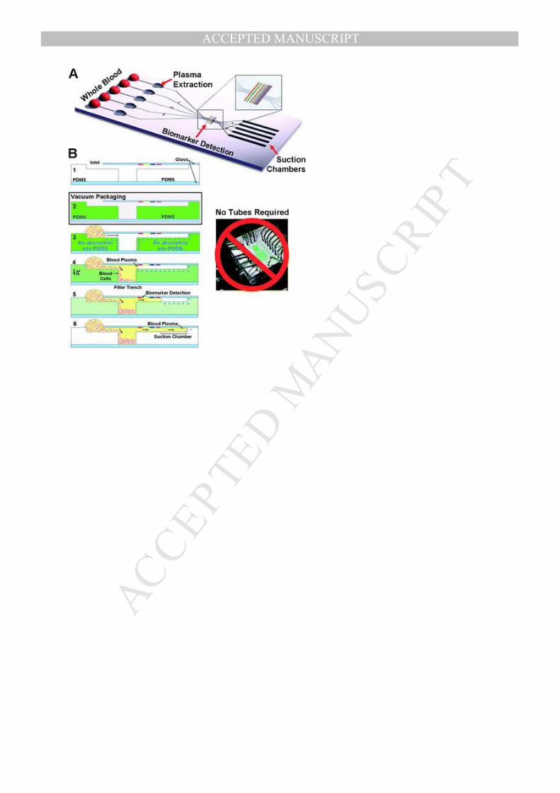
MA
NU
SC
RIP
T
AC
CE
PTE
D
ACCEPTED MANUSCRIPT
ACCEPTED MANUSCRIPT

MA
NU
SC
RIP
T
AC
CE
PTE
D
ACCEPTED MANUSCRIPT
ACCEPTED MANUSCRIPT