bronchogenic cysts in adults: ct mr and pathologic findings · than muscle on t1 -weighted mr...
TRANSCRIPT

Journal of the Korean Radiol ogical Society, 1995: 32(3) ’ 423- 428
Bronchogenic Cysts in Adults: CT, MR, and Pathologic Findings1
Jeung Sook Kim, M.D. , Joong MoAhn, M.D., Kyung Soo lee, M.D.
Bronchogenic cysts can arise either in the mediastinum or in the lung parenchyma. On both CT and T1 -weighted MR images, the cyst contents can show a variable signal characteristics . However, on T2 -weighted MR images, the cyst show high signal intensity. Although unusual , the cyst may communicate with the tracheobronchial tree and demonstrate partial or complete air content . With hemorrhage, fluid-fluid levels can also be seen within the cyst. Pathologi- cally,
the diagnosis of bronchogenic cyst rests on demonstration of tissues normally fou nd in the tracheobronchial tree within the cyst wal l.
Index Words : Mediastinum , neoplasms Lung , cysts Lung , CT Mediastinum. CT
INTRODUCTION
Bronchogenic cysts are closed epithelial-lined structures which are believed to represent localized airway segments which have branched abnormally and separated from the tracheobronchial tree during embryogenesis (1 , 2). If the abnormality occurs during early gestation , mediastinal bronchogenic cysts occur. Lung parenchymal bronchogenic cysts result from abnormal branching later in gestation. When examined with computed tomography (CT) , most bronchogen ic cysts are round and show homogeneous attenuation. However, the measured attenuation values of the cyst fluid can range from water (Iess than 20 HU) to soft tissue (greater than 20 HU) (3 , 4). Approximately 50 % of bronchogenic cysts will demonstrate water attenuation values allowing confident diagnosis on CT.
On magnetic resonance (MR) , bronchogenic cysts demonstrate variable signal intensity on T1 - weighted sequences , but characteristically have high signal intensity on T2 -weighted sequences (5, 6). Although bronchogenic cysts are pathologically benign , they may be associated with pericardial defects (7) , localized hypoplasia of the lung parenchyma (8) , or superim-
' Department ofD iagnostic Imag ing, Sam sung M edical Center
Rece ived December 28 , 1994 ; Accepted March 2, 1995
Address reprint requests to : Kyung Soo Lee, M.D. , Departm ent 01 Diagnosti c
Imaging Samsung Medical Center , n 50 Irwon.dong, Kangnam-ku , Seoul , 135
230 Korea. Tel. 82.2. 3410.2516 Fax. 82- 2. 341 0- 2559
posed infection (1). The aim of this pictorial essay is to illustrate the spectrum of findings associated with bronchogenic cysts on CT and MR images and on pathologic specimens
MATERIALS and METHODS
We reviewed all cases of bronchogenic cysts diagnosed at two institutions (Vancouver General Hospital and Samsung Medical Center) between January 1986 and December 1994. Twenty one bronchogenic cysts were identified. Pathologic confirmation was obtained in 11 cases with surgical excision . In the remaining ten cases , the diagnosis was based on an appropriate clinical presentation and characteristic CT or MR findings. The patients included four men and 17 women ranging in age from 19 to 74 years (average , 44 years) In 19 cases , chest CT was performed on a GE 9800 scanner (GE Medical Systems, Milwaukee) using standard technical parameters (120 kVp , 200 mA, 2 sec scan) and 10 mm collimated sections at 10 mm spacings. Intravenous contrast was administered in 14 cases.
In 12 patients , MRI was performed on a 1.5T clinical imager (GE Signa, GE Medical Systems, Milwaukee) using cardiac gated spin-echo sequences in a variety of imaging planes. In all studies , both T1 - weighted (TR 645 -1 000 msec, TE 20 msec) and proton density and T2 -weighted images (TR 2000-2769msec, TE 20 - 30 /60-120msec) were obtained. Respiratory compensation , two or fou r excitations , and a 192 by 256 matrix
낌

Journal of the Korean Radiological Society, 1995; 32(3) ‘ 423- 428
were used. Spatial presaturation was employed in selected imaging planes to reduce the artifact from flowing blood
AII pathologic specimens were reviewed by a single experienced surgical pathologist.
Fig . 1. Bronchogenic cyst in a 58-year-old woman. Contrast enhanced CT scan obtained at thoracic inlet shows spherical mass with homogeneous fluid attenuation (8 HU) in right paratracheal region
a
c
b
RESULTS
Mediastinal bronchogenic cysts Seventeen of 21 bronchogenic cysts were located
within the mediastinum. Ten cysts were situated within 3 cm of the tracheal carina, with the remaining seven in the lower paraesophageal region. Mediastinal bronchogenic cysts ranged in size from 2.1 to 6.0 cm in their largest dimension (average , 4.6 cm). CT scans were available in 15 of 17 cases. On CT the cyst fluid had an average attenuation value ranging from 2.4 to 73 HU. Six cysts showed attenuation similar to that of water (Iess than 20 HU) (Figs. 1, 2) and nine had attenuation similar to that of soft tissue (greater than 20 HU) (Fig. 3, 4). Calcification was not detected in any of these cysts
MRI was performed in 12 mediastinal bronchogenic cysts. On T1 - weighted images, six cysts had signal intensity similar to that of muscle, and six had signal intensity greater than muscle. In ten cases in which both CT and MR were performed , cysts with water attenuation on CT (Iess than 20 HU) had signal intensity on T1 - weighted MR sequences similar to that of muscle (Fig . 2). Conversely , cysts with soft tissue attenuation on CT (greater than 20 HU) had higher signal intensity
Fig. 2. B ronchogenic cyst associated with partial pericardial de-fect in a 26-year-old woman a. Contrast enhanced CT scan performed at subcarinal level shows dumbbell shaped mass with low attenuation. b. Transverse T1 -weighted (TR 645, TE 20) MR image demonstrates mass with signal intensity similar to that of chest wall muscle. There is marked narrowing of right pul monary artery c. Transverse T2-weighted (TR 2581 , TE 90) MR image demonstrates homogeneous high signal intensity of mass
424 -

than muscle on T1 - weighted MR images (Fig. 4). These
findings are consistent with previous reports of high
signal intensity on T1 - weighted MR images in bron-
Jeung Sook Kim, et al : Bronchogenic Cysts in Adults
chogenic cysts with high protein content (9). Although variable signal intensity was seen on T1 -
weighted images, all mediastinal bronchogenic cysts
a b Fig. 3 . Mediastinal bronchogenic cyst in a 19-year-old woman
a. Contrast enhanced CT scan obtained at level of right atrium shows slightly inhomogeneous mass with attenuation similar to that of
soft tissu e
b. Photograph of the gross specimen demonstrates spherical cystic mass with smooth glistening capsule. On needle puncture mucoid
fluid was obtained (arrow).
c
Fig. 4. Bronchogenic cyst in a 35-year-old woman
a. Contrast enhanced CT scan obtained at level of bronchus
intermedius shows pear-shaped mass with soft tissue attenu
ation
b. Coronal T1 - weighted (TR 923, TE 20) MR image shows sub
carinal mass with high signal intensity. Carinal angle is widened
bymass
c. Transverse T2 - weighted (TR 2769, TE 100) MR image obtained
at same level as “ a " demonstrates homogeneous high signal in
tensity mass
… ι
Journal of the Korean Radiological Society, 1995; 32(3) : 423- 428
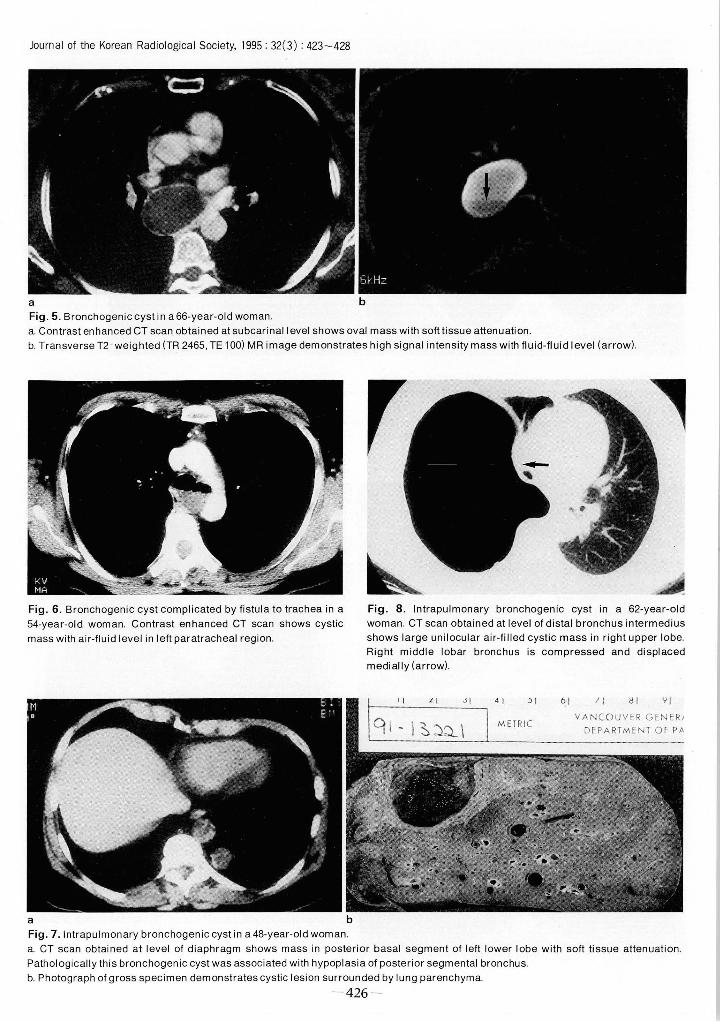
a b Fig. 5 . Bronchogenic cyst in a 66-year-old woman a. Contrast enhanced CT scan obtained at subcarinallevel shows oval mass with softtissue attenuation. b. Transverse T2 - weighted (TR 2465 , TE 1 00) MR image demonstrates high signal intensity mass with fluid-fl uid I evel (arrow)‘
Fig. 6 . Bronchogenic cyst complicated by fist미 a to trachea in a Fig. 8. Intrapulmonary bronchogenic cyst in a 62-year-old 54-year-old woman. Contrast enhanced CT scan shows cystic woman. CT scan obtained at level of distal bronchus intermedius mass with air-fluid level in left paratracheal region. shows large unilocular air-filled cystic mass in right upper lobe
Right middle lobar bronchus is compressed and displaced medi ally (arrow)
11 ι JI
a b Fig . 7. Intrapulmonary bronchogenic cyst in a 48-year-old woman
:> I 61 ! 1 !J 1 YI
VANCOUVER GE NER J DEPARTMENT Of P;'
a. CT scan obtained at level of diaphragm shows mass in posterior basal segment of left lower lobe with soft tissue attenuation Pathologically this bronchogenic cyst was associated with hypoplasia of posterior segmental bronchus
b. Photograph of gross specimen demonstrates cystic lesion surrounded by lung parenchyma
- 426 -

Jeung Sook Kim, et al : Bronchogenic Cysts in Adults
i a b Fig . 9. Congenital cystic adenomatoid malformation appearing as multiseptated cystic mass in a 35-year-old woman a. 10 mm collimation CTscan obtained at level of inferior p비 monary vein shows multiseptated cystic mass containing multilple air-fluid levels in left lower lobe. Partial atelectasis of remaining left lower lobe is observed posteriorly b. Photograph of gross specimen of lesion shows multiseptated cystic mas in anteromedial basal segment of left lower lobe
demonstrated high signal intensity on T2 - weighted images. In addition , in one case T2 - weighted images showed a fluid-fluid level within the cyst (Fig. 5) which was not identifiable on either T1 - weighted MR image or on CT. At surgery, this cyst contained dark brown fluid suggesting previous hemorrhage.
In one case , a mediastinal bronchogenic cyst was complicated by fistula formation to the distal trachea resulting in an air-fluid level (Fig. 6). Another mediastinal bronchogenic cyst was associated with a partial pericardial defect which created an unusual dumbbell shaped cyst with both intra- and extrapericardial components (Fig. 2). The intrapericardial component compressed the right pulmonary artery , with resultant diminished blood flow to the right lung on a quantitative nuclear medicine perfusion scan.
Intrapulmonary bronchogenic cysts Four of 21 bronchogenic cysts were intrapulmonary.
These cysts ranged in size from 2.5 to 16 cm in diameter (average, 7 cm). These cysts were located in the right upper lobe (n=1 ), Ieft upper lot갱 (n=1) , right lower lobe (n=1) and the left lower lobe (n=1) , respectively. Three cysts contained fluid and one was completely air filled. On CT, fluid-filled cysts showed attenuation similar to that of soft tissue (Fig. 7). A calcified wall was seen in one of the fluid-filled cysts. The air-filled cyst had a smooth thin wall (Fig. 8) . MRI was not obtained in any case of intrap비 monary bronchogenic cyst. Intrap비
monary bronchogenic cyst should be differentiated from congenital cystic adenomatoid malformation on CT scan (Fig. 9) .
PATHOLOGY
Pathologically , all bronchogenic cysts were spheri cal unilocular masses covered with a smooth and
glistening capsule (Fig. 3, 7). They contained thick mucus and were lined by ciliated columnar epithelium with focal or extensive areas of squamous metaplasia and chronic inflammatory infiltrates. The pathologic diagnosis of bronchogenic cyst rested on the identifi- cation of one or more of the tissues normally found in the tracheobronchial tree within the cyst wall , such as hyaline cartilage, smooth muscle, bronchial mucus glands or nerve trunks
Acknowledgments: We sincerely thank Drs. Müller and Mayo at Vancouver General hospital to permit us to include their cases of bronchogenic cysts in this essay.
REFERENCES
1. St. Georges R, Deslauriers J, Duranceau A, et al. Clinical spectrum of bronchogenic cysts 01 the mediastinum and lung in the adult. Ann Thorac Surg 1991 ; 52 : 6-13
2. Salyer DC, Salyer WR, Eggleston JC. Benign developmental cysts 01 the mediastinum. Arch Pathol Lab Med 1977 ;101 ‘
136-139 3. Mendelson DS, Rose JS, E/remidis SC, Kirschner PA , Cohen BA
Bronchogenic cysts with high CT numbers. AJR 1983 ; 140 463-465
4. Yernault J-C, Kuhn G, Dumortier P, Rocmans P, Ketelbant P,
Vuyst PD “ Solid" mediastinal bronchogenic cyst : mineralogic analysis. AJR 1986; 146: 73-74
5. Naidich DP , Rumancik WM ‘ Ettenger NA, et al. Congenital anomal ies ofthe lungs in adults : MR diagnosis. AJR 1988 ; 151 1-19
6. Nakata H, Egashira K, Watanabe H, et al. MRI 01 bronchogenic cysts. JComput AssístTomogr1993 ; 17 : 267-270
7. Kwak DL, Stork WJ , Greenberg SD. Partial delect 01 the pericardium associated with a bronchogeniccyst. Radíology 1971 ; 1 01 287-288
8. Panicek DM , Heitzman ER , Randall PA, et al. The continuum 01 P비monary developmental anomalies. RadíoGraphícs 1987 ; 1 747-772
427 -

Journal of the Korean Radiological Society, 1995 : 32(3) : 423-428
9. Mitchell DG, Burk DL, Vinisty S, Rifkin MD. The biophysical basis 831-837
01 tissue contrast in extracranial MR imaging. AJR 1987:149
대 한 밤사 선 의 학회 지 1995: 32(3) : 423-428
성언 기관지 낭종:전산화단충촬영,자기공명영상및 병리소견1
I 삼성의료원 진단방사선과
김 정 숙·안 중 모·이 경 수
성인에서 병리 (n=l l) 및 특징적 임상소견과 영상소견 (n= lO) 으로 진단된 21예의 기관지낭종의 전산화단층촬영, 자기
공명영상 및 병리소견을 보고한다. 17예는 종격동에서 4려l는 폐실질에서 기인했으며 전산화단층촬영이나 T1 강조 자기공명
영상에서 낭종의 내용물은 다앙한 음영소견을 보였다. 하지만 T2 강조 자기공명영상에서는 모든 예에서 높은 음영을 보였다.
비록 드물지만 기관지 낭종은 기관 및 기관지와 교통할 수도 있으며 이 경우 낭종내 공기음영이 보일 수 있다. 낭종내 출혈
의 fluid-fluid level 도 볼 수 있다. 병리적 진 단은 남종벽내에서 기관 및 기관지의 정상 조직을 발견함으로써 가능하다.
428 -