bronchiolitis obliterans, bronchiectasis, and other ...j. clin. path., 1971, 24, 72-82 bronchiolitis...
TRANSCRIPT

J. clin. Path., 1971, 24, 72-82
Bronchiolitis obliterans, bronchiectasis, and othersequelae of adenovirus type 21 infection inyoung childrenD. M. 0. BECROFT
From the Princess Mary Hospital for Children, Auckland, New Zealand
SYNOPSIS A remarkably high incidence of bronchiectasis and other pulmonary sequelae was
observed in young children affected during an epidemic of severe lower respiratory tract infectionsapparently caused by adenovirus type 21. The histopathological findings are described in four cases
in which one or both lungs were obtained for examination at intervals ranging from two months to
three years after the acute infections. Widespread bronchiolar obliteration (bronchiolitis obliterans)was a striking finding in all four. The severity of bronchial inflammation and of bronchiectasis was
proportional to the time elapsed since the acute infections. Bronchiolar obliteration is a likely sequelof the necrotizing bronchiolitis which may occur during acute adenovirus infections. The role ofbronchiolar obliteration in the pathogenesis of bronchiectasis and other chronic lung disease isdiscussed. Adenoviruses may be a major cause of post-infectious bronchiectasis in childhood.
The symptoms of bronchiectasis in childhoodfrequently date from an acute respiratory infection,and in four large series these infections were con-sidered the likely initiating factor in 48% to 77% ofcases (Field, 1949; Strang, 1956; Swierenga, 1957;Clark, 1963). However, pulmonary sequelae arerare in these common infections, and the exactaetiology of illnesses recalled as 'pneumonia','bronchitis', 'measles', or 'whooping cough' is oftenin doubt in retrospective studies. Therefore thecontribution, individually and collectively, of thecommon bacterial and viral pathogens to subsequentchronic lung disease remains ill defined. The moreprecise diagnostic methods now available permitprospective studies, but few have been reported. Anepidemic of adenovirus type 21 infection in Aucklandduring 1965 provided an opportunity for such astudy. Over a six-month period 43 children aged 3 to18 months were admitted to hospital with infectionsdiagnosed initially as bronchiolitis, bronchitis, orbronchopneumonia. The distinctive feature of theepidemic was the protracted course of the illnessesin most children, waxing and waning over severalweeks. Lang, Howden, Laws, and Burton (1969)have described the clinical, virological, and radio-logical findings in 25 of these patients in whom therewas good evidence of advenovirus infection, thisevidence including the isolation of adenovirus typeReceived for publication 9 June 1970.
21 from 20 cases. Only eight of the 25 patients con-sidered by Lang et al (1969) appeared to recovercompletely. One child died during the acute stagesof the illness and 16 (64%) had residual lung changes.Five developed bronchiectasis. Of those with residuallung changes, one died and two have had pneu-monectomies. The pathological findings in thesethree children and in a third fatality from the largergroup of 43 cases will be described in this report.Widespread bronchiolar obliteration was found inall four and the relationship of this change to theknown acute effects of adenovirus infection and tothe induction of further sequelae will be discussed.
Case Reports
All four infants were admitted to The Princess MaryHospital for Children within a three-week period inAugust 1965.
CASE 1This 5-month-old Polynesian boy was admittedbecause of cough and wheeze for four days. He hadhad a morbilliform rash one week before. A diag-nosis of bronchiolitis, pneumonia, and mild otitismedia was made on admission. Radiographs of thechest showed slight overinflation and increasedmarkings in both lung fields and consolidation ofthe right lower lobe. His weight was at the 25th
72
on April 1, 2020 by guest. P
rotected by copyright.http://jcp.bm
j.com/
J Clin P
athol: first published as 10.1136/jcp.24.1.72 on 1 February 1971. D
ownloaded from

Bionchiolitis obliterans, bronchiectasis, and other sequelae ofadenovirus type 21 infection in young children 73
percentile for age. The white blood count was10,500/cmm (neutrophils 62%) and there was amild hypochromic anaemia. Staphylococcus pyogeneswas cultured from the throat. He was treated withpenicillin and then with cloxacillin and ampicillin.Fever subsided gradually over 10 days, but he con-tinued to wheeze and oxygen was given intermit-tently for cyanosis. After four weeks, wheezingbecame exacerbated and he required oxygen con-tinuously for several days. Radiographs at this timeshowed collapse of the lower lobe of the right lungand overinflation elsewhere. His condition fluctuated,but wheeze was always present and oxygen wasrequired intermittently. Antispasmodics and anti-biotics were continued with little effect. Sweatelectrolyte levels were normal. Sixty-one days afterhis admission there was a further exacerbation ofsymptoms which, in contrast to earlier episodes, wasaccompanied by high fever. Further treatment withantibiotics and intravenous hydrocortisone had noeffect and he died on the next day.
Macroscopic findings at necropsyThe pleural cavities were normal. The right lungweighed 120 g, the left lung 110 g. Both lungs werevoluminous except for the right lower lobe whichwas collapsed and firm. In the remaining lobes paleemphysematous areas alternated with others whichwere firm and intensely congested or haemorrhagic.The bronchi in these lobes contained bloodstainedfluid. The mucosa of bronchi throughout the lungswas thickened and congested but there was nobronchiectasis. Hilar lymph nodes were slightlyenlarged and fleshy. The heart weighed 50 g. Therewas dilatation of the right ventricle and some hyper-trophy of the ventricular wall. All other organs werenormal.
CASE 2This 9-month-old Maori boy was admitted with athree-day history of cough and wheezing. He hadbeen in hospital with similar symptoms one monthbefore when a diagnosis of acute wheezing bronchitishad been made, but he had appeared to recovercompletely and had remained well for three moreweeks. A diagnosis of bronchopneumonia was madeon the second admission. The radiographs of thechest showed overinflation of the lungs and inter-stitial inflammatory changes, but after a few dayshe developed clinical and radiological signs of awidespread patchy pneumonic consolidation in bothlungs. His weight was at the 10th percentile. He hadhypochromic anaemia with a haemoglobin of6-9 g/100 ml and a white blood count of 7,200/cmm(68% neutrophils). No bacterial pathogens wereisolated from a throat swab. He was treated with
cloxacillin and ampicillin and the pneumoniaresolved slowly during the next three weeks. How-ever, wheeze persisted and he was cyanosed at times.Continuous wheezing and overinflation of the lungswere the major clinical and radiological features ofthe remainder of his illness. He was afebrile most ofthe time and antibiotics, antispasmodics, and adrenalcorticosteroids had no effect. Serum gamma globulinlevels were normal. Five weeks after admission hedeveloped a paroxysmal tachycardia and two weekslater there was a cardiac arrest. After he was resusci-tated his respirations were assisted with intermittentpositive pressure for one week and he had a trach-eostomy. He did not regain consciousness and diedin respiratory and cardiac failure four months afterhis second admission.
Macroscopic findings at necropsyThe pleural cavities were normal. The trachea belowthe tracheostomy and the main bronchi containedpus. The right lung weighed 130 g, the left lung 100 g.Both upper lobes were voluminous due to emphy-
Fig. 1. The cut surface of the formalin-fixed right lungof case 2, four months after the acute infection. Thicken-ing of bronchial walls, bronchiectasis, fibrosis, emphysemaand a bleb of bronchiolar origin are apparent.
on April 1, 2020 by guest. P
rotected by copyright.http://jcp.bm
j.com/
J Clin P
athol: first published as 10.1136/jcp.24.1.72 on 1 February 1971. D
ownloaded from

D. M. 0. Becroft
sema which included a few small blebs up to 4 mmin diameter (Fig. 1). There were small consolidatedfoci near the hilum of these lobes and more extensiveareas of consolidation or collapse in the remaininglobes. Both lower lobes were reduced in volume andthe bronchi were crowded together. Large andmedium-sized bronchi in all lobes had thickenedwalls and irregular pus-filled lumina and there wasobvious bronchiectasis in the partly collapsed lowerlobes.The heart weighed 100 g and there was hyper-
trophy of the right ventricular wall. The brainweighed only 680 g and showed extensive gliosis andcyst formation through the grey matter of thecerebral cortex and basal nuclei. The brain stem wasnormal as were other organs.
CASE 3Nine days before admission this 11-month-oldCaucasian boy developed a rash and was consideredto have measles. The rash subsided. but was followedby fever, cough, and dyspnoea. On admission, ribswere retracted, and moist sounds were heard onauscultation all over the chest. A diagnosis ofpneumonia was made. A radiograph of the chestshowed abnormal markings compatible with peri-bronchial inflammatory changes. His weight was atthe 3rd percentile and he had a mild hypochromicanaemia. The white blood count was 27,400/cmm(91 % neutrophils). No bacterial pathogens wereisolated from a throat swab. He was treated withpenicillin and chloramphenicol, but fever persistedand after three days his condition deteriorated, withsevere wheezing and signs suggestive of right heartfailure. Radiographs showed collapse-consolidationof the right lung with mediastinal displacement. Hewas treated with digoxin, cloxacillin, and strepto-mycin, and slowly improved. Fever subsided within aweek of his admission but wheezing continued andthere were residual changes in the radiographs of there-expanded right lung when he was discharged after26 days in hospital. He was readmitted only fourdays later because of a recurrence of fever and severedyspnoea and wheezing. Radiographs of the chestshowed generalized overinflation and an increase inthe inflammatory changes in both lung fields. Hewas treated with cloxacillin and ampicillin and,although fever settled after three days, dyspnoeaincreased and he became cyanosed. Radiographsagain showed collapse of the right lung andmediastinal displacement, but there was no re-expansion after removal of thick secretions atbronchoscopy. He improved slowly and was dis-charged again after 11 weeks in hospital, but hadto be readmitted on several occasions because ofexacerbations of the respiratory symptoms. Further
Fig. 2. A bronchogram of the collapsed right lung ofcase 3, six months after the acute infection, showingwidespread saccular and varicose bronchiectasis.
bronchoscopies did not relieve the collapse-con-solidation of the right lung, and a bronchogram,performed six months after the onset of his illness,showed extensive bronchiectasis of this lung (Fig. 2).After one year a right pneumonectomy was per-formed at another hospital. After the pneumonec-tomy he had one further hospital admission with arespiratory infection, but then did not attend forfollow up.
Macroscopic findingsThe resected right lung was collapsed and markedlycongested. The right main bronchus was normal,but the major branches were obviously dilated,particularly in the lower lobe where the subdivisionscontained pus. The tracheobronchial lymph nodeswere enlarged and fleshy.
CASE 4This 10-month-old Polynesian boy had a history ofcough, anorexia, and drowsiness for eight days. Forthree days he had been treated with tetracycline for
74
on April 1, 2020 by guest. P
rotected by copyright.http://jcp.bm
j.com/
J Clin P
athol: first published as 10.1136/jcp.24.1.72 on 1 February 1971. D
ownloaded from

Bronchiolitis obliterans, bronchiectasis, andother sequelae ofadenovirus type 21 infection in young children 75
bronchitis and pneumonia and he was admittedhaving had a convulsion. On admission he had signsof consolidation at the base of the right lung andradiographs confirmed the presence of broncho-pneumonic changes in the right lung and in the leftlower zone. His weight was at the 25th percentile.He had a hypochromic anaemia, with a haemoglobinof 5-5 g/100 ml. The white blood count was 11,400/cmm (19% neutrophils). No bacterial pathogenswere isolated from a throat swab. Sweat, electrolyte,and serum gamma globulin levels were normal. Hewas treated with penicillin, cloxacillin, and ampicil-lin. The cerebrospinal fluid was normal, butsymptoms of central nervous system disease were ofmajor concern during the next three weeks. He hadpersistent fits and a paresis of the right arm fromwhich he recovered slowly. As the consolidationcleared from the right lung the radiological appear-ances changed to overinflation on this side. Therewas intermittent wheezing and cyanosis. Then, fourweeks after admission, radiographs showed collapseof the right lower lobe, followed two weeks later bycollapse of the whole of the right lung. The lung didnot expand after aspiration of secretions at broncho-scopy and tracheostomy. Bronchography, performed12 weeks after admission, showed saccular bron-chiectasis affecting all segments of the collapsedright lung and the posterior basal segment of theleft lower lobe.
CASE 5A child whose clinical course and radiologicalchanges were described by Lang et al (1969) as theircase 2 has had a right pneumonectomy nearly fiveyears after the acute illness. The lung weighed 140 gand was examined in greater detail than was pbs-sible in previous cases. The lower lobe expanded tonormal volume when inflated with air at a pressureof 40 cm H20. The upper and middle lobes wereatelectatic and could not be inflated. The possibilityof a persisting virus infection was investigated byestablishing fibroblast cultures from bronchialmucosa and alveolar tissue. Neither cell line showedcytopathic changes after six weeks in culture.Numerous sections of the lung were examined, manyserially. There was severe fibrous thickening andchronic inflammation of the walls of small bronchiin all lobes. Most of the terminal bronchioles werecompletely obliterated and replaced by radiatingfibrous scars accompanying smaller branches of thepulmonary arteries. As in the previous cases, manyof these scars contained remnants of smooth muscleand elastic tissue. Although obliteration was equallysevere in all lobes, most respiratory bronchioles,alveolar ducts, and alveoli in the lower lobe werewell aerated, whereas the upper and middle lobes
were atelectatic and hypoplastic. The radiologicalfindings had suggested that this extensive oblitera-tion had occurred during the first few weeks of thechild's illness. There appears to have been a narrowmargin between the degree of obliteration requiredto produce chronic atelectasis and that allowingsufficient collateral ventilation for aeration to bemaintained.He remained in hospital for many months having
occasional exacerbations of respiratory symptoms.The right lung remained collapsed and a rightpneumonectomy was performed three years later.He was well, but had signs persisting in the left chest,one year after the operation.
Macroscopic findingsThe excised lung measured 11 x 9 x 5 cm in theinflated state. Large firm lymph nodes were presentanterior to the entering bronchi, the largest 2 cmacross. The bronchi in all lobes were considerablydilated and thick walled, the dilatation extending tothe pleural surfaces (Fig. 3). Fibrous adhesionscovered the pleural surface and there had beenrecent haemorrhage into the remaining lungparenchyma.
Fig. 3. Severe bronchiectasis and peribronchial fibrosisin the right pneumonectomy specimen from case 4 threeyears after the acute infection.
Histology
Several blocks of tissue were taken from each lobeof the lungs of case 1 and one or more blocks fromeach lobe of case 2. Single blocks of tissue wereavailable from the right middle and lower lobes ofcase 3 and from all three lobes of the right lung ofcase 4. Standard histological procedures were used
on April 1, 2020 by guest. P
rotected by copyright.http://jcp.bm
j.com/
J Clin P
athol: first published as 10.1136/jcp.24.1.72 on 1 February 1971. D
ownloaded from

D. M. 0. Becroft
for a variety of stains applied to sections from bothparaffin-embedded and frozen material. Serialsections were examined from selected blocks of thelungs of cases 1 and 2.
THE LOWER RESPIRATORY TRACTSLarge and medium-sized bronchiThe thickening of the bronchial walls in each casewas due to chronic inflammation and fibrosis whichinvolved all layers to some extent, but ranged inseverity from a predominantly mucosal change incase 1 to an advanced full-thickness disorganizationtypical of severe bronchiectasis in case 4. Thechanges in cases 2 and 3 were of intermediateseverity and there was a clear correlation betweenthe amount of destructive change and the time sincethe initial infection. The lamina propria of themucosa in each case was expanded by vascularfibrous connective tissue. Plasma cells and lympho-cytes infiltrated this tissue, often heavily, butlymphoid follicles were seen in case 4 only. Themucosal surfaces of many large bronchi in cases 2,
P.zir M.: - F " _.I W la W
Fig. 4. Case 2. The proliferating epithelium lining alarge pus-filled bronchus. The mitoses include a tri-aster.H&E x370.
3, and 4 were raised into a series of tall folds andpapillary projections and many bronchial luminawere filled with purulent exudate. The greater partof the bronchial epithelium was abnormal. The mostfrequent change was simple hypertrophy with hyper-plasia of goblet cells, but metaplasia into a many-layered, stratified, and otherwise undifferentiatedepithelium was a feature of the mucosa of all largebronchi in case 2. This epithelium also showed focalulceration, intense mitotic activity, and a fewatypical metaphases (Fig. 4). No viral inclusionswere detected in the bronchial epithelium, nor atother levels of the respiratory tract in any of the fourcases.
In case 1 the deeper layers of the bronchial wallswere infiltrated by lymphocytes and plasma cells,but there was little fibrosis. In cases 2 and 3 thecellular infiltration was more intense and there wasa variable fibrosis extending into the peribronchialtissues. Muscle and elastic tissue was disorganizedin the walls of the more dilated bronchi but cartilagewas unaffected. Cartilage, muscle, elastic tissue, andglands were largely destroyed in the walls of thegreatly dilated bronchi of case 4. Peribronchialfibrosis and an inflammatory reaction, whichincluded many lymphoid follicles, had incorporatedmuch of the surrounding lung parenchyma in thiscase.The loss of glands in case 4 was in keeping with
the severity of other changes in the bronchial walls,but in the other cases glandular involvement ap-peared disproportionately severe. This applied par-ticularly in case 1 where many peripherally placedlobules were replaced by fibrous tissue containingchronic inflammatory cells, leaving only remnantsof acinar spaces lined by undifferentiated cuboidalcells.
Small bronchi and bronchiolesObliteration or stenosis of bronchioles and smallbronchi was the most striking histological finding inthese cases, particularly in cases 1 and 2 in whichboth lungs were available for full examination.Obliterative changes were found in all sections fromthese cases and affected most of the smaller air pas-sages over large portions of all lobes. The appear-ances illustrated in Fig. 5 were typical of theoccluded air passages in cases 1, 2 and 3, in whichthe mucosa was usually completely destroyed andthe lumina filled with vascular fibrous tissue. Onlythe preservation of typically arranged smooth muscleand elastic fibres at the periphery allowed the identi-fication of many of these structures. Others con-tained clumps of epithelial cells or gland-like spaces.Inflammatory cells, usually lymphocytes and plasmacells, were sparse, Serial sections showed that the
76
on April 1, 2020 by guest. P
rotected by copyright.http://jcp.bm
j.com/
J Clin P
athol: first published as 10.1136/jcp.24.1.72 on 1 February 1971. D
ownloaded from

Bronchiolitis obliterans, bronchiectasis, and other sequelae ofadenovirus type 21 infection in young children 77
Fig. 5b
Fig. 5. Obliterative changes typical of the majorityof bronchioles from (a) case 1, (b) case 2, and (c) case 3,two, four, and 12 months respectively after acuteinfections. A fragment of epithelium remains in (b) butotherwise the smooth muscle is the only identifyingfeature. H & E (all x 120).
Fig. Sc5
on April 1, 2020 by guest. P
rotected by copyright.http://jcp.bm
j.com/
J Clin P
athol: first published as 10.1136/jcp.24.1.72 on 1 February 1971. D
ownloaded from
D. M. 0. Becroft
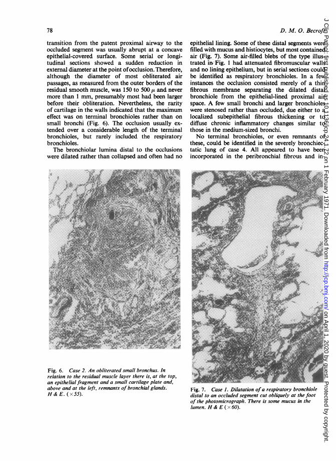
transition from the patent proximal airway to theoccluded segment was usually abrupt at a concaveepithelial-covered surface. Some serial or longi-tudinal sections showed a sudden reduction inexternal diameter at the point ofocclusion. Therefore,although the diameter of most obliterated airpassages, as measured from the outer borders of theresidual smooth muscle, was 150 to 500 ,u and nevermore than 1 mn, presumably most had been largerbefore their obliteration. Nevertheless, the rarityof cartilage in the walls indicated that the maximumeffect was on terminal bronchioles rather than onsmall bronchi (Fig. 6). The occlusion usually ex-tended over a considerable length of the terminalbronchioles, but rarely included the respiratorybronchioles.The bronchiolar lumina distal to the occlusions
were dilated rather than collapsed and often had no
Fig. 6. Case 2. An obliterated small bronchus. Inrelation to the residual muscle layer there is, at the top,an epithelial fragment and a small cartilage plate and,above and at the left, remnants of bronchial glands.H & E. (x 55).
epithelial lining. Some of these distal segments werefilled with mucus and histiocytes, but most containedair (Fig. 7). Some air-filled blebs of the type illus-trated 'HI'Fig. 1 had attenuated fibromuscular wallsand no lining epithelium, but in serial sections couldbe identified as respiratory bronchioles. In a fewinstances the occlusion consisted merely of a thinfibrous membrane separating the dilated distalbronchiole from the epithelial-lined proximal airspace. A few small bronchi and larger bronchioleswere stenosed rather than occluded, due either to alocalized subepithelial fibrous thickening or todiffuse chronic inflammatory changes similar tothose in the medium-sized bronchi.No terminal bronchioles, or even remnants of
these, could be identified in the severely bronchiec-tatic lung of case 4. All appeared to have beenincorporated in the peribronchial fibrous and in-
4-'
Fig. 7. Case 1. Dilatation ofa respiratory bronchioledistal to an occluded segment cut obliquely at the footof the photomicrograph. There is some mucus in thelumen. H & E ( x 60).
78
on April 1, 2020 by guest. P
rotected by copyright.http://jcp.bm
j.com/
J Clin P
athol: first published as 10.1136/jcp.24.1.72 on 1 February 1971. D
ownloaded from

Bronchiolitis obliterans, bronchiectasis, and other sequelae ofadenovirus type 21 infection in young children 79
flammatory tissue which replaced much of the lungand extended to near the pleural surfaces. Respira-tory bronchioles, some mucus-filled, remained in thesmall amount of surviving alveolar tissue.
AlveoliThe greater part of the alveolar tissue was abnormal.Emphysema alternated with smaller areas ofatelectasis and fibrosis in relation to the extensivebronchiolar obliteration in the lungs of cases 1 and 2.Emphysema was particularly extensive in the upperlobes of cases 1 and 2 and was of obstructive typewith little or no destruction of alveolar walls. Thefibrotic areas in the lungs of case 2, and more par-ticularly in case 1, often had a lobular distribution.Alveolar walls were diffusely thickened, the liningcells were cuboidal, and the alveolar lumens werefilled with clumps of large macrophages many ofwhich contained cholesterol. In case 1 both thefibrotic and non-fibrotic lung tissue showed focalinfiltration by neutrophils, extensive fresh haemor-rhage, and fibrinous exudation, changes which weretaken as evidence for a terminal acute infection.In all cases the alveoli surrounding inflamed oroccluded bronchioles and bronchi were lined bycuboidal cells. Apart from this change the alveolartissue in the few sections examined from case 3showed atelectasis only. The massive peribronchialfibrosis in case 4 included many alveolar remnantslined by cuboidal cells, while the residual alveolartissue, situated mainly in the subpleural regions,was normal apart from a minor degree of collapseand recent haemorrhage.
VesselsThere was an endarteritis severely narrowing manysmall pulmonary arteries in case 2 and similar
changes involving both pulmonary and bronchialarteries of case 4. Vessels in other cases were normal.
Tracheo-bronchial lymph nodesIn all cases these showed well developed, but non-specific, reactive changes.
OTHER ORGANS OF CASES 1 AND 2Apart from chronic inflammation and squamousmetaplasia in the tracheal mucosa there were nochanges of note in the upper respiratory tracts. Thecortical grey matter of the brain of case 2 showedextensive gliosis and cyst formation consistent withanoxic injury. Other organs were unremarkable.
Virology
The methods of virological investigation and addi-tional bacteriological and epidemiological datahave already been presented by Lang et al (1969).The results of virological studies on the four casesof this report are presented in the Table. Type 21adenoviruses were isolated from throat swabs takenfrom cases 2, 3, and 4 within a few days of theiradmission to hospital. The isolation from case 2 wasat his second admission and, although relapses werecommon during the epidemic, the interval betweenthe two admissions in this case was more than threeweeks and the two were assumed to be unrelated.High titres of complement-fixing antibody to adeno-viruses were demonstrated in convalescent serafrom cases 2, 3, and 4. These results were suggestiveof recent infection, but an actual rise in titre fromthe acute phase was demonstrated in case 4 only.No other viruses were isolated, and the high titresof antibody to para-influenza virus type 3 in con-
Case Virus Isolations Complement-fixing Antibodies (Reciprocals of Dilutions)No.
Specimen Day' Virus Acute Serum Convalescent SerumObtained Isolated
Dayl Adeno- RSV Pl P2 P3 Day' Adeno- RSV Pl P2 P3Obtained virus Obtained virus
1 Lung 62 - 3 <16 <16 <8 <8 <16 NT NT NT NT NT NTFaeces 62
2 Throat swab 5 Adenovirus 6 NT <16 NT NT NT 34 128 <16 <8 <8 128type 21
3 Throat swab I Adenovirustype 21
Throat swab 31 - NT NT NT NT NT NT 32 4096 >16 >8 >8 >256Throat swab 110 Adenovirus
type 214 Throat swab 3 Adenovirus 3 <16 <16 <8 <8 <16 512 <16 <8 <8 128
type 21
Table Results of virological studies'Days after admission.NT = not tested.PSV Respiratory syncytial virus.RI. P2, P3 - Parainfluenza viruses types 1, 2, and 3 respectively.
on April 1, 2020 by guest. P
rotected by copyright.http://jcp.bm
j.com/
J Clin P
athol: first published as 10.1136/jcp.24.1.72 on 1 February 1971. D
ownloaded from

D. M. 0. Becroft
valescent sera from these and other cases in theepidemic are unexplained.
Discussion
The pathological changes in these four children areconsidered to be the sequelae of infections byadenovirus type 21 which were acquired from twomonths to three years previously in individual cases.All were admitted to hospital at the height of theepidemic of adenovirus type 21 infection withclinical features resembling those of the other casesdescribed by Lang et al (1969). Adenovirus type 21was isolated and supporting serological evidenceobtained in the three cases in which these investiga-tions were carried out during the early part of theillnesses.One feature common to the early clinical course
of each case was that some radiological and clinicalimprovement had occurred before a serious impair-ment of ventilatory capacity became evident. Allhad severe wheezing and were cyanosed withoutoxygen at a time when fever had subsided and radio-graphs of the lungs showed hyperinflation but littlecollapse or consolidation. The persistence of thesesigns of obstructive lung disease in cases 1 and 2until their deaths and for months in cases 3 and 4correlates well with the dominant pathological find-ing of total or partial obliteration at bronchiolarand small bronchial level.
'Bronchiolitis obliterans', with a similar biphasicclinical course, has been described after exposure totoxic gases, after influenza. and other unspecifiedinfections, and also in cases where no cause wasapparent (Spencer, 1968). Laurent, Dalloz, andNezelof (1966) reported the isolation of adenovirustype 5 from a 10-month-old child dying of bronchio-litis obliterans but, nevertheless, favoured theinhalation of some unknown toxin as the cause. Theassociation with adenovirus infection cannot beeasily dismissed in the present series of cases.Furthermore, the obliterative changes would be thesequelae predicted from what is known of the acuteeffects of adenovirus infections on the lower respira-tory tract.The histological findings in five cases of fatal
infection by adenovirus type 7 in Auckland havealready been described (Becroft, 1967). In this paperreferences were given to descriptions of a similarpathology found in more than 60 infants in whomd,eath was attributed to infections by adenovirus ofserotypes 7 or 3, while later reports have addedmore cases and expanded the age range affected andthe number of serotypes concerned (Levin, Dietrich,and Guillory, 1967; Angella and Connor, 1968;Steen-Johnsen, Orstavik, and Attramadal, 1969).
The characteristic features have been a combinationof necrotizing bronchitis, bronchiolitis, and pneu-monia, necrosis of bronchial mucous glands, andthe presence of intranuclear inclusions in manysurviving cells. In the Auckland cases with type 7infection no respiratory epithelium had survived insome blocks of lung tissue and many bronchiolarand bronchial lumina were filled with exudate fromthe ulcerated surfaces. The failure of reconstitutionof ulcerated epithelium and the organization ofexudate retained preferentially in smaller air pas-sages wou!d lead to obliterative changes similar tothose described above. Progression of this change,with cicatrization, episodic obstruction by retainedsecretions, and secondary infections could accountfor a fluctuating clinical course in the absence ofpersisting viral infection.The necrosis which has been observed in the acute
stages of adenovirus infection has not extendeddeeply to involve the cartilage or muscle layers. Thiscan be correlated with the preservation of smoothmuscle in the obliterated bronchioles of the casesof this report and the absence of major destructivechanges in the deeper layers of the bronchial walls.Obliteration of bronchial glands was dispropor-tionately severe in these cases and there is likelycorrelation with the consistent finding of necrosis ofthese glands in the acute type 7 infections (Becroft,1967). A prior necrotizing pneumonia, resemblingthat found in the acute infections, is a possible causeof the alveolar fibrosis in cases 1 and 2 above, butthe fibrotic areas also had some features of obstruc-tive pneumonitis, including cholesterol accumula-tion, and may have been secondary to bronchiolarocclusion (Spencer, 1968).Although the pathological findings in these cases
appear a logical sequel to acute necrotizing changessimilar to those caused by other adenovirus sero-types, and although there is no reason to believethat type 21 should differ in its acute affects, therehas been no direct demonstration of the latter. Thefew reports of adenovirus type 21 infections suggestthat the virus resembles other adenovirus serotypesin usually causing only minor upper respiratory,febrile, and ocular infections (Bell, Rota, andMcComb, 1960; van der Veen and Dijkman, 1962).Lower respiratory tract infections have been de-scribed (van der Veen and Dijkman, 1962) and singlefatal cases have been reported by Clarke, Corner,Gambier, Macrae, and Peacock (1964) and byCrandell, Dowdle, Holcomb, and Dahl (1968), butthe extensive pneumonia in the former was not de-scribed in detail and central nervous system diseasespredominated in the latter.The occurrence of clinical and radiological
sequelae in 60% of the surviving infants in the Auck-
80
on April 1, 2020 by guest. P
rotected by copyright.http://jcp.bm
j.com/
J Clin P
athol: first published as 10.1136/jcp.24.1.72 on 1 February 1971. D
ownloaded from

Bronchiolitis obliterans, bronchiectasis, and other sequelae of adenovirus type 21 infection in young children 81
land epidemic also appears to be exceptional fortype 21 infection and for adenovirus infectionsgenerally. A few cases with persisting pulmonarylesions have been described (Lamy, Frezal, andCohen-Solal, 1963; Clarke et al, 1964; Steen-Johnsen et al, 1969), but sequelae were not men-tioned in reports of other adenovirus epidemics(Sterner, 1962). The consistent finding of bronchiolarand bronchial obliteration in the pathologicalmaterial from the Auckland epidemic suggests thatthis was of major importance in the pathogenesis ofother pulmonary sequelae. The persistence ofatelectasis, obstructive pneumonitis, or emphysemawould be determined by the degree of bronchiolarobliteration and by the adequacy of collateral air-ways (McLean, 1958). In cases 1 and 2 emphysemawas the more usual finding distal to the occludedairways, implying that collateral pathways werepatent. Of the survivors, 10 (40%) had radiographsshowing permanently increased lung markings, withor without crowding of bronchi suggesting fibrosisor atelectasis. Three more, including cases 3 and 4,had massive collapse of a lung persisting despitevigorous treatment, presumably because collateralairways were inadequate. The sequence of consolida-tion, followed by overinflation and then massivecollapse of the right lung of case 4, is believed toreflect progression of obliterative changes duringresolution of the bronchopneumonia. Bronchiolarobliteration in infancy, with subsequent inhibitionof normal development of the lung, has been pro-posed as a cause of McLeod's syndrome of unilateralradiolucency of the lung (Reid, Simon, Zorab, andSeidelin, 1967). Although radiographs of somesurvivors showed focal or generalized emphysemano typical examples of McLeod's syndrome wereseen during the period of follow up.
Bronchiectasis was demonstrated at broncho-graphy in six1 of the surviving cases at intervals ofthree months to one year after the acute infection,and was found at the necropsy on case 2 four monthsafter the onset. Although bronchi in lobes known tobe chronically atelectatic were most severely affected,non-atelectatic segments were also involved in allcases, and bilateral changes were demonstratedwhenever both lungs were examined. The bronchiec-tasis was usually varicose or saccular and there wasa consistent lack of filling of the periphery of theaffected segments by contrast media, even in thoseexamined early and showing only slight dilatation.This change, sometimes due to retained secretions,but usually indicating bronchial and bronchiolarobliteration, has been well documented in manyradiological and pathological studies of bronchiec-tatic lungs (Reid, 1950; Culiner, 1963). The patho-'One case has been added to the five listed by Lang et al (1969).
logical findings in the present series of cases areconsidered to give support to the conclusion ofChurchill (1949) that 'obliterative bronchitis andbronchiolitis with or without atelectasis may besignificant in the development of certain types ofbronchiectasis and not merely be a by-product ofthe pathologic process'. Bronchiolar obliterationcould contribute to both factors considered bySpencer (1968) to be the main causes of bronchiec-tasis: first, to absorption, collapse, and fibrosis, andsecond, to inflammation, by predisposing anatomic-ally and physiologically to the stagnation of secre-tions. Thus, in addition to the bronchiolar obliterationthere was in case 1, two months after the acuteinfection, only moderate bronchial inflammation;case 2 after four months had severe bronchialinflammation and early bronchiectasis; case 3 afterone year had established bronchiectasis withoutgross destructive changes; and in case 4 after threeyears there was severe destructive bronchiectasis offollicular type in which the obliterative changescould easily be considered as secondary.No opinion can be offered on the role of persisting
viral infection in these cases. The virus was isolatedfrom case 3 four months after his acute illness, butwas not recovered from the lungs of case 1 after twomonths. The reactive changes in lymphoid tissueappeared non-specific, and although the atypicalepithelial proliferation in case 2 had some resem-blance to the virus-induced effect observed in theacute stages of adenovirus type 7 infection (Becroft,1967), no viral inclusions were detected.These cases provide strong evidence for an
adenoviral aetiology of some cases of childhoodbronchiectasis and for a pathogenesis based on aninitial necrotizing bronchitis and bronchiolitis. Themaximum incidence of the respiratory illnesseswhich have initiated symptoms in post-infectiousbronchiectasis has, in most series, been in the firsttwo years of life. Adenoviruses are an importantcause of lower respiratory tract infections at thisage, and of the other respiratory pathogens onlyinfluenza virus is recognized as causing epithelialnecrosis of comparable extent. Adenoviruses may,therefore, be the major cause of post-infectiousbronchiectasis, a possibility which has been sug-gested previously on the basis of serological studiesby MacFarlane and Sommerville (1957) and Rytel,Connor, Welch, Kraybill, Edwards, Rosenbaum.Frank, and Miller (1964). Morbilliform rashes andother exanthemata, a pertussis-like syndrome, and asimultaneous infection with measles virus have allbeen described in association with adenovirus infec-tion (Chany, Lepine, Lelong, Le-Tan-Vinh, Satge,and Virat, 1958; St. Geme and Prince, 1966; Collier,Connor, and Irving, 1966; Lang et al, 1969). Conse-
on April 1, 2020 by guest. P
rotected by copyright.http://jcp.bm
j.com/
J Clin P
athol: first published as 10.1136/jcp.24.1.72 on 1 February 1971. D
ownloaded from

D. M. 0. Becroft
quently, adenoviruses have also to be considered as
the possible aetiological agents in cases of bronchiec-tasis developing after apparent measles or whoopingcough. Clearly, only prospective studies based on
accurate initial diagnoses can give reliable informa-tion on the infectious causes of bronchiectasis.
I am grateful to Dr Grahame Fox, Dr Alice Bush,and Dr Ruthven Lang for permission to publishdetails of cases in their care; to Dr J. F. Burton forthe results of virological studies; to Dr F. H. Simsfor access to the descriptions of the surgical speci-mens and to the histological material from cases 3and 4; to Dr Geoffrey Dodd for reviewing radio-graphs; and to Mr A. D. Fraser and Mr R. J.Patterson for expert assistance with photographicand histological procedures respectively.
References
Angella, J. J., and Connor, J. D. (1968). Neonatal infection caused byadenovirus type 7. J. Pediat., 72, 474-478.
Becroft, D. M. 0. (1967). Histopathology of fatal adenovirus infectionof the respiratory tract in young children. J. clin. Path., 20,56 1-569.
Bell, S. D., Jr., Rota, T. R., and McComb, D. E. (1960). Adenovirusisolated from Saudi Arabia. III. Six new serotypes. Amer. J.trop. Med. Hyg., 9, 523-526.
Clark, N. S. (1963). Bronchiectasis in childhood. Brit. med. J., 1, 80-88.Clarke, S. K. R., Corner, B. D., Gambier, D. M., Macrae, J., and
Peacock, D. B. (1964). Viruses associated with acute respiratoryinfections. Brit. med. J., 1, 1536-1539.
Chany, C., Lepine, P., Lelong, M., Le-Tan-Vinh, Satg6, P., and Virat,J. (1958). Severe and fatal pneumonia in infants and youngchildren associated with adenovirus infections. Amer. J. Hyg.,67, 367-378.
Churchill, E. D. (1949). The segmental and lobular physiology andpathology of the lung. J. thorac. Surg., 18, 279-293.
Collier, A. M.,Connor, J. D.,and Irving, W. R. Jr. (1966). Generalisedtype 5 adenovirus infection associated with the pertussis syn-drome. J. Pediat., 69, 1073-1078.
Crandell, R. A., Dowdle, W. R., Holcomb, T. M., and Dahl, E. V.(1968). A fatal illness associated with two viruses: an inter-mediate adenovirus type (21-16) and influenza A. J. Pediat., 72,467-473.
Culiner, M. M. (1963). Obliterative bronchitis and bronchiolitis withbronchiectasis. Dis. Chest., 44, 351-361.
Field, C. E. (1949). Bronchiectasis in childhood. 1. Clinical survey of160 cases. Pediatrics, 4, 21-46.
Lamy, M., Frezal, J., and Cohen-Solal, J. (1963). Pneumopathiechronique associ6e a une infection par adenovirus. Arch. franc.Pediat., 20, 612-620.
Lang, W. R., Howden, C. W., Laws, J., and Burton, J. F. (1969).Bronchopneumonia with serious sequelae in children withevidence of adenovirus type 21 infection. Brit. med. J., 1, 73-79.
Laurent, M., Dalloz, J. C., and Nezelof, C. (1966). La bronchioliteobliterante: a propos d'un cas frappant un neurrisson de 10mois. Arch. Anat. Path., 14, 253-259.
Levin, S., Dietrich, J., and Guillory, J. (1967). Fatal nonbacterialpneumonia associated with adenovirus type 4. J. Amer. m7ed.Ass., 201, 975-977.
Macfarlane, P. S., and Sommerville, R. G. (1957). Non-tuberculousjuvenile bronchiectasis: a virus disease? Lancet, 1, 770-771.
McLean, K. H. (1958). The pathogenesis of pulmonary emphysema.Amer. J. Med., 25, 62-74.
Reid, L.McA. (1950). Reduction in bronchial subdivision in bronchiec-tasis. Thorax, 5, 233-247.
Reid, L., Simon, G., Zorab, P. A., and Seidelin, R. (1967). The devel-opment of unilateral hypertransradiancy of the lung. Brit. J.Dis. Chest, 61, 190-192.
Rytel, M. W., Conner, G. H.,Welch, C. C., Kraybill, W. H., Edwards,E. A., Rosenbaum, M. J., Frank, P. F., and Miller, L. F. (1964).Infectious agents associated with cylindrical bronchiectasis.Dis. Chest, 46, 23-28.
St. Geme, J. W., Jr., and Prince, J. T. (1966). Mixed systemic virusinfection: a postulate for alteration of host resistance toadenovirus infection. J. Pediat., 69, 653-655.
Spencer, H. (1968). Pathology of the Lung, 2nd ed. Pergamon Press,Oxford.
Steen-Johnsen, J., Orstavik, 1., and Attramadal. A. (1969). Severeillnesses due to adenovirus type 7 in children. Acta paediat.Scand., 58, 157-163.
Sterner, G. (1962). Adenovirus infection in childhood. Acta paediat.(Uppsala), Suppl., 142.
Strang, C. (1956). The fate of children with bronchiectasis. Ann. intern.Med., 44, 630-656.
Swierenga, J. (1957). Childhood bronchiectasis. Dis. Chest, 32, 154-161.
Van der Veen, J., and Dijkman, J. H. (1962). Association of type 21adenovirus with acute respiratory illness in military recruits.Amer. J. Hyg., 76, 149-159.
82
on April 1, 2020 by guest. P
rotected by copyright.http://jcp.bm
j.com/
J Clin P
athol: first published as 10.1136/jcp.24.1.72 on 1 February 1971. D
ownloaded from