broadband shifts in local field potential power spectra are
TRANSCRIPT

Behavioral/Systems/Cognitive
Broadband Shifts in Local Field Potential Power Spectra AreCorrelated with Single-Neuron Spiking in Humans
Jeremy R. Manning,1 Joshua Jacobs,2 Itzhak Fried,3,4,5,6 and Michael J. Kahana2
1Neuroscience Graduate Group and 2Department of Psychology, University of Pennsylvania, Philadelphia, Pennsylvania 19104, 3Division of Neurosurgeryand 4Semel Institute of Neuroscience and Human Behavior, University of California, Los Angeles, California 90095, and 5Functional Neurosurgery Unit,Tel-Aviv Medical Center, and 6Sackler Faculty of Medicine, Tel-Aviv University, Tel-Aviv 69978, Israel
A fundamental question in neuroscience concerns the relation between the spiking of individual neurons and the aggregate electricalactivity of neuronal ensembles as seen in local field potentials (LFPs). Because LFPs reflect both spiking activity and subthreshold events,this question is not simply one of data aggregation. Recording from 20 neurosurgical patients, we directly examined the relation betweenLFPs and neuronal spiking. Examining 2030 neurons in widespread brain regions, we found that firing rates were positively correlatedwith broadband (2–150 Hz) shifts in the LFP power spectrum. In contrast, narrowband oscillations correlated both positively andnegatively with firing rates at different recording sites. Broadband power shifts were a more reliable predictor of neuronal spiking thannarrowband power shifts. These findings suggest that broadband LFP power provides valuable information concerning neuronal activitybeyond that contained in narrowband oscillations.
IntroductionA large and growing literature has documented the existenceof oscillatory activity from large neuronal ensembles in bothhumans and animals, and has shown that these brain oscilla-tions are related to a wide variety of behavioral and cognitivestates (Kahana et al., 2001; Buzsaki, 2004, 2006; Crone et al.,2006; Jacobs and Kahana, 2009; Jerbi et al., 2009). Elucidatingthe relation between large-scale neural patterns and the activ-ities of individual neurons is critical for gaining a deep under-standing of the brain and how it supports behavior (Kreiman,2007). To this end, several studies of humans and animals haveexamined the relation between large-scale recordings, such aslocal field potentials (LFPs), and the activities of individualneurons.
Recording from monkey visual cortex, Rasch et al. (2008)found that increases in gamma activity (40 –90 Hz) and decreasesin delta and theta activity (1–10 Hz) in the LFP signal were cor-related with increased neuronal firing rates. Examining primaryauditory cortex as human neurosurgical patients viewed briefmovies, Mukamel et al. (2005) also observed that increasedgamma activity and decreased theta activity correlated with neu-ronal firing. The positive correlation between gamma activity andneuronal spiking is a robust finding reported in a variety of stud-ies (Fries et al., 2001; Pesaran et al., 2002).
Reports of strong correlations between neuronal firing andnarrowband activity (i.e., oscillations) have supported the viewthat oscillations reflect synchronized spike timing in large neuro-nal ensembles (Singer and Gray, 1995; Logothetis, 2003; Fries etal., 2007). This follows in part from the temporal-binding hy-pothesis (von der Malsburg, 1981), which proposes that synchro-nized neural activity can solve the “binding problem” by linkingmultiple neuronal signals (Koffka, 1935; Kohler, 1947; Kanisza,1979; Pal and Pal, 1993). However, several studies suggest thatapparent correlations between spikes and gamma-band LFP ac-tivity are actually due to broadband LFP patterns, rather thanband-specific oscillations (Mitzdorf, 1985; Juergens et al., 1999;Cruikshank et al., 2002; Kaur et al., 2004; Kreiman et al., 2006;Chen et al., 2007; Miller et al., 2007b). Furthermore, task-relatedmodulations in human LFP activity have been observed through-out a broad frequency range extending to !150 –200 Hz, show-ing that the human brain exhibits broadband phenomena inaddition to narrowband oscillations (Lachaux et al., 2005; Tanjiet al., 2005; Canolty et al., 2006). Other work suggests that broad-band changes in LFP activity are related to neuronal spiking(Miller et al., 2009; Milstein et al., 2009). However, no previouswork has directly compared neuronal spiking in humans withsimultaneous broadband LFP activity.
Here we examined the relation between LFPs and single-neuron activity in 20 neurosurgical patients during a virtual-navigation task. We recorded action potentials from 2030neurons from diverse brain regions including the neocortex andmedial temporal lobe. Consistent with previous studies, wefound a population of narrowband-shift neurons, which variedtheir firing in proportion to LFP power at specific frequencybands. Narrowband-shift neurons were present throughout thebrain, but were especially prevalent in the frontal cortex andamygdala. In addition, we observed a larger population of broad-
Received April 21, 2009; revised Sept. 24, 2009; accepted Sept. 28, 2009.We are grateful for the support of National Institutes of Health Grants MH61975, MH062196, T32 NS054575,
NS033221, and F32 NS50067-01A1; National Science Foundation Grant SBE0354378; and Dana Foundation GrantIntracranial EEG for Theta Rhythm Contingency During Cognitive Tasks. We thank S. Polyn and C. Weidemann forvaluable feedback on an earlier draft of this manuscript, A. Ekstrom for useful discussion on our approach, andL. Lohnas for help with calculations.
Correspondence should be addressed to Michael J. Kahana, Department of Psychology, University of Pennsylva-nia, 3401 Walnut Street, Room 303C, Philadelphia, PA 19104. E-mail: [email protected].
DOI:10.1523/JNEUROSCI.2041-09.2009Copyright © 2009 Society for Neuroscience 0270-6474/09/2913613-08$15.00/0
The Journal of Neuroscience, October 28, 2009 • 29(43):13613–13620 • 13613

band-shift neurons, which varied their firing with the overallheight of the LFP power spectrum at all frequencies. Broadband-shift neurons appeared in all examined brain regions, but wereespecially prevalent in the medial temporal lobe. Broadband in-creases in LFP power were almost exclusively positively corre-lated with single-neuron firing, providing a robust estimate ofneuronal firing. We propose that, when estimating local neuronalfiring using LFPs, researchers should examine broadband powerin addition to power contained in narrow frequency bands.
Materials and MethodsElectrophysiological recordings. We examined intracranial brain record-ings from 20 neurosurgical patients undergoing treatment for drug-resistant epilepsy. During each 25– 60 min recording session, patientsplayed a virtual-navigation game, Yellow Cab, in which they assume therole of a taxi driver and chauffeur (virtual) passengers to their desireddestinations. While playing this game, patients learn the virtual environ-ment’s layout (Newman et al., 2007) and display brain oscillations atvarious frequencies related to learning and sensorimotor integration(Caplan et al., 2003; Ekstrom et al., 2005).
Patients are implanted with 6 –12 neurosurgical depth electrodes byclinical teams. LFPs recorded from these electrodes are used to map theseizure focus and identify functional brain regions for potential subse-quent surgical resection. In addition, extending from the tip of eachdepth electrode is a set of nine small microwires. (The ninth wire is therecording reference for the other eight.) Each microwire is 40 !m indiameter and records from a small, local region of cortex. By recordingfrom the microwires at 32 kHz, we observe both high-frequency single-neuron spiking (Fried et al., 1999) and lower-frequency LFPs (Mukamelet al., 2005; Jacobs et al., 2007). Using the WaveClus software package(Quiroga et al., 2004), we identified the action potentials of 0 –3 neuronsper microwire, for a total of 2030 neurons across 20 subjects. Recordingswere obtained in widespread brain regions including the frontal cortex,posterior cortex (occipital and parietal cortices), amygdala, hippocam-pus, and parahippocampal region (Witter, 2002). To make the LFP datamore computationally tractable, we downsampled our recordings to 2kHz. We then applied a second-order Butterworth notch filter at 60 Hz toremove line noise. We computed the smoothed firing rate for each timepoint by convolving each neuron’s spike train with a Gaussian kernel(half-width " 500 ms). To prevent the low-frequency components of theaction-potential waveform from contaminating the LFP signal, we re-placed the data samples in the #2 to 8 ms window around each spike witha linear interpolation of the underlying LFP signal (Jacobs et al., 2007).These recordings were obtained for a previous study (Jacobs et al., 2007),but the analyses reported here are completely novel.
LFP feature extraction. We measured oscillatory power in the LFP sig-nal using Morlet wavelets (wave number " 4) at 50 log-spaced frequen-cies between 2 and 150 Hz (2 $ 10 0.0383x for x ! {0 . . . 49}). Becauseoscillatory power at a given frequency is " 2 distributed (Percival andWalden, 1993; Henrie and Shapley, 2005), we log-transformed thewavelet-calculated powers to make the distributions more normal. Toaccount for interelectrode impedance differences, we normalized thepowers recorded at each electrode such that the mean power spectrumwas centered at 0 with an SD of 1.
To analyze the relation between LFP spectral power and spiking activ-ity, we next divided each recording session into 500 ms epochs. Thisepoch length was chosen to provide a reasonable balance between tem-poral resolution (which we sought to maximize) and correlations acrosssuccessive measurements (which we sought to minimize). To eliminatethe effect of nonbiological noise on our analysis, we removed epochs withfiring rates above the 99th percentile. For computing summary statistics,we calculated the mean power contained in the following narrow fre-quency bands: delta (2– 4 Hz), theta (4 – 8 Hz), alpha (8 –12 Hz), beta(12–30 Hz), and gamma (30 –150 Hz). In addition to measuring LFPpower at narrow frequency bands, we computed broadband power.Broadband power refers to LFP voltage fluctuations that occur at a broadrange of frequencies, consistent with the voltage following a random-walk process (Annibaldi and Hopcraft, 2002), unlike true oscillations
limited to a narrow frequency range. To measure the broadband powerfor each individual epoch, we fit a line to that epoch’s wavelet-calculatedpower spectrum via a robust regression (Holland and Welsch, 1977).Robust regression fits the majority of data points closely and is relativelyunaffected by outliers; in contrast, a standard least-squares regressionwould be more affected by data points that did not fall on the line. This iscritical for distinguishing broadband and narrowband effects because itminimizes the impact of narrowband oscillations, which appear as localpeaks in the LFP power spectrum, toward the computed broadbandpower. We use the term “broadband power” to refer to the mean heightof the robust-regression-fitted line in each epoch.
Regression framework. Our primary objective was to examine how thefiring rates of individual neurons related to narrowband changes (i.e.,oscillations) and broadband changes in the LFP. To quantify the extent towhich each of these patterns predicted neuronal firing, we used a series ofleast-squares regressions. For each neuron, we set up five bivariate simul-taneous regressions of the form R " #0 % #BB % #FF, where R was avector containing the estimated firing rate for each epoch, F was a vectorcontaining the average power in each epoch for one narrowband fre-quency (delta, theta, alpha, beta, or gamma), and B was a vector contain-ing broadband power. Before computing the mean power in each narrowfrequency band for each electrode, we z-transformed the power distribu-tion at each individual frequency to have a mean of 0 and an SD of 1. Thisstep ensured that the individual frequencies in each band contributedequally, despite the overall 1/f a shape of the power spectrum. The regres-sion was performed five times (once for each frequency band), and theregression coefficients, #0, #B, and #F, were fit separately each time.When the #B coefficients from all five regressions were significantly dif-ferent from zero (see bootstrap procedure, below), and all had the samesign, we designated the neuron as a broadband-shift neuron. This tech-nique ensured that, for broadband-shift neurons, broadband power ex-plained a significant portion of the firing-rate variance beyond any onenarrow frequency band alone.
We designated a neuron as a narrowband-shift neuron if exactly one#F was significantly different from zero (see bootstrap procedure, be-low). In addition, a neuron was also labeled as a narrowband-shift neu-ron if it had exactly two significant #F values with the same sign inneighboring frequency bands (e.g., #" & 0 and ## & 0). We added thissecond condition so that our analysis was not biased against neuronswhose firing rates were related to oscillations that spanned the frequency-band boundaries. In this way, the firing rates of narrowband-shift neu-rons were correlated with oscillations in one narrow frequency band.Note that a single neuron could be tagged as both a broadband-shiftneuron and a narrowband-shift neuron. As a potential alternative to thebivariate regression framework presented here, we also considered theuse of a single simultaneous regression for each neuron (using broad-band power and each of the five frequency bands as regressors). How-ever, because the different frequency bands were highly colinear, resultsusing this alternative approach were difficult to interpret.
Multiple comparisons. Our framework for classifying neurons asbroadband- and narrowband-shift neurons relied on the outcomes ofseveral potentially correlated statistical tests. To address this issue weused a bootstrap procedure to calculate a p threshold to use for eachstatistic. We computed this threshold in a way that allowed us to set asingle false-positive rate for the entire procedure (i.e., across all five re-gressions). We used a time-shifting bootstrap to estimate the probabilityof falsely labeling each neuron as a narrowband-shift neuron and as abroadband-shift neuron. For each neuron, we generated 1000 simulatedfiring rate vectors by circularly shifting the values of the original vector bya random number of elements. For each time-shifted vector, we re-ranthe original regression analyses providing, for each neuron, 1000 sets ofbootstrap regression coefficients and p values. We used these bootstrapsto determine, for each neuron, two p thresholds— one for identifyingnarrowband-shift neurons and one for identifying broadband-shiftneurons—that gave a 5% false-positive rate for each designation. Al-though individual LFP recordings may exhibit differing correlations be-tween the powers at neighboring frequencies, our procedure ensured thateach neuron’s false-positive rate is fixed at 5% for each effect.
13614 • J. Neurosci., October 28, 2009 • 29(43):13613–13620 Manning et al. • Broadband Shifts

ResultsUsing recordings of 2030 neurons from 20 neurosurgical patients(supplemental Table S1, available at www.jneurosci.org as sup-plemental material), we analyzed the relation between the firingrates of these neurons and simultaneous variations in the LFP. Inparticular, we determined how moment-to-moment variationsin two distinct aspects of LFP power related to simultaneouschanges in the firing rates of nearby neurons (supplemental Fig.S1, available at www.jneurosci.org as supplemental material). Inaddition to examining LFP oscillations in narrow frequencyranges, as traditionally done in this type of work (Kahana, 2006;Fries et al., 2007), we also analyzed a novel measure of broadbandLFP activity. Broadband activity refers to changes in the LFPpower spectrum that simultaneously appear at all frequencies,rather than being limited to a narrow frequency band. We mea-sured broadband power using a technique that measures theoverall level of the LFP power spectrum while ignoring narrow-band oscillatory peaks (see Materials and Methods). This proce-dure ensures that whenever we observed a neuron whose spikingwas correlated with broadband power, this activity was truly re-lated to the overall level of the LFP power spectrum, rather thanto oscillations at specific bands. We ruled out that broadband LFPpower shifts are a consequence of low-frequency components ofthe spike waveform appearing in the LFP (see supplemental ma-terial, available at www.jneurosci.org). Before presenting aggre-gate statistics for the full dataset, we provide some sample data toillustrate the major phenomena of interest.
Figure 1 illustrates the relation between broadband powershifts and spiking activity recorded from an electrode in theamygdala of Patient 3. Figure 1a shows the normalized LFPpower spectrum (black lines) and the mean broadband LFPpower (red lines) for each of 30 consecutive 500 ms epochs. Fig-ure 1b shows the neuron’s spiking (black tick marks) and meanfiring rate (blue lines) for these same epochs. Across these epochs,variations in broadband power were strongly correlated with si-multaneous variations in the neuron’s firing rate (Pearson’s r "0.92, p & 10#12). Note that both broadband power and neuronalfiring rate exhibited local maxima at 0.5, 4, 6.5, and 10 s, and bothhad local minima at 1.5 and 5 s. Critically, variations in LFPpower were not limited to particular narrow frequency bands,but rather appeared as overall broadband shifts in the entirepower spectrum (Fig. 1a, brown lines).
To determine whether this pattern was robust across the en-tire recording session, we examined the mean broadband LFPpower and the mean firing rate for each of the 3477 half-secondepochs we recorded for this neuron. Data from each epoch ap-pear as a point in Figure 2a, where the horizontal coordinateindicates the firing rate and the vertical coordinate indicates thenormalized broadband power. Across the entire recording ses-sion, these points were clustered along the diagonal, indicatingthat neuronal firing rate was positively correlated with LFPbroadband power (Pearson’s r " 0.6, p & 10#10). Figure 2b de-picts this relation in a different manner, showing the mean LFPpower spectra for each of five groups of epochs where this neuronhad different firing rates (different colors in Fig. 2a). As thisneuron’s firing rate increased, the LFP power spectrum exhibiteda proportional upward shift at all frequencies.
We next sought to identify all neurons in our dataset whosefiring rates varied with broadband power (as in the exampleabove) or with narrowband power (as documented in the previ-ous literature). Because broadband power is influenced by each
a
b
Figure 1. LFP power and neuronal firing time series. Each box details the activity in one 500 ms epoch. a, This panel illustrates how various features of the LFP change over time. In each epoch,the black lines indicate the overall LFP power spectrum, brown lines indicate robust-fit lines, and the horizontal red lines indicate mean broadband powers. b, This panel illustrates changes inneuronal firing rate concurrent with changes in the LFP power spectrum. Black vertical ticks represent the times when individual spikes occurred, dark blue lines indicates the smoothed firing rate(see Materials and Methods), and horizontal blue lines indicate mean firing rates in each epoch. Mean broadband power is shown in b (horizontal red lines) on a different scale (indicated at right).
a b
Figure 2. A representative neuron exhibiting a positive correlation between firing rate andbroadband LFP power. a, Broadband power and firing rate for the neuron analyzed in Figure 1.Each 500 ms epoch of the recording session is represented by one colored dot. The color of eachdot represents its relative firing rate. Warm colors depict epochs with high firing rates, and coolcolors indicate epochs with low firing rates. The dashed black line shows an ordinary least-squares regression to these data. b, Average LFP power spectra for epochs with differentfiring rates. The same color scheme is used in both panels. As firing rate increases, thepower spectrum exhibits a positive shift at all observed frequencies. The thickness of eachline represents '1 SEM.
Manning et al. • Broadband Shifts J. Neurosci., October 28, 2009 • 29(43):13613–13620 • 13615
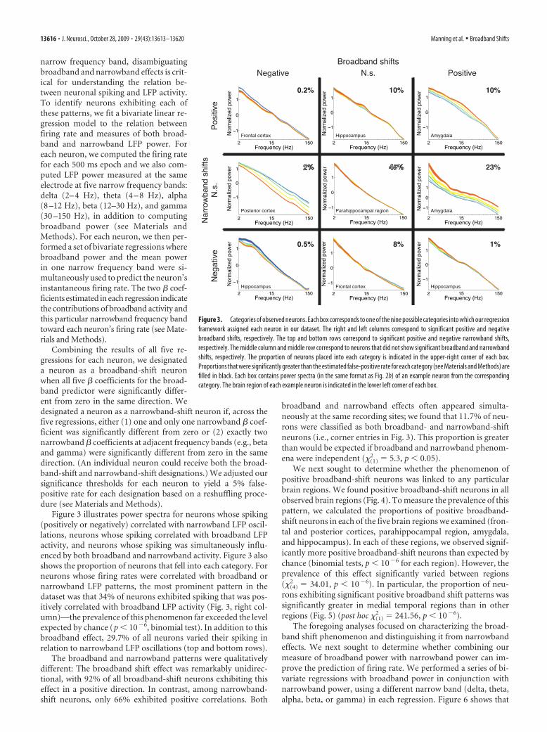
narrow frequency band, disambiguatingbroadband and narrowband effects is crit-ical for understanding the relation be-tween neuronal spiking and LFP activity.To identify neurons exhibiting each ofthese patterns, we fit a bivariate linear re-gression model to the relation betweenfiring rate and measures of both broad-band and narrowband LFP power. Foreach neuron, we computed the firing ratefor each 500 ms epoch and we also com-puted LFP power measured at the sameelectrode at five narrow frequency bands:delta (2– 4 Hz), theta (4 – 8 Hz), alpha(8 –12 Hz), beta (12–30 Hz), and gamma(30 –150 Hz), in addition to computingbroadband power (see Materials andMethods). For each neuron, we then per-formed a set of bivariate regressions wherebroadband power and the mean powerin one narrow frequency band were si-multaneously used to predict the neuron’sinstantaneous firing rate. The two # coef-ficients estimated in each regression indicatethe contributions of broadband activity andthis particular narrowband frequency bandtoward each neuron’s firing rate (see Mate-rials and Methods).
Combining the results of all five re-gressions for each neuron, we designateda neuron as a broadband-shift neuronwhen all five # coefficients for the broad-band predictor were significantly differ-ent from zero in the same direction. Wedesignated a neuron as a narrowband-shift neuron if, across thefive regressions, either (1) one and only one narrowband # coef-ficient was significantly different from zero or (2) exactly twonarrowband # coefficients at adjacent frequency bands (e.g., betaand gamma) were significantly different from zero in the samedirection. (An individual neuron could receive both the broad-band-shift and narrowband-shift designations.) We adjusted oursignificance thresholds for each neuron to yield a 5% false-positive rate for each designation based on a reshuffling proce-dure (see Materials and Methods).
Figure 3 illustrates power spectra for neurons whose spiking(positively or negatively) correlated with narrowband LFP oscil-lations, neurons whose spiking correlated with broadband LFPactivity, and neurons whose spiking was simultaneously influ-enced by both broadband and narrowband activity. Figure 3 alsoshows the proportion of neurons that fell into each category. Forneurons whose firing rates were correlated with broadband ornarrowband LFP patterns, the most prominent pattern in thedataset was that 34% of neurons exhibited spiking that was pos-itively correlated with broadband LFP activity (Fig. 3, right col-umn)—the prevalence of this phenomenon far exceeded the levelexpected by chance (p & 10#6, binomial test). In addition to thisbroadband effect, 29.7% of all neurons varied their spiking inrelation to narrowband LFP oscillations (top and bottom rows).
The broadband and narrowband patterns were qualitativelydifferent: The broadband shift effect was remarkably unidirec-tional, with 92% of all broadband-shift neurons exhibiting thiseffect in a positive direction. In contrast, among narrowband-shift neurons, only 66% exhibited positive correlations. Both
broadband and narrowband effects often appeared simulta-neously at the same recording sites; we found that 11.7% of neu-rons were classified as both broadband- and narrowband-shiftneurons (i.e., corner entries in Fig. 3). This proportion is greaterthan would be expected if broadband and narrowband phenom-ena were independent ("(1)
2 " 5.3, p & 0.05).We next sought to determine whether the phenomenon of
positive broadband-shift neurons was linked to any particularbrain regions. We found positive broadband-shift neurons in allobserved brain regions (Fig. 4). To measure the prevalence of thispattern, we calculated the proportions of positive broadband-shift neurons in each of the five brain regions we examined (fron-tal and posterior cortices, parahippocampal region, amygdala,and hippocampus). In each of these regions, we observed signif-icantly more positive broadband-shift neurons than expected bychance (binomial tests, p & 10#6 for each region). However, theprevalence of this effect significantly varied between regions("(4)
2 " 34.01, p & 10#6). In particular, the proportion of neu-rons exhibiting significant positive broadband shift patterns wassignificantly greater in medial temporal regions than in otherregions (Fig. 5) (post hoc "(1)
2 " 241.56, p & 10#6).The foregoing analyses focused on characterizing the broad-
band shift phenomenon and distinguishing it from narrowbandeffects. We next sought to determine whether combining ourmeasure of broadband power with narrowband power can im-prove the prediction of firing rate. We performed a series of bi-variate regressions with broadband power in conjunction withnarrowband power, using a different narrow band (delta, theta,alpha, beta, or gamma) in each regression. Figure 6 shows that
Figure 3. Categories of observed neurons. Each box corresponds to one of the nine possible categories into which our regressionframework assigned each neuron in our dataset. The right and left columns correspond to significant positive and negativebroadband shifts, respectively. The top and bottom rows correspond to significant positive and negative narrowband shifts,respectively. The middle column and middle row correspond to neurons that did not show significant broadband and narrowbandshifts, respectively. The proportion of neurons placed into each category is indicated in the upper-right corner of each box.Proportions that were significantly greater than the estimated false-positive rate for each category (see Materials and Methods) arefilled in black. Each box contains power spectra (in the same format as Fig. 2b) of an example neuron from the correspondingcategory. The brain region of each example neuron is indicated in the lower left corner of each box.
13616 • J. Neurosci., October 28, 2009 • 29(43):13613–13620 Manning et al. • Broadband Shifts

broadband and gamma-band power were the two dominant LFPmeasures that positively correlated with firing rate. The propor-tions of neurons exhibiting these two effects were comparable toone another and were both significantly greater than the propor-tions of significant positive or negative correlations observed atother frequency bands ("(1)
2 ( 65 and p & 10#10 for all compar-isons). In addition, we observed that in the delta, theta, and alphabands, a greater number of neurons showed negative correlationswith firing rate than positive correlations, which is consistentwith recent literature (Mukamel et al., 2005). A notable feature ofthe broadband effects is that they are predominantly unidirec-tional, with nearly all (92%) significant correlations being in thepositive direction. In contrast, for neurons showing significantgamma-firing-rate correlations, only 63% showed positive cor-relations. Because the broadband effects were significantly moreunidirectional than narrowband gamma effects ("(1)
2 " 171.4,p & 10#10), our results indicate that broadband power is a morespecific predictor of increased neuronal spiking than narrow-band oscillations.
To further illustrate the robust nature of the broadband phe-nomenon, we computed the average power spectra across all2030 neurons in our dataset (Fig. 7a). Although only 37% of theneurons included in this figure demonstrated significant firingrate-related broadband shifts in their LFP power spectra (Fig. 7b),there is nevertheless a clear overall effect of increasing power at allfrequency bands with increased neuronal firing. However, re-peating this analysis after excluding all broadband-shift neurons(Fig. 3, left and right columns) reveals an effect of increasedgamma power with increased firing rate (Fig. 7c). Thus, the pop-ulation of non-broadband-shift neurons recovers the gamma-firing-rate relation described in the prior literature (Mukamel etal., 2005).
DiscussionWe examined the relation between the firing of individual neu-rons and simultaneous LFP activity. In previous work, LFP re-cordings have typically been analyzed in terms of narrowbandoscillations, which indicate that nearby neurons are synchro-
Figure 4. Broadband-shift neurons throughout the brain. Each row shows two exampleneurons from a single brain region. Within each row, each plot illustrates the power spectrafrom an example neuron, in the same format as Figure 2b. (Each of these neurons were catego-rized as positive broadband-shift neurons, corresponding to the middle-right box of Fig. 3.)
Figure 5. Percentage of positive broadband-shift neurons observed in each brain region.Dark gray bars correspond to neocortical regions: frontal cortex (Fr) and posterior cortex (Cx).Light gray bars correspond to medial temporal lobe structures: amygdala (Amyg), hippocampus(Hippo), and parahippocampal region (Par). Positive broadband-shift neurons were more con-centrated in the medial temporal lobe than in the neocortex. The dotted horizontal black linemarks the false-detection rate for positive broadband-shift neurons. Error bars indicate 95%confidence intervals based on the binomial distribution.
Figure 6. LFP components that predict firing rate. Dark gray bars indicate the percentage ofneurons in each region that exhibited positive correlations between firing rate and a particularLFP feature; light gray bars show the percentage of neurons in each region that exhibitednegative correlations. The bars on the left indicate the proportions of neurons whose firing rateswere correlated with power in each narrow frequency band: delta (2– 4 Hz), theta (4 – 8 Hz),alpha (8 –12 Hz), beta (12–30 Hz), and gamma (30 –150 Hz). Each neuron may be counted in atmost one direction (i.e., either positive or negative) per narrow frequency band. The bars on theright indicate the proportions of neurons whose firing rates were correlated with broadbandpower (i.e., broadband-shift neurons).
Manning et al. • Broadband Shifts J. Neurosci., October 28, 2009 • 29(43):13613–13620 • 13617

nously spiking at a particular frequency(Buzsaki, 2006; Fries et al., 2007; Jensenet al., 2007). For many of the neuronsthat we recorded, narrowband oscillationscorrelated significantly with firing rate,especially in the gamma frequency band.We also analyzed LFP recordings using ameasure of broadband activity, whichidentifies fluctuations that simultaneouslyappear at all frequencies. This analysis re-vealed that increases in the power ofbroadband LFP activity positively corre-lated with the spiking of nearby neurons.(We verified that these broadband in-creases in LFP power were not the result ofthe appearance of spike waveforms in our recordings; see supple-mental material, available at www.jneurosci.org.) We observedthis broadband effect in multiple patients and brain regions, andfound that it was most prevalent in medial temporal lobe struc-tures. Our findings indicate that both narrowband and broad-band components of the LFP can be used to estimate the firingrates of nearby neurons, even though LFP recordings do not ac-tually display the waveforms of individual action potentials. Be-low we discuss these findings in relation to the recent literature onnarrowband and broadband LFP phenomena.
Narrowband effectsA wide range of studies have examined the relation between nar-rowband LFP oscillations and the firing of nearby neurons, iden-tifying an array of patterns that often differ across species,frequencies, brain regions, and behaviors. However, two com-mon trends have emerged.
Generally in neocortex, the power of low-frequency (less than!10 Hz) oscillations are negatively correlated with neuronal fir-ing rates, while the power of high-frequency (more than !30 Hz)oscillations are typically positively correlated with neuronal firingrates (Mukamel et al., 2005; Rasch et al., 2008). These resultscomport well with the observation of task-related increases in theamplitude of high-frequency activity and decreases in the ampli-tude of lower-frequency activity (Crone et al., 1998a,b; Miller etal., 2007b). In the present study, we recorded from distributedregions throughout the brains of neurosurgical patients andfound narrowband effects that were consistent with this litera-ture. In particular, we found that LFP oscillations at low frequen-cies (delta, theta, and alpha bands) were negatively correlatedwith neuronal spiking, while high-frequency LFP oscillationswere most often positively correlated with neuronal spiking (betaand gamma bands). However, we also found a significant num-ber of neurons whose firing rates were negatively correlated withgamma oscillations (Fig. 6). Thus, there is considerable variabil-ity in the relation between narrowband LFP oscillations and neu-ronal spiking across neurons.
Broadband effectsOur primary finding is that neuronal spiking is positively corre-lated with broadband LFP power. Broadband changes in LFPpower are qualitatively different from narrowband changes:whereas narrowband power changes reflect modulations in oscil-latory activity, broadband power changes generally reflect mod-ulations in the variance of the LFP time series (Annibaldi andHopcraft, 2002). Previous research has identified broadbandpower changes in brain regions that are thought to be involvedwith particular behaviors. One example is a study by Miller et al.
(2007b), which showed that finger and tongue movements areaccompanied by highly localized broadband LFP power increasesin human motor cortex (also see Miller et al., 2008, 2009). Thesestudies show that broadband LFP shifts can be difficult to identifyin practice because they often occur simultaneously with powerdecreases at alpha and theta frequencies [e.g., see Miller et al.(2007b), their Figs. 1, 2]. Furthermore, oscillatory correlates ofspatial and verbal memory processes are not always specific toa given frequency band, but rather appear at a very broadrange of frequencies, consistent with a broadband effect(Ekstrom et al., 2007; Sederberg et al., 2007). Although fewother studies have discussed their findings in terms of broad-band patterns, a close inspection of the data from several stud-ies reveals examples of behavior-related broadband LFPpatterns. For example, Edwards et al. (2005) report that unex-pected auditory stimuli were followed by LFP changes in left-frontal and temporal cortex that appear to be broadband innature [Edwards et al. (2005), their Fig. 3]. Furthermore, thedata reported by Lachaux et al. (2005) are consistent with thepresence of positive broadband power shifts in the fusiformgyrus during face viewing [Lachaux et al. (2005), their Fig. 2,top; also their Fig. 5, patient 3]. A third example can be foundin Belitski et al.(2008), which examined LFP power spectrafrom macaque visual cortex during movie watching. Figure 2ain their manuscript shows a positive broadband shift in theLFP power spectrum during movies, relative to a baseline pe-riod. Finally, Ball et al. (2008) show data suggesting thatbroadband patterns can be observed noninvasively using scalpelectroencephalography (EEG). For example, they show re-cordings from an electrode above motor cortex that exhibits abroadband power increase (coupled with a low-frequency nar-rowband decrease) after movement [Ball et al. (2008), theirFig. 1a]. While each of these studies implies that broadbandpower indicates when a particular brain region is active, ours isthe first to show that increases in broadband activity directlyrelate to simultaneous increases in neuronal spiking.
Given our finding that neuronal spiking is correlated withbroadband power, one may wonder why many studies reporttask-related high-frequency (gamma) modulation rather than re-porting broadband effects (Lachaux et al., 2005; Belitski et al.,2008; Ball et al., 2008). As described by Miller et al. (2007b),task-related spectral changes often appear as simultaneous low-frequency decreases coupled with broadband increases in power.Due to these low-frequency decreases, in practice, broadbandpower increases are often most visible in the gamma band. Thusmany of the gamma responses reported in the literature mayactually be the result of broadband power increases coupled withlow-frequency narrowband power decreases.
a b c
Figure 7. Average power spectra for different subsets of neurons. a, Average power spectra for all 2030 neurons in our dataset.The power spectra are normalized by subtracting the mean of the lowest firing rate power spectrum (dark blue) from all five curvesin each panel. The thickness of each line represents '1 SEM. As in Figure 2, cooler colors correspond to lower firing rates, whilewarmer colors correspond to higher relative firing rates. Each neuron contributes exactly once to each curve, at each frequency.b, Average power spectra for the 759 broadband-shift neurons. c, Average power spectra for the 1271 neurons that did not exhibitthe broadband-shift effect.
13618 • J. Neurosci., October 28, 2009 • 29(43):13613–13620 Manning et al. • Broadband Shifts

An important feature of our methodology is the robust, non-linear regression (Holland and Welsch, 1977) technique we usedfor distinguishing simultaneous changes in broadband and nar-rowband LFP activity. Because this regression is resistant to out-liers, its estimate of broadband power is only minimally affectedby the presence or absence of narrowband oscillations, whichappear as narrowband peaks in the power spectrum. In contrast,linear methods might interpret the appearance of large narrow-band oscillations as increased broadband power. This issue canbe important, because broadband and narrowband changes oftenoccur simultaneously and in opposite directions (Miller et al.,2007b, 2009; Ball et al., 2008), which can make these effects dif-ficult to tease apart.
Broader implicationsA particularly exciting implication of this study is that increases(Miller et al., 2007b) and decreases (Lachaux et al., 2008) inbroadband power, which can be observed from both invasive(Edwards et al., 2005; Lachaux et al., 2005; Ekstrom et al., 2007;Miller et al., 2007b, 2009; Sederberg et al., 2007) and noninvasive(Ball et al., 2008) recordings, may be used as a proxy for neuronalspiking. This is especially important for researchers using macro-electrode recordings to study the neural basis of human behavior,because such recordings cannot resolve single-neuron spiking,but can record these broadband shifts. To the extent that thebroadband effects observed here generalize to activity recorded atbroader spatial scales, one could use broadband activity to mea-sure correlates of neuronal spiking in scalp recorded EEG andMEG signals, or even to modulate neuronal spiking using real-time feedback methods (Lachaux et al., 2007; Miller et al., 2007a).In this way, the broadband power measure proposed here maylead to new discoveries concerning how neuronal activity under-lies complex human behavior and cognition.
ReferencesAnnibaldi SV, Hopcraft KI (2002) Random walks with power-law fluctua-
tions in the number of steps. J Phys A Math Gen 35:8635– 8645.Ball T, Demandt E, Mutschler I, Neitzel E, Mehring C, Vogt K, Aertsen A,
Schulze-Bonhage A (2008) Movement related activity in the highgamma range of the human EEG. Neuroimage 41:302–310.
Belitski A, Gretton A, Magri C, Murayama Y, Montemurro MA, LogothetisNK, Panzeri S (2008) Low-frequency local field potentials and spikes inprimary visual cortex convey independent visual information. J Neurosci28:5696 –5709.
Buzsaki G (2004) Large-scale recording of neuronal ensembles. Nat Neuro-sci 7:446 – 451.
Buzsaki G (2006) Rhythms of the brain. New York: Oxford UP.Canolty RT, Edwards E, Dalal SS, Soltani M, Nagarajan SS, Kirsch HE, Berger
MS, Barbaro NM, Knight RT (2006) High gamma power is phase-locked to theta oscillations in human neocortex. Science 313:1626 –1628.
Caplan JB, Madsen JR, Schulze-Bonhage A, Aschenbrenner-Scheibe R, New-man EL, Kahana MJ (2003) Human theta oscillations related to senso-rimotor integration and spatial learning. J Neurosci 23:4726 – 4736.
Chen CM, Lakatos P, Shah AS, Mehta AD, Givre SJ, Javitt DC, Schroeder CE(2007) Functional anatomy and interaction of fast and slow visual path-ways in macaque monkeys. Cereb Cortex 17:1561–1569.
Crone NE, Miglioretti DL, Gordon B, Sieracki JM, Wilson MT, Uematsu S,Lesser RP (1998a) Functional mapping of human sensorimotor cortexwith electrocorticographic spectral analysis. I. Alpha and beta event-related desynchronization. Brain 121:2271–2299.
Crone NE, Miglioretti DL, Gordon B, Lesser RP (1998b) Functional map-ping of human sensorimotor cortex with electrocorticographic spectralanalysis. II. Event-related synchronization in the gamma band. Brain121:2301–2315.
Crone NE, Sinai A, Korzeniewska A (2006) High-frequency gamma oscilla-tions and human brain mapping with electrocorticography. Prog BrainRes 159:275–295.
Cruikshank SJ, Rose HJ, Metherate R (2002) Auditory thalamocortical syn-aptic transmission in vitro. J Neurophysiol 87:361–384.
Edwards E, Soltani M, Deouell LY, Berger MS, Knight RT (2005) Highgamma activity in response to deviant auditory stimuli recorded directlyfrom human cortex. J Neurophysiol 94:4269 – 4280.
Ekstrom AD, Caplan JB, Ho E, Shattuck K, Fried I, Kahana MJ (2005) Hu-man hippocampal theta activity during virtual navigation. Hippocampus15:881– 889.
Ekstrom A, Viskontas I, Kahana M, Jacobs J, Upchurch K, Bookheimer S,Fried I (2007) Contrasting roles of neural firing rate and local field po-tentials in human memory. Hippocampus 17:606 – 617.
Fried I, Wilson CL, Maidment NT, Engel J Jr, Behnke E, Fields TA, Mac-Donald KA, Morrow JW, Ackerson L (1999) Cerebral microdialysiscombined with single-neuron and electroencephalographic recording inneurosurgical patients. J Neurosurg 91:697–705.
Fries P, Reynolds JH, Rorie AE, Desimone R (2001) Modulation of oscilla-tory neuronal synchronization by selective visual attention. Science291:1560 –1563.
Fries P, Nikolic D, Singer W (2007) The gamma cycle. Trends Neurosci30:309 –316.
Henrie JA, Shapley R (2005) LFP power spectra in V1 cortex: the gradedeffect of stimulus contrast. J Neurophysiol 94:479 – 490.
Holland PW, Welsch RE (1977) Robust regression using iteratively re-weighted least-squares. Commun Stat Theory Methods A 6:813– 827.
Jacobs J, Kahana MJ (2009) Neural representations of individual stimuli inhumans revealed by gamma-band ECoG activity. J Neurosci29:10203–10214.
Jacobs J, Kahana MJ, Ekstrom AD, Fried I (2007) Brain oscillations controltiming of single-neuron activity in humans. J Neurosci 27:3839 –3844.
Jensen O, Kaiser J, Lachaux JP (2007) Human gamma-frequency oscilla-tions associated with attention and memory. Trends Neurosci30:317–324.
Jerbi K, Ossandon T, Hamame CM, Senova S, Dalal SS, Jung J, Minotti L,Bertrand O, Berthoz A, Kahane P, Lachaux JP (2009) Task-relatedgamma-band dynamics from an intracerebral perspective: review andimplications for surface EEG and MEG. Hum Brain Mapp 30:1758 –1771.
Juergens E, Guettler A, Eckhorn R (1999) Visual stimulation elicits lockedand induced gamma oscillations in monkey intracortical- and EEG-potentials, but not in human EEG. Exp Brain Res 129:247–259.
Kahana MJ (2006) The cognitive correlates of human brain oscillations.J Neurosci 26:1669 –1672.
Kahana MJ, Seelig D, Madsen JR (2001) Theta returns. Curr Opin Neuro-biol 11:739 –744.
Kanisza G (1979) The organization of vision. New York: Praeger.Kaur S, Lazar R, Metherate R (2004) Intracortical pathways determine
breadth of subthreshold frequency receptive fields in primary auditorycortex. J Neurophysiol 91:2551–2567.
Koffka K (1935) Principles of gestalt psychology. New York: Harcourt,Brace and World.
Kohler W (1947) Gestalt psychology. New York: Liveright.Kreiman G (2007) Brain science: from the very small to the very large. Curr
Biol 17:R768 –R770.Kreiman G, Hung CP, Kraskov A, Quiroga RQ, Poggio T, DiCarlo JJ (2006)
Object selectivity of local field potentials and spikes in the macaque infe-rior temporal cortex. Neuron 49:433– 445.
Lachaux JP, George N, Tallon-Baudry C, Martinerie J, Hugueville L, MinottiL, Kahane P, Renault B (2005) The many faces of the gamma band re-sponse to complex visual stimuli. Neuroimage 25:491–501.
Lachaux JP, Jerbi K, Bertrand O, Minotti L, Hoffmann D, Schoendorff B,Kahane P (2007) A blueprint for real-time functional mapping via hu-man intracranial recordings. PLoS One 2:e1094.
Lachaux JP, Jung J, Mainy N, Dreher JC, Bertrand O, Baciu M, Minotti L,Hoffmann D, Kahane P (2008) Silence is golden: transient neural deac-tivation in the prefrontal cortex during attentive reading. Cereb Cortex18:443– 450.
Logothetis NK (2003) The underpinnings of the BOLD functional magneticresonance imaging signal. J Neurosci 23:3963–3971.
Miller KJ, den Nijs M, Shenoy P, Miller JW, Rao RP, Ojemann JG (2007a)Real-time functional brain mapping using electrocorticography. Neuro-image 37:504 –507.
Miller KJ, Leuthardt EC, Schalk G, Rao RPN, Anderson NR, Moran DW,
Manning et al. • Broadband Shifts J. Neurosci., October 28, 2009 • 29(43):13613–13620 • 13619

Miller JW, Ojemann JG (2007b) Spectral changes in cortical surface po-tentials during motor movement. J Neurosci 27:2424 –2432.
Miller KJ, Shenoy P, den Nijs M, Sorensen LB, Rao RPN, Ojemann JG (2008)Beyond the gamma band: the role of high-frequency features in move-ment classification. IEEE Trans Biomed Eng 55:1634 –1637.
Miller KJ, Zanos S, Fetz EE, den Nijs M, Ojemann JG (2009) Decoupling thecortical power spectrum reveals real-time representation of individualfinger movements in humans. J Neurosci 29:3132–3137.
Milstein J, Mormann F, Fried I, Koch C (2009) Neuronal shot noise andBrownian 1/f 2 behavior in the local field potential. PLoS One 4:e4338.
Mitzdorf U (1985) Current source-density method and application in catcerebral cortex: investigation of evoked potentials and EEG phenomena.Physiol Rev 65:37–100.
Mukamel R, Gelbard H, Arieli A, Hasson U, Fried I, Malach R (2005) Cou-pling between neuronal firing, field potentials, and fMRI in human audi-tory cortex. Science 309:951–954.
Newman EL, Caplan JB, Kirschen MP, Korolev IO, Sekuler R, Kahana MJ(2007) Learning your way around town: how virtual taxicab drivers learnto use both layout and landmark information. Cognition 104:231–253.
Pal NR, Pal SK (1993) A review of image segmentation techniques. PatternRecognit Lett 26:1277–1294.
Percival DB, Walden AT (1993) Spectral analysis for physical applications.Cambridge, UK: Cambridge UP.
Pesaran B, Pezaris JS, Sahani M, Mitra PP, Andersen RA (2002) Temporalstructure in neuronal activity during working memory in macaque pari-etal cortex. Nat Neurosci 5:805– 811.
Quiroga RQ, Nadasdy Z, Ben-Shaul Y (2004) Unsupervised spike detectionand sorting with wavelets and superparamagnetic clustering. NeuralComput 16:1661–1687.
Rasch MJ, Gretton A, Murayama Y, Maass W, Logothetis NK (2008) Infer-ring spike trains from local field potentials. J Neurophysiol 99:1461–1476.
Sederberg PB, Schulze-Bonhage A, Madsen JR, Bromfield EB, McCarthy DC,Brandt A, Tully MS, Kahana MJ (2007) Hippocampal and neocorticalgamma oscillations predict memory formation in humans. Cereb Cortex17:1190 –1196.
Singer W, Gray CM (1995) Visual feature integration and the temporal cor-relation hypothesis. Annu Rev Neurosci 18:555–586.
Tanji K, Suzuki K, Delorme A, Shamoto H, Nakasato N (2005) High-frequency $-band activity in the basal temporal cortex during picture-naming and lexical-decision tasks. J Neurosci 25:3287–3293.
von der Malsburg C (1981) The correlation theory of brain function. Inter-nal Report 81-2. Gottingen, Germany: Max-Planck-Institute for Biophys-ical Chemistry.
Witter M (2002) The parahippocampal region: past, present and future. In:The parahippocampal region: organization and role in cognitive func-tions (Witter M, Wouterlood F, eds), pp 3–19. Oxford, UK: Oxford UP.
13620 • J. Neurosci., October 28, 2009 • 29(43):13613–13620 Manning et al. • Broadband Shifts