briarcliff high school, briarcliff manor, ny, usa hhs … use of red/nir light to stimulate,...
TRANSCRIPT

Photobiomodulation and the brain: a new paradigm
Madison Hennessy1 and Michael R Hamblin2,3,4
1Briarcliff High School, Briarcliff Manor, NY, USA
2Wellman Center for Photomedicine, Massachusetts General Hospital, Boston, MA, USA
3Department of Dermatology, Harvard Medical School, Boston, MA, USA
4Harvard-MIT Division of Health Sciences and Technology, Cambridge, MA, USA
Abstract
Transcranial photobiomodulation (PBM) also known as low level laser therapy (tLLLT) relies on
the use of red/NIR light to stimulate, preserve and regenerate cells and tissues. The mechanism of
action involves photon absorption in the mitochondria (cytochrome c oxidase), and ion channels in
cells leading to activation of signaling pathways, up-regulation of transcription factors, and
increased expression of protective genes. We have studied PBM for treating traumatic brain injury
(TBI) in mice using a NIR laser spot delivered to the head. Mice had improved memory and
learning, increased neuroprogenitor cells in the dentate gyrus and subventricular zone, increased
BDNF and more synaptogenesis in the cortex. These highly beneficial effects on the brain suggest
that the applications of tLLLT are much broader than at first conceived. Other groups have studied
stroke (animal models and clinical trials), Alzheimer’s disease, Parkinson’s disease, depression,
and cognitive enhancement in healthy subjects.
Keywords
Transcranial photobiomodulation; low level laser therapy; brain disorders; traumatic brain injury; stroke; Alzheimer’s disease; psychiatric diseases
1 Introduction
Light in the visible and near-infrared spectrum makes up the major part of solar radiation
and is also provided by artificial light sources. Since light is widely present throughout our
world, approaches that harness it for the purposes of medical treatment is hardly a new
phenomenon. Photobiomodulation (PBM) involves the utilization of light from the visible
and near infrared portions of the spectrum at a relatively low power density that is
insufficient to heat or burn tissue. Although PBM often uses lasers as the light source, its
method of action is not thermal or ablative in any way. Rather, the photons themselves
stimulate chemical changes within the cells, provoking biological reactions that benefit the
body in a variety of ways - including the triggering of neuroprotective responses,
improvements in metabolism, blood flow, and neurogenesis, and decreases in inflammation
and oxidative stress [1].
HHS Public AccessAuthor manuscriptJ Opt. Author manuscript; available in PMC 2018 January 01.
Published in final edited form as:J Opt. 2017 January ; 19(1): 013003–. doi:10.1088/2040-8986/19/1/013003.
Author M
anuscriptA
uthor Manuscript
Author M
anuscriptA
uthor Manuscript

The medical benefits of low level laser therapy (LLLT) were first discovered in 1967 by Dr.
Endre Mester at the Semmelweis Medical University in Hungary. Mester was trying to
repeat an experiment originally described by McGuff in Boston USA, who had successfully
used a ruby laser to cure malignant tumors in rats [2]. However, Mester’s laser possessed
only a very small fraction of the power possessed by McGuff’s laser. Thus, he was
unsuccessful in curing any tumors. However, he did observe a heightened rate of hair growth
and better wound healing in the rats in which he had surgically implanted tumors, the first
indication that low level laser light (rather than high power thermal lasers) could have its
own distinct applications in medicine [3, 4]. Over the almost 50 years since then, LLLT or
PBM has come into use for the treatment of pain [5] and wounds [6] and for cosmetic
purposes such as decreasing the severity of wrinkles [7] and regrowing hair [8]. Based on the
knowledge of its protective, restorative and healing abilities, research was eventually
conducted into whether PBM could be used in the treatment of acute stroke [9], and from
there onwards for a wide variety of other neurological conditions [10].
Generally, transcranial PBM (the name applied to PBM specifically when the light is applied
to the head to treat neurological conditions) procedures involve the placement of a light
source (or multiple light sources) on one or several areas of the head, with the goal of
stimulating a certain part of the brain, which varies depending on the illness or disorder
being treated. The light can originate from a laser or a light emitting diode (LED), and can
be pulsed or continuous. Several parameters are typically employed to fully describe PBM
protocols. One is the wavelength of light being used. Wavelength is important, as only
certain ranges have any effect. Light in the 700–750 nm range, for example, tends to have
little impact, whereas the impact of light in the 600–690 nm or 760–900 nm is far greater.
Another important parameter is the irradiance or power density (mW/cm2), which equals the
power of the light source in Watts divided by the area over which the light hits the body. A
similar concept is energy density (sometimes called the fluence), which is provided by
Joules/area (J/cm2). It should be noted that if the spot is relatively small (<< 1 cm2), the
power density and energy density are likely to be misleading. For instance if the spot is only
1 mm2, a calculation might lead to claims that 1W/cm2 was delivered from a 10 mW laser.
Additionally it is important to note that the treatment duration, and the frequency of
repetition can have influence on the success (or lack thereof). Finally there is some evidence
that pulsing the light can have a better effect than continuous wave (CW) light [11, 12].
Parameters are of vital importance in PBM due to its characteristic biphasic dose response
effect [13, 14]. That is, PBM can have vastly different effects depending on such factors as
wavelength, irradiance, energy density, and treatment duration. This biphasic effect adheres
to the “Arndt-Schultz Law”, which states that if weak stimuli provoke a significant rise in
activity, then strong stimuli will not continue to provoke ever-increasing rises. Instead, they
only provoke an increase until a certain peak level is reached, after which point the activity
decreases until it returns to baseline, and may even get to the point where a negative (or
inhibitory) response is achieved [13, 14].
As of now, transcranial PBM research still remains in a state of relative infancy. While its
positive effects have been demonstrated countless times in animal models, they have yet to
be proven in broad-scope clinical testing. However, the research that does exist is very
promising, strongly indicating that PBM could be a viable treatment option for a broad range
Hennessy and Hamblin Page 2
J Opt. Author manuscript; available in PMC 2018 January 01.
Author M
anuscriptA
uthor Manuscript
Author M
anuscriptA
uthor Manuscript

of neurological diseases including stroke, traumatic brain injury, Parkinson’s disease,
Alzheimer’s disease, and depression, in addition to providing cognitive enhancement for
healthy subjects [10].
2 Molecular Mechanisms of Photobiomodulation
2.1 Mitochondria and Cytochrome-C Oxidase
Cytochrome C oxidase (Cox) is the terminal enzyme in the electron transport chain. The
electron transport chain, through a series of redox reactions, facilitates the transfer of
electrons across the inner membrane of the mitochondria. As the terminal enzyme, otherwise
known as “complex IV”, Cox mediates the transfer of electrons from cytochrome C to
molecular oxygen (see Figure 1). In many publications, Cox has been shown to be an
acceptor and transducer of signals activated by light in the red and near-infrared regions of
the spectrum [15]. Specifically, exposure to this type of light, via PBM, seems to provoke an
increase in the availability of electrons for the reduction of molecular oxygen in the catalytic
center of Cox, increasing mitochondrial membrane potential (MMP), and increasing levels
of adenosine triphosphate (ATP), cyclic adenosine monophosphate (cAMP), and reactive
oxygen species (ROS), all of which indicate heightened mitochondrial functioning and
cellular metabolism [1].
Although other portions of the electron transfer chain, such as complexes I–IV and succinate
dehydrogenase do experience increased activity as a result of PBM, Cox is still believed to
be the primary photoacceptor. This notion is supported by the fact that low-level irradiation
such as PBM causes increased oxygen consumption, and that the majority of oxygen
consumption occurs at complex IV. It is additionally supported by the fact that the presence
of sodium azide, a Cox inhibitor, renders the effects of PBM null [16].
It has been proposed that the reason PBM increases Cox activity is through the
photodissociation of NO from Cox. Evidenced by the increased levels of NO following
PBM, it is possible that PBM reverses the mitochondrial inhibition of cellular respiration
that exists as a result of excessive NO binding . According to this theory, NO would be
photodissociated from its binding sites at the copper and heme iron centers in Cox. Thus,
NO would no longer prevent binding to oxygen, and enzymatic activity would be restored to
normal levels, allowing for an influx of oxygen, the resumption of respiration and a burst of
generation of ROS. NO can also be photo-released from other intracellular sites, including
nitrosylated myoglobin and hemoglobin, producing similarly positive effects [17].
On a slightly broader scale, PBM is also believed to trigger retrograde mitochondrial
signaling [18]. This term refers to the communication between the mitochondria and the
nucleus of a cell (normally signals pass the other way from the nucleus to other organelles).
The aforementioned mitochondrial changes result in an altered mitochondrial ultrastructure,
which drives the communication. As a result, membrane permeability and ion flux at the cell
membrane are altered, in turn leading to production of reactive oxygen species (ROS), and
altered activity of redox-sensitive transcription factors such as activator protein-1 (AP1) and
NF-κB [19].
Hennessy and Hamblin Page 3
J Opt. Author manuscript; available in PMC 2018 January 01.
Author M
anuscriptA
uthor Manuscript
Author M
anuscriptA
uthor Manuscript

2.2 Light/Heat Gated Ion Channels
Although evidence proving that light-gated or heat-gated ion channels could be mechanisms
of action in PBM is sparse at present, it is gradually increasing [20]. PBM is thought likely
to affect transient receptor potential (TRP) channels. First discovered in a Drosophila mutant
as the mechanism responsible for the vision of insects, some TRP channels are now known
to be sensitive to light or heat [21], in addition to a wide variety of other stimuli [22]. TRP
channels are calcium channels, and are modulated by phosphoinositides [23].
The majority of research relating PBM to light/heat-gated ion channels has concentrated on
the TRPV “vanilloid” subfamily of TRP channels. Evidence from studies [24–27] have led
to the general consensus that TRP channels are most likely to be activated by green light or
infrared light. However, because green light lacks the same penetrating ability of infrared or
near-infrared light, it lacks practical clinical application. However, Ryu et al. found that
exposure to infrared (2,780 nm) wavelength light attenuated TRPV1 activation, causing a
decrease in generation of pain stimuli. A similar, but far less dramatic antinociceptive effect
was also observed when TRPV4 was exposed to light of the same wavelength. TRPV4 was
also shown to be responsive to 1,875 nm pulsed light [26]. Since water is the primary
absorber of infrared in this region and is also by far the most prevalent single molecule in
living systems, it is possible that minute alterations in temperature (low enough to produce
no detectable tissue heating) could alter protein conformation and open the channel.
3. Tissue optics, light delivery, light sources
3.1 Does light penetrate into the brain?
It could be reasonably claimed that the brain is one of the most interesting organs, for those
working in the field of tissue optics. Tisse optics describes how visible and infrared light of
different wavelengths interacts with different tissues, and involves measuring tissue optical
properties, namely, absorption coefficient, scattering coefficient, and anisotropy factor. One
reason the brain is so interesting is the intense interest in near-infrared spectroscopy (NIRS)
and diffuse optical imaging. Functional NIRS (fNIRS) using 700–900 nm light has become
established as a brain imaging technique that can be compared to functional magnetic
resonance imaging (fMRI) that has made such a revolutionary impact upon neurosceince in
récent years [28]. Different laboratories have investigated the penetration of light of different
wavelengths though the scalp and the skull, and to what depths into the brain this light can
penetrate. Haeussinger et al estimated that the mean penetration depth (5% remaining
intensity) of NIR light through the scalp and skull was 23:6+0:7 mm [29]. Jagdeo et al [30]
used human cadaver heads (skull with intact soft tissue) to measure penetration of 830 nm
light, and found penetration depended on the anatomical region of the skull (0.9% at the
temporal region, 2.1% at the frontal region, and 11.7% at the occipital region). Red light
(633 nm) hardly penetrated at all. Tedord et al [31] also used human cadaver heads to
compare penetration of 660 nm, 808 nm, and 940nm light. They found 808 nm light was
best and could reach a depth in the brain of 40–50mm.
Hennessy and Hamblin Page 4
J Opt. Author manuscript; available in PMC 2018 January 01.
Author M
anuscriptA
uthor Manuscript
Author M
anuscriptA
uthor Manuscript

3.2 Systemic effects?
Another manner in which PBM may affect the brain is through the provocation of broader
systemic effects. That is, when PBM is used to target a specific area other than the brain,
such as lower back pain, the brain can benefit remotely from the changes being caused. For
example, PBM stimulates macrophages and mast cells, which can benefit the brain. Also,
regardless of the treatment site, PBM can lead to the down-regulation of pro-inflammatory
cytokines and the up-regulation of anti-inflammatory cytokines on a body-wide basis,
meaning that the brain will still benefit from the positive effects despite not being the
specific target for treatment. Another possibility is that PBM can trigger the production of a
yet-unidentified extracellular signaling molecule from the mitochondria (for instance),
which is then transported to enable the remote action of PBM on brain cells and other non-
local cells. It should be remembered that blood platelets have large stores of ATP, and that
low platelet ATP content has been linked with dépression [32].
It is in fact very likely that the beneficial effects of PBM on the brain cannot be entirely
explained by penetration of photons through the scalp and skull into the brain itself. There
have been some studies that have explicitly addressed this exact issue. In a study of PBM for
Parkinson’s disease in a mouse model [33]. Mitrofanis and colleagues compared delivering
light to the mouse head, and also covered up the head with aluminum foil so that they
delivered light to the remainder of the mouse body. They found that there was a highly
beneficial effect on neurocognitive behavior with irradiation to the head, but nevertheless
there was also a statistically significant (although less pronounced benefit, referred to by
these authors as an ‘abscopal effect”) when the head was shielded from light [34]. Moreover
Oron and co-workers [35] have shown that delivering NIR light to the mouse tibia (using
either surface illumination or a fiber optic) resulted in improvement in a transgenic mouse
model of Alzheimer’s disease (AD). Light was delivered weekly for 2 months, starting at 4
months of age (progressive stage of AD). They showed improved cognitive capacity and
spatial learning, as compared to sham-treated AD mice. They proposed that the mechanism
of this effect was to stimulate c-kit-positive mesenchymal stem cells (MSCs) in autologous
bone marrow (BM) to enhance the capacity of MSCs to infiltrate the brain, and clear β-
amyloid plaques [36]. It should be noted that the calvarial bone marrow of the skull contains
substantial numbers of stem cells [37].
3.3 Where should the light be delivered?
The obvious answer is that if one wants to affect the brain one should deliver light to the
head, and more specifically, this often imeans the forehead as there is no hair present. Hair
can act as a significant attenuator of light in the visible and NIR regions. Moreover light that
hits the forehead penetrates to the frontal lobes of the cortex, a part of the brain that is
responsible for motor function, problem solving, spontaneity, memory, language, initiation,
judgement, and impulse control. However other investigators have come to the conclusion
that other specific parts of the head (and therefore the underlying brain) should be
specifically targeted by spatially directed spots of light. Margaret Naeser has been a leading
proponent of this point of view and believes that the default mode network is critically
important in the brain’s response to PBM [38]. A paper from Russia [39] treated patients
with Alzheimer’s disease by threading a fiber optic all the way from the femoral artery to the
Hennessy and Hamblin Page 5
J Opt. Author manuscript; available in PMC 2018 January 01.
Author M
anuscriptA
uthor Manuscript
Author M
anuscriptA
uthor Manuscript

middle cerebral artery to deliver red laser light (20mW) into the brain. As mentioned above
the Mitrofanis laboratory in Australia has studied Parkinson’s disease in animal models, and
after implanting a fiber optic into the mouse brain [40], they repeated the procedure in
Macaque monkeys with Parkinson’s disease caused by MPTP administration [41].
Chinese investigators [42] and the Vielight Company in Canada [43] have long studied
intranasal PBM, in which a low power LED or laser diode (<20mW) is clipped to the inside
of one nostril, and have even claimed that therapeutic doses of light can penetrate to the
brain. However it can be readily appreciated that systemic blood absorption, or even
activation of olefactory ensheathing cells (a type of stem cell resident in the nasal mucosa
[44]) could also play a role in the beneficial effects of intranasal PBM on the brain.
Laser acupuncture is often used as an alternative or as an addition to traditional Chinese
acupuncture using needles [45]. Many of the applications of laser acupuncture have been for
conditions that affect the brain [46] such as Alzheimer’s disease [47] and autism [48] that
have all been investigated in animal models. Moreover laser acupuncture has been tested
clinically [49].
3.4 Light sources
A wide array of different light sources (lasers and LEDs) have been employed for tPBM.
One of the most controversial questions which remains to be conclusively settled, is whether
a coherent monochromatic laser is superior to non-coherent LEDs that typically have a 30
nm band-pass (full width half maximum). Although wavelengths in the NIR region (800–
1100 nm) have been the most often used, red wavelengths have sometimes been used either
alone, or in combination with NIR. Power levels also vary markedly from Class IV lasers
with total power outputs in the region of 10W [50], to lasers with more modest power levels
(circa 1W). LEDs can also have widely varying total power levels depending on the size of
the array and the number and power of the individual diodes (now diodes are available with
a power of 3W each). Power densities can also vary quite substantially from the Photothera
laser [51] and other class IV lasers [52], which required active cooling or constant
movement (~700 mW/cm2), to LEDs in the region of 10–30 mW/cm2.
4 Mechanisms of PBM Applied to the Brain
4.1 Metabolism
Improved metabolic functioning is one of the most easily recognizable effects of PBM, and
increased intracellular ATP production is one of its most strongly supported mechanisms of
action. Using phosphorus magnetic resonance spectroscopy (P-MRS), significant increases
in nucleotide triphosphate (NTP), a marker of cellular energy availability, have been
observed following PBM [53] strongly indicating the increase in cellular ATP production to
be a result of PBM. This is significant, as lowered ATP production is a hallmark of many
neurological conditions such as major depressive disorder, traumatic brain injury,
Parkinson’s disease, and Alzheimer’s disease, the details of which will be discussed at a
later point.
Hennessy and Hamblin Page 6
J Opt. Author manuscript; available in PMC 2018 January 01.
Author M
anuscriptA
uthor Manuscript
Author M
anuscriptA
uthor Manuscript

4.2 Blood Flow
The production of NO as result of PBM is similarly well recorded [54]. NO is a major
neuronal signaling molecule which, among other functions, possesses the ability to trigger
vasodilation. To do so, it first stimulates soluble guanylate cyclase to form cyclic-GMP
(cGMP). The cGMP then activates protein kinase G, leading to the reuptake of Ca2+ and the
opening of calcium-activated potassium channels. Due to the subsequent fall in
concentration of Ca2+, myosin light-chain kinase is prevented from phosphorylating the
myosin molecule, causing the smooth muscle cells in the lining of blood vessels and
lymphatic vessels to become relaxed. This vasodilation then promotes improved circulation,
which in turn leads to improved cerebral oxygenation in a similar manner to that observed
with pulsed electromagnetic fields [55]. As is the case with any other part of the body,
improvement in blood flow significantly aids the functioning of the area in which it occurs.
4.3 Neuroprotection
A wide variety of evidence suggests that PBM can be utilized for neuroprotection--
essentially, as a preemptive measure to protect cells from future damage and reduce ongoing
damage and promote their survival and longevity. One way it achieves this result is by
inhibiting the activity of glycogen synthase kinase 3β (GSK3β). To do so, it activates
protein kinase B (AKT) which increases the phosphorylation level of its Ser9 amino acid
residue, which then allows the N-terminus of GSK3β to bind with its own binding site. One
result of this is the accumulation and translocation to the nucleus of β-catenin, which ceases
to be underphosphorylated and therefore becomes more viable when GSK3β activity is
inhibited. Once allowed to accumulate in the nucleus, β-catenin relies on the increased
activity of TCF/LEF transcription factors to promote cellular survival [56]. This inhibition of
GSK3β also helps to prevent apoptosis, the normal cell death that occurs as an organism
grows. GSK3β is believed to act as a mediator between AKT and Bax, a protein which is
translocated to the nucleus in the presence of pro-apoptotic stimuli to trigger the beginning
of the process. However, when GSK3β is inhibited, the communication pathway between
AKT and Bax is cut off. As a result, the signals for Bax translocation are inhibited [57]. This
pathway is shown in Figure 2.
PBM also demonstrates neuroprotective qualities in the form of protection against
senescence. It has been observed to activate the extracellular signal-related kinase (ERK)/
forkhead box protein M1 (FOXM1) pathway. The FOXM1 protein regulates the progression
from the G1 to the S phase of the cell cycle, and via the activation of the ERK/FOXM1
pathway, PBM leads to the greater translocation of ERK to the nucleus and the greater
accumulation of FOXM1 in the nucleus. This, in turn, causes reduced expression of the p21
protein and mitotic arrest in the G1 phase therefore slowing the overall progression of
cellular senescence [58].
PBM has also been shown to be effective in protecting cells from the harmful effects of
toxins. In a study done by Eells [59], irradiation with 670 nm light was successful in causing
the recovery of retinal function and the prevention of histological damage in rodent models
exposed to methanol. This is likely due to the fact that methanol generates the toxic
metabolite formic acid, an inhibitor of Cox, and PBM is a known stimulator of Cox. A study
Hennessy and Hamblin Page 7
J Opt. Author manuscript; available in PMC 2018 January 01.
Author M
anuscriptA
uthor Manuscript
Author M
anuscriptA
uthor Manuscript

by Wong-Riley et al [60] on the effects of PBM given post-exposure to toxins such as
potassium cyanide and tetrodotoxin produced similarly successful results, especially when
rats were irradiated with 670 and 830 nm light, the peaks of the Cox absorption spectrum.
This further indicates that the antitoxin effect of PBM can be traced to its stimulation of
Cox.
PBM is also effective in prevention of the harmful effects associated with potassium
cyanide. When pretreated with 670 nm light, Liang et al. in 2006 found that neuronal
expression of Bax induced by cyanide was decreased, preventing the subsequent apoptosis
[56].
In addition, PBM has demonstrated the rather unique quality of affecting cells in different
states of health in different ways, essentially modifying the cell in whatever way might be
necessary to promote its survival. For instance, in normal cells the absorption of light by
Cox leads to an increase in MMP and a brief surge in ROS production. However, in cells
where MMP is low due to existing oxidative stress, excitotoxicity, or inhibition of electron
transport, ROS are produced from dysfunctional mitochondria. In this case light absorption
leads to an increase of MMP towards normal levels and thereby a decrease in ROS
production [61]. Similarly, the typical response to PBM in healthy cells is an uptick in
intracellular Ca2+ [61]. However, in cells that already contain excessive levels of Ca2+ as
seen in excitotoxicity, PBM provokes the opposite reaction, lowering intracellular calcium
and promoting cell survival [62].
4.4 Oxidative Stress
Oxidative stress occurs when there exists an imbalance between the production of reactive
oxygen species (ROS) and the ability of the body to counteract their effects using
antioxidants, leading to ROS becoming harmful when they are present in excess and for a
long time. Many sources have linked oxidative stress to various neurological conditions,
such as major depressive disorder [63], traumatic brain injury and Alzheimer’s disease [64].
In studies of the effect of PBM on traumatized muscle, PBM has been shown to be effective
in regulating the amount of cytokine-inducible nitric oxide synthase (iNOS) produced by the
cell. This is important because excessive amounts of iNOS can lead to the excessive
production of NO, which would then lead to production of the reactive nitrogen species,
peroxynitrite, leading to an increase in oxidative stress. Specifically, iNOS could overwhelm
the positive effects of other forms of NO synthase such as endothelial nitric oxygen synthase
(eNOS), which is the species primarily responsible for the vasodilating effects of PBM [65–
67]
PBM has also been shown to cause increases in angiogenesis, leading to further
improvements in blood flow. As demonstrated by Cury, PBM at 780 nm and 40 J/cm2
triggered an increase in the expression of the protein HIF1α and of vascular endothelial
growth factor and a decrease in matrix metalloproteinase 2 activity, all of which were found
to induce angiogenesis [68]. Additionally, in an in vitro study of the effects of red/NIR light
on red blood cells done by Mungrue et al., NIR light was found to be quite effective in
Hennessy and Hamblin Page 8
J Opt. Author manuscript; available in PMC 2018 January 01.
Author M
anuscriptA
uthor Manuscript
Author M
anuscriptA
uthor Manuscript

protecting red blood cells from oxidation, which is a common occurrence in brains
compromised by conditions such as MDD [65].
4.5 Anti-Inflammatory effects
Inflammation is a of one of the innate immune system’s defenses against foreign bodies
such as bacteria and viruses. On a cellular level, it occurs when the transcription factor NF-
κB is activated. While acute inflammation can often have positive effects, chronic
inflammation can have very negative effects. Many diseases, including neurodegenerative
diseases and mood disorders, can be traced at least in part to chronic inflammation.
One way PBM helps to quell inflammation is through the inhibition of the cyclooxygenase 2
(COX-2) enzyme. Lim et al. found that 635 nm light irradiation at low power was able to
cause such inhibition by decreasing intracellular ROS. Inhibition of COX-2 via
pharmaceutical means is widely supported at current, with COX-2 inhibitors making up a
significant portion of the market for non steroidal anti-inflammatory drugs (NSAIDS) [69].
Through PBM, essentially the same result is being accomplished, just with a different
stimulus.
PBM can also modulate cellular levels of free NF-κB. When light is absorbed in the
mitochondria there is a brief burst of ROS (as mentioned above). These ROS can activate
protein kinase D that can go on to signal in the NF-κB system [70]. NF-κB is found in the
cytosol bound to IκB, an inhibitor protein. Pro-inflammatory stimuli have the ability to
activate IκB kinase, an upstream signaling regulator that causes the degradation of IκB.
Once the IκB has been degraded, the NF-κB is free to translocate to the nucleus, where it
triggers the expression of pro-inflammatory genes [19]. These signaling pathways are shown
in Figure 3. However, Lim et al. found evidence suggesting that treatment with 635 nm light
inhibited the NF-κB pathway, leading to the down regulation of pro-inflammatory gene
expression [69].
Additionally, PBM possesses the ability to modulate levels of cytokines, proteins that act as
important signaling molecules for the immune system. PBM has been shown to modulate
levels of both pro and anti- inflammatory cytokines, though to explian the prevention of
inflammation, its ability to modulate levels of tumor necrosis factor (TNFα) and other pro-
inflammatory cytokines is especially useful.
4.6 Neurogenesis and Synaptogenesis
One of the most notable and potentially significant effects of PBM in the brain is its ability
to promote both synaptogenesis and neurogenesis. This is vitally important, as many brain
conditions, including TBI, neurodegenerative diseases, and mood disorders can be traced,
either partially or in full, to atrophy, cell death, and poor neuronal connections in certain
regions of the brain. If PBM possesses the ability to counter these effects by facilitating
neural regeneration and rewiring, it could prove to be extremely promising as a novel
method of treating such conditions.
One manner in which PBM promotes neuronal connectivity is BDNF (brain derived
neurotrophic factor) regulation. BDNF is a protein found in the nervous system, which helps
Hennessy and Hamblin Page 9
J Opt. Author manuscript; available in PMC 2018 January 01.
Author M
anuscriptA
uthor Manuscript
Author M
anuscriptA
uthor Manuscript

to maintain existing neurons and to encourage the growth of new neurons and synapses.
Specifically, it is believed to modulate dendritic structure to facilitate improved synaptic
transmission. PBM has been shown to slow attenuation of BDNF via the ERK/CREB
pathway, thus positively affecting dendritic morphogenesis and improved neuronal
connectivity [71]. BDNF is also a mediator of the downstream protein synapsin 1, which
improves synaptogenesis by accelerating the development of neuronal fibers and
maintaining synaptic contact [72]. In a study done by Meng et al [71] denser branches and
increased interconnectivity between fibers were observed in neural tissue of embryonic rats
following irradiation with 780 nm light, indicating increased action of these proteins. BDNF
has also been linked to improvements in neuroplasticity and adaptation, which is especially
important in cases of traumatic brain injury and stroke [73].
PBM has also proven to be effective as a tool for manipulating stem cells, so that they may
be optimized for repairing damaged brain tissue and for improving neurogenesis overall.
Specifically, it has been shown to increase cell migration, differentiation, proliferation, and
viability all of which are important for the success of any sort of stem cell therapy. In the
brain, PBM has the potential to activate neural stem cells, which typically lay dormant in
certain areas of the brains of complex organisms once the organism has reached maturity.
Once activated, the neural stem cells could facilitate the regeneration of damaged tissue.
PBM additionally possesses the ability to provoke increased production of neuroprogenitor
cells, which are similar in their functionality to neural stem cells and would have positive
effects on neurogenesis [74].
5 PBM for Stroke
Strokes result when hemorrhages or blockages reduce blood flow to part of the brain,
causing inflammation, hypoxia, and subsequent cell death. Because PBM had been shown to
have positive effects on inflammation, oxidative stress, and neurogenesis, among other
things, investigators hypothesized that it could be utilized to reduce damage in acute stroke
patients.
5.1 Animal Studies
Initial preclinical testing began on animal models, typically investigating the ability of PBM
to prevent long-term neurological damage following an induced acute stroke. Typical
parameters tested were 810 nm NIR light at a calculated power on the brain surface (7.5
mW/cm2). One group, Lapchak et al., found in two separate studies that motor functioning
[75] and clinical behavior ratings [76] were improved in a rabbit small clot embolic stroke
model that had been irradiated 6–24 hours post embolization. Additionally, Oron et al. found
in 2006 [77] that PBM applied to rat models post stroke led to significantly reduced long-
term neurological deficits. They also found that numbers of newly-formed neuronal cells and
migrating cells were increased post-irradiation. Successful studies such as these led to the
general consensus that PBM irradiation applied in the short term post-stroke reduces
subsequent neurological damage, and that further development through clinical trials would
be beneficial.
Hennessy and Hamblin Page 10
J Opt. Author manuscript; available in PMC 2018 January 01.
Author M
anuscriptA
uthor Manuscript
Author M
anuscriptA
uthor Manuscript

5.2 Clinical Trials
The first major studies concerning PBM for stroke were the NeuroThera Effectiveness and
Safety Trials (NEST), a series of 3 clinical trials which included a total of 1410 subjects
receiving irradiation at 808 nm and 1 J/cm2 (on the brain) between 16 and 18 hours
following an ischemic stroke. Although evidence proving the efficacy of treatment was
inconclusive in NEST-3, there was a significant trend for improvement post-treatment in
both NEST-1 and NEST-2. In NEST-1, 70% of patients receiving genuine treatment had
successful outcomes (defined as a 9 point decrease on the National Institutes of Health
Stroke Scale (NIHSS) 90 days post treatment), compared to 51% of subjects who received
an identical sham treatment. In NEST-2, a higher percentage of patients receiving genuine
treatment (36.3%) achieved a favorable outcome (defined as a score less than 16 the
NIHSS), as compared to 30.9% of patients who received sham treatment. While this was not
considered statistically significant, it still represented a positive trend. The third and last
clinical trial, NEST-3, was planned to enrol 1000 patients, but the study was prematurely
terminated by the DSMB for futility (an expected lack of statistical significance) [78]. The
failure of NEST3 may have been due to the insufficient light pénétration to the brain, the
decision to use only one tPBM treatment instead of a series of treatments [79]. Moreover,
the optimum brain areas to be treated in acute stroke remain to be determined. Additionally,
in all three studies PBM was demonstrated to be safe and more or less free of adverse side
effects.
Research has also been done into whether laser PBM, rather than needles, can be utilized to
stimulate acupuncture points for the treatment of paralysis in chronic stroke patients. In one
study [80] seven patients were tested, with five seeing some increase (11–28%) in isolated
motion of certain joints. Generally, the treatment was found to be most effective in those
with mild to moderate paralysis, as opposed to severe. Norman Doidge, in Toronto, Canada
has described the use of PBM as a component of a neuroplasticity approach to rehabilitate
chronic stroke patients [81].
6 Traumatic Brain Injury (TBI)
6.1 Acute TBI
The majority of studies regarding the effects of PBM on acute TBI have been conducted on
animal models. For example, in a study by Xuan et al [74]., mouse models who had been
subjected to controlled cortical impact received either one or three 810 nm laser treatments
following induction of TBI. The results were very promising, indicating that PBM had
triggered increased rates of neurogenesis and synaptogenesis in the mouse models (see
Figure 4). This finding was supported by immunofluorescence staining of sections of brain
tissue from mice sacrificed at different time intervals post-treatment, which revealed markers
of significant increases in the formation of neuroprogenitor cells, of cellular migration, and
of BDNF levels between 7 and 28 days following PBM treatment. Additionally, mice that
had received PBM saw greater improvement on the Morris Water Maze Test, indicating
improved spatial memory, learning and cognitive perception. Another interesting observation
from this study was the effect of lesion size in the brains of mouse models, or rather its non-
effect. Despite lesion size growing over the four-week testing period, cognitive functioning
Hennessy and Hamblin Page 11
J Opt. Author manuscript; available in PMC 2018 January 01.
Author M
anuscriptA
uthor Manuscript
Author M
anuscriptA
uthor Manuscript

improved (it should be noted that this was true of all animals, regardless of treatment
received). However, the rate of improvement was accelerated in mice that received genuine
treatment as opposed to sham). This observation indicates that PBM likely aided in the
process of neuroplastic adaptation, or the process by which the uninjured portion of the brain
takes over the functions of the injured portion. Evidence from this and other similar studies
seem to point to the conclusion that PBM used as a treatment for acute TBI is effective
because it accelerates the process of regeneration and repair within the brain, thus reducing
long term damage and improving cognitive functioning overall.
6.2 Chronic TBI
Several common neurological characteristics have been observed in sufferers of chronic
TBI. One is a long-term increase in microglial activity, leading to chronic inflammation [82].
Another is a decrease in brain interconnectivity due to abnormalities in white matter axons
[83]. Specifically, chronic TBI patients seem to present with deficits in the functioning of
three systems-- the default mode network (DMN), the central executive network (CEN), and
the salience network (SN) (see Figure 5D). Typically, patients have impaired ability to
deactivate the DMN, meaning that rapid switching between networks cannot occur,
hindering overall cognitive performance. TBI is also linked with chronic sleep disturbances,
which result in deficiencies in cognitive functioning, and with lower levels of ATP
production in the damaged cortical tissue. However, PBM has been demonstrated to be
effective in treating the majority of these symptoms in a variety of models. As previously
discussed, PBM reduces inflammation, improves neural interconnectivity (via
synaptogenesis), and heightens ATP production.
Initial clinical studies of PBM for chronic TBI in humans have been promising. For
example, research conducted by Naeser et al. revealed that after receiving a total of 18 LED
treatments at 22.2 mW/cm2, subjects saw improvements in executive functioning and verbal
memory, as noted by improved scores on the Stroop test for executive functioning, Trial 4
inhibition switching, which measures ability to regulate between networks and yielded a p
value of .003; and the California Verbal Learning Test II, Long Delay and Free Recall,
which yielded a p value of .006 [38]. Additionally, the Naeser lab has carried out pilot
research demonstrating that fMRI scans of the brains of chronic aphasia patients both before
and after a series of 18 LED treatments indicate increased connectivity between nodes in all
three networks affected by TBI. Although at present the majority of studies of PBM for TBI
tend to be limited by size, results so far have been quite positive, and indicative of the fact
that the anti-inflammatory and regenerative properties of PBM could prove to be very
effective in treating TBI.
7 Parkinson’s disease
Parkinson’s disease is caused by the gradual degeneration of dopaminergic neurons in the
substantia nigra pars compacta (SNc), leading to decreased control of movement over time.
In relation to Parkinson’s, PBM has primarily been studied for its neuroprotective abilities.
Several groups have already conducted research into the effects of PBM on mouse models of
Parkinson’s disease. Mitrofanis’ group found that dopaminergic cells in the SNc were
Hennessy and Hamblin Page 12
J Opt. Author manuscript; available in PMC 2018 January 01.
Author M
anuscriptA
uthor Manuscript
Author M
anuscriptA
uthor Manuscript

protected from toxicity caused by MPTP (a drug used to induce Parkinson’s symptoms in
animal models) if PBM was administered following injection [84]. The same group found
that, in tau transgenic mice, PBM decreased expression of hyperphosphorylated tau,
reducing tau-caused oxidative stress subsequent cellular degeneration [85]. They went on to
test a surgically implanted intracranial fiber designed to deliver either 670 nm LED (0.16
mW) or 670 nm laser (67 mW) into the lateral ventricle of the brain in MPTP-treated mice
[40]. Both low power LED and high power laser were effective in preserving SNc cells, but
the laser was considered to be unsuitable for long term use (6 days) due to excessive heat
production. As mentioned above, these authors also reported a protective effect of abscopal
light exposure (head shielded) in this mouse model [33]. Recently this group has tested their
implanted fiber approach in a model of Parkinson’s disease in adult Macaque monkeys
treated with MPTP [86]. Clinical evaluation of Parkinson’s symptoms (posture, general
activity, bradykinesia, and facial expression) in the monkeys were improved at low doses of
light (24 J or 35 J) compared to high doses (125 J) [87]. Consistent across the majority of
these studies, though, has been the finding that PBM led to increased levels of tyrosine
hydroxylase (TH+), which is itself indicative of improvements in the functioning of
dopaminergic cells. The pathology of Parkinson’s disease has also been linked to
mitochondrial dysfunction, which has been proven to be counteracted by PBM. Essentially,
it seems to be a logical conclusion that PBM could be quite effective in the treatment of
Parkinson’s disease in human patients.
8 Alzheimer’s disease
Alzheimer’s disease is a neurodegenerative disorder characterized by progressive memory
loss and cognitive dysfunction [88]. While the exact cause remains unknown, there are
several definitive characteristics. For example, hypometabolism and atrophy of such regions
as the cerebral cortex, the temporal lobe, and the parietal lobe is extremely common. The
brains of Alzheimer’s patients are also easily distinguishable for their numerous amyloid
plaques caused by the buildup of beta-amyloid peptide (β42), around the cells, and
neurofibrillary tangles, caused by accumulation of hyperphosphorylated tau protein within
the cells. Vascular abnormalities have also been implicated in Alzheimer’s etiology [89],
together with toxicity caused by metals such as copper, zinc and aluminum [90]
8.1 Animal Models
Previous findings regarding the neuroprotective and regenerative effects of PBM have led to
numerous studies in animal models of its applicability for the treatment of Alzheimer’s
disease. For example, De Taboada et al. tested the effects of PBM in a transgenic mouse
model of Alzheimer’s disease (amyloid-β protein precursor, AβPP). Beginning at three
months of age, PBM was administered three times a week. It was found that, in mice who
had received treatment, A-beta plaque numbers were decreased and amyloid levels within
the brain were reduced. Importantly tPBM mitigated the behavioral effects seen with
advanced amyloid deposition and reduced the expression of inflammatory markers in the
AβPP transgenic mice [91]. Purushothuman et al studied two different mouse models of
AD-related pathologies: the K369I tau transgenic model (K3), engineered to develop
neurofibrillary tangles, and the APPswe/PSEN1dE9 transgenic model (APP/PS1),
Hennessy and Hamblin Page 13
J Opt. Author manuscript; available in PMC 2018 January 01.
Author M
anuscriptA
uthor Manuscript
Author M
anuscriptA
uthor Manuscript

engineered to develop amyloid plaques [92]. Mice were treated with tPBM 20 times over a
four-week period and histochemistry was used to quantify AD-related pathological
hallmarks and other markers of cell damage in the neocortex and hippocampus. In the K3
mice, pBM gave a reduction in hyperphosphorylated tau, neurofibrillary tangles and
oxidative stress markers (4-hydroxynonenal and 8-hydroxy-2’-deoxyguanosine) to near
wildtype levels in the neocortex and hippocampus, and with a restoration of expression of
the mitochondrial marker cytochrome c oxidase in surviving neurons. In the APP/PS1 mice,
PBM led to a reduction in the size and number of amyloid-β plaques in the neocortex and
hippocampus. In a follow-up study these workers reported very similar findings in the
cerebellum [93]
There have additionally been studies wherein it was found that animal models saw
improvements in metabolic activity in areas of the brain typically impacted by Alzheimer’s
after receiving PBM. For instance, Rojas et al. [94] found that PBM enhanced cortical
oxygenation and metabolic capacity in the brains of rats, in addition to improving memory.
Indeed, it seems that PBM has the ability to target Alzheimer’s disease from several
different angles, making it out to be very viable as a potential treatment.
8.2 Clinical Trials
As of yet, clinical trials of PBM for Alzheimer’s remain in the very preliminary phases.
However, what little research that currently exists is quite promising. The biggest clinical
study came from Russia where investigators used an intravenous fiberoptic approach that
was threaded upwards from the femoral artery until it reached The majority of human
studies of PBM have investigated intranasal PBM, a form of PBM where the light originates
from a source placed inside the nasal cavity, because it is believed to allow for better access
to the regions of the brain most directly affected by Alzheimer’s. No broad studies of this
sort have yet been conducted, however, there have been several extensive case studies. Lim
followed four Alzheimer’s patients over the course of a year, with PBM treatment occurring
in daily 25 minute sessions using the Vielight Neuro and Intranasal device (see Figure 5).
Three patients received treatment with pulsed light emitting diodes at 810 nm and 13
mW/cm2. A fourth received continuous LED treatment at 633 nm and 7.5 mW/cm2. The
caregivers of all four patients reported significant improvements in cognitive ability over the
course of a year. While two provided only qualitative observations, the other two were rated
on the Mini Mental State Examination before and after the treatment period. Both saw
notable improvements in score, with one even being upgraded to the status of practically “no
cognitive impairment” from his baseline “significant cognitive impairment”. While the effect
of treatment on levels of amyloid beta plaques and neurofibrillary tangles cannot be
determined within the scope of the research, the noted improvements in memory and
cognition seem to suggest that, at the very least, PBM was slowing the neurodegenerative
process. Given the frequency of treatment, and the fact that patients who did not maintain a
firm treatment regimen did not see as much success, it would be a reasonable conclusion to
draw that the positive effects of PBM seen here can certainly be traced, at least in part, to its
pro-metabolic properties [43].
Hennessy and Hamblin Page 14
J Opt. Author manuscript; available in PMC 2018 January 01.
Author M
anuscriptA
uthor Manuscript
Author M
anuscriptA
uthor Manuscript

An interesting paper from Russia [39] described the use of intravascular PBM to treat 89
patients with AD who received PBM (46 patients) or standard treatment with memantine
and rivastigmine (43 patients). The PBM consisted of threading a fiber-optic through a
cathéter in the fémoral artery and advancing it to the to the distal site of the anterior and
middle cerebral arteries and delivering 20 mW of red laser for 20–40 minutes. The PBM
group had improvement in cerebral microcirculation leading to permanent (from 1–7 years)
reduction in dementia and cognitive recovery.
9 Depression and Other Psychiatric Disorders
Major depressive disorder (MDD) is a psychological condition characterized by persistent
low mood and anhedonia. It is believed to be caused by inadequate levels of monoamines--
the neurotransmitters serotonin, norepinephrine, and dopamine. This leads to the arising of a
variety of neurological symptoms, all of which contribute to the severity of the disease. For
example, hypometabolism of the limbic system and the frontal lobe has been found to be
very common in depressed patients [95]. Suppression of neurogenesis in the dentate gyrus
[96] and hippocampal atrophy are also very common [97] and can often be traced in part to
increased levels of proinflammatory cytokines [98]. Additionally, MDD patients are believed
to be more susceptible to oxidative stress [99]. As previously discussed, PBM has been
proven to combat these symptoms, heightening brain metabolism by stimulating ATP
production, lowering levels of proinflammatory cytokines, promoting neurogenesis, and
reducing oxidative stress.
While the frequency of studies regarding PBM for depression remains low thus far, and
while those that have taken place have been limited by a small sample size, there have been
some very promising initial results. For example, Schiffer et al. exposed 10 treatment-
resistant depressed patients to a single PBM treatment, an 810 nm, 250 mW/cm2 LED
source placed at two locations on the forehead for four minutes each (see Figure 6). It was
found that, after two weeks, the mean Hamilton Depression Rating Scale (HDRS) score for
the group had decreased by about 10 points (23.9 +/−8.8 SD to 13.2) and 60% had achieved
remission, although by the four week mark symptoms had begun to reappear [100].
Additionally, Cassano et al. [101] studied the effects of multiple PBM treatments
administered over three weeks, and compared these results to those of three weeks of sham
treatment, with a double-blind status being maintained for the duration of the seven week
period (a washout week was designated between the two three week treatment periods). The
light parameters utilized were 808nm wavelength, 700 mW/cm2 irradiance, and 84 J/cm2
fluence (see figure 7). At completion of the study, two out of four patients had achieved
remission, and the mean HDRS score had decreased from the baseline of 19.8 +/− 4.35 to 13
+/− 5.35. There have also been several instances [38, 52, 102] where TBI patients receiving
PBM saw decreases in their comorbid depression.
10 Brain Enhancement in Normal Subjects
While many studies have noted the positive effects of PBM on cognition and memory
retention, very few have studied it for the sole purpose of improving the cognitive
functioning of healthy subjects. The majority of data comes from a double-blind, placebo
Hennessy and Hamblin Page 15
J Opt. Author manuscript; available in PMC 2018 January 01.
Author M
anuscriptA
uthor Manuscript
Author M
anuscriptA
uthor Manuscript

controlled study conducted by Barrett and Gonzalez-Lima [103, 104], who tested the effect
of PBM on the memory and attention of a class of 40 undergraduate students. Subjects
received treatment with 1064 nm light at an irradiance of 250 mW/cm2 and a fluence of 60
J/cm2 at two different sites, or an identical sham treatment for four minutes at each site. The
area of the brain targeted was the right frontal pole of the cerebral cortex. After two weeks, it
was found that subjects who received genuine treatment saw noticeable cognitive
improvements. For instance, they saw faster reaction times (as measured by a psychomotor
vigilance test), and performed better on a memory test that involved viewing images of red
and yellow squares and identifying their matches out of several options following a short
waiting period. The genuine treatment group had a greater number of correct responses and
took less time to select an image. Barrett and Gonzalez-Lima also noted that the genuine
treatment group seemed less affected by day-to-day anxieties and stressors. Overall, it is
quite logical that PBM would positively affect healthy patients. While PBM produces some
effects that would only benefit an ill patient, many of its effects, including reducing
oxidative stress and increasing blood flow, are quite applicable to the broader population.
11 Future Outlook
The future outlook for photobiomodulation and the brain is, taken overall, highly promising.
Due to the extremely positive results obtained in studies in animal models, and the small
clinical trials that have been conducted thus far, broader clinical testing of PBM and its
applications for neurological conditions is certainly warranted, as much as it is necessary if
PBM is to ever become a widely accessible treatment. It is worth mentioning here that trials
of pharmaceutical drugs to treat brain damage due to stroke [105] or TBI [106] have largely
been unsuccessful. Moreover, despite huge amounts of funding and research by both
academic labs and industry, progress in discovering drugs to halt Alzheimer’s and
Parkinson’s diseases has not been dramatic [107]. Perhaps it is time to give tPBM serious
trials for these indications considering its established safety and notable lack of adverse
effects, and relative cost-effective nature? Although psychiatric drugs such as anti-
depressants are well established and among some of the world’s biggest selling
pharmaceuticals, their rate of effectiveness is disappointing and they can have high rates of
distressing side-effects [108, 109]. Although the testing of tPBM for depression has a lot
further to go, the availability of inexpensive home-use LED devices suggests that tPBM
could also be used for this indication.
Acknowledgments
MRH was supported by US NIH grants R01AI050875 and R21AI121700, Air Force Office of Scientific Research grant FA9550-13-1-0068, by US Army Medical Research Acquisition Activity grant W81XWH-09-1-0514, and by US Army Medical Research and Materiel Command grant W81XWH-13-2-0067.
Bibliography
1. De Freitas LF, Hamblin MR. Proposed Mechanisms of Photobiomodulation or Low-Level Light Therapy. IEEE Journal of Selected Topics in Quantum Electronics. 2016; 22:7000417. [PubMed: 28070154]
Hennessy and Hamblin Page 16
J Opt. Author manuscript; available in PMC 2018 January 01.
Author M
anuscriptA
uthor Manuscript
Author M
anuscriptA
uthor Manuscript

2. McGuff PE, Deterling RA Jr, Gottlieb LS. Tumoricidal effect of laser energy on experimental and human malignant tumors. The New England journal of medicine. 1965; 273:490–492. [PubMed: 5318702]
3. Mester E, Lud√°ny G, Sellyei M, Szende B, Tota J. The simulating effect of low power laser rays on biological systems. Laser Rev. 1968; 1:3.
4. Mester E, Szende B, Gartner P. The effect of laser beams on the growth of hair in mice. Radiobiol Radiother (Berl). 1968; 9:621–626. [PubMed: 5732466]
5. Chow RT, Johnson MI, Lopes-Martins RA, Bjordal JM. Efficacy of low-level laser therapy in the management of neck pain: a systematic review and meta-analysis of randomised placebo or active-treatment controlled trials. Lancet. 2009; 374:1897–1908. [PubMed: 19913903]
6. Houreld NN. Shedding light on a new treatment for diabetic wound healing: a review on phototherapy. ScientificWorldJournal. 2014; 2014:398412. [PubMed: 24511283]
7. Avci P, Gupta A, Sadasivam M, Vecchio D, Pam Z, Pam N, Hamblin MR. Low-level laser (light) therapy (LLLT) in skin: stimulating, healing, restoring. Semin Cutan Med Surg. 2013; 32:41–52. [PubMed: 24049929]
8. Avci P, Gupta GK, Clark J, Wikonkal N, Hamblin MR. Low-level laser (light) therapy (LLLT) for treatment of hair loss. Lasers Surg Med. 2013
9. Huang YY, Gupta A, Vecchio D, de Arce VJ, Huang SF, Xuan W, Hamblin MR. Transcranial low level laser (light) therapy for traumatic brain injury. J Biophotonics. 2012; 5:827–837. [PubMed: 22807422]
10. Naeser MA, Hamblin MR. Potential for Transcranial Laser or LED Therapy to Treat Stroke, Traumatic Brain Injury, and Neurodegenerative Disease. Photomed Laser Surg. 2011; 29:443–446. [PubMed: 21728786]
11. Ando T, Xuan W, Xu T, Dai T, Sharma SK, Kharkwal GB, Huang YY, Wu Q, Whalen MJ, Sato S, Obara M, Hamblin MR. Comparison of therapeutic effects between pulsed and continuous wave 810-nm wavelength laser irradiation for traumatic brain injury in mice. PLoS ONE. 2011; 6:e26212–e26220. [PubMed: 22028832]
12. Hashmi JT, Huang YY, Sharma SK, Kurup DB, De Taboada L, Carroll JD, Hamblin MR. Effect of pulsing in low-level light therapy. Lasers Surg Med. 2010; 42:450–466. [PubMed: 20662021]
13. Huang YY, Chen AC, Carroll JD, Hamblin MR. Biphasic dose response in low level light therapy. Dose Response. 2009; 7:358–383. [PubMed: 20011653]
14. Huang YY, Sharma SK, Carroll JD, Hamblin MR. Biphasic dose response in low level light therapy - an update. Dose Response. 2011; 9:602–618. [PubMed: 22461763]
15. Karu TI. Multiple roles of cytochrome c oxidase in mammalian cells under action of red and IR-A radiation. IUBMB Life. 2010; 62:607–610. [PubMed: 20681024]
16. Poyton RO, Ball KA. Therapeutic photobiomodulation: nitric oxide and a novel function of mitochondrial cytochrome c oxidase. Discov Med. 2011; 11:154–159. [PubMed: 21356170]
17. Gladwin MT, Shiva S. The ligand binding battle at cytochrome c oxidase: how NO regulates oxygen gradients in tissue. Circ Res. 2009; 104:1136–1138. [PubMed: 19461104]
18. Karu TI. Mitochondrial signaling in mammalian cells activated by red and near-IR radiation. Photochem Photobiol. 2008; 84:1091–1099. [PubMed: 18651871]
19. Chen AC, Arany PR, Huang YY, Tomkinson EM, Sharma SK, Kharkwal GB, Saleem T, Mooney D, Yull FE, Blackwell TS, Hamblin MR. Low-Level Laser Therapy Activates NF-kB via Generation of Reactive Oxygen Species in Mouse Embryonic Fibroblasts. PLoS ONE. 2011; 6:e22453. [PubMed: 21814580]
20. Zhang D, Spielmann A, Wang L, Ding G, Huang F, Gu Q, Schwarz W. Mast-cell degranulation induced by physical stimuli involves the activation of transient-receptor-potential channel TRPV2. Physiol Res. 2012; 61:113–124. [PubMed: 21574765]
21. Feng Q. Temperature sensing by thermal TRP channels: thermodynamic basis and molecular insights. Curr Top Membr. 2014; 74:19–50. [PubMed: 25366232]
22. Nishida M, Hara Y, Yoshida T, Inoue R, Mori Y. TRP channels: molecular diversity and physiological function. Microcirculation. 2006; 13:535–550. [PubMed: 16990213]
23. Rohacs T. Phosphoinositide regulation of TRP channels. Handb Exp Pharmacol. 2014; 223:1143–1176. [PubMed: 24961984]
Hennessy and Hamblin Page 17
J Opt. Author manuscript; available in PMC 2018 January 01.
Author M
anuscriptA
uthor Manuscript
Author M
anuscriptA
uthor Manuscript
24. Yang WZ, Chen JY, Yu JT, Zhou LW. Effects of low power laser irradiation on intracellular calcium and histamine release in RBL-2H3 mast cells. Photochem Photobiol. 2007; 83:979–984. [PubMed: 17645673]
25. Ryu JJ, Yoo S, Kim KY, Park JS, Bang S, Lee SH, Yang TJ, Cho H, Hwang SW. Laser modulation of heat and capsaicin receptor TRPV1 leads to thermal antinociception. J Dent Res. 2010; 89:1455–1460. [PubMed: 20935279]
26. Albert ES, Bec JM, Desmadryl G, Chekroud K, Travo C, Gaboyard S, Bardin F, Marc I, Dumas M, Lenaers G, Hamel C, Muller A, Chabbert C. TRPV4 channels mediate the infrared laser-evoked response in sensory neurons. J Neurophysiol. 2012; 107:3227–3234. [PubMed: 22442563]
27. Gu Q, Wang L, Huang F, Schwarz W. Stimulation of TRPV1 by Green Laser Light. Evid Based Complement Alternat Med. 2012; 2012:857123. [PubMed: 23365602]
28. Cui X, Bray S, Bryant DM, Glover GH, Reiss AL. A quantitative comparison of NIRS and fMRI across multiple cognitive tasks. Neuroimage. 2011; 54:2808–2821. [PubMed: 21047559]
29. Haeussinger FB, Heinzel S, Hahn T, Schecklmann M, Ehlis AC, Fallgatter AJ. Simulation of near-infrared light absorption considering individual head and prefrontal cortex anatomy: implications for optical neuroimaging. PLoS One. 2011; 6:e26377. [PubMed: 22039475]
30. Jagdeo JR, Adams LE, Brody NI, Siegel DM. Transcranial red and near infrared light transmission in a cadaveric model. PLoS One. 2012; 7:e47460. [PubMed: 23077622]
31. Tedford CE, DeLapp S, Jacques S, Anders J. Quantitative analysis of transcranial and intraparenchymal light penetration in human cadaver brain tissue. Lasers Surg Med. 2015; 47:312–322. [PubMed: 25772014]
32. Sommer AP, Trelles MA. Light pumping energy into blood mitochondria: a new trend against depression? Photomed Laser Surg. 2014; 32:59–60. [PubMed: 24476496]
33. Johnstone DM, el Massri N, Moro C, Spana S, Wang XS, Torres N, Chabrol C, De Jaeger X, Reinhart F, Purushothuman S, Benabid AL, Stone J, Mitrofanis J. Indirect application of near infrared light induces neuroprotection in a mouse model of parkinsonism - an abscopal neuroprotective effect. Neuroscience. 2014; 274:93–101. [PubMed: 24857852]
34. Johnstone DM, Mitrofanis J, Stone J. Targeting the body to protect the brain: inducing neuroprotection with remotely-applied near infrared light. Neural Regen Res. 2015; 10:349–351. [PubMed: 25878572]
35. Farfara D, Tuby H, Trudler D, Doron-Mandel E, Maltz L, Vassar RJ, Frenkel D, Oron U. Low-level laser therapy ameliorates disease progression in a mouse model of Alzheimer’s disease. J Mol Neurosci. 2015; 55:430–436. [PubMed: 24994540]
36. Oron A, Oron U. Low-Level Laser Therapy to the Bone Marrow Ameliorates Neurodegenerative Disease Progression in a Mouse Model of Alzheimer’s Disease: A Minireview. Photomed Laser Surg. 2016
37. Iwashita T, Tada T, Zhan H, Tanaka Y, Hongo K. Harvesting blood stem cells from cranial bone at craniotomy--a preliminary study. J Neurooncol. 2003; 64:265–270. [PubMed: 14558603]
38. Naeser MA, Zafonte R, Krengel MH, Martin PI, Frazier J, Hamblin MR, Knight JA, Meehan WP 3rd, Baker EH. Significant improvements in cognitive performance post-transcranial, red/near-infrared light-emitting diode treatments in chronic, mild traumatic brain injury: open-protocol study. J Neurotrauma. 2014; 31:1008–1017. [PubMed: 24568233]
39. Maksimovich IV. Dementia and Cognitive Impairment Reduction after Laser Transcatheter Treatment of Alzheimer’s Disease. World J Neurosci. 2015; 5
40. Moro C, Massri NE, Torres N, Ratel D, De Jaeger X, Chabrol C, Perraut F, Bourgerette A, Berger M, Purushothuman S, Johnstone D, Stone J, Mitrofanis J, Benabid AL. Photobiomodulation inside the brain: a novel method of applying near-infrared light intracranially and its impact on dopaminergic cell survival in MPTP-treated mice. J Neurosurg. 2014; 120:670–683. [PubMed: 24160475]
41. Moro C, El Massri N, Darlot F, Torres N, Chabrol C, Agay D, Auboiroux V, Johnstone DM, Stone J, Mitrofanis J, Benabid AL. Effects of a higher dose of near-infrared light on clinical signs and neuroprotection in a monkey model of Parkinson’s disease. Brain Res. 2016; 1648:19–26. [PubMed: 27396907]
Hennessy and Hamblin Page 18
J Opt. Author manuscript; available in PMC 2018 January 01.
Author M
anuscriptA
uthor Manuscript
Author M
anuscriptA
uthor Manuscript

42. Liu TCY, Cheng L, Su WJ, Zhang YW, Shi Y, Liu AH, Zhang LL, Qian ZY. Randomized, Double-Blind, and Placebo-Controlled Clinic Report of Intranasal Low-Intensity Laser Therapy on Vascular Diseases. Int J Photoenergy. 2012; 2012:489713–489718.
43. Lim, L. The Potential of Treating Alzheimer’s disease with Intranasal Light Therapy. Toronto: MedicLights Research Inc; 2013.
44. Duan D, Lu M. Olfactory mucosa: a rich source of cell therapy for central nervous system repair. Rev Neurosci. 2015; 26:281–293. [PubMed: 25781675]
45. Quah-Smith I, Williams MA, Lundeberg T, Suo C, Sachdev P. Differential brain effects of laser and needle acupuncture at LR8 using functional MRI. Acupunct Med. 2013; 31:282–289. [PubMed: 23920052]
46. Quah-Smith I, Sachdev PS, Wen W, Chen X, Williams MA. The brain effects of laser acupuncture in healthy individuals: an FMRI investigation. PLoS One. 2010; 5:e12619. [PubMed: 20838644]
47. Sutalangka C, Wattanathorn J, Muchimapura S, Thukham-Mee W, Wannanon P, Tong-un T. Laser acupuncture improves memory impairment in an animal model of Alzheimer’s disease. J Acupunct Meridian Stud. 2013; 6:247–251. [PubMed: 24139462]
48. Khongrum J, Wattanathorn J. Laser Acupuncture Improves Behavioral Disorders and Brain Oxidative Stress Status in the Valproic Acid Rat Model of Autism. J Acupunct Meridian Stud. 2015; 8:183–191. [PubMed: 26276454]
49. Quah-Smith I, Suo C, Williams MA, Sachdev PS. The Antidepressant Effect of Laser Acupuncture: A Comparison of the Resting Brain’s Default Mode Network in Healthy and Depressed Subjects During Functional Magnetic Resonance Imaging. Med Acupunct. 2013; 25:124–133. [PubMed: 24761169]
50. Henderson TA. Multi-watt near-infrared light therapy as a neuroregenerative treatment for traumatic brain injury. Neural Regen Res. 2016; 11:563–565. [PubMed: 27212912]
51. Hacke W, Schellinger PD, Albers GW, Bornstein NM, Dahlof BL, Fulton R, Kasner SE, Shuaib A, Richieri SP, Dilly SG, Zivin J, Lees KR, Committees N. Investigators, Transcranial laser therapy in acute stroke treatment: results of neurothera effectiveness and safety trial 3, a phase III clinical end point device trial. Stroke. 2014; 45:3187–3193. [PubMed: 25293665]
52. Henderson TA, Morries LD. Near-infrared photonic energy penetration: can infrared phototherapy effectively reach the human brain? Neuropsychiatr Dis Treat. 2015; 11:2191–2208. [PubMed: 26346298]
53. Cassano P, Petrie SR, Hamblin MR, Henderson TA, Iosifescu DV. Review of transcranial photobiomodulation for major depressive disorder: targeting brain metabolism, inflammation, oxidative stress, and neurogenesis. Neurophotonics. 2016; 3:031404. [PubMed: 26989758]
54. Sharma SK, Kharkwal GB, Sajo M, Huang YY, De Taboada L, McCarthy T, Hamblin MR. Dose response effects of 810 nm laser light on mouse primary cortical neurons. Lasers Surg Med. 2011; 43:851–859. [PubMed: 21956634]
55. Bragin DE, Statom GL, Hagberg S, Nemoto EM. Increases in microvascular perfusion and tissue oxygenation via pulsed electromagnetic fields in the healthy rat brain. J Neurosurg. 2015; 122:1239–1247. [PubMed: 25343187]
56. Liang J, Liu L, Xing D. Photobiomodulation by low-power laser irradiation attenuates Abeta-induced cell apoptosis through the Akt/GSK3beta/beta-catenin pathway. Free Radic Biol Med. 2012; 53:1459–1467. [PubMed: 22917976]
57. Zhang L, Zhang Y, Xing D. LPLI inhibits apoptosis upstream of Bax translocation via a GSK-3beta-inactivation mechanism. J Cell Physiol. 2010; 224:218–228. [PubMed: 20333643]
58. Ling Q, Meng C, Chen Q, Xing D. Activated ERK/FOXM1 pathway by low-power laser irradiation inhibits UVB-induced senescence through down-regulating p21 expression. J Cell Physiol. 2014; 229:108–116. [PubMed: 23804320]
59. Eells JT, Henry MM, Summerfelt P, Wong-Riley MT, Buchmann EV, Kane M, Whelan NT, Whelan HT. Therapeutic photobiomodulation for methanol-induced retinal toxicity. Proc Natl Acad Sci U S A. 2003; 100:3439–3444. [PubMed: 12626762]
60. Wong-Riley MT, Liang HL, Eells JT, Chance B, Henry MM, Buchmann E, Kane M, Whelan HT. Photobiomodulation directly benefits primary neurons functionally inactivated by toxins: role of cytochrome c oxidase. J Biol Chem. 2005; 280:4761–4771. [PubMed: 15557336]
Hennessy and Hamblin Page 19
J Opt. Author manuscript; available in PMC 2018 January 01.
Author M
anuscriptA
uthor Manuscript
Author M
anuscriptA
uthor Manuscript

61. Huang YY, Nagata K, Tedford CE, McCarthy T, Hamblin MR. Low-level laser therapy (LLLT) reduces oxidative stress in primary cortical neurons in vitro. J Biophotonics. 2013; 6:829–838. [PubMed: 23281261]
62. Huang YY, Nagata K, Tedford CE, Hamblin MR. Low-level laser therapy (810 nm) protects primary cortical neurons against excitotoxicity in vitro. J Biophotonics. 2014; 7:656–664. [PubMed: 24127337]
63. Maurya PK, Noto C, Rizzo LB, Rios AC, Nunes SO, Barbosa DS, Sethi S, Zeni M, Mansur RB, Maes M, Brietzke E. The role of oxidative and nitrosative stress in accelerated aging and major depressive disorder. Prog Neuropsychopharmacol Biol Psychiatry. 2016; 65:134–144. [PubMed: 26348786]
64. Tramutola A, Lanzillotta C, Perluigi M, Butterfield DA. Oxidative stress, protein modification and Alzheimer disease. Brain Res Bull. 2016
65. Mungrue IN, Husain M, Stewart DJ. The role of NOS in heart failure: lessons from murine genetic models. Heart Fail Rev. 2002; 7:407–422. [PubMed: 12379825]
66. Ahmed I, Bose SK, Pavese N, Ramlackhansingh A, Turkheimer F, Hotton G, Hammers A, Brooks DJ. Glutamate NMDA receptor dysregulation in Parkinson’s disease with dyskinesias. Brain. 2011; 134:979–986. [PubMed: 21371994]
67. Assis L, Moretti AI, Abrahao TB, de Souza HP, Hamblin MR, Parizotto NA. Low-level laser therapy (808 nm) contributes to muscle regeneration and prevents fibrosis in rat tibialis anterior muscle after cryolesion. Lasers Med Sci. 2013; 28:947–955. [PubMed: 22898787]
68. Cury V, Moretti AI, Assis L, Bossini P, Crusca Jde S, Neto CB, Fangel R, de Souza HP, Hamblin MR, Parizotto NA. Low level laser therapy increases angiogenesis in a model of ischemic skin flap in rats mediated by VEGF, HIF-1alpha and MMP-2. J Photochem Photobiol B. 2013; 125:164–170. [PubMed: 23831843]
69. Lim W, Kim J, Kim S, Karna S, Won J, Jeon SM, Kim SY, Choi Y, Choi H, Kim O. Modulation of lipopolysaccharide-induced NF-kappaB signaling pathway by 635 nm irradiation via heat shock protein 27 in human gingival fibroblast cells. Photochem Photobiol. 2013; 89:199–207. [PubMed: 22892019]
70. Storz P, Mitochondrial ROS--radical detoxification. mediated by protein kinase D. Trends Cell Biol. 2007; 17:13–18. [PubMed: 17126550]
71. Meng C, He Z, Xing D. Low-level laser therapy rescues dendrite atrophy via upregulating BDNF expression: implications for Alzheimer’s disease. J Neurosci. 2013; 33:13505–13517. [PubMed: 23946409]
72. Marte A, Messa M, Benfenati F, Onofri F. Synapsins Are Downstream Players of the BDNF-Mediated Axonal Growth. Mol Neurobiol. 2016
73. YYW Huang Q, Xuan W, Ando T, Xu T, Sharma SK, Kharkwal GB, Hamblin MR. Low Level Light Therapy for Traumatic Brain Injury.
74. Xuan W, Vatansever F, Huang L, Hamblin MR. Transcranial low-level laser therapy enhances learning, memory, and neuroprogenitor cells after traumatic brain injury in mice. J Biomed Opt. 2014; 19:108003. [PubMed: 25292167]
75. Lapchak PA, Salgado KF, Chao CH, Zivin JA. Transcranial near-infrared light therapy improves motor function following embolic strokes in rabbits: an extended therapeutic window study using continuous and pulse frequency delivery modes. Neuroscience. 2007; 148:907–914. [PubMed: 17693028]
76. Lapchak PA, Wei J, Zivin JA. Transcranial infrared laser therapy improves clinical rating scores after embolic strokes in rabbits. Stroke. 2004; 35:1985–1988. [PubMed: 15155955]
77. Oron A, Oron U, Chen J, Eilam A, Zhang C, Sadeh M, Lampl Y, Streeter J, DeTaboada L, Chopp M. Low-level laser therapy applied transcranially to rats after induction of stroke significantly reduces long-term neurological deficits. Stroke. 2006; 37:2620–2624. [PubMed: 16946145]
78. Zivin JA, Sehra R, Shoshoo A, Albers GW, Bornstein NM, Dahlof B, Kasner SE, Howard G, Shuaib A, Streeter J, Richieri SP, Hacke W. N.-. investigators, NeuroThera(R) Efficacy and Safety Trial-3 (NEST-3): a double-blind, randomized, sham-controlled, parallel group, multicenter, pivotal study to assess the safety and efficacy of transcranial laser therapy with the NeuroThera(R)
Hennessy and Hamblin Page 20
J Opt. Author manuscript; available in PMC 2018 January 01.
Author M
anuscriptA
uthor Manuscript
Author M
anuscriptA
uthor Manuscript

Laser System for the treatment of acute ischemic stroke within 24 h of stroke onset. Int J Stroke. 2014; 9:950–955. [PubMed: 23013107]
79. Lapchak PA, Boitano PD. Transcranial Near-Infrared Laser Therapy for Stroke: How to Recover from Futility in the NEST-3 Clinical Trial. Acta Neurochir Suppl. 2016; 121:7–12. [PubMed: 26463915]
80. Naeser MA, Stiassny-Eder D, Galler V, Hobbs J, Bachman D, Lannin L. Laser acupuncture in the treatment of paralysis in stroke patients: a CT scan lesion site study. Am J Acupuncture. 1995; 23:13–28.
81. Doidge, N. The Brain’s Way of Healing: Remarkable Discoveries and Recoveries from the Frontiers of Neuroplasticity. New York, NY: Viking Press; 2015.
82. Karve IP, Taylor JM, Crack PJ. The contribution of astrocytes and microglia to traumatic brain injury. Br J Pharmacol. 2016; 173:692–702. [PubMed: 25752446]
83. Kinnunen KM, Greenwood R, Powell JH, Leech R, Hawkins PC, Bonnelle V, Patel MC, Counsell SJ, Sharp DJ. White matter damage and cognitive impairment after traumatic brain injury. Brain. 2011; 134:449–463. [PubMed: 21193486]
84. Moro C, Torres N, El Massri N, Ratel D, Johnstone DM, Stone J, Mitrofanis J, Benabid AL. Photobiomodulation preserves behaviour and midbrain dopaminergic cells from MPTP toxicity: evidence from two mouse strains. BMC Neurosci. 2013; 14:40. [PubMed: 23531041]
85. Purushothuman S, Nandasena C, Johnstone DM, Stone J, Mitrofanis J. The impact of near-infrared light on dopaminergic cell survival in a transgenic mouse model of parkinsonism. Brain Res. 2013; 1535:61–70. [PubMed: 23998985]
86. El Massri N, Moro C, Torres N, Darlot F, Agay D, Chabrol C, Johnstone DM, Stone J, Benabid AL, Mitrofanis J. Near-infrared light treatment reduces astrogliosis in MPTP-treated monkeys. Exp Brain Res. 2016
87. Moro C, Massri NE, Darlot F, Torres N, Chabrol C, Agay D, Auboiroux V, Johnstone DM, Stone J, Mitrofanis J, Benabid AL. Effects of a higher dose of near-infrared light on clinical signs and neuroprotection in a monkey model of Parkinson’s disease. Brain Res. 2016
88. Scheltens P, Blennow K, Breteler MM, de Strooper B, Frisoni GB, Salloway S, Van der Flier WM. Alzheimer’s disease. Lancet. 2016; 388:505–517. [PubMed: 26921134]
89. Wijesinghe P, Shankar SK, Yasha TC, Gorrie C, Amaratunga D, Hulathduwa S, Kumara KS, Samarasinghe K, Suh YH, Steinbusch HW, De Silva KR. Vascular Contributions in Alzheimer’s Disease-Related Neuropathological Changes: First Autopsy Evidence from a South Asian Aging Population. J Alzheimers Dis. 2016
90. Shcherbatykh I, Carpenter DO. The role of metals in the etiology of Alzheimer’s disease. J Alzheimers Dis. 2007; 11:191–205. [PubMed: 17522444]
91. De Taboada L, Yu J, El-Amouri S, Gattoni-Celli S, Richieri S, McCarthy T, Streeter J, Kindy MS. Transcranial laser therapy attenuates amyloid-beta peptide neuropathology in amyloid-beta protein precursor transgenic mice. J Alzheimers Dis. 2011; 23:521–535. [PubMed: 21116053]
92. Purushothuman S, Johnstone DM, Nandasena C, Mitrofanis J, Stone J. Photobiomodulation with near infrared light mitigates Alzheimer’s disease-related pathology in cerebral cortex - evidence from two transgenic mouse models. Alzheimers Res Ther. 2014; 6:2. [PubMed: 24387311]
93. Purushothuman S, Johnstone DM, Nandasena C, Eersel J, Ittner LM, Mitrofanis J, Stone J. Near infrared light mitigates cerebellar pathology in transgenic mouse models of dementia. Neurosci Lett. 2015; 591:155–159. [PubMed: 25703226]
94. Rojas JC, Bruchey AK, Gonzalez-Lima F. Low-level light therapy improves cortical metabolic capacity and memory retention. J Alzheimers Dis. 2012; 32:741–752. [PubMed: 22850314]
95. Pandya M, Altinay M, Malone DA Jr, Anand A. Where in the brain is depression? Curr Psychiatry Rep. 2012; 14:634–642. [PubMed: 23055003]
96. Sahay A, Drew MR, Hen R. Dentate gyrus neurogenesis and depression. Prog Brain Res. 2007; 163:697–722. [PubMed: 17765746]
97. Czeh B, Michaelis T, Watanabe T, Frahm J, de Biurrun G, van Kampen M, Bartolomucci A, Fuchs E. Stress-induced changes in cerebral metabolites, hippocampal volume, and cell proliferation are prevented by antidepressant treatment with tianeptine. Proc Natl Acad Sci U S A. 2001; 98:12796–12801. [PubMed: 11675510]
Hennessy and Hamblin Page 21
J Opt. Author manuscript; available in PMC 2018 January 01.
Author M
anuscriptA
uthor Manuscript
Author M
anuscriptA
uthor Manuscript

98. Miller AH, Raison CL. The role of inflammation in depression: from evolutionary imperative to modern treatment target. Nat Rev Immunol. 2016; 16:22–34. [PubMed: 26711676]
99. Sarandol A, Sarandol E, Eker SS, Erdinc S, Vatansever E, Kirli S. Major depressive disorder is accompanied with oxidative stress: short-term antidepressant treatment does not alter oxidative-antioxidative systems. Hum Psychopharmacol. 2007; 22:67–73. [PubMed: 17299810]
100. Schiffer F, Johnston AL, Ravichandran C, Polcari A, Teicher MH, Webb RH, Hamblin MR. Psychological benefits 2 and 4 weeks after a single treatment with near infrared light to the forehead: a pilot study of 10 patients with major depression and anxiety. Behav Brain Funct. 2009; 5:46. [PubMed: 19995444]
101. Cassano P, Cusin C, Mischoulon D, Hamblin MR, De Taboada L, Pisoni A, Chang T, Yeung A, Ionescu DF, Petrie SR, Nierenberg AA, Fava M, Iosifescu DV. Near-Infrared Transcranial Radiation for Major Depressive Disorder: Proof of Concept Study. Psychiatry J. 2015; 2015:352979. [PubMed: 26356811]
102. Morries LD, Cassano P, Henderson TA. Treatments for traumatic brain injury with emphasis on transcranial near-infrared laser phototherapy. Neuropsychiatr Dis Treat. 2015; 11:2159–2175. [PubMed: 26347062]
103. Barrett DW, Gonzalez-Lima F. Transcranial infrared laser stimulation produces beneficial cognitive and emotional effects in humans. Neuroscience. 2013; 230:13–23. [PubMed: 23200785]
104. Gonzalez-Lima F, Barrett DW. Augmentation of cognitive brain functions with transcranial lasers. Front Syst Neurosci. 2014; 8:36. [PubMed: 24672439]
105. Kaur H, Prakash A, Medhi B. Drug therapy in stroke: from preclinical to clinical studies. Pharmacology. 2013; 92:324–334. [PubMed: 24356194]
106. Maas AI, Roozenbeek B, Manley GT. Clinical trials in traumatic brain injury: past experience and current developments. Neurotherapeutics. 2010; 7:115–126. [PubMed: 20129503]
107. Kulshreshtha A, Piplani P. Current pharmacotherapy and putative disease-modifying therapy for Alzheimer’s disease. Neurol Sci. 2016; 37:1403–1435. [PubMed: 27250365]
108. Potocnjak I, Degoricija V, Vukicevic Baudoin D, Culig J, Jakovljevic M. Cardiovascular side effects of psychopharmacologic therapy. Int J Cardiol. 2016; 219:367–372. [PubMed: 27352209]
109. Givens CJ. Adverse Drug Reactions Associated with Antipsychotics, Antidepressants, Mood Stabilizers, and Stimulants. Nurs Clin North Am. 2016; 51:309–321. [PubMed: 27229284]
Hennessy and Hamblin Page 22
J Opt. Author manuscript; available in PMC 2018 January 01.
Author M
anuscriptA
uthor Manuscript
Author M
anuscriptA
uthor Manuscript

Figure 1. Photobiomodulation is absorbed by cytochrome c oxidase (unit IV) in the mitochondrial
électron transport chain leading to an increase in oxygen consumption and ATP synthesis.
ATP, adenosine triphosphate; NADH, nicotinamide adénine dinucleotide reduced form; NIR,
near-infrared red
Hennessy and Hamblin Page 23
J Opt. Author manuscript; available in PMC 2018 January 01.
Author M
anuscriptA
uthor Manuscript
Author M
anuscriptA
uthor Manuscript

Figure 2. Signaling pathway leading from light absorption to anti-apoptosis and neuroprotection.
AKT, alternative term for protein kinase B; Bax, BCL2-associated X protein; GSK3β,
glycogen synthase kinase 3β, PBM, photobiomodulation
Hennessy and Hamblin Page 24
J Opt. Author manuscript; available in PMC 2018 January 01.
Author M
anuscriptA
uthor Manuscript
Author M
anuscriptA
uthor Manuscript
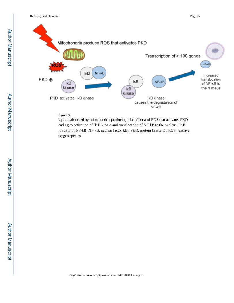
Figure 3. Light is absorbed by mitochondria producing a brief burst of ROS that activates PKD
leading to activation of Ik-B kinase and translocation of NF-kB to the nucleus. Ik-B,
inhibitor of NF-kB; NF-kB, nuclear factor kB ; PKD, protein kinase D ; ROS, reactive
oxygen species.
Hennessy and Hamblin Page 25
J Opt. Author manuscript; available in PMC 2018 January 01.
Author M
anuscriptA
uthor Manuscript
Author M
anuscriptA
uthor Manuscript

Figure 4. In a mouse model of traumatic brain injury caused by controlled cortical impact exposure of
the head to 810 nm laser leads to improved neurological performance. At the same time
there is upregulation of BDNF, neurogenesis and synaptogenesis in the brain. BDNF, brain
derived neurotrophic factor; PBM, photobiomodulation.
Hennessy and Hamblin Page 26
J Opt. Author manuscript; available in PMC 2018 January 01.
Author M
anuscriptA
uthor Manuscript
Author M
anuscriptA
uthor Manuscript

Figure 5. (A) Vielight 810 Intranasal; (B) Vielight Neuro left view ; (C) Vielight Neuro right view; (D)
1. Mesial préfrontal cortex ; 2. Precuneus ; 3. Posterior congulate cortex; 4. Inferior pariétal
lobe; 5. Hippocampus.
Hennessy and Hamblin Page 27
J Opt. Author manuscript; available in PMC 2018 January 01.
Author M
anuscriptA
uthor Manuscript
Author M
anuscriptA
uthor Manuscript

Figure 6. 810 nm LED application to the forehead for treatment of dépression and anxiety.
Hennessy and Hamblin Page 28
J Opt. Author manuscript; available in PMC 2018 January 01.
Author M
anuscriptA
uthor Manuscript
Author M
anuscriptA
uthor Manuscript

Figure 7. 810 nm laser application to the forehead for treatment of dépression.
Hennessy and Hamblin Page 29
J Opt. Author manuscript; available in PMC 2018 January 01.
Author M
anuscriptA
uthor Manuscript
Author M
anuscriptA
uthor Manuscript